ORIGINAL ARTICLE
New species and reports of rust fungi (Basidiomycota,
Uredinales) of South America
Reinhard Berndt&Anja Rössel&Francisco Freire
Received: 12 July 2006 / Revised: 12 December 2006 / Accepted: 13 December 2006 / Published online: 30 January 2007 # German Mycological Society and Springer-Verlag 2007
Abstract Four new species of rust fungi (Basidiomycota, Uredinales) are proposed: Edythea soratensis on Berberis phyllacantha (Berberidaceae), Prospodium bicristatum on Mansoa sp. (Bignoniaceae), Uromyces cearensis on Ipo-moea sp. (Convolvulaceae) and Uredo cavernula on Ribes weberbaueri (Grossulariaceae). U. cavernula most proba-bly belongs to the genus Goplana. Phakopsora phyllanthi and Phakopsora vernoniae are newly reported for Brazil and the New World. The uredinial stage of the latter is the same as Uredo toroiana, known so far from Hispaniola in the Caribbean. The parasitic mycelium of Esalque holwayi was studied. It is strictly intracellular but comprises well-defined haustoria and intracellular hyphae.
Introduction
Rust specimens collected recently in Brazil and Peru and a herbarium specimen originating from Bolivia were found to represent species that are new to science. The present paper aims to contribute to the knowledge of the rust mycobiota of these countries by a detailed description and illustrations of the new species.
Materials and methods
Spores and hand sections of herbarium material were mounted in lactophenol and gently heated to boiling. The preparations were examined with C. Zeiss “Axioskop” or “Axiophot” light microscopes, and photographs were taken with a C. Zeiss MC-80 camera on Kodak Ektachrome 64 Professional slide film. All micrographs were taken using DIC optics. At least 30 spores of each spore stage were measured and the arithmetic mean (“mean”) calculated. Results
Edythea soratensis Ritschel sp. nov.
Etymology: named after the place of collection, the town Sorata in Bolivia (Fig.1a–c).
Fungus parasiticus hospitum non vel paullum commutat. Telia abaxialia, ca. 60–120 μm diam., pallide aurantiaca ad ochracea in herbario sicco, laxe dispersa vel in gregibus 0.6–1 mm diam.; teliosporae subglobosae ad late ellipsoi-deae, verticaliter septatae, basaliter et apicaliter complana-tae, non vel paullum constrictae ad septum, (27)29–37 (39)×(29)32–35(39) μm (medium 32.5×34 μm), pariete pallide flavobrunneo, levi, ca. 1–2 μm crasso, propter
Taxonomical novelties Edythea soratensis Ritschel
Prospodium bicristatum R. Berndt and F. Freire Uredo cavernula R. Berndt and R.S. Peterson Uromyces cearensis R. Berndt and F. Freire R. Berndt (*)
Herbarium turicense,
Institute of Integrative Biology (IBZ), ETH Zurich, CHN G-28.1, Universitätstr. 16,
CH-8092 Zurich, Switzerland e-mail: [email protected] A. Rössel
Systematic Botany and Mycology, Botanical Institute, University of Tübingen,
Auf der Morgenstelle 1, 72076 Tubingen, Germany
e-mail: [email protected] F. Freire
EMBRAPA/CNPAT,
Rua Dra. Sara Mesquita, 2270, Planalto Pici, Fortaleza, Ceara 60.511-110, Brazil e-mail: [email protected]
septum usque ad 3μm crasso, poris germinationis obscuris, apicaliter ad subapicaliter sitis, pedicellis tenue tunicatis, hyalinis, 10–20 μm longis et 7–10 μm latis, frequenter infra hilum frangentibus; mesosporae unicellulatae, sub-globosae ad ellipsoideae, (22)25–28(30)×(25)27–29 (30) μm (medium 26.5×28.5 μm), pariete pallide flavo-brunneo, levi, aequaliter ca. 3μm crasso, poris germinatio-nis conspicuis, apicaliter ad subapicaliter sitis, pedicellis ut in teliosporis. Urediniosporae absunt.
In foliis Berberidis phyllacanthae Rusby (Berberidaceae). Host not visibly or hardly affected by parasite. Telia abaxial, ca. 60–120 μm diameter, pale orange to ochra-ceous in dried herbarium specimen, loosely scattered or in groups of 0.6–1 mm diameter; teliospores subglobose to broadly ellipsoid, vertically septate, basally and apically flattened, not or only slightly constricted at the septum, (27)29–37(39)×(29)32–35(39) μm (mean 32.5×34 μm), wall pale yellowish brown, smooth, ca. 1–2 μm thick, thickened near the septum up to 3 μm; germ pores obscure, situated apically to subapically; pedicels thin walled, hyaline, 10–20 μm long and 7–10 μm wide, usually breaking off near the hilum; mesospores one-celled, subglobose to elliptic, (22)25–28(30)×(25)27–29 (30) μm (mean 26.5×28.5 μm), wall pale yellowish brown, smooth, evenly ca. 3 μm thick; germ pores conspicuous, apical to subapical, pedicels as in the teliospores. Urediniospores not observed.
On leaves of Berberis phyllacantha Rusby (Berberidaceae). Holotype (PUR 68851): Bolivia, Sorata, on B. phylla-cantha, 27 Apr. 1920, leg. E.W.D. and M.M. Holway (Plants of South America no. 579, collected by E.W.D. and M.M. Holway), det. H.S. Jackson.
E. soratensis is based on a specimen from Sorata, Bolivia, determined as Edythea quitensis (Lagerh.) Jacks. and Holw. by Jackson (1931a). The specimen differs from the type of E. quitensis by the size of the teliospores as well as by the occurrence of one-celled mesospores. E. quitensis, first described as Uropyxis quitensis by Arthur (1918), is based on material from Quito, Ecuador, collected by G. Lagerheim in 1891. In the original description, the urediniospores are described to measure 19–23×23– 24 μm and the teliospores to be 20–23 μm wide and 21– 26 μm long. According to our investigation of the type material, the urediniospores measure 22.5–27×22.5–25 μm (mean 24.5×23 μm) and the teliospores 22.5–27×25– 30 μm (mean 25.0×27.0 μm). The teliospores of the specimen from Sorata are distinctly larger (mean 32.5× 34 μm) and differ also from those of two other species of Edythea, Edythea berberidis and Edythea tenella H.S. Jacks. and Holw., in size and shape. Mesospores have hitherto not been observed in members of the genus Edythea. Therefore, the new species E. soratensis is described. The type collection of E. soratensis also contains
Fig. 1 Edythea soratensis. a View upon telium from above. b Two-celled teliospores. c One-Two-celled mesospores. Scale bars = 10μm
aecidial aecia. It is doubtful, however, that they belong to E. soratensis, as they are not associated to the telia.
Material investigated
E. quitensis, Ecuador, Quito, on Berberis sp., Apr. 1891, leg. G. Lagerheim (PUR 68850, holotype). E. quitensis, Ecuador, Quito, on Berberis sp., 18 Aug. 1920, leg. E.W.D. and M.M. Holway, det. H.S. Jackson (PUR 68852; Reliquiae Holwayanae 228, Plants of South America no. 918). E. quitensis, Ecuador, Cuenca, on Berberis sp., 10 Sep. 1920, leg. E.W.D. and M.M. Holway, det. H.S. Jackson (PUR 68853; Plants of South America no. 980). E. quitensis, Ecuador, Provincia Quito, in monte Pichincha, on Berberis schwerini C.K. Schneid., 17 Sep. 1937, leg. et det. H. Sydow (PUR N 3389; Sydow, Fungi exotici exsiccati no. 1097). E. quitensis, Bolivia, Cochabamba, 3,500 m altitude, on Berberis boliviana Rusby, June 1946, leg. M. Cardenas (PUR 78800). E. quitensis, Ecuador, ca. 13 km from Piñas, on the Piñas-Loja road, on the border of
El Oro and Loja Provinces, elevation ca. 7,000 ft, on Berberis sp., 30 July 1975, leg. K.P. Dumont, S.E. Carpenter, P. Buriticá (PUR 86592, 86593). E. berberidis, Ecuador, Tahatanga, on Berberis glaucescens St. Hil., Sep. 1891, leg. G. Lagerheim (PUR 68849, holotype). E. tenella, Bolivia, Sorata, on Berberis divaricata Rusby, 22 Apr. 1920, leg. E.W.D. and M.M. Holway, det. H.S. Jackson (PUR 68854, Plants of South America no. 564, holotype).
Prospodium bicristatum R. Berndt and F. Freire, sp. nov.
Etymology: named after the two ridges of interconnected spines crowning the urediniospore apex (Figs. 2, 3 and
4a–d).
Spermogonia et aecia ignota. Uredinia foliicola, abax-ialia, parva, cyathiformia, paraphysibus peripheralibus circumdata; paraphyses anguste cylindricae vel conicae ad aciculares, ca. 30–45×5–9 μm, rectae vel irregulariter deformatae, stramineae, crasse tunicatae, subacutae; uredi-niosporae asymmetricae, obovoideae vel late ellipsoideae aspectu frontali (versus porum germinationis), rotundato-triangulares aspectu laterali, 24–29×23.5–28 μm (medium 27.0×25.9μm), pariete brunneo, ca. 1.5 μm crasso, sparse potius delicateque echinulato praeter tonsuras duas in poris
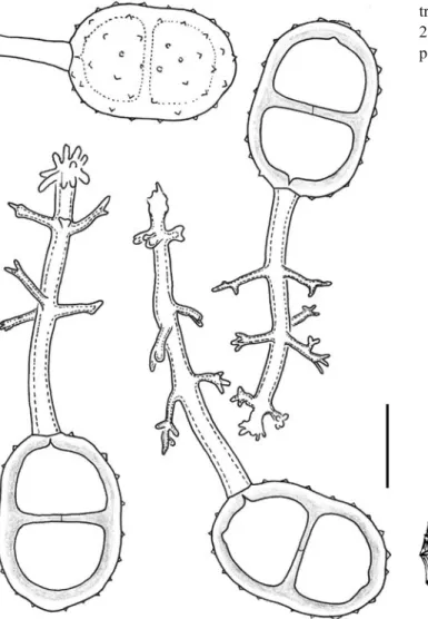
Fig. 2 Prospodium bicristatum. Teliospores with appendaged
germinationis et cristam spinibus coniunctis, delicatis, usque ad 3 μm longis, quam ab hilo ad hilum evolutum est et post bifurcationem apicem bifariam cingit, poris germinationis duobus, plusminusve aequatorialibus et oppositis, inconspicue papillatis. Telia urediniis similia; teliosporae bicellulares, late ellipsoideae, utrinque rotunda-tae, ad septum non vel aegre constricrotunda-tae, 38–42×28–32 μm (medium 39.9×30.0μm), pariete castaneo, ca. 4 μm crasso, bistrato (indistincte in sporis maturis), strato interiori brunneo, exteriorem, quod dilutius est, aequanti, sparse et aequaliter verrucoso verrucis acutis, late conicis (inter se ca. 5–7 μm distantibus), poris germinationis indistinctis, verosimiliter (sub)apicalibus et hilum juxta, pedicellis basaliter vel leniter oblique, rariter laterale insertis, sub-hyalinis, moderate crasse tunicatis, 2–3 verticillis appendi-cis, apicaliter sparse ramificatis, praeditis.
In foliis Mansoae sp. (Bignoniaceae).
Spermogonia and aecia unknown. Uredinia abaxial on leaves, distinctly basket-like, shortly stalked, surrounded by peripheral paraphyses; paraphyses narrowly cylindrical to conical or needle-shaped, ca. 30–45×5–9 μm, straight or
slightly bent, straw-coloured, thick-walled, subacute; ure-diniospores asymmetrical: obovoid to broadly ellipsoid in view parallel to the axis between germ pores, rounded-triangular when the direction of the view is perpendicular to the axis between the pores, 24–29×23.5–28 μm (mean 27.0×25.9 μm); spore wall yellowish brown, ca. 1.5 μm thick, sparsely and rather finely echinulate, except for two large smooth areas around the germ pores and two opposite ridges of interconnected bigger spines that originate at the hilum and branch in the distal part of the spore to garland the spore apex; spines of the ridges rather delicate but up to 3 μm long, connected by a translucent membrane that appears to be finely layered; germ pores two, more or less equatorial and opposite, with inconspicuous subhyaline papillae. Telia like the uredinia; teliospores bi-cellular, broadly ellipsoid, rounded at both ends, not or only slightly constricted at the septum, 38–42×28–32 μm (mean 39.9× 30.0μm); spore wall chestnut brown, ca. 4 μm thick, two-layered (layering indistinct in mature spores), with equally thick layers, the outer one being lighter brown than the inner one, sparsely and evenly verrucose by flatly conical
Fig. 4 a–d Prospodium bicris-tatum. a Basket-like telium with peripheral thick-walled paraph-yses. b Teliospores. c Uredinio-spores with spines and crests. d Urediniospores in optical sec-tion. Scale bars = 10μm. e, f Uromyces cearensis. e Telio-spores. f Urediniospores among teliospores. Scale bars = 10μm
but pointed warts (points of warts ca. 5–7 μm apart); germ pores indistinct but most probably (sub)apical and close to the hilum; pedicels attached basally or slightly laterally on proximal spore cell, rarely shifted sidewards by up to 90°, subhyaline and slightly thick-walled, adorned with two to three well-developed whorls of appendages that stick out more or less rectangularly and branch sparsely in their distal part.
On leaves of Mansoa sp. (Bignoniaceae).
Holotype (UB): Brazil, Ceará State, Marco City (03°06′ 44″S, 40°05′31″W), on Mansoa sp., 3 Dec. 2004, leg. F. Freire (isotype HeRB 8536, located in Z + ZT).
P. bicristatum is different from the other known rust fungus parasitizing Mansoa, Prospodium laevigatum Hennen and Sotão, which has strongly constricted smooth teliospores. It also differs from other described Prospodium spp. especially by the characteristically ornamented uredi-niospores that do not show a distinctly two-layered wall.
Uromyces cearensis R. Berndt and F. Freire, sp. nov.
Etymology: after the Brazilian state of Ceará where the fungus was collected (Figs.4e,f,5and 6).
Spermogonia et aecia ignota. Uredinia absentia, sed copia urediniosporarum saturate ferruginea supra telios adest; urediniosporae obovoideae, late ellipsoideae, 33.5– 40×28–34 μm (medium 36.9×30.8 μm), pariete ca. 3.5– 4.5 μm crasso, bistrato, strato exteriori subhyalino, strato interiori crassiori aureoque, sparse et aequaliter echinulato spinis potius grossis, inter se ca. 3.5–6 μm distantibus, poris germinationis 3–4, plusminusve aequatorialibus et aequidistantibus, epapillatis. Telia in foliis amphigena sparsa, gregibus densis vel solutis, minuta ad parva (0.2– 0.8 mm diam.), atra, celeriter exposita, pulverulenta; teliosporae ovoideae, late ellipsoideae vel subglobosae, 30.5–38.5×26.5–32 μm (medium 34.4×28.8 μm), pariete ca. 3.5–4 μm crasso, apicaliter usque ad 6.5–8 μm, obscure badio, aequaliter potius sparseque verrucoso verrucis humile conicis, poro germinationis apicali vel subapicali, cum papilla lata, dilutiore brunnea, nonnunquam infirme evoluta, pedicellis subhyalinis, juxta hilum dilute brunneis, collabentibus, sporis brevibus vel aequantibus, levibus, proximaliter granulosis.
In foliis tomentosis Ipomoeae sp. indeterminatae (Convolvulaceae).
Spermogonia and aecia unknown. Uredinia lacking, but urediniospores present on several telia, deeply ferrugineous; urediniospores obovoid or broadly ellipsoid, 33.5–40×28– 34μm (mean 36.9×30.8 μm), wall about 3.5–4.5 μm thick, two-layered with an outer subhyaline layer and a thicker, golden inner layer, sparsely and evenly echinulate with rather coarse spines (spines ca. 3.5–6 μm apart); three to
Fig. 6 Urediniospores of Uromyces cearensis. Scale bar = 20μm Fig. 5 Teliospores of Uromyces cearensis. Scale bar = 20μm
four germ pores, more or less equatorial and equidistant, without papillae but outer wall layer slightly thickened at pores. Telia amphigenous on leaves, scattered in loose or dense groups; individual sori tiny to small, 0.2–0.8 mm diameter, sooty black, early naked and pulverulent; telio-spores ovoid, broadly ellipsoid to subglobose, 30.5–38.5× 26.5–32 μm (mean 34.4×28.8 μm); spore wall about 3.5– 4μm thick, apically thickening to 6.5–8 μm, dark chestnut brown, evenly and rather sparsely verrucose with flat conical warts; germ pore apical or subapical, covered by a broad, lighter brown papilla that may not always be clearly differentiated; pedicels collapsing, short (up to as long as spores), generally smooth but proximally granular, subhya-line except for a light brown ring proximal to the hilum.
On an undetermined species of Ipomoea (Convolvulaceae) with tomentose leaves.
Holotype (UB): Brazil, Ceará State, Monsenhor Tabosa City (04°46′74″S, 40°07′71″W), on Ipomoea sp., 10 Sep. 2004, leg. F. Freire (isotype HeRB 8529, located in Z + ZT). The Convolvulaceae are hosts for a considerable number of Uromyces species (Monoson and Schlesser1980). None of their New World representatives combine the characters observed in U. cearensis. The most similar species appears to be Uromyces greenstockii Doidge from South Africa but it has smaller teliospores with subhyaline, hemispherical papillae. Uredinia are unknown in U. greenstockii.
Uredo cavernula R. Berndt and R.S. Peterson sp. nov.
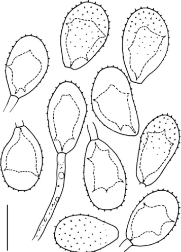
Etymology: describing the shape of the lumen of the urediniospores (Fig.7).
Uredinia in pagina abaxiali foliorum sparsa, maculis stramineis usque ad remisse purpureo-luteolis insidentia, rotundata et pulvinata, ca. 0.4–0.7 mm diam., in herbario sicco ochroleuca vel cerina; urediniosporae obovoideae, (24)27–33(35)×20–25 μm (medium 30.6×22.2 μm), pari-ete subhyalini ad hyalini, apicaliter 6–10 μm crasso, lateraliter irregulariter (2)3–5 μm crasso, foveolis profundis sparsis (poris germinationis?) praedito, echinulato spinis moderate delicatis, inter se ca. 2–3.5 μm distantibus, hilum versus decrescentibus, uno tertiis parte levi, sporae delicate pedicellatae pedicellis fragilibus hyalinis.
In foliis Ribis weberbaueri Janzc. (Grossulariaceae). Uredinia scattered abaxially on leaves, on straw-col-oured to faint purplish-yellow leaf spots, rounded, pulvi-nate, ca. 0.4–0.7 mm diameter; in herbarium specimen cream-coloured to apricot; urediniospores obovoid, (24)27– 33(35)× 20–25 μm (mean 30.6×22.2 μm); spore wall subhyaline to hyaline, apically thickened to 6–10 μm, laterally irregularly (2)3–5 μm thick, with several scattered deep pits (germ pores?), echinulate by moderately delicate spines ca. 2–3.5 μm apart, echinulation fading towards hilum and more or less lacking in proximal third of spore
surface, delicately stalked by hyaline, slender pedicels that tend to break off close to the hilum.
On leaves of R. weberbaueri Janzc. (Grossulariaceae). Holotype (Z + ZT): Peru, Depto. La Libertad, Prov. Santiago de Chuco, road from Santiago de Chuco to Huamachuco (7°58.435′S, 78°12.906′W), altitude 4,110 m, 19 Apr. 2004, leg. M. Weigend and Ch. Schwarzer.
U. cavernula is very similar to the uredinial stage of Goplana ribis-andicolae R. Berndt (Berndt1999). It differs from that species by thicker-walled spores whose proximal third is smooth. We are convinced that U. cavernula will prove to belong to Goplana once the telial stage will have been discovered.
Esalque holwayi (H.S. Jacks.) Hennen, Figueiredo and de Carvalho
Syn. Triactella holwayi H.S. Jacks.
Teliospores of E. holwayi are very similar to those of Triphragmium. Although aware of this fact, Jackson
(1931b) assigned his new species to the genus Triactella
mainly because of the leguminous host. Triactella was later shown to be a synonym of Hapalophragmium. As T.
holwayi is different from Hapalophragmium, Hennen et al. (2000) transferred it to a new genus, Esalque. However, like Jackson, they did not find morphological characters that would have shown convincingly that E. holwayi was different from Triphragmium.
While studying a uredinial specimen of E. holwayi from Brazil, we noticed that the parasitic mycelium of the rust was entirely intracellular and comprised“ordinary” septate hyphae, haustorial mother cells (HMC) and stalked haus-toria (Fig.8). Where the septate hyphae entered an adjacent host cell, they did not only breach the host cell wall but formed HMC adjacent to the penetration site as well. These gave rise to delicate, more or less cylindrical or allantoid haustoria in the neighbouring host cell. The nuclei
remained within the HMC and did not migrate into the haustorial bodies.
Similar mycelia with intracellular HMC are widespread in Raveneliaceae (Berndt1997) and are known for a single member of Phakopsoraceae, Physopella zeae (Mains) Cummins and Ramachar [=Phakopsora zeae (Mains) Buriticá] (Heath and Bonde 1988). The type of parasitic mycelium therefore supports the assumption of Jackson
(1931b) and Hennen et al. (2000) that Esalque belongs to
Raveneliaceae.
Brazil, State of Ceará, Guaramiranga City, on Caesalpi-nia cf. ferrea Mart., 6 Jan. 2005, leg. F. Freire, det. R. Berndt (HeRB 8541, located at Z + ZT).
Phakopsora phyllanthi Dietel in the Neotropics
A recent collection of Phakopsora on Phyllanthus from Brazil was determined as P. phyllanthi, a species, to our knowledge, not hitherto reported to occur in the New World. The species can be easily distinguished from the other New World Phakopsoras on Phyllanthus by its paraphysate uredinia of the “Malupa”-type (Buriticá
1999). A specimen collected in Venezuela on Phyllanthus sp. and another one from Ecuador on Phyllanthus stipulatus (Ruf.) G. Webster (H. Bauch, personal communication) could also be assigned to this species. P. phyllanthi may therefore have a wide distribution in tropical America.
Material investigated
Brazil, Ceará State, Fortaleza City, on Phyllanthus acidus Skeels. (Euphorbiaceae), 10 Jan. 2006, leg. F. Freire, det. R. Berndt (HeRB 8581, at Z + ZT). Venezuela, Miranda State, at Mamporal near Laguna de Tacarigua, on Phyllanthus sp., 13 Dec. 1993, leg. et det. R. Berndt (HeRB A-113, at Z + ZT).
Fig. 8 Esalque holwayi. Host cells with parasitic mycelium consisting of intracellular hyphae that form haustorial mother cells (stars) and haustoria (arrows). Scale bar = 10μm
Table 1 Comparison of Phakopsora vernoniae with Uredo toroiana
Urediniospores Paraphyses
Phakopsora vernoniae, after original description
Globose, elongate or obovate, 20–29×15–20 μm, spore wall 1–1.5 μm thick, echinulate, pores obscure
Peripheral, inwardly curved, 30–40 μm long, at the apex capitate-thickened to 10–15 μm broad, wall hyaline to yellowish-brown, 1.5μm thick, apically to 3 μm P. vernoniae,
Brazilian specimen (HeRB 8521)
Obovoid, ellipsoid to broadly ellipsoid or subclavate, 19–29.5×16.5–20 μm (mean 23.9×17.8 μm), spore wall ca. 1μm thick, subhyaline to ochraceous, moderately densely and finely echinulate
(ca. 1.5–2.2 μm between spines), pores obscure
Peripheral, cylindrical to subclavate, 1–2 septate, more or less inwardly curved, with a slightly thickened (1.5–2.5 μm) ochraceous to golden wall
Uredo toroiana (PUR 47272)
Obovoid or broadly ellipsoid, 22.5–30.5×16.5–20 μm (mean 25.4×18.2μm; 22–30×19–26 μm according to Kern), spore wall ca. 1.5μm thick, moderately densely echinulate with slender and sharp ca. 1μm long spines, pores obscure
Peripheral, subclavate, 1–2 septate, mostly inwardly bent, with golden to ochraceous, 2.5–3 μm thick wall
Phakopsora vernoniae Jørstad in Brazil
Syn. Uredo toroiana Kern
A rust fungus collected in Brazil on Vernonia sp. (Asteraceae) revealed paraphysate uredinia together with very few telia of Phakopsora. The fungus fits the description of P. vernoniae Jørstad in all characters and is thus assigned to the latter species (Gjærum 1986; Jørstad 1956). P. vernoniae has not been known in the New World so far. According to the description, U. toroiana from Santo Domingo (Hispaniola) is similar to the uredinial stage of P. vernoniae, although it is described to have broader urediniospores (Kern 1928). We investi-gated U. toroiana and were unable to find essential differences to the uredinial stage of P. vernoniae. Thus, U. toroiana is regarded as a synonym of the latter. The minor differences observed between P. vernoniae from Brazil and U. toroiana (the latter with slightly bigger urediniospores, slightly thicker spore wall and slightly coarser echinulation) are believed to lie within the variation of the species (Table1).
Material investigated
P. vernoniae, Brazil, Ceará State, Monsenhor Tabosa City, on Vernonia sp., 10 Sep. 2004, leg. F. Freire, det. R. Berndt (HeRB 8521, at Z + ZT). U. toroiana, Dominican Republic, Prov. Espaillat, Valle del Cibao, Moca, Estación Nacional Agronómica, 1930, leg. Ciferri and Ekman. Ciferri, Mycoflora Domingensis Exsiccata no. 193 (PUR 47272).
Acknowledgment We thank M. Weigend for sending rust-infected Ribes to R.S. Peterson and for determining the host. We are also grateful to the director and curator of PUR (Arthur Herbarium, Purdue University) for the loan of specimens.
References
Arthur JC (1918) Uredinales of the Andes, based on collections by Dr. and Mrs. Rose. Bot Gaz 65:460–474
Berndt R (1997) Morphology of the haustoria of Ravenelia and Kernkampella spp. Mycol Res 101:23–34
Berndt R (1999) Neotropical rust fungi: new species and observations. Mycologia 91:1045–1059
Buriticá P (1999) La familia Phakopsoraceae en el neotrópico III, géneros: Batistospora y Phakopsora. Rev Acad Colomb Cienc Exactas Fis Nat 23:271–305
Gjærum HB (1986) East African rusts (Uredinales), mainly from Uganda. 5. On families belonging to Gamopetalae. Mycotaxon 27:507–550
Heath MC, Bonde MR (1988) The temporal relationship between the development of intracellular hyphae and haustoria by Physopella zeae in Zea mays. Can J Bot 66:742–744
Hennen JF, Figueiredo MB, de Carvalho AA Jr (2000) Esalque holwayi gen. et comb. nov., a rust of Brazilian ironwood (Caesalpinia species). Mycologia 92:312–316
Jackson HS (1931a) The rusts of South America based on the Holway collections III. Mycologia 23:96–116
Jackson HS (1931b) The rusts of South America based on the Holway collections IV. Mycologia 23:332–364
Jørstad I (1956) Reliquiae Lagerheimianae, African Uredinales. Ark Bot (ser. 2) 3:563–598
Kern FD (1928) Fungi of Santo Domingo—II. Uredinales. Mycologia 20:60–82
Monoson HL, Schlesser PE (1980) Uromyces on New-World Convolvulaceae. Mycologia 72:817–820