Abstract. Earlier studies have shown that modification of
the octapeptide octreotide in positions 3 and 8 may result
in compounds with increased somatostatin receptor
affin-ity that, if radiolabelled, display improved uptake in
so-matostatin receptor-positive tumours. The aim of a recent
research study in our laboratory was to employ the
paral-lel peptide synthesis approach by further exchanging the
amino acid in position 3 of octreotide and coupling the
macrocyclic chelator
DOTA(1,4,7,10-tetraazacyclodode-cane-1,4,7,10-tetraacetic acid) to these peptides for
label-ling with radiometals like gallium-67 or -68, indium-111,
yttrium-90 and lutetium-177. The purpose was to find
ra-diopeptides with an improved somatostatin receptor
bind-ing profile in order to extend the spectrum of targeted
tu-mours. A first peptide, [
111In,
90Y-DOTA]-1-Nal
3-octreo-tide (
111In,
90Y-DOTA-NOC), was isolated which showed
an improved profile. In
III-DOTA-NOC exhibited the
fol-lowing IC
50values (nM) when studied in competition
with [
125I][Leu
8,
D-Trp22, Tyr
25]somatostatin-28 (values
for Y
III-DOTA-NOC are shown in parentheses): sstr2,
2.9±0.1 (3.3±0.2); sstr3, 8±2 (26±1.9); sstr5, 11.2±3.5
(10.4±1.6). Affinity towards sstr1 and 4 was very low or
absent. In
III-DOTA-NOC is superior to all
somatostatin-based radiopeptides having this particular type of binding
profile, including DOTA-lanreotide, and has three to four
times higher binding affinity to sstr2 than In
III,Y
III-DOTA-Tyr
3-octreotide (In
III,Y
III-DOTA-TOC). In
addi-tion, [
111In]DOTA-NOC showed a specific and high rate
of internalization into AR4-2J rat pancreatic tumour cells
which, after 4 h, was about two times higher than that of
[
111In]DOTA-TOC and three times higher than that of
[
111In]DOTA-octreotide ([
111In]DOTA-OC). The
internal-ized radiopeptides were externalinternal-ized intact upon 2 h of
internalization followed by an acid wash. After 2–3 h of
externalization a plateau is reached, indicating a
steady-state situation explained by reactivation of the receptors
followed by re-endocytosis. Biodistribution studies in CA
20948 tumour-bearing rats showed rapid clearance from
all sstr-negative tissues except the kidneys. At 4 h the
up-take of [
111In]DOTA-NOC in the tumour and sstr-positive
tissues, such as adrenals, stomach and pancreas, was
three to four times higher than that of [
111In]DOTA-TOC.
Differential blocking studies indicate that this is at least
partially due to the uptake mediated by sstr3 and sstr5.
These very promising preclinical data justify the use of
this new radiopeptide for imaging and potentially internal
radiotherapy studies in patients.
Keywords: Somatostatin receptor subtypes – Indium-111
– Yttrium-90 – Tumour targeting – Peptide receptors
Eur J Nucl Med Mol Imaging (2003) 30:1338–1347
DOI 10.1007/s00259-003-1255-5
Introduction
Radiopeptides are becoming of increasing interest in
tu-mour targeting for either the localization or the internal
radiotherapy of neoplasms [1, 2, 3, 4, 5, 6, 7, 8, 9].
Ana-logues of the somatotropin release inhibiting factor
Abbreviations of the common amino acids are in accordance withthe recommendations of IUPAC-IUB [IUPAC-IUB Commission of Biochemical Nomenclature (CBN), Symbols for amino-acid de-rivatives and peptides, recommendations 1971. Eur J Biochem 1972; 27:201–207].
Helmut R. Mäcke (
✉
)Division of Radiological Chemistry,
Institute of Nuclear Medicine, Department of Radiology, University Hospital Basel, Petersgraben 4,
4031 Basel, Switzerland e-mail: [email protected]
Tel.: +41-61-2654699, Fax: +41-61-2655559
Original article
DOTA-NOC, a high-affinity ligand of somatostatin
receptor subtypes 2, 3 and 5 for labelling
with various radiometals
Damian Wild1, Jörg S. Schmitt1, Mihaela Ginj1, Helmut R. Mäcke1, Bert F. Bernard2, Eric Krenning2, Marion de Jong2,
Sandra Wenger3, Jean-Claude Reubi3
1 Division of Radiological Chemistry, Institute of Nuclear Medicine, Department of Radiology, University Hospital Basel, Basel, Switzerland 2 Department of Nuclear Medicine, University Hospital Rotterdam, Rotterdam, The Netherlands
3 Institute of Pathology, University of Bern, Bern, Switzerland
Received: 3 February 2003 / Accepted: 24 May 2003 / Published online: 21 August 2003 © Springer-Verlag 2003
(SRIF), somatostatin, radiolabelled with a variety of
gam-ma-, positron- and beta-emitters, are the prototypes of
such peptides. Somatostatin is a cyclic peptide hormone
that occurs naturally in two bioactive molecular forms:
somatostatin-14 and its N-terminally extended form,
so-matostatin-28. It exerts different biological effects in
dif-ferent parts of the body such as the brain, the pituitary,
the pancreas, the gut and some components of the
im-mune system. The effects include inhibition of hormone
secretion and modulation of neurotransmission and cell
proliferation. These actions are mediated by specific,
G-protein-coupled receptors. Today five different
somato-statin receptor subtypes have been characterized and
cloned (sstr1–5). They are responsible for different
bio-logical responses. As some of these receptors are
over-expressed in several human tumours, especially
neuroen-docrine tumours and their metastases, these tumours can
be visualized in vivo by radiometal chelator conjugated
somatostatin analogues like [
111In-DTPA-D-Phe
1]-octreo-tide (OctreoScan) [10]. It has also been shown that
so-matostatin receptor scintigraphy using this agent is the
most sensitive method for localization of primary and
metastatic disease in endocrine pancreatic tumours and
carcinoids except insulinomas [11]. New conjugates may
show higher sensitivity with regard to the localization of
tumours and metastases, e.g.
99mTc-depreotide is
regis-tered in many countries and appears to show good
perfor-mance in the evaluation of solitary pulmonary nodules
[12], in breast tumours and even in melanoma [13]. On
the other hand, a recent study comparing OctreoScan
with
99mTc-depreotide in 44 patients with neuroendocrine
tumours showed that the
111In-labelled peptide yielded a
far higher detection rate for neuroendocrine tumours,
es-pecially for liver metastases [14].
111In]DOTA-TOC and
[
90Y]DOTA-TOC (DOTA =
1,4,7,10-tetraazacyclodode-cane-1,4,7,10-tetraacetic acid) have been shown to be
ef-fective targeting and therapeutic agents in animal models
and patients [15, 16, 17, 18, 19, 20, 21, 22, 23]. In
addi-tion, replacement of the alcohol group at the C-terminus
of the octapeptide by a carboxylic acid group led to
in-creased sstr2 affinity [24], and [
177Lu-DOTA]-D-Phe
1-Tyr
3-Thr
8-octreotide ([
177Lu]DOTA-TATE) showed
high-er tumour uptake than [
111In-DTPA]-octreotide in six
pa-tients with somatostatin receptor-positive tumours [25].
These new radiopeptides show distinctly higher sstr2
af-finity compared with OctreoScan. Nevertheless, they bind
with high affinity only to sstr2: their affinity to sstr5 is
low, and that to sstr3 almost negligible; no affinity of
these new compounds was found to sstr1 and sstr4.
Al-though the majority of tumours studied with radiolabelled
somatostatin analogues express mainly sstr2 [26], recent
literature data indicate that also sstr1 and sstr3–5 may be
present in some human tumours. For example, in binding
studies using [
125I]-RC-160
(D-Phe-Cys-Tyr-D-Trp-Lys-Val-Cys-Trp-NH
2; K
D=6.55 nM) as the radioligand,
Halmos et al. [27] found somatostatin receptors to be
present on 76% of human epithelial ovarian cancers. By
use of in situ hybridization, Reubi et al. found m-RNAs
of sstr1, 2 and 3 in a variety of human tumours, including
GH-adenoma [28]. Forssell-Aronsson et al. reported the
lack of sstr2 (except for medullary thyroid carcinoma)
with Northern blot in most of the thyroid tumours studied
in 68 patients. Nevertheless, all tumour types regularly
expressed sstr1, sstr3, sstr4 and sstr5 [29]. Raderer et al.
could visualize primary pancreatic adenocarcinoma in
vi-vo with [
111In]DOTA-lanreotide (LAN;
D-2-Nal-Cys-Tyr-D-Trp-Lys-Val-Cys-Thr-NH2) but not with [
111In]DTPA-octreotide [30]. Traub et al. reported [
111In]DOTA-LAN
to have a high sensitivity for detection of lung cancer
[31]. However, their claim that this peptide targets
sstr2–5 with high affinity and sstr1 with lower affinity
could not be confirmed by Reubi et al. [24]. To extend
the biological activity profile of radiolabelled
somatosta-tin analogues, we started a programme to synthesize
ra-diopeptides with affinity to all somatostatin receptor
sub-types in order to potentially extend the spectrum of
acces-sible tumours in diagnosis and internal radiotherapy. A
first compound resulted from a parallel synthesis
ap-proach exchanging the amino acid in position 3 of
octreo-tide; this led to a radiopeptide, [
111In/
90Y-DOTA]-1-Nal
3-octreotide ([
111In/
90Y]DOTA-NOC), which had improved
affinity to sstr2 and high affinity to sstr3 and sstr5 when
compared with our lead compound [
111In/
90Y]DOTA-TOC.
Here we present the preclinical evaluation of this
pep-tide with regard to binding affinity, rate of internalization
and biodistribution in a tumour-bearing rat model and
compare its properties with those of [
111In/
90Y-DOTA
0-Tyr
3]-octreotide and [
111In/
90Y-DOTA
0]-octreotide.
Materials and methods
All chemicals were obtained from commercial sources and used without further purification. H-Thr(tBu)-ol-(2-chloro-trityl)-resin was obtained from Advanced ChemTech (Giessen, Germany) and Fmoc (9-fluorenylmethoxycarbonyl) amino acids were purchased from NovaBiochem AG (Läufelfingen, Switzerland), Bachem (Bubendorf, Switzerland) and Neosystems (France). 111InCl
3was
obtained from Mallinckrodt Medical (Petten, The Netherlands). The prochelator DOTA(tBu)3 (4,7,10-tricarboxymethyl-tert-butyl ester 1,4,7,10-tetraazacyclododecane-1-acetate) was synthesized according to Heppeler et al. [32] or purchased from Macrocyclics (Richardson, Tex., USA). The reactive side chains of the amino acids were masked with one of the following groups: Cys, acet-amidomethyl; Lys, t-butoxycarbonyl; Thr, t-butyl; Trp, t-butoxy-carbonyl. Analytical reversed-phase high-performance liquid chro-matography (RP-HPLC) was carried out on a Hewlett Packard 1050 HPLC system (Waldbronn, Germany) equipped with a multi-wavelength detector and a flow-through Berthold LB506C1 gam-ma-detector. Preparative HPLC was done on a Bischof HPLC system (Metrohm AG, Switzerland) with HPLC-pumps 2250 and a Lambda 1010 UV detector (Metrohm AG, Switzerland). CC250/4 Nucleosil 120-3C18 columns from Macherey-Nagel were used for analytical HPLC, and a VP250/21 Nucleosil 200-5C15 column for preparative HPLC. The gradient systems
consist-ed of mixtures of water with 0.1% trifluoroacetic acid (TFA) (sol-vent A) and acetonitrile (sol(sol-vent B). Quantitative gamma-counting was performed on a COBRA 5003 gamma-system well counter from Packard Instrument Company (Switzerland). Electrospray ionization mass spectrometry (ESI-MS) was carried out with a Finnigan SSQ 7000 spectrometer (Bremen, Germany).
Synthesis. The peptide-chelator conjugates were synthesized by standard Fmoc solid phase synthesis [33] on 2-chlorotritylchloride resin (substitution 0.8 mmol/g) on a Rink peptide-synthesizer Switch 24 (RinkCombichem Technologies, Bubendorf, Switzer-land), according to the general procedure described previously [32]. The last step was the coupling of the prochelator DOTA(tBu)3 to the N-terminus of the peptide. Cleavage of the fully protected conjugates from the resin, oxidative cyclization using iodine, de-protection and HPLC purification led to compounds 1–3 (Table 1, Fig. 1), which could be labelled with “cold” or radioactive In3+or
Y3+(InIII, YIII). All compounds were lyophilized after purification
and characterized by ESI-MS and RP-HPLC. In each case the MS spectra consisted of a major [M+2Na]2+ion peak and some other
smaller peaks corresponding to [M+Na]+and [M+3Na]3+ions. All
peptide-chelator conjugates had a purity >95% confirmed by RP-HPLC. The results are given in Table 1. The peptide-DOTA conju-gates obtained were designated DOTA-NOC [DOTA0-D-Phe1
-1-Nal3]-octreotide), DOTA-OC [DOTA0-octreotide]) and
DOTA-TOC [DOTA0-Tyr3]-octreotide).
The DOTA-SRIF analogues were complexed with InCl3 (an-hyd.) and Y(NO3)3·5 H2O, using the following procedure: ca. 20 µg of the respective DOTA-peptide was heated along with 1.5 eq. of the corresponding metal salt in 150µl 0.2 M sodium ac-etate buffer (pH 5) for 25 min. After cooling, 20µl 0.1 M DTPA (pH 5) was added to complex free metal ions. This mixture was loaded onto a SepPak C18cartridge (Millipore, Switzerland), acti-vated using 5 ml MeOH followed by 10 ml H2O. [MIII(DTPA)]2−
was washed from the cartridge using water. The MIII
-DOTA-pep-tide was eluted with methanol and obtained in >97% purity after evaporation of methanol. The radiopeptides were synthesized ac-cording to Heppeler et al. [32] and obtained in >99% radiochemi-cal purity at specific activities of >37 GBq/µmol peptide.
For internalization experiments, the DOTA-peptides were la-belled to a specific activity of about 37 GBq/µmol peptide; excess InCl3was then added and the mixture was purified on a SepPak C18 cartridge as described above to afford [111In/InIII]DOTA-peptides.
Determination of the somatostatin receptor affinity profiles. Cells stably expressing human sstr1–5 were grown as described previ-ously [24]. All culture reagents were supplied by GIBCO/BRL and Life Technologies (Grand Island, N.Y.). Cell membrane pellets
were prepared and receptor autoradiography was performed on pel-let sections (mounted on microscope slides) as described in detail previously [24]. For each of the tested compounds, complete dis-placement experiments were performed with the universal somato-statin radioligand [125I][Leu8,D-Trp22,Tyr25]somatostatin-28 using
increasing concentrations of the unlabelled peptide ranging from 0.1 to 1,000 nM. Somatostatin-28 was run in parallel as control us-ing the same increasus-ing concentrations. IC50values were calculated after quantification of the data using a computer-assisted image processing system. Tissue standards (autoradiographic [125
I]micro-scales Amersham, UK) containing known amounts of isotopes, cross-calibrated to tissue-equivalent ligand concentrations, were used for quantification [24]. The concentrations of the peptide so-lutions were measured by UV-spectroscopy (εNOC,280 mm=9,855). Cell culture and radioligand internalization studies. Sst2receptor expressing AR4-2J cells were obtained from Novartis Pharma (Basel, Switzerland). The AR4-2J cell line was maintained by se-rial passage in mono-layers in Dulbecco’s Modified Eagle’s Medi-um (DMEM), supplemented with 10% fetal bovine serMedi-um, amino acids, vitamins and penicillin-streptomycin, in a humidified 5% CO2/air atmosphere at 37°C. Viability of the cells and cell num-bers were counted under a microscope with a “Neubauer’s count-ing chamber”. For all cell experiments, the cells were seeded at a density of 0.8–1.1 million cells/well in six-well plates and incubat-ed overnight with internalization buffer to obtain a good cell ad-herence. The loss of cells during the internalization experiments was below 10%. When different radiolabelled peptides were com-pared in cell experiments, the same cell suspension-containing plates were used. Furthermore, the internalization rate was linearly corrected to 1 million cells/well in all cell experiments.
Medium was removed from the six-well plates and cells were washed once with 2 ml of internalization buffer (DMEM, 1% fetal bovine serum, amino acids and vitamins, pH 7.4). Furthermore, 1.5 ml internalization buffer was added to each well and the plates were incubated at 37°C for about 1 h. Thereafter approximately 500,000 cpm or 0.02 MBq/well 111In/115In-labelled peptides
(2.5 pmol/well) to a final concentration of 1.67 nM were added to the medium and the cells were incubated at 37°C for the indicated time periods in triplicate. Internalization was also studied using three dif-ferent concentrations of [111In/115In]DOTA-NOC (0.15 pmol/well or
0.1 nM, 2.5 pmol/well or 1.67 nM, and 10 pmol/well or 6.67 nM). To determine non-specific membrane binding and internalization, cells were incubated with radioligand in the presence of 1µM octreotide. Cellular uptake was stopped by removing medium from the cells and by washing twice with 1 ml of ice-cold phosphate-buffered saline (PBS). Acid wash for 10 min with a 0.1 M glycine buffer pH 2.8 on ice was also performed twice. This was shown previously to be
suffi-Fig. 1. Structural formulae of
the DOTA-conjugated peptides. 1, DOTA-1-Nal3-octreotide
(DOTA-NOC); 2, oct-reotide (OC); 3, DOTA-Tyr3-octreotide (DOTA-TOC)
cient to remove >90% of receptor-bound radioligand. This procedure was performed to distinguish between membrane-bound (acid-re-leasable) and internalized (acid-resistant) radioligand. Finally, the cells were treated with 1 N NaOH. The culture medium, the receptor-bound and the internalized fraction were measured radiometrically in a gamma-counter (Packard, Cobra II).
Radioligand externalization studies. AR4-2J cells (1 million) were incubated with 2.5 pmol/well or 1.67 nM [111In/115In]-labelled
DOTA-NOC, DOTA-TOC or DOTA-OC for 120 min, then the medium was removed and the wells were washed twice with 1 ml ice-cold PBS. In each experiment an acid wash for 10 min on ice with a glycine buffer of pH 2.8 was performed to remove the re-ceptor-bound ligand. Cells were then incubated again at 37°C with fresh externalization buffer (DMEM containing 1% fetal bovine serum pH 7.4). After different time points the external medium was removed for quantification of radioactivity in a gamma-coun-ter and replaced with fresh 37°C exgamma-coun-ternalization medium. Ingamma-coun-ternal- Internal-ized ligand was extracted in 1 N NaOH, removed and quantified in a gamma-counter. The recycled fraction was expressed as a per-centage of the total internalized amount per 1 million cells, and the stability of the externalized peptides was determined using HPLC after removal of the solvent by a centrifugal evaporator. Biodistribution. Animal experiments were performed in compli-ance with the regulations of our institutions and with generally ac-cepted guidelines governing such work. Male Lewis rats (200–250 g) bearing the CA20948 pancreatic tumour (0.4–3.5 g) were used in the experiments. Rats were injected under ether ana-esthesia with 2–3 MBq of 0.34 nmol (0.5µg total peptide mass) [111In]DOTA-NOC in 0.5 ml saline into the dorsal vein of the
pe-nis. At several time points, rats were sacrificed under ether anaes-thesia. Organs and blood were collected and the radioactivity in these samples was determined using a gamma-counter.
In order to determine the non-specific uptake of the radiopep-tides, rats were injected with 0.5 mg octreotide in 0.5 ml saline as a co-injection with the radioligand.
To study the sstr2-, 3- and 5-related specific uptake of [111In]DOTA-NOC in the SRIF receptor-positive tissues, blocking
studies were designed with two different somatostatin analogues: DTPA-TATE (sstr2-selective ligand) and InIII-DOTA-NOC (sstr2,
3, and 5 affinity). Twenty-five micrograms of these peptides was co-injected with 2–3 MBq [111In]DOTA-NOC (0.34 nmol in
0.5 ml saline) into the dorsal vein of the penis of non-tumour-bear-ing male Lewis rats. Rats were sacrificed at 24 h and the organs of interest collected and counted for radioactivity.
Statistical methods. Student’s t test was used to determine statisti-cal significance. Differences at the 95% confidence level (P<0.05) were considered significant.
Results
Synthesis and radiolabelling
The three DOTA-coupled octapeptides (Fig. 1) were
ob-tained by parallel synthesis on a trityl chloride resin and
are part of a small library. The peptides not reported here
will be discussed in a more comprehensive chemistry
publication. The overall yield of the DOTA peptides was
about 30% based on the first Fmoc cleavage. The
pep-tides were prepared and purified to >95% purity by
HPLC analysis.
Uncomplexed and metal ion-complexed DOTA
pep-tides were characterized by HPLC, by ESI-MS and by
the retained affinity to the somatostatin receptors. Some
selected analytical data are given in Table 1. Labelling
Table 1. Analytical data for each of the compounds
Compound number Compound name Mass spectrum HPLCa
Calculated M (Da) Observed M+Na+(Da) T
R(min) Purity (%)
1 DOTA-NOC 1,454.6 1,477.7 20.77 98.5
2 DOTA-OC 1,404.6 1,428.3 18.27 99.2
3 DOTA-TOC 1,420.6 1,444.3 16.1 99.0
aElution system: flow 0.75 ml/min; A, 0.1% TFA in H
2O; B, MeCN; linear gradient: 0–30 min, 10% B to 60% B. TR, elution time
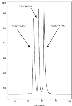
Fig. 2. Elution profile of [111In]DOTA-TOC, [111In]DOTA-OC and
[111In]DOTA-NOC on the HPLC system described in Materials and
Fig. 3. Comparison of the internalization rate of [111
In]DOTA-NOC (■), [111In]DOTA-TOC (●) and [111In]DOTA-OC (▲) into
AR4-2J cells. Values and standard deviations are the result of three independent experiments with triplicates in each experiment and are expressed as specific internalization (percentage of dose added to 1 million cells at 1.67 nM concentration, 37°C)
was performed in acetate buffer (pH 5, 0.4 M) by heating
at 95°C for 25 min, affording labelling yields with
111In
and
90Y of >99% at a specific activity of >37 GBq/
µ
mol
peptide. The HPLC elution profile of the three
111In-la-belled peptides is shown in Fig. 2. The respective RF
values are 17.03 min for [
111In]DOTA-TOC, 18.83 min
for [
111In]DOTA-OC and 20.88 min for [
111In]DOTA-NOC (for a gradient see the footnote to Table 1),
indicat-ing that this is the order of increasindicat-ing lipophilicity.
Receptor binding and affinity profiles
Table 2 shows the IC
50values of the three radiopeptides
studied in this work as their Y
III(In
III) complexed
ver-sions and of Y
III-DOTA-LAN for the five somatostatin
receptor subtypes. The values were obtained by
perform-ing complete displacement experiments with the
univer-sal somatostatin radioligand [
125I][Leu
8, D-Trp
22, Tyr
25]-somatostatin-28 on membranes from cells expressing the
receptor subtypes and were compared with the data for
somatostatin-28.
All compounds bound specifically to sstr2 with IC
50values between 3 and 23 nM. High specific binding
af-finities to sstr3 were also found for In
III-DOTA-NOC
(IC
50=8±2 nM), Y
III-DOTA-NOC (IC
50
=26±1.9 nM) and
Y
III-DOTA-OC (IC
50
=27±8 nM). Y
III-DOTA-TOC and
Y
III-DOTA-LAN showed only very low affinities to sstr3
(IC
50≥
300 nM).
All metallopeptides also showed specific binding to
sstr5; Y
III-DOTA-TOC bound with IC
50
=204±92 nM and
Y
III-DOTA-OC with IC
50
=58±22 nM whereas Y
III-DOTA-LAN (IC
50=16.3±3.4 nM) and Y
III-DOTA-NOC
(IC
50=10.4±1.6 nM) showed rather high affinities to sstr5.
In vitro internalization studies in AR4-2J cells
Figure 3 shows the results in respect of the
time-dependent internalization of [
111In/
115In]DOTA-NOC,
[
111In/
115In]DOTA-TOC and [
111In/
115In]DOTA-OC into
AR4-2J rat pancreatic tumour cells during a 240-min
in-Table 2. Affinity profiles (IC50) for human sst 1–5 receptors
Compound hsst1 hsst2 hsst3 hsst4 hsst5 SS-28 3.8±0.3 (10) 2.5±0.3 (11) 5.7±0.6 (10) 4.2±0.3 (11) 3.7±0.4 (11) InIII-DOTA-NOC >10,000 (3) 2.9±0.1 (3) 8±2 (3) 227±18 (3) 11.2±3.5 (3) YIII-DOTA-NOC >1,000 (3) 3.3±0.2 (3) 26±1.9 (3) >1,000 (3) 10.4±1.6 (3) YIII-DOTA-TOC >10,000 (6) 11.4±1.7 (6) 389±136 (5) >10,000 (6) 204±92 (6) YIII-DOTA-OC >10,000 (5) 20±2.2 (5) 27±8 (5) >1000 (5) 58±22 (4) YIII-DOTA-LAN >10,000 (4) 22.8±4.9 (4) 290±105 (4) >1000 (4) 16.3±3.4 (4)
IC50values are in nM (mean±SEM). Number of independent stud-ies is given in parentheses. SS-28 was used as control
Using an assay based on rat brain cortex membranes and [125
I]-Tyr3-octreotide as radioligand, InIII-DOTA-D-Phe1-octreotide and
InIII-DOTA-Tyr3-octreotide were shown to be equipotent to the
corresponding YIII-DOTA-peptides (data not shown or Heppeler et
al. [32])
Fig. 4. Comparison of the percentage (± standard deviation) of
ra-diopeptide internalized into AR4-2J cells (1 million cells, 37°C) after 30 min with different radiopeptide concentrations. Symbols are as in Fig. 3
cubation period at 37°C. About 85%–95% of totally
in-ternalized ligand was specifically inin-ternalized. At
30 min and 1.67 nM concentration, [
111In]DOTA-NOC
showed 7.2%±1.0% specific cell uptake of the total
ac-tivity administered which increased to 24.8%±1.6%
up-take at 4 h. A tendency to reach a plateau was found at
24 h for all three peptides (data not shown). The uptake
of [
111In]DOTA-OC at 30 min was only 1.0%±0.2% and
increased to 7.5%±0.3% at 4 h, whereas [
111In]DOTA-TOC showed 2.0%±0.7% internalization at 30 min,
ris-ing to 11.6%±0.8% at 4 h. The percentage of
internal-ized peptide measured at 30 min as a function of
con-centration is shown in Fig. 4.
The rate of externalization is shown in Fig. 5. In these
experiments,
111In labelled peptides were allowed to
in-ternalize for 120 min; cells were then washed twice with
PBS before removing the receptor-bound ligand with the
glycine buffer. Medium was then added and removed
af-ter 10 min, 30 min, 60 min, 120 min and 240 min and
measured for radioactivity. Up to 60 min, the three
pep-tides showed insignificant differences in the
externaliza-tion rate. Thereafter the extent of externalized ligand was
highest for [
111In]DOTA-OC, with about 60% at 4 h
compared with 47%±4% for [
111In]DOTA-NOC and
Fig. 5. Comparison of the externalization rate (± standard
devia-tion) after 120-min internalization of the three radiopeptides. Sym-bols are as in Fig. 3
Table 3. Biodistribution and tissue radioactivity ratios in organs, blood and CA 20948 tumour of rats 4 h, 24 h and 48 h after injection of
[111In]DOTA-NOC and [111In]DOTA-TOC
Time Organ [111In]DOTA-TOC P [111In]DOTA-NOC [111In]-DOTA-NOC
unblocked, n=3 rats unblocked, n=5 rats blockedc, n=3 rats
4 h Blooda 0.014±0.001 24 h Blooda 0.002±0.000 0.009 0.003±0.0005 0.003±0.0001 4 h Kidneysa 1.91±0.07 24 h Kidneysa 2.32±0.13 <0.0001 1.73±0.14 1.88±0.02 48 h Kidneysa 1.81±0.14 4 h Livera 0.18±0.018 24 h Livera 0.05±0.000 0.0004 0.11±0.020 0.10±0.008 24 h Spleena 0.04±0.000 0.0001 0.05±0.009 0.05±0.007 4 h Femurb 0.127±0.010 24 h Femurb 0.05±0.010 0.0004 0.098±0.016 0.022±0.005 48 h Femurb 0.084±0.014 4 h Pancreasb 2.57±0.08 <0.0001 7.75±1.39 24 h Pancreasb 1.70±0.13 0.01 2.93±0.69 0.33±0.05 4 h Adrenalsb 3.62±0.14 <0.0001 7.43±0.42 24 h Adrenalsb 3.19±0.27 <0.0001 7.21±1.02 1.32±0.28 48 h Adrenalsb 1.80±0.13 <0.0001 5.85±0.60 4 h Stomachb 0.91±0.12 24 h Stomachb 0.25±0.04 0.0014 0.56±0.13 0.08±0.01 4 h Pituitaryb 1.48±0.07 019 ns 1.66±0.20 24 h Pituitaryb 1.22±0.03 0.064 ns 1.89±0.55 0.21±0.06 4 h Tumourb 1.15±0.16 0.0004 3.86±0.63 24 h Tumourb 1.12±0.11 0.021 2.08±0.62 0.36±0.10 48 h Tumourb 0.51±0.04 <0.0001 2.04±0.29
Tumour to tissue ratios
24 h Tumour/blood 520 693
24 h Tumour/liver 22.4 18.9
4 h Tumour/kidneys 2.02
24 h Tumour/kidneys 0.48 1.20
48 h Tumour/kidneys 1.12
Values are the mean of % ID/g±SE
aSomatostatin receptor-negative organs bSomatostatin receptor-positive organs
cBlocked with 0.5 mg octreotide as a co-injection with the
43%±2% for [
111In]DOTA-TOC. At 4 h the
externaliza-tion curve of all peptides showed a plateau. Study of the
chemical structure of the externalized [
111In]DOTA-NOC
and [
111In]DOTA-TOC by HPLC gave no indication of
metabolites. The peptides externalized were intact.
Biodistribution studies in rats
The 4-h, 24-h and 48-h uptake values of [
111In]DOTA-NOC and [
111In]DOTA-TOC in sst receptpositive
or-gans, including pancreas, adrenals, pituitary, stomach
and CA 20948 rat pancreatic tumour, as well as the
kid-neys, liver, spleen, femur and blood are shown in
Ta-ble 3. Both radiopeptides displayed rapid blood
clear-ance with less than 0.02% ID/g remaining in the blood at
4 h. There was also fast clearance from all sstr-negative
tissues except the kidneys. The excretion of both
pep-tides was mainly by the kidneys. [
111In]DOTA-NOC had
a significantly higher uptake than [
111In]DOTA-TOC in
all sstr-positive tissues at all time points, e.g. at 4 h
(tu-mour, 3.9%±0.6% ID/g vs 1.15%±0.16% ID/g; pancreas,
7.75%±1.4% ID/g vs 2.57%±0.08% ID/g; adrenals,
7.43%±0.42% ID/g vs 3.62%±0.14% ID/g; pituitary,
1.66%±0.20% ID/g vs 1.48%±0.07% ID/g).
To estimate the uptake in sstr-positive organs which may
be due to receptor subtype expression other than sstr2, in
vi-vo blocking studies were performed in normal rats using
different blocking agents like [DTPA
0-Tyr
3-Thr
8]-octreotide
(DTPA-TATE), an sstr2-specific ligand (IC
50=3.9±1 nM),
and In
III-DOTA-NOC (IC
50
: sstr2=2.9±0.1 nM, sstr3=
8±2 nM, sstr5=11.2±3.5 nM). In the adrenals, blocking with
only 25
µ
g of In
III-DOTA-NOC showed a very efficient
re-duction of about 95% whereas 25
µ
g of the sstr2-selective
ligand DTPA-TATE resulted in only about 75% blocking.
A significantly higher blocking effect was also found
with In
III-DOTA-NOC in the pancreas, pituitary and
stomach (data not shown).
Discussion
Receptor scintigraphy with
111In-DTPA-octreotide has
become the “gold standard” for the localization, staging
and management of neuroendocrine tumours [34]. The
high sensitivity of somatostatin receptor scintigraphy
and its ability to change the management of patients with
neuroendocrine tumours has been demonstrated in
sever-al studies [35, 36, 37, 38, 39]. The same agent has been
used for receptor-mediated radionuclide therapy with
some success if injected in high doses (6 GBq every 4
weeks) for total doses of up to about 100 GBq [40, 41,
42]. This agent only binds with reasonably high affinity
to sstr2 and with low affinity to sstr5 [24]; in addition,
111In is not an ideal therapeutic radionuclide. Therefore
several groups have developed agents based on
somato-statin for improved targeting with positron emitters like
68
Ga,
64Cu and the gamma emitter
111In, and most
impor-tantly for labelling with therapeutic radionuclides like
90Y and
177Lu. The most successful peptides have been
[Tyr
3]-octreotide (TOC) and [Tyr
3, Thr
8]-octreotide
(oct-reotate = TATE) coupled to the macrocyclic chelator
DOTA (DOTA-TOC, DOTA-TATE) [32, 43, 44, 45].
These agents bind with high affinity only to sstr2.
Al-though sstr2 is probably the most abundantly expressed
SRIF receptor in human cancer [26], subtypes sstr1,
sstr3, sstr4 and sstr5 may also be of interest. So, our
ap-proach is focussed on the identification of analogues
with a pan-somatostatin binding profile carrying
func-tional groups for radiolabelling [46] and we have started
a programme to use parallel synthesis methods to
pro-duce DOTA-coupled octapeptides with the aim of
im-proving the affinity to subtypes other than 2 while
main-taining the sstr2 affinity. In a series of compounds we
found that [DOTA
0-1-naphthyl
3]-octreotide shows
prom-ising properties owing to its high affinity to sstr2, with
an IC
50value of 3.3±0.2 nM if complexed to Y
IIIand
2.9±0.1 nM if complexed to In
III, which is three- to
four-fold higher than the corresponding value for Y
III-DOTA-TOC (IC
50=11.4±1.7 nM). The new metallopeptides are
equipotent to somatostatin-28 on sstr2 and a factor of 7
more potent than Y
III-DOTA-LAN. Moreover, they show
good affinity to sstr3 and are significantly more potent at
sstr5 than Y
III-DOTA-LAN. These data do not confirm
the pan-somatostatin-like binding affinities of
DOTA-LAN published earlier by Smith-Jones et al. [47]. In
ad-dition, In
III- and Y
III-DOTA-NOC show the highest
af-finities on sstr3. A metal ion dependence is found at
sstr3 and sstr4, In
III-DOTA-NOC being about three to
four times more potent than Y
III-DOTA-NOC. A
poten-tial explanation for this phenomenon is the difference in
the coordination geometry of the two DOTA-metal
IIIcomplexes, which was documented using
1H-NMR
spec-troscopy and X-ray crystallography [48]. Y
III-DOTA-OC
and Y
III-DOTA-NOC are equipotent on sstr3 and about a
factor of 6 less potent than the endogenous ligand. There
is no metal ion dependence in affinity to sstr5.
Cell uptake and release
In order to obtain a defined and homogeneous
metallopep-tide, [
111In]DOTA-NOC was complexed with “cold” In
IIIto yield [
111In,
115In]DOTA-NOC. At 4 h of
internaliza-tion, [
111In/
115In]DOTA-NOC showed a factor of 2 higher
specific cell uptake than [
111In/
115In]DOTA-TOC and a
factor of about 3 higher than [
111In/
115In]DOTA-OC at
1.67 nM peptide concentration per 1 million cells. The
dif-ference was even more pronounced at 0.1 nM
concentra-tion (Fig. 4). This order follows the receptor affinity of the
three radiopeptides, indicating that receptor affinity is the
major factor determining the rate of internalization.
As the addition of excess cold octreotide inhibits 90%
of the uptake, it can be considered as specific and
recep-affinity to sstr2, sstr3 and sstr5. The higher blocking
effi-ciency of In
III-DOTA-NOC in the adrenals, pancreas,
stomach and pituitary may indicate that part of the
radio-ligand uptake is due to the improved receptor subtype
profile. These organs have previously been shown to
ex-press different receptor subtypes, at least at the mRNA
level: sstr2: adrenals, pituitary and pancreas; sstr3:
pitui-tary, pancreas and stomach; sstr5: adrenals, pituipitui-tary,
pancreas and stomach [51].
In conclusion, we have developed a new radiopeptide
based on somatostatin which promises to target a broader
range of somatostatin receptors and concomitantly a
larger spectrum of tumours. These preclinical data
indi-cate that [
111In]DOTA-NOC is superior to existing and
well-studied radiolabelled somatostatin analogues.
In-deed, the predictions from these preclinical studies have
been confirmed in initial clinical studies in which
excel-lent images of thyroid cancer patients have been
ob-tained. We assume that [
90Y]/[
177Lu]DOTA-NOC will
have similar favourable properties.
Acknowledgements. M. Ginj, D. Wild, J. Schmitt and H. Maecke acknowledge support from the Swiss National Science Foundation project No. 31-52969.97, BBW No. C00.0091 and BBT project 4668.1 EUS. The support provided by Novartis Pharma in respect of MS and NMR is gratefully acknowledged.
This work was performed within the COST B12 action.
References
1. Behr TM, Béhé M, Becker W. Diagnostic applications of ra-diolabeled peptides in nuclear endocrinology. Q J Nucl Med 1999; 43:268–280.
2. Breeman WAP, de Jong M, Kwekkeboom DJ, et al. Somato-statin receptor-mediated imaging and therapy: basic science, current knowledge, limitations and future perspectives. Eur J Nucl Med 2001; 28:1421–1429.
3. Fischman A, Babich J, Strauss H. A ticket to ride: peptide ra-diopharmaceuticals. J Nucl Med 1993; 34:2253–2263. 4. Heppeler A, Froidevaux S, Eberle A, Maecke H. Receptor
tar-geting for tumor localization and therapy with radiopeptides. Curr Med Chem 2000; 7:971–994.
5. Lamberts SW, Van der Lely AJ, De Herder WW, Hofland LJ. Octreotide. N Engl J Med 1996; 25:246–254.
6. Lister-James J, Moyer B, Dean T. Small peptides radiolabeled with 99mTc. Q J Nucl Med 1996; 40:221–233.
7. Liu S, Edwards D. 99mTc-labeled small peptides as diagnostic
radiopharmaceuticals. Chem Rev 1999; 99:2235–2268. 8. Reubi JC. Neuropeptide receptors in health and disease: the
molecular basis for in vivo imaging. J Nucl Med 1995; 36: 1825–1835.
9. Thakur M. Radiolabelled peptides: now and the future. Nucl Med Comm 1995; 16:724–732.
10. Krenning EP, Kwekkeboom DJ, Bakker WH, et al. Somatosta-tin receptor scintigraphy with [111In-DTPA-D-Phe1]- and [123
I-Tyr3]-octreotide: the Rotterdam experience with more than
1000 patients. Eur J Nucl Med 1993; 20:716–731.
11. Gibril F, Reynolds JC, Doppman JL, et al. Somatostatin recep-tor scintigraphy: its sensitivity compared with that of other
im-tor mediated. In the time interval of the study, no steady
state was reached, but the distinct leveling off of
[
111In/
115In]DOTA-NOC uptake indicates that steady
state was closely approached; we explain this by the
on-set of efflux of radiopeptides that were shown to be
structurally intact.
If, upon internalization of the radioligand for 2 h, the
cells were exposed to the culture medium, a
time-depen-dent efflux of the radiopeptides could again be observed,
indicating rapid recycling to the extracellular medium. A
steady state was reached already after 2–3 h of release
(Fig. 5). This is in agreement with data that we have
published previously on [
67Ga]NODAGA-TOC [49]. We
interpret this as beginning reactivation of the receptors
by the intact externalized peptides and concomitant
re-endocytosis. The finding that the weakest binder
[
111In]DOTA-OC apparently shows the most efficient
ex-ternalization fits with this explanation. The fact that the
externalized peptides are still intact upon release is
an-other indication that this is the correct interpretation. It is
also in keeping with the conclusion drawn from data
ob-tained by Koenig et al. [50].
Biodistribution studies
The biodistribution studies in CA20948-bearing tumour
rats demonstrated superior uptake of [
111In]DOTA-NOC
compared with [
111In]DOTA-TOC at 4 h, 24 h and 48 h in
receptor-positive normal tissues (except the pituitary) and
the tumour. An estimated area under the curve showed an
improvement of approximately 2.5-fold in the tumour.
This improvement is likely due to the improved sst2, sst3
and sst5 receptor affinity and the significantly faster rate
of internalization, as exemplified in the AR4-2J cell line.
The octreotide co-injection experiment demonstrated that
the uptake is specific and receptor mediated.
Radiometal labelled radiopeptides show high and
per-sistent kidney uptake, limiting their therapeutic potential.
One of the goals in the design of new somatostatin-based
radioligands is to reduce their uptake in the kidney.
In-deed, the tumour-to-kidney ratio of [
111In]DOTA-NOC is
improved 2.5-fold compared with [
111In]DOTA-TOC.
Despite the distinctly higher lipophilicity of
[
111In]DOTA-NOC over [
111In]DOTA-TOC, the uptake
in the liver and the intestines is surprisingly low. In
addi-tion, the long residence time of the new radiopeptide in
the tumour indicates that it is not only suitable for
imag-ing but also efficacious in targeted radiotherapy when
la-belled with
90Y and/or
177Lu provided that there is no
significant difference among these M
IIIradiometals.
To understand the contribution of the high uptake
val-ues in SRIF receptor-positive organs due to the different
subtype affinities of [
111In]DOTA-NOC, the uptake in
these tissues was studied using different blocking agents,
namely DTPA-TATE, an sstr2-specific ligand with
IC
50=3.9±1 nM, and In
III-DOTA-NOC, which has a high
aging methods in detecting primary and metastatic gastrino-mas—a prospective study. Ann Intern Med 1996; 125:24–26. 12. Blum JE, Handmaker H, Rinne NA. The utility of a
somato-statin-type receptor binding peptide radiopharmaceutical (P829) in the evaluation of solitary pulmonary nodules. CHEST 1999; 115:224–232.
13. Virgolini I, Leimer M, Handmaker H, et al. Somatostatin re-ceptor subtype specificity and in vivo binding of a novel tu-mor tracer, 99mTc-P829. Cancer Res 1998; 58:1850–1859.
14. Lebtahi R, Le Cloirec J, Houzard C, et al. Detection of neuro-endocrine tumors: 99mTc-P829 scintigraphy compared with 111In-pentetreotide scintigraphy. J Nucl Med 2002; 43:889–
895.
15. de Jong M, Bakker WH, Krenning EP, et al. Yttrium-90 and indium-111 labelling, receptor binding and biodistribution of [DOTA0,D-Phe1,Tyr3]octreotide, a promising somatostatin
ana-logue for radionuclide therapy. Eur J Nucl Med 1997; 24: 368–371.
16. Stolz B, Weckbecker G, Smith-Jones PM, Albert R, Raulf F, Bruns C. The somatostatin receptor-targeted radiotherapeutic [90Y-DPTA-DPhe1,Tyr3]octreotide (90Y-SMT 487) eradicates
experimental rat pancreatic CA 20948 tumours. Eur J Nucl Med 1998; 25:668–674.
17. Otte A, Herrmann R, Heppeler A, et al. Yttrium-90-DOTATOC: first clinical results. Eur J Nucl Med 1999; 26:1439–1447. 18. Otte A, Mueller-Brand J, Dellas S, Nitzsche E, Herrmann R,
Maecke H. Yttrium-90-labelled somatostatin-analogue for cancer treatment. Lancet 1998; 351:417–418.
19. Cremonesi M, Ferrari M, Zoboli S, et al. Biokinetics and do-simetry in patients administered with 111In-DOTA-Tyr3
-octreo-tide: implications for internal radiotherapy with 90
Y-DOTA-TOC. Eur J Nucl Med 1999; 26:877–886.
20. Waldherr C, Pless M, Maecke H, et al. Tumor response and clinical benefit in neuroendocrine tumors after 7.4 GBq 90
Y-DOTATOC. J Nucl Med 2002; 43:610–616.
21. Paganelli G, Zoboli S, Cremonesi M, et al. Receptor-mediated radiotherapy with 90Y-DOTA-Phe1-Tyr3-octreotide. Eur J Nucl
Med 2001; 28:426–434.
22. Waldherr C, Pless M, Maecke H, Haldemann A, Mueller-Brand J. The clinical value of [90Y-DOTA]-D-Phe1-Tyr3
-oct-reotide (90Y-DOTATOC) in the treatment of neuroendocrine
tumours: a clinical phase II study. Ann Oncol 2001; 12: 942–945.
23. de Jong M, Breeman WA, Bernard BF, et al. Tumor response after [90Y-DOTA0,Tyr3]octreotide radionuclide therapy in a
transplantable rat tumor model is dependent on tumor size. J Nucl Med 2001; 42:1841–1846.
24. Reubi J, Schaer J, Waser B, et al. Affinity profiles for human somatostatin receptor sst1–sst5 of somatostatin radiotracers selected for scintigraphic and radiotherapeutic use. Eur J Nucl Med 2000; 27:273–282.
25. Kwekkeboom DJ, Bakker WH, Kooij PP, et al. [177
Lu-DOTA0,Tyr3]octreotate: comparison with [111DTPA0
]octreo-tide in patients. Eur J Nucl Med 2001; 28:1319–1325. 26. Reubi JC, Waser B, Schaer J, Laissue JA. Somatostatin
recep-tor sst1–sst5 expression in normal and neoplastic human tis-sues using receptor autoradiography with subtype-selective li-gands. Eur J Nucl Med 2001; 28:836–846.
27. Halmos G, Sun B, Schally AV, Hebert F, Nagy A. Human ovarian cancers express somatostatin receptors. J Clin Endo-crinol Metab 2000; 85:3509–3512.
28. Reubi JC, Schaer J, Waser B, Mengod G. Expression and lo-calization of somatostatin receptor sstr1, sstr2, and sstr3
mes-senger RNAs in primary human tumors using in situ hybrid-ization. Cancer Res 1994; 54:3455–3459.
29. Forssell-Aronsson E, Nilsson O, Benjegard SA, et al. 111
In-DTPA-D-Phe1-octreotide binding and somatostatin receptor
subtypes in thyroid tumors. J Nucl Med 2000; 41:636–642. 30. Raderer M, Pangerl T, Leimer M, et al. Expression of human
somatostatin receptor subtype 3 in pancreatic cancer in vitro and in vivo. J Nat Cancer Inst 1998; 90:1666–1668.
31. Traub T, Petkov V, Ofluoglu S, et al. 111In-DOTA-lanreotide
scintigraphy in patients with tumors of the lung. J Nucl Med 2001; 42:1309–1315.
32. Heppeler A, Froidevaux S, Maecke HR, et al. Radiometal-la-belled macrocyclic chelator-derivatised somatostatin analogue with superb tumour targeting properties and potential for re-ceptor-mediated internal radiotherapy. Chem Eur J 1999; 5:1974–1981.
33. Atherton E, Sheppard R. Fluorenylmethoxycarbonyl-poly-amide solid phase peptide synthesis. General principles and development. Oxford: Oxford Information Press, 1989. 34. Oberg K. Established clinical use of octreotide and lanreotide
in oncology. Chemotherapy 2001; 47:40–53.
35. Chiti A, Fanti S, Savelli G, et al. Comparison of somatostatin receptor imaging, computed tomography and ultrasound in the clinical management of neuroendocrine gastro-entero-pancre-atic tumours. Eur J Nucl Med 1998; 25:1396–1403.
36. Lebtahi R, Cadiot G, Sarda L, et al. Clinical impact of somato-statin receptor scintigraphy in the management of patients with neuroendocrine gastroenteropancreatic tumors. J Nucl Med 1997; 38:853–858.
37. Cadiot G, Bonnaud G, Lebtahi R, et al. Usefulness of somato-statin receptor scintigraphy in the management of patients with Zollinger-Ellison syndrome. Gut 1997; 41:107–114. 38. Termanini B, Gibril F, Reynolds JC, et al. Value of
somatosta-tin receptor scintigraphy: a prospective study in gastrinoma of its effect on clinical management. Gastroenterology 1997; 112:335–347.
39. Gibril F, Doppman JL, Reynolds JC, et al. Bone metastases in patients with gastrinomas: a prospective study of bone scan-ning, somatostatin receptor scanscan-ning, and MRI in their detec-tion, their frequency, location and effect of their detection on management. J Clin Oncol 1998; 16:1040–1053.
40. Krenning EP, de Jong M, Kooij PP, et al. Radiolabelled so-matostatin analogue(s) for peptide receptor scintigraphy and radionuclide therapy. Ann Oncol 1999; 10 (Suppl 2):S23–S29. 41. Janson E, Eriksson B, Oberg K. Treatment with high dose
[111In-DTPA-D-Phe1]-octreotide in patients with
neuroendo-crine tumors. Acta Oncol 1999; 38:373–377.
42. Valkema R, de Jong M, Bakker WH, et al. Phase 1 study of pep-tide receptor radionuclide therapy with [111In-DTPA0]-octreotide:
the Rotterdam experience. Semin Nucl Med 2002; 32:110–123. 43. de Jong M, Breeman WA, Bernard BF, et al. [177
Lu-DOTA0,Tyr3]Octreotate for somatostatin receptor-targeted
ra-dionuclide therapy. Int J Cancer 2001; 92:628–633.
44. Lewis J, Lewis M, Srinivasan A, Schmidt MA, Wang J, Anderson CJ. Comparison of four 64Cu-labeled somatostatin
analogues in vitro and in tumor-bearing rat model: evaluation of new derivatives for positron emission tomography imaging and targeted radiotherapy. J Med Chem 1998; 42:1341–1347. 45. Froidevaux S, Heppeler A, Eberle A, et al. Preclinical
compar-ison in AR4-2J tumor bearing-mice of four radiolabeled 1,4,7,10-tetraazacyclododecane-1,4,7,10-tetraacetic acid-so-matostatin analogs for tumor diagnosis and internal radiothera-py. Endocrinology 2000; 141:3304–3312.
49. Eisenwiener KP, Prata MIM, Buschmann I, et al. NODAGATOC, a new chelator-coupled somatostatin analogue labeled with [67/68Ga] and [111In] for SPECT, PET, and targeted therapeutic
ap-plications of somatostatin receptor (hsst2) expressing tumors. Bioconj Chem 2002; 13:530–541.
50. Koenig JA, Kaur R, Dodgeon I, Edwardson JM, Humphrey PPA. Fates of endocytosed somatostatin sst2 receptor and as-sociated agonists. Biochem J 1998; 336:291–298.
51. Raulf F, Pérez J, Joyer D, Bruns C. Differential expression of five somatostatin receptor subtypes, sstr1-5, in the CNS and peripheral tissue. Digestion 1994; 55 (Suppl 3):46–53. 46. Reubi JC, Eisenwiener KP, Rink H, Waser B, Maecke H.
A new peptide somatostatin agonist with high affinity to all five somatostatin receptors. Eur J Pharmacol 2002; 456: 45–49.
47. Smith-Jones PM, Bischof C, Leimer M, et al. DOTA-lanreo-tide: a novel somatostatin analog for tumor diagnosis and ther-apy. Endocrinology 1999; 140:5136–5148.
48. Maecke H, Scherer G, Heppeler A, Hennig M. Is In-111 an ideal surrogate for Y-90? If not why? Eur J Nucl Med 2001; 28:967.
![Figure 3 shows the results in respect of the time- time-dependent internalization of [ 111 In/ 115 In]DOTA-NOC, [ 111 In/ 115 In]DOTA-TOC and [ 111 In/ 115 In]DOTA-OC into AR4-2J rat pancreatic tumour cells during a 240-min](https://thumb-eu.123doks.com/thumbv2/123doknet/14845470.627116/5.892.72.824.861.1004/figure-shows-results-respect-dependent-internalization-pancreatic-tumour.webp)
![Table 3. Biodistribution and tissue radioactivity ratios in organs, blood and CA 20948 tumour of rats 4 h, 24 h and 48 h after injection of [ 111 In]DOTA-NOC and [ 111 In]DOTA-TOC](https://thumb-eu.123doks.com/thumbv2/123doknet/14845470.627116/6.892.117.389.75.308/table-biodistribution-tissue-radioactivity-ratios-organs-tumour-injection.webp)