ORIGINAL ARTICLE
Efficacy of taurolidine against periodontopathic
species
—an in vitro study
Sigrun Eick&Sabrina Radakovic&Wolfgang Pfister&
Sandor Nietzsche&Anton Sculean
Received: 1 February 2011 / Accepted: 11 May 2011 / Published online: 24 May 2011 # Springer-Verlag 2011
Abstract The antimicrobial effect of taurolidine was tested against periodontopathic species in comparison to chlo-rhexidine digluconate in the presence or absence of serum. Minimal inhibitory concentrations (MIC), microbiocidal concentrations (MBC), as well as killing were determined against 32 different microbial strains including 3 Porphyr-omonas gingivalis, 3 Aggregatibacter actinomycetemcomi-tans, and 15 potentially superinfecting species with and without 25% v/v human serum. The MIC50 of taurolidine
against the tested microbial strains was 0.025% and the MIC90 0.05%. The respective values for the MBCs were
0.05% and 0.1%. Addition of 25% serum (heat-inactivated) did not change the MIC and MBC values of taurolidine. In contrast, MICs and MBCs of chlorhexidine (CHX) increased by two steps after addition of serum. Taurolidine killed microorganisms in a concentration and time-dependent manner, the killing rate of 1.6% taurolidine was 99.08%±2.27% in mean after 2 h. Again, killing activity of taurolidine was not affected if serum was added, whereas addition of inactivated serum clearly reduced the killing rate of all selected bacterial strains by CHX. Therefore, taurolidine possesses antimicrobial properties which are not reduced in the presence of serum as a main
component in gingival crevicular fluid and wound fluid. Taurolidine may have potential as an antimicrobial agent in non-surgical and surgical periodontal treatment.
Keywords Taurolidine . Chlorhexidine . Periodontopathic bacteria . Serum
Taurolidine is a derivative of the amino acid taurine. Taurine is not incorporated into proteins. It plays a role in the development of the central nervous system, the retina membrane stabilization, and immune response. The level of the enzyme cysteine sulfinic acid decarboxylase which is required for biosynthesis of taurine is low in humans. Therefore, the intake of taurine occurs via food, i.e., it is found especially in seafood and meat [1]. Decreased plasma levels of taurine have been reported in trauma and sepsis [2]. Taurolidine acts as an antibacterial agent and prevents adhesion of bacteria to epithelial cells [3]. Taurolidine inhibits production of proinflammatory cytokines, such as interleukin (IL)-1 and tumor necrosis factor (TNF)-α [4]. Animal studies with cats fed a taurine-free diet resulted in a significant reduction of phagocytosis rate of Staphylococ-cus epidermidis [5]. Hamsters treated with supplementary taurine did not develop typical oxidant tissue damage after NO2exposure [6]. Taurine reacts with HOCl and produces
the less reactive and more stable taurine chloramines [1]. The activity of taurolidine is not influenced by organic matter such as blood [7]. The systemic toxicity of taurolidine appears to be low indicated by the finding that healthy volunteers were administered 5 g of taurolidine per day without serious adverse effects [8]. In vitro, toxic effects were observed after 30 min exposure of 0.5% taurolidine to chondrocytes but still being less in compar-ison to 3% hydrogen peroxide and 0.04% polyhexanide [9].
S. Eick (*)
:
S. Radakovic:
A. SculeanDepartment of Periodontology, Dental School, University of Bern, Freiburgstrasse 7,
CH-3010 Bern, Switzerland e-mail: sigrun.eick@zmk.unibe.ch W. Pfister
Institute of Medical Microbiology, University Hospital of Jena, Jena, Germany
S. Nietzsche
Center for Electron Microscopy, University Hospital of Jena, Jena, Germany
Taurolidine is used in the prevention of catheter-related infections [10] and in bone infections [11]. A possible application is discussed in the treatment of tumors since it has been shown that taurolidine promotes apoptosis of tumor cells [12].
Due to its properties, taurolidine may be also a promising disinfectant in the therapy of periodontitis and peri-implantitis. However, at present, chlorhexidine (CHX) represents the gold standard among the disinfectants used for the treatment of periodontal and peri-implant infections. Chlorhexidine is active against many microbial species [13– 15], but it may exert some cytotoxic effect as shown in some in vitro studies [16, 17]. Furthermore, the antimicrobial activity of chlorhexidine is strongly reduced in the presence of serum [18], which in turn, limits its effectiveness in the subgingival environment. Therefore, there is a stringent need to search for novel agents exhibiting an antimicrobial activity in the presence of serum or blood thus having a potential for clinical application in the subgingival environment. Al-though taurolidine may represent a potential agent against periodontopathic bacteria, the available data are still very limited. Therefore, the aim of this in vitro study was to evaluate the antimicrobial effect of taurolidine against selected periodontopathic species in comparison to chlorhex-idine digluconate in the presence or absence of serum.
Materials and methods Antimicrobials
Taurolidine was available as a 2% solution, which, according to the manufacturer's information, was the high-est concentration to be solubilized (Geistlich Pharma AG, Wolhusen, Switzerland), while 0.1% chlorhexidine digluc-onate (CHX) solution (B. Braun, Melsungen, Germany) was used as a positive control. Different dilutions with dH2O were made from these concentrations. Sodium
chloride (0.9%) served as the negative control. Microorganisms
In total, 32 different microbial strains were included in the study. Seventeen of these strains belonged to species clearly involved in pathogenesis of periodontitis (“real” periodon-topathogens); the 15 others may play a role as potentially superinfecting species. Among them, 11 bacterial strains were Gram-positive, 20 Gram-negative, and 1 strain was a yeast. Beside laboratory strains, clinical isolates were included. Three P. gingivalis strains, three A. actino-mycetemcomitans strains, as well as six potentially super-infecting species, originated from clinical samples obtained from patients with severe periodontitis (Table1).
All the strains were precultivated 24–72 h prior to the experiments. Modified tryptic soy agar [19] (“real” perio-dontopathic bacteria) and trypic soy agar were used as cultivation media. A. actinomycetemcomitans strains and Streptococcus constellatus ATCC 27823 were always incubated with 5% CO2, the other “real”
periodontopath-ogens were incubated anaerobically and all potentially superinfecting species within normal atmospheric condi-tions, each at 37°C.
Minimal inhibitory and minimal microbiocidal concentrations
Micro-broth dilution technique was used to determine minimal inhibitory concentrations (MICs). After subculti-vation of bacterial strains (and a yeast species) and checking of purity, a defined inoculum was added to a broth containing defined concentrations of the antimicro-bials. The range of the tested final concentrations were 0.0004–0.025% for chlorhexidine digluconate and 0.00625–0.4% for taurolidine. Mueller-Hinton broth was used for the superinfecting species and Wilkins-Chalgren broth added by 2.5 mg l−1 vitamin K, 5 mg l−1 hemin, 10 mg l−1 N-acetylmuramic-acid, and 5% blood was the medium used for the others. After an incubation time of 42 h (18 h aerobes), the growth of microbes was analyzed by visual checking of turbidity. The MIC represented the lowest concentration without visible turbidity. MIC50 and
MIC90data are the values for MICs where 50% and 90% of
the strains were inhibited in their growth. In case of Wilkins-Chalgren broth with all the additives, it was difficult to register visually turbidity. Here, subcultivation of all wells was made, and MIC was defined as the concentration showing clearly less growth than the control. For determination of minimal microbiocidal (mostly bacte-ricidal) concentration (MBC), non-turbid cultures were subcultivated on agar plates without the addition of any antimicrobial agent. After incubation, MBCs were mea-sured as the lowest concentration without any colonies on agar plates. MBC50 and MBC90 represent the
concentra-tions where 50% and 90% of the strains were completely killed.
Determination of MICs and MBCs were repeated in the presence of 25% v/v inactivated human serum (Sigma-Aldrich, Steinheim, Germany). Serum had been inactivated by heating to 56°C for 30 min to determine exclusively the inhibitory effect of serum proteins and to exclude a killing by the complement cascade as a component of the serum. Native serum (without inactivation) was additionally used for six selected species to consider the possible bactericidal effect of complement in addition to the serum proteins. The following bacterial strains were included in these experi-ments: A. actinomycetemcomitans J7, Fusobacterium
nucleatum ATCC 25586, P. gingivalis ATCC 33277, S. constellatus ATCC 27823, Enterobacter cloacae JGr1, and Staphylococcus aureus MRSA VA25607/2.
Killing activity
A defined inoculum of microorganisms (about 104) in 0.9% w/v NaCl with 0.01% (w/v) yeast extract was added by taurolidine and CHX in different concentrations (CHX— 0.005%, 0.02%, 0.008%; taurolidine—0.1%, 0.4%, 1.6%). After 1 and 2 h of incubation, the numbers of viable bacteria were determined by enumeration of colony form-ing units. Determination of killform-ing rates was repeated for six species in the presence of 25% v/v inactivated and native
serum. The same bacterial strains which were included in the MIC and MBC experiments with native serum were tested. Here, the lowest concentration of each antimicrobial was chosen, only P. gingivalis ATCC 33277 was tested in the presence of 0.4% taurolidine; without serum, 0.1% taurolidine did not show any killing effect.
All experiments were made in independent duplicate. Methods were adapted to the guidelines of Clinical and Laboratory Standards Institute [20,21].
Scanning electron microscopy photographs
Four strains (F. nucleatum ATCC 25586, P. gingivalis ATCC 33277, A. actinomycetemcomitans J7, S. aureus
Species Origin Gram
“Real” periodontopathic
Fusobacterium nucleatum ATCC 25586 Laboratory strain Negative
Prevotella intermedia ATCC 25611 Laboratory strain Negative
Porphyromonas gingivalis ATCC 33277 Laboratory strain Negative
P. gingivalis M5-1-2 Clinical isolate Negative
P. gingivalis MaRL Clinical isolate Negative
P. gingivalis J430-1 Clinical isolate Negative
Tannerella forsythia ATCC 43037 Laboratory strain Negative
Aggregatibacter actinomycetemcomitans ATCC 33384 Laboratory strain Negative
A. actinomycetemcomitans J1 Clinical isolate Negative
A. actinomycetemcomitans J2 Clinical isolate Negative
A. actinomycetemcomitans J7 Clinical isolate Negative
Campylobacter rectus ATCC 33238 Laboratory strain Negative
Eikenella corrodens ATCC 23834 Laboratory strain Negative
Capnocytophaga gingivalis ATCC 33624 Laboratory strain Negative
Eubacterium nodatum ATCC 33099 Laboratory strain Positive
Parvimonas micra ATCC 33270 Laboratory strain Positive
Streptococcus constellatus ATCC 27823 Laboratory strain Positive Potentially superinfecting species
Enterobacter cloacae JGr1 Clinical isolate Negative
Klebsiella pneumonia JGr2 Clinical isolate Negative
Pseudomonas aeruginosa DSM 50071 Laboratory strain Negative
Escherichia coli VA25304/2-09 (ESBL) Clinical isolate Negative
E. coli VA25488/1-09 (ESBL) Clinical isolate Negative
E. coli BK 20303-09 (ESBL) Clinical isolate Negative
Enterococcus faecalis ATCC 29212 Laboratory strain Positive
Staphylococcus aureus ATCC 29213 Laboratory strain Positive
S. aureus ATCC 43300 (MRSA) Laboratory strain Positive
S. aureus MR8126-09 (MRSA) Laboratory strain Positive
S. aureus VA25607/2-09 (MRSA) Clinical isolate Positive
Enterococcus faecium VA23477/1-09 Clinical isolate Positive
E. faecium UR17400-09 Clinical isolate Positive
E. faecium ST10343-09 Clinical isolate Positive
Candida albicans ATCC 76615 Laboratory strain Yeast
Table 1 Tested microorganisms and their characteristics (origin, Gram characteristics)
VA25607/2-09) were chosen for scanning electron micros-copy (SEM) photographs. An overnight culture was placed on slides and exposed to aqueous solutions containing 0.08% CHX and 1.6% taurolidine solutions and dH2O
(control) for 1 h at 37°C. After removal of the solution, the bacteria were carefully washed with dH2O and initially
fixed in 2% glutaraldehyde in cacodylate buffer for 30 min, washed twice with cacodylate buffer and dehydrated using a graded ethanol series (10 min each concentration). Following critical point drying, samples were sputtercoated with gold and examined with a ZEISS LEO-1530 Gemini (Carl Zeiss NTS GmbH, Oberkochen, Germany) equipped with a field emission electron gun at 10 keV.
Results
Determination of minimal inhibitory and minimal microbiocidal concentrations with and without serum The MIC50of CHX against the tested microbial strains was
0.0008% and the MIC90 0.0016%. The respective values
for MBC50 and MBC90 were 0.0008% and 0.0063%.
Addition of 25% serum (heat-inactivated) enhanced the MIC and MBC values by up to three steps. The MIC50and
MIC90 with serum were 0.0063% and 0.0125%. The
MBC50 and MBC90 values were 0.0125% and 0.025%.
The MIC50 of taurolidine against the tested microbial
strains was 0.025% and the MIC90 0.05%. The respective
values for the microbiocidal concentrations were 0.05% and 0.1%. Addition of 25% serum (heat-inactivated) did not enhance the MIC and MBC values in general. The MIC50
and MIC90, as well as the MBC50 with serum, were the
same as without. Only the MBC90 value was one step
higher and reached 0.2% (Fig.1).
Additionally, the effect of native (non-inactivated) serum was tested on six selected species. In mean, MIC of CHX increased by two steps after addition of non-activated serum. Using native serum resulted in the same MICs for four strains, unexpectedly, a higher MIC was registered for S. constellatus ATCC 27823 compared to inactivated serum. MBCs of CHX increased at least by two steps after addition of non-activated serum. Using native serum resulted in the same MBCs for three strains, lower MBCs
Fig. 1 Cumulative minimal inhibitory concentration (MIC) and minimal bactericidal concentrations (MBC) of chlorhexidine digluco-nate and taurolidine against all tested strains (n=32) with and without serum. MICs and MBCs were determined by using micro-broth dilution technique. Serum was added in a final concentration of 25% v/v after being inactivated to exclude a potential bactericidal effect of complement
Table 2 Minimal inhibitory concentrations of chlorhexidine digliuconate against selected species without, as well as with inactive and native serum
Strain MIC/MBC of chlorhexidine without serum with inactivated serum with native serum
A. actinomycetemcomitans J7 0.0016/0.0016 0.0063/0.0063 0.0063/0.0063
F. nucleatum ATCC 25586 ≤0.0004/≤0.0004 0.0031/0.0031 0.0031/0.0031
P. gingivalis ATCC 33277 ≤0.0004/≤0.0004 0.0008/0.0016 0.0016/0.0016
S. constellatus ATCC 27823 0.0004/0.0004 0.0016/0.0031 0.0250/0.0500
E. cloacae JGr1 0.0063/0.0063 0.0250/0.1000 0.0250/0.0250
S. aureus MRSA VA25607/2 0.0016/0.0031 0.0063/0.0125 0.0063/0.0063
Minimal inhibitory concentrations (MICs) and minimal bactericidal concentrations (MBCs) were determined by using micro-broth dilution technique. Serum was added in a final concentration of 25% v/v native and after being inactivated to exclude a potential bactericidal effect of complement
were registered for the two involved potentially super-infecting species and S. constellatus ATCC 27823 was less susceptible compared to inactivated serum (Table2). The MICs of taurolidine did not increase after addition of inactivated serum, contrary S. aureus MRSA VA25607/2 was slightly more susceptible. Using native serum resulted in the same MICs for four strains, the MICs of two other strains changed only slightly by one step (one MIC was higher, the other lower). In general, MBCs of taurolidine did not change after addition of inactivated serum; only the MBC of S. aureus MRSA VA25607/2 was one step higher than without. Using native serum resulted in the same MBCs for two strains, lower MBCs were registered for the two involved potentially superinfecting species as well as for P. gingivalis ATCC 33277, and only S. constellatus ATCC 27823 was slightly less susceptible compared to the inactivated serum (Table3).
Killing
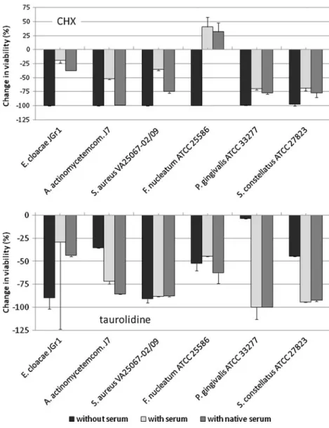
CHX was highly efficient in killing the tested micro-organisms. In mean, the killing rate was more than 99% for all three tested concentrations after 2 h. For taurolidine, a concentration and time-dependent effect was visible. Nev-ertheless, the killing rate of the highest tested concentration of 1.6% was 99.08%±2.27% after 2 h (Fig. 2.). All concentrations of CHX and the highest tested concentration of taurolidine (1.6%) completely erradicated most of the strains after 2 h. Here, taurolidine eliminated all Gram-negative potentially superinfecting strains (Table4). Addi-tion of inactivated serum clearly reduced the killing rate of all selected bacterial strains by CHX; moreover, F. nucleatum ATCC 25586 was obviously stimulated to multiply. Compared to the inactivated serum, native serum increased the killing rates of all species. Serum did not influence clearly the killing ability of taurolidine, except for E. cloacae JGR1, where it was reduced (Fig.3).
SEM photographs
Three Gram-negative and one Gram-positive species were chosen for electron microscopy. Gram-negative bacteria
having an outer membrane seemed to be more damaged by the antimicrobials. After exposure to chlorhexidine, a flow out of substances is clearly visible. In part, bacteria seemed to have burst. The damages were not as obvious after exposure to taurolidine. The surface appeared to be rough; impressions suggest also a flow out of inner particles, but to a much less extent, compared to chlorhexidine. The surface of S. aureus VA25607-02/09 (MRSA) appeared unchanged after exposure of the antimicrobials. An outer membrane is missing in Gram-positives, and the inner membrane is covered by a thick peptidoglycan layer. Thus, possible damages to the inner membrane might not be visible (Fig. 4).
Discussion
The antimicrobial activity of taurolidine has been known for more than 20 years. It was first used in the prevention and treatment of peritonitis [22]. In the present study, we compared taurolidine with chlorhexidine digluconate which
Table 3 Minimal inhibitory concentrations of taurolidine against selected species without, as well as with inactive and native serum Strain MIC/MBC of taurolidine without serum with inactivated serum with native serum
A. actinomycetemcomitans J7 0.0125/0.0125 0.0125/0.0125 <=0.0063/0.0125
F. nucleatum ATCC 25586 0.0125/0.0125 0.0125/0.0125 0.0125/0.0125
P. gingivalis ATCC 33277 0.0125/0.0250 0.0125/0.0250 0.0125/0.0125
S. constellatus ATCC 27823 ≤0.0063/0.0063 ≤0.0063/0.0063 0.0125/0.0125
E. cloacae JGr1 0.0500/0.1000 0.0500/0.1000 0.0500/0.0500
S. aureus MRSA VA25607/2 0.0500/0.0500 0.0250/0.1000 0.0250/0.0250
Fig. 2 Changes in viabilities of microorganisms by different chlorhexidine digluconate and taurolidine concentrations after 1 and 2 h. Reduction of viability is shown. That means,−100% represents a complete elimination. Changes of viability was calculated for each strain separetely; the mean and standard deviation for all 32 investigated strains are presented
is one of the best documented antimicrobial agents in dentistry [23]. The MICs of taurolidine were all below 5% of the normally used concentration of that substance with the exception of Candida albicans ATCC 76615. This confirms the findings from an earlier study which deter-mined MIC values against seven oral species; among them, one F. nucleatum and one Prevotella intermedia strain [24].
Similar to this study [24], lower MIC values were found for chlorhexidine compared to taurolidine.
With the exception of the C. albicans strain which was found being completely resistant to taurolidine, minimal microbiocidal concentrations of that antimicrobial were in mean one to two steps higher than the MICs. Additionally to C. albicans, taurolidine showed only a limited
bacteri-Microbial strains (number) CHX (%) Tauroline (%)
0.005 0.02 0.08 0.1 0.4 1.6 “Real” periodontopathogens Gram-positive bacteria (3) 1 2 3 0 1 1 Gram-negative bacteria (14) 11 14 14 1 6 11 Potentially superinfecting Gram-positive bacteria (9) 4 9 9 0 4 8 Gram-negative bacteria (5) 2 4 4 0 0 5 Candida albicans (1) 1 1 1 0 0 0
Table 4 Number of strains which were totally killed after addition of the antimicrobials for 2 h
Microorganisms (104) were added by taurolidine and CHX in different. After 2 h of incubation, the numbers of viable bacteria were determined by enumeration of colony forming units. The number of strains which were totally killed is listed (detection level, ten microorganisms)
Fig. 3 Influence of serum (inactivated and native) on killing rates of selected bacterial strains by 0.005% chlorhexidine digluconate and 0.1% (P. gingivalis 0.4%) taurolidine after 1 h. Concentrations of the disinfectants have been chosen in relation to the MIC values. Serum was added in a final concentration of 25% v/v. Comparison of native with inactivated serum shows the potential bactericidal effect of complement. Changes of viabil-ity are shown, negative values mean killing efficacy of the compounds, whereas positive values suggest a growth-promoting effect
cidal effect on E. faecalis ATCC 29213, which is known as the test strain for antiseptics; the MBC for that species was 0.4% being 40% of the normal concentration. Thus, the present results confirm earlier MIC and MBC values reported for taurolidine, even a larger difference between MIC and MBC values for E. faecalis [25].
Focusing on the included species, different aspects need to be pointed out. First of all, taurolidine was more active against the “real” periodontopathogens compared to the potentially superinfecting species. This might be attributed to the more or less incomplete activity against the included C. albicans strain. Secondly, taurolidine was found to be similarly active against Gram-positive and Gram-negative bacterial strains. This is in accordance with an earlier study
where aerobic and anaerobic species originated from dentoalveolar infections were tested [26]. Taurolidine is described as an unstable molecule in aqueous solution, while masked formaldehyde is released which inactivates endotoxin [27]. Furthermore, it was suggested that other mechanisms such as interaction with peptidoglycan con-tribute to the antimicrobial action [28]. SEM photographs of Gram-negative species appear to support an interaction with compounds of the cell wall.
In the MIC assays, the chosen highest test concentrations being in the range of about 20% of the concentrations in commercially available products were selected because of the necessity to add nutrient broth and bacterial suspension. This reflects also a general rule in pharmacology for
Fig. 4 Fusobacterium nucleatum ATCC 25586, Porphyromonas gingivalis ATCC 33277, Aggregatibacter actinomycetemcomitans J7, and Staphylococcus aureus (MRSA) VA25607-02/09 (from top to
bottom) after 1 h exposure of 0.08% chlorhexidine (middle) and 1.6% taurolidine (right) as well as a control (left)
antibiotics that MICs should exceed the in vitro concentra-tion by the factors 2 to 8 [29]. In the killing assays, up to 80% of the application concentration was tested. At that concentration, both taurolidine and chlorhexidine reduced the viability of the microorganisms by more than 99% after 2 h in general. Most of the used microbial strains were completely killed. But taurolidine exerted a clear concentration and time-dependent effect. Killing was enhanced after 2 h. At a lower concentration of taurolidine, the antimicrobial activity was often completely blocked. Based on the findings, it may be anticipated that a sufficient high concentration of taurolidine needs to be ensured for several hours, while a dilution of taurolidine, e.g., by the flow of gingival crevicular fluid should be avoided as much as possible. Thus, from a clinical point of view, the choice of an optimal carrier device, ensuring a long-lasting, sufficiently high concentration of the active substance, appears to be a key factor, which may significantly affect the therapeutic use. In the treatment of periodontal infections, the topical application of antibiotics incorporated in various types of controlled released devices, i.e., such as gels [30, 31] or microspheres [32] represents an important therapeutic modality.
Gingival crevicular fluid contains up to 35% of the albumin found in serum [33]. After non-surgical and surgical periodontal treatment, serum-rich wound fluid is produced. Therefore, the activity of antimicrobials in the presence of serum plays an important role. Our results clearly indicate that taurolidine was active in a serum-rich environment, while on the contrary, the efficacy of chlorhexidine was dramatically decreased. This finding may suggest that taurolidine may be also applied in a serum-rich environment, e.g., after periodontal surgery. An enhanced bactericidal activity of taurolidine in the presence of serum as described before [34] was not always found. This might be associated with a resistance of many periodontopathogens against killing by complement [35, 36]. In killing assays, both the potential inhibiting effect of serum proteins and the complement activity are combined with the efficacy of the antimicrobials. In the present study, only selected strains were tested, and it can be suggested that the complement effect does not overcome an inhibition by the other serum compounds.
Serum may also be an important component of subgin-gival plaque being a biofilm. The special conditions of biofilms were not addressed in this study. An earlier made study found a plaque-growth reducing effect of taurolidine which was less pronounced in comparison to chlorhexidine [37]. Recently, experiments in healthy volunteers have shown a decreased viability in supragingival plaque after 2 min of rinsing with taurolidine; again, the potential antimicrobial effect was not as high as after rinsing with chlorhexidine [38]. In that study, the bactericidal effect was determined immediately after the 2-min exposure.
Con-cluding from our results showing a time-dependent antimi-crobial effect of taurolidine at a later time-point differences might be less remarkable between the two antimicrobials. A depot effect can be suggested for taurolidine, as a lock solution, prevents successful infections in catheters [39]. Biofilms are organized microbial communities surrounded by a biopolymer matrix and characterized by slow growth, increasing mutation frequency, and maximum tolerance to antibiotics [40]; the special conditions of biofilms should be addressed in subsequent studies testing taurolidine.
Pathogenesis of periodontitis comprises the microbial challenge and the host response with an inflammatory and immune response directed to cope with the challenge. The host response results in production of many proinflamma-tory cytokines involved in inflammation and bone loss [41]. It is well-established that periodontopathic bacteria are able to attach and to invade epithelial cells [42,43], while polymorphonuclear neutrophils interacting with periodontopathic bacteria release reactive oxidative spe-cies [44], which in turn damage the soft and hard periodontal tissues. Taurolidine has been demonstrated to inhibit adhesion of Escherichia coli, Staphylococcus saprophyticus, and C. albicans to mucosal epithelial cells [3] and of Gram-negative species to plastic surfaces [39]. Taurolidine derivatives decrease production of proinflam-matory cytokines and oxidants [45]. Thus, the positive immunomodulatory properties of taurolidine might be of importance in periodontitis and should be analyzed in further in vitro and in vivo studies.
Taken together, the present findings suggest that: (a) taurolidine possesses antimicrobial properties which are not reduced in the presence of serum which is a main component in gingival crevicular and wound fluid and (b) the demonstrated in vitro antimicrobial properties of taurolidine warrant further evaluation in non-surgical and surgical periodontal therapy.
Acknowledgments The authors are grateful to Regula Hirschi and Marianne Weibel for their excellent assistance in performing the in vitro assays. We are indebted to Richard Miron (University of Bern) for the language corrections. This work was funded by Geistlich Pharma AG, Wolhusen, Switzerland.
Conflict of interest The authors declare that they have no conflicts of interest.
References
1. Schuller-Levis GB, Park E (2003) Taurine: new implications for an old amino acid. FEMS Microbiol Lett 226:195–202
2. Engel JM, Muhling J, Weiss S, Karcher B, Lohr T, Menges T, Little S, Hempelmann G (2006) Relationship of taurine and other amino acids in plasma and in neutrophils of septic trauma patients. Amino Acids 30:87–94
3. Gorman SP, McCafferty DF, Woolfson AD, Jones DS (1987) Reduced adherence of micro-organisms to human mucosal epithelial cells following treatment with Taurolin, a novel antimicrobial agent. J Appl Bacteriol 62:315–320
4. Bedrosian I, Sofia RD, Wolff SM, Dinarello CA (1991) Taurolidine, an analogue of the amino acid taurine, suppresses interleukin 1 and tumor necrosis factor synthesis in human peripheral blood mononuclear cells. Cytokine 3:568–575 5. Schuller-Levis G, Mehta PD, Rudelli R, Sturman J (1990)
Immunologic consequences of taurine deficiency in cats. J Leukoc Biol 47:321–331
6. Gordon RE, Heller RF (1992) Taurine protection of lungs in hamster models of oxidant injury: a morphologic time study of paraquat and bleomycin treatment. Adv Exp Med Biol 315:319– 328
7. Brearley S, George RH (1980) The rate of antimicrobial action of noxythiolin and taurolin. J Hosp Infect 1:201–209
8. Gong L, Greenberg HE, Perhach JL, Waldman SA, Kraft WK (2007) The pharmacokinetics of taurolidine metabolites in healthy volunteers. J Clin Pharmacol 47:697–703
9. Rohner E, Kolar P, Seeger JB, Arnholdt J, Thiele K, Perka C, Matziolis G (2011) Toxicity of antiseptics against chondrocytes: what is best for the cartilage in septic joint surgery? Int Orthop (in press)
10. Bradshaw JH, Puntis JW (2008) Taurolidine and catheter-related bloodstream infection: a systematic review of the literature. J Pediatr Gastroenterol Nutr 47:179–186
11. Burri C (1990) Treatment of local infection with taurolin. Aktuelle Probl Chir Orthop 34:78–84
12. Jacobi CA, Menenakos C, Braumann C (2005) Taurolidine—a new drug with anti-tumor and anti-angiogenic effects. Anticancer Drugs 16:917–921
13. De Soete M, Mongardini C, Peuwels M, Haffajee A, Socransky S, van Steenberghe D, Quirynen M (2001) One-stage full-mouth disinfection. Long-term microbiological results analyzed by checkerboard DNA-DNA hybridization. J Periodontol 72:374– 382
14. Cosyn J, Sabzevar MM (2007) Subgingival chlorhexidine varnish administration as an adjunct to same-day full-mouth root planing. II. Microbiological observations. J Periodontol 78:438–445 15. Cosyn J, Wyn I, De Rouck T, Sabzevar MM (2007) Subgingival
chlorhexidine varnish administration as an adjunct to same-day full-mouth root planing. I. Clinical observations. J Periodontol 78:430–437
16. Hidalgo E, Dominguez C (2001) Mechanisms underlying chlorhexidine-induced cytotoxicity. Toxicol In Vitro 15:271– 276
17. Cabral CT, Fernandes MH (2007) In vitro comparison of chlorhexidine and povidone-iodine on the long-term proliferation and functional activity of human alveolar bone cells. Clin Oral Investig 11:155–164
18. Oosterwaal PJ, Mikx FH, van den Brink ME, Renggli HH (1989) Bactericidal concentrations of chlorhexidine-digluconate, amine fluoride gel and stannous fluoride gel for subgingival bacteria tested in serum at short contact times. J Periodontal Res 24:155– 160
19. Feres M, Haffajee AD, Allard K, Som S, Goodson JM, Socransky SS (2002) Antibiotic resistance of subgingival species during and after antibiotic therapy. J Clin Periodontol 29:724–735
20. CLSI (2007) Clinical and Laboratory Standards Institute (formerly NCCLS). Methods for antimicrobial susceptibility testing of anaerobic bacteria; Approved standard – seventh edition. CLSI document 27:M11-A7
21. CLSI (2009) Clinical and Laboratory Standards Institute (formerly NCCLS). Performance standards for antimicrobial disk suscepti-bility tests, 5th ed. CLSI document 29:M02-A9
22. Linder MM, Ott W, Wesch G, Wicki O, Marti MC, Moser G (1981) Therapy of purulent peritonitis. Documentation of 78 cases and experience with taurolin (author's transl). Langenbecks Arch Chir 353:241–250
23. Baehni PC, Takeuchi Y (2003) Anti-plaque agents in the prevention of biofilm-associated oral diseases. Oral Dis 9(Suppl 1):23–29
24. Reynolds S, Moran J, Addy M, Wade WG, Newcombe R (1991) Taurolin as an oral rinse. I. Antimicrobial effects in vitro and in vivo. Clin Prev Dent 13:13–17
25. Shah CB, Mittelman MW, Costerton JW, Parenteau S, Pelak M, Arsenault R, Mermel LA (2002) Antimicrobial activity of a novel catheter lock solution. Antimicrob Agents Chemother 46:1674– 1679
26. Zimmermann M, Preac-Mursic V (1993) The antimicrobial actions of taurolin and other preparations on the pathogenic spectrum in dentoalveolar infections. Int J Clin Pharmacol Ther Toxicol 31:130–136
27. Gidley MJ, Sanders JK, Myers ER, Allwood MC (1981) The mode of antibacterial action of some 'masked' formaldehyde compounds. FEBS Lett 127:225–227
28. Caruso F, Darnowski JW, Opazo C, Goldberg A, Kishore N, Agoston ES, Rossi M (2010) Taurolidine antiadhesive properties on interaction with E. coli; its transformation in biological environment and interaction with bacteria cell wall. PLoS ONE 5:e8927
29. Newman MG, van Winkelhoff AJ (2001) Antibiotic and antimi-crobial use in dental practice.Second edition. Quintessence Publishing Co, Inc, Chicago, Berlin, London, Tokyo, Paris, Barcelona, Sao Paulo, Moscow, Prague, and Warsaw
30. Pradeep AR, Sagar SV, Daisy H (2008) Clinical and microbi-ologic effects of subgingivally delivered 0.5% azithromycin in the treatment of chronic periodontitis. J Periodontol 79:2125– 2135
31. Stoller NH, Johnson LR, Trapnell S, Harrold CQ, Garrett S (1998) The pharmacokinetic profile of a biodegradable controlled-release delivery system containing doxycycline compared to systemically delivered doxycycline in gingival crevicular fluid, saliva, and serum. J Periodontol 69:1085–1091
32. Van Dyke TE, Offenbacher S, Braswell L, Lessem J (2002) Enhancing the value of scaling and root-planing: arestin clinical trial results. J Int Acad Periodontol 4:72–76
33. Tew JG, Marshall DR, Burmeister JA, Ranney RR (1985) Relationship between gingival crevicular fluid and serum anti-body titers in young adults with generalized and localized periodontitis. Infect Immun 49:487–493
34. Blenkharn JI (1990) In-vitro antibacterial activity of noxythiolin and taurolidine. J Pharm Pharmacol 42:589–590
35. Potempa M, Potempa J, Kantyka T, Nguyen KA, Wawrzonek K, Manandhar SP, Popadiak K, Riesbeck K, Eick S, Blom AM (2009) Interpain A, a cysteine proteinase from Prevotella intermedia, inhibits complement by degrading complement factor C3. PLoS Pathog 5:e1000316
36. Potempa M, Potempa J, Okroj M, Popadiak K, Eick S, Nguyen KA, Riesbeck K, Blom AM (2008) Binding of complement inhibitor C4b-binding protein contributes to serum resistance of Porphyromonas gingivalis. J Immunol 181:5537–5544
37. Reynolds S, Moran J, Wade WG, Addy M, Newcombe R (1991) Taurolin as an oral rinse. II. Effects on in vitro and in vivo plaque regrowth. Clin Prev Dent 13:18–22
38. Arweiler NB, Auschill TM, Sculean A (2011) Antibacterial effect of taurolidine (2%) on established dental plaque biofilm. Clin Oral Investig (in press)
39. Solomon LR, Cheesbrough JS, Ebah L, Al-Sayed T, Heap M, Millband N, Waterhouse D, Mitra S, Curry A, Saxena R, Bhat R,
Schulz M, Diggle P (2010) A randomized double-blind controlled trial of taurolidine-citrate catheter locks for the prevention of bacteremia in patients treated with hemodialysis. Am J Kidney Dis 55:1060–1068
40. Hoiby N, Bjarnsholt T, Givskov M, Molin S, Ciofu O (2010) Antibiotic resistance of bacterial biofilms. Int J Antimicrob Agents 35:322–332
41. Cochran DL (2008) Inflammation and bone loss in periodontal disease. J Periodontol 79:1569–1576
42. Lamont RJ, Chan A, Belton CM, Izutsu KT, Vasel D, Weinberg A (1995) Porphyromonas gingivalis invasion of gingival epithelial cells. Infect Immun 63:3878–3885
43. Eick S, Rodel J, Einax JW, Pfister W (2002) Interaction of Porphyromonas gingivalis with KB cells: comparison of different clinical isolates. Oral Microbiol Immunol 17:201–208
44. Guentsch A, Puklo M, Preshaw PM, Glockmann E, Pfister W, Potempa J, Eick S (2009) Neutrophils in chronic and aggressive periodontitis in interaction with Porphyromonas gingivalis and Aggregatibacter actinomycetemcomitans. J Periodontal Res 44:368–377
45. Marcinkiewicz J, Kurnyta M, Biedron R, Bobek M, Kontny E, Maslinski W (2006) Anti-inflammatory effects of taurine derivatives (taurine chloramine, taurine bromamine, and taurolidine) are medi-ated by different mechanisms. Adv Exp Med Biol 583:481–492