Preparation of Surfactant-free Nanoparticles of Methacrylic Acid Copolymers
Used for Film Coating
Submitted: July 12, 2005; Accepted: May 2, 2006; Published: July 28, 2006
Cung An Nguyen,1Yvette Niamien Konan-Kouakou,1,2 Eric Allémann,1,3 Eric Doelker,1 David Quintanar-Guerrero,4Hatem Fessi,5 and Robert Gurny1
1
School of Pharmaceutical Sciences, University of Geneva, University of Lausanne, Department of Pharmaceutics and Biopharmaceutics, 1211 Geneva 4, Switzerland
2
Present address: Swiss Federal Institute of Technology (EPFL), CH-1015 Lausanne, Switzerland
3Present address: Bracco Research SA, CH-1228 Plan-les-Ouates, Switzerland 4
Depto de postgrado en farmácia, FES-Cuautitlán-UNAM, CP 54704, México
5
Claude Bernard University Lyon I, 69622 Villeurbanne Cedex, France
ABSTRACT
The aim of the present study was to prepare surfactant-free pseudolatexes of various methacrylic acid copolymers. These aqueous colloidal dispersions of polymeric materials for oral administration are intended for film coating of solid dosage forms or for direct manufacturing of nanoparticles. Nano-particulate dispersions were produced by an emulsification-diffusion method involving the use of partially water-miscible solvents and the mutual saturation of the aqueous and organic phases prior to the emulsification in order to reduce the initial thermodynamic instability of the emulsion. Because of the self-emulsifying properties of the methacrylic acid copoly-mers, it was possible to prepare aqueous dispersions of col-loidal size containing up to 30% wt/vol of Eudragit RL, RS, and E using 2-butanone or methyl acetate as partially water-miscible solvents, but without any surfactant. However, in the case of the cationic Eudragit E, protonation of the tertiary amine groups by acidification of the aqueous phase was nec-essary to improve the emulsion stability in the absence of surfactant and subsequently to prevent droplet coalescence during evaporation. In addition, a pseudolatex of Eudragit E was used to validate the coating properties of the formulation for solid dosage forms. Film-coated tablets of quinidine sul-fate showed a transparent glossy continuous film that was firmly attached to the tablet. The dissolution profile of quini-dine sulfate from the tablets coated with the Eudragit E pseu-dolatex was comparable to that of tablets coated with an acetonic solution of Eudragit E. Furthermore, both types of coating ensured similar taste masking. The emulsification-evaporation method used was shown to be appropriate for the preparation of surfactant-free colloidal dispersions of the 3 types of preformed methacrylic acid copolymers; the
dispersions can subsequently be used for film coating of solid dosage forms.
KEYWORDS: Pseudolatex, methacrylic acid copolymers, Eudragit, emulsification-diffusion method, nanoparticles, film coating, quinidine sulfateR
INTRODUCTION
The first methods used to produce nanoparticles were de-veloped by polymer chemists in the field of latex engineer-ing and were later adapted for pharmaceutical applications. Nanoparticles are prepared by several techniques involving either in situ polymerization of monomers (latex) or disper-sion of preformed polymers (pseudolatex or artificial latex). Methods using well-characterized polymers may be pre-ferred, given the presence of toxic residues (namely un-reacted monomer, initiator, and emulsifier molecules) in the polymerization reactions.1,2The emulsification-evaporation method for pharmaceutical use was proposed first by Gurny et al3 and is based on a patent by Vanderhoff et al4 where a polymer is dissolved in a volatile water-immiscible organic solvent. The organic solution is dispersed in an aqueous phase containing an emulsifier and easily forms an oil-in-water (o/w) emulsion. However, the addition of sta-bilizers is often required to avoid the formation of polymer flakes. Continuous emulsification under stirring prevents coa-lescence of the organic droplets and can be further improved by sonication or microfluidization.1,5-8 The emulsification-evaporation method is interesting for many reasons: the use of pharmaceutically acceptable organic solvents, high yields, good reproducibility, and easy scaling up. A fur-ther step has been achieved with the development of the emulsification-diffusion method described by Leroux et al9 and Quintanar-Guerrero et al.10,11 This process involves (1) the mutual saturation of the organic and the aqueous phases prior to the emulsification, (2) the dispersion of a partially water-miscible solvent with the dissolved polymer Corresponding Author: Robert Gurny, University of
Geneva, School of Pharmaceutical Sciences, Quai Ernest-Ansermet 30, 1211 Geneva 4, Switzerland. Tel: + 41 22 379 61 46; Fax: + 41 22 379 65 67; E-mail: robert.gurny@ pharm.unige.ch
into an aqueous phase containing a stabilizer, and (3) the addition of a large amount of pure water that provokes the diffusion of the solvent and the aggregation of the polymer as nanoparticles.10 With this method, it is necessary to remove a considerable amount of water to obtain a high polymer concentration.5
It is, however, worth noting that both methods use a surfac-tant in order to emulsify and prevent the aggregation during the diffusion process. In this respect, it is of utmost interest to develop preparation procedures that avoid surfactants. The aim of the present study was to use the emulsification-diffusion technique to produce surfactant-free nanoparticles of methacrylic acid copolymers (Eudragit RL, RS, and E) in a high concentration for film coating. The influence of some process parameters on the nanoparticle size, such as the type of polymer and its concentration in the organic phase, the stirring rate, the pH, and the type and concen-tration of stabilizing agents, has been investigated. A pseu-dolatex of Eudragit E prepared with the proposed method was used for the coating of solid dosage forms to assess the film-forming properties and to verify the surface morphol-ogy of tablets by scanning electron microscopy.
MATERIALS AND METHODS
Materials
Quinidine sulfate was purchased from Hanseler (Herisau, Switzerland). Eudragit E, RL, and RS with an average mo-lecular weight of 150 kDa were a gift from Degussa (Darm-stadt, Germany). Eudragit E is a cationic copolymer based on dimethylaminoethyl methacrylate and neutral meth-acrylic esters. Eudragit RS and RL are copolymers of meth-acrylic and methacrylic acid esters containing between 4.5% and 6.8%, and 8.8% and 12%, of quaternary ammonium groups, respectively. The chemical structures of methacrylate co-polymers used are shown in Figure 1. In the case of
Eu-dragit E pseudolatex, 2 nonionic stabilizing agents were tested: poloxamers 188 and 407 (BASF, Ludwigshafen, Ger-many). Two partially water-miscible solvents of analytical grade, namely 2-butanone (water solubility = 275 mg/mL, boiling point 80-C, ρ = 0.80 g/mL) and methyl acetate (water solubility = 280 mg/mL, bp 57-C, ρ = 0.93 g/mL, experimental determinations), were used as solvents for the Eudragit grades and purchased from Fluka (Buchs, Switzerland). Acetic acid of analytical grade was added to adjust the pH in the aqueous phase in the emulsion, and tributyl citrate was used as a plasticizer for the coating of tablets. Other chemicals were of analytical grade and used as received.
Methods
Pseudolatex Preparation
Nanoparticles were produced according to the emulsification-diffusion method described previously,5,12where 2-butanone (or methyl acetate) and water were mutually saturated be-fore use to ensure the initial thermodynamic equilibrium of both liquids. Typically, 4 g of Eudragit were dissolved in 20 mL of the water-saturated 2-butanone. This organic so-lution was emulsified with 40 mL of 2-butanone-saturated aqueous solution under mechanical stirring at 2000 rpm for 15 minutes. Batches of Eudragit E emulsions were pre-pared using either decreasing amounts of poloxamer as a stabilizer or acetic acid to reduce the pH of the aqueous phase prior to the emulsification to improve the stability of the emulsion. Extraction of the solvent from the nanodrop-lets was achieved by evaporation at 40-C in a rotary evap-orator under reduced pressure (200 mmHg) for 15 minutes, leading to the formation of nanoparticles by precipitation of the macromolecules. The recovered solvent was reused to prepare new batches, except for those with acetic acid. The mean particle size and polydispersity index (PI) of the nanoparticles were determined 3 times by photon correla-tion spectroscopy using a Zetasizer 5000 (Malvern, Worces-tershire, UK). Using the same instrument mounted with an electrophoretic cell, the zeta potential (ζ) of Eudragit nanoparticles was determined from the electrophoretic mo-bility measurements at 25-C. Scanning electron micros-copy (SEM) (JEOL, Tokyo, Japan) was used to examine the surface of the nanoparticles. Samples were prepared on metallic studs and coated with a gold film (20 nm).
Interfacial Tension Determination
The interfacial tension at the solvent-water interface was determined by the Du Noüy method using a digital-tensiometer K10 (Krüss, Hamburg, Germany) with a plati-num ring. The temperature was kept constant at 20-C by circulating thermostated water through a jacketed vessel
containing the sample. Each determination was repeated in triplicate, and the average results were reported. The experimental uncertainty of interfacial tension determina-tion was ~0.1 mN/m.
Preparation and Characterization of Coated Tablets Convex tablets of 10 mm in diameter (curvature radius 8.5 mm) containing 15 mg of quinidine sulfate were pre-pared. The concentration of Eudragit E in the pseudolatex (80 mL) was adjusted to 10% (wt/vol). This suspension was used as a coating dispersion in a Hi-Coater HCT-20 Mini (Gebrüder Lödige, Paderborn, Germany) equipped with a Gilson Minipuls 3 pump (Villiers-le-Bel, France). Tributyl citrate (0.8 g) was added as a plasticizer to the pseudolatex 10 minutes prior to the coating under magnetic stirring. Details of the operating conditions appear in Table 1. A second batch of tablets was coated using the same pro-cedure with a 10% (wt/vol) acetonic solution of Eudragit E added with 1% (wt/vol) tributyl citrate. The operating con-ditions were identical to the suspension, except for the dry-ing air temperature, which was lowered to 40-C.
Coated tablets were examined for their mean disintegration time and their dissolution rate. The disintegration time was determined according to the European Pharmacopeia with HCl 0.1M. The dissolution test was performed with 6 in-dividual tablets in 900 mL of HCl 0.1M at 37-C using the European Pharmacopoeia paddle apparatus at 50 rpm. The amount of quinidine sulfate dissolved was measured spectrophotometrically at 251 nm using an automated sam-pling system (Hewlett Packard 8453). Coated tablets were evaluated with regard to duration of taste-masking time by a group of 6 volunteers selected according to the European Pharmacopeia for their sensitivity to bitterness using qui-nine hydrochloride solutions. Tablets were kept immobile in the oral cavity until dissolution of the coating and sub-sequent bitterness perception. Tests were performed in
trip-licate. The surface morphology and the film thickness of the coated tablets were examined by SEM.
RESULTS AND DISCUSSION
The proposed emulsification-diffusion method without emul-sifier enabled the preparation of nanoparticles of Eudragit RL, RS, and E at high concentrations of 20% (wt/vol) with 2-butanone or methyl acetate as partially water-miscible solvents (Table 2). The particle size data suggest that it is possible to form pseudolatexes from a conventional o/w emulsion without emulsifier by a simple extraction of the solvent from the nanodroplets. The displacement of the solvent in a rotary evaporator under reduced pressure led to the in situ precipitation of the polymer and the formation of stable nanoparticles. In the case of Eudragit E, particles of colloidal size free of stabilizer were obtained only by lowering the pH to obviate some solubility of the polymer in the aqueous phase.
The preparation of a stable emulsion is crucial in order to avoid the coalescence of nanodroplets that occurs during the emulsification and evaporation step. In particular, spe-cific requirements must be satisfied, such as a small differ-ence in density between the dispersed and the continuous phase, a high viscosity of the continuous phase, a low in-terfacial tension, the existence of an electrical double layer at the oil/water interface according to the Derjaguin-Landau-Verwey-Overbeek (DLVO) theory, and the mechanical strength of the adsorbed layer at the oil/water interface.13 In these experiments, the density and the viscosity could not account for the emulsion stability. Therefore, a low inter-facial tension, the electrical double layer, and the mechanical strength of the adsorbed layer may be the most important parameters to increase the stability of the emulsion.
Role of the Interfacial Tension
From the photon correlation spectroscopy measurements, the size of nanoparticles of Eudragit RL prepared using 2-butanone and methyl acetate were, respectively, 66 and 75 nm, with a PI smaller than 0.14, indicating a narrow unimodal pattern (Table 2). With Eudragit RS, the aver-age sizes were 121 nm (2-butanone) and 127 nm (methyl acetate), with a PI around 0.17. These data are supported by the interfacial tension values, which were always less than 1 mN/m (1.8 mN/m for the water–methyl acetate sys-tem), indicating that using partially miscible water solvents actually decreases the interfacial tension. For comparison, the interfacial tension between a poorly soluble solvent such as chloroform (7 mg/mL) and water was 33 mN/m. As a result, the preparation of Eudragit RL pseudolatex using chloroform as a solvent was not possible and led to a mas-sive aggregation of particles (Table 2). Saito et al13 and
Table 1. Operating Conditions for the HCT-20 Mini
Tablet Charge 350 g of 10-mm Convex
Tablets Containing 15 mg of Quinidine Sulfate Each Eudragit E pseudolatex
20% (wt/vol)
80 mL
Pan rotation 10 rpm
Preheating time 5 minutes
Drying air temperature 55-60-C
Feed, and spraying air pressure 2 mL/min at 0.8 kg/cm2
Exhaust air 33-35-C
Final drying time 5 minutes
Coating time 50 minutes
recently Chernysheva et al14 have confirmed that lowering the interfacial tension is effective for increasing the o/w emulsion stability.
In the same experimental conditions, when water and methyl acetate were not mutually saturated prior to the emulsifica-tion, 2 populations of nanoparticles (with modes of 360 and 120 nm, total average size = 134 nm, PI = 0.248) were observed in the pseudolatex of Eudragit RS (see batch 5 in Table 2). This indicated that saturating the dispersed and the external phases can decrease the initial thermodynamic instability when both phases are mixed together.
Role of the Electrical Double Layer
The presence of Eudragit RS or RL in the solvent reduced the interfacial tension values below the limit of determi-nation (1 mN/m) of the tensiometer. The low interfacial ten-sion may be explained by the formation of electrical double layers of ions and charged adsorption layers by Eudragit quaternary ammonium groups. The hydrophilic and lipo-philic moieties of Eudragit RS or RL caused the polymer chains to orient themselves at the interface between the or-ganic phase and the aqueous phase.
From the o/w emulsion standpoint, polycationic copoly-mers such as Eudragit RS and RL may provide even more stability. Because of the quaternary ammonium groups of the macromolecules, the oil droplets are covered with a positively charged surface, repelling each other and further
stabilizing the emulsion. Zeta potential (ζ ) determinations confirmed that Eudragit RL and RS nanoparticles were positively charged, with values of 64.5 ± 2.4 mV and 57.1 ± 2.2 mV, respectively. The difference in size of nanoparticles of Eudragit RL (66 and 75 nm) and Eudragit RS (121 and 127 nm) may be explained by the higher density of qua-ternary ammonium groups in Eudragit RL. These results are in accordance with Lehmann et al,15 who have established excellent self-emulsifying properties of Eudragit RL and RS, recently confirmed by the work of Chernysheva et al.14
Role of the Adsorbed Layer
Tertiary amine groups in Eudragit E preferably are ori-ented at the liquid-liquid interface. This can provide the polymer with some hydrophilic character (Figure 1). Fur-thermore, it was possible to obtain homogeneous emulsions of Eudragit E, but the droplets coalesced during the evap-oration step, because of insufficient stabilization. The pseu-dolatexes obtained showed dispersed microparticles of around 2.2 µm (PI = 0.381). Polymer flakes were also visible on the surface of these batches (Table 2). Increasing the stirring rate from 2000 rpm to 5000 rpm decreased neither the size of particles nor the PI. The ζ value of these microparticles was as low as 18.5 ± 2.3 mV, indicating that the electrical repulsion played a minor role in the stabilization of the emulsion. It was concluded that the mechanism involved was mainly the steric repulsion, which acts by an excess of polymer chains and the elastic deformation. First, polymer
Table 2. Eudragit Pseudolatexes Produced* Batch
No Eudragit Type Solvent
Acetic Acid (mL) (pH Measured) Stabilizer (% wt/vol) Mean Size (nm) ± SD† Polydispersity Index Aggregation‡ 1 RL 2-Butanone — 0 66 ± 9 0.133 – 2 RL MeOAc — 0 75 ± 11 0.139 – 3 RS 2-Butanone — 0 121 ± 10 0.161 – 4 RS MeOAc — 0 127 ± 12 0.176 – 5§ RS MeOAc — 0 134 ± 15 0.248 – 6 E 2-Butanone — 0 2240 ± 112 0.381 + 7║ E 2-Butanone — 0 2160 ± 141 0.355 + 8 E 2-Butanone — F-68 (1.0) 399 ± 35 0.202 + 9 E 2-Butanone — F-127 (1.0) 304 ± 17 0.149 – 10 E 2-Butanone — F-127 (0.5) 366 ± 22 0.196 + 11 E 2-Butanone 0.5 (pH = 6.3) 0 279 ± 17 0.208 + 12 E 2-Butanone 1 (pH = 5.0) 0 245 ± 8 0.142 – 13 E (10%) 2-Butanone 1 (pH = 5.0) 0 241 ± 7 0.139 – 14 E (30%) 2-Butanone 1.08 (pH = 5.0) 0 353 ± 15 0.251 – 15 E (40%) 2-Butanone 1.15 (pH = 5.0) 0 1350 ± 55 0.307 – 16 E MeOAc 1 (pH = 5.0) 0 256 ± 6 0.167 – 17 RL Chloroform — 0 — — +++
*Organic phase: 20 mL (polymer 20% wt/vol); aqueous phase: 40 mL; stirring rate: 2000 rpm. MeOAc indicates methyl acetate. †n = 3.
‡Polymer aggregation on the surface after evaporation: (–) not observed, (+) observed, and (+++) massive. §No mutual saturation of internal and external phases prior to emulsification.
chains dissolved in droplets may present their hydrophilic moieties at the interface of both phases; when 2 particles approach each other the polymer layers start to overlap. This leads to a local decrease in water concentration and sets up an osmotic pressure gradient. Water flows in to dilute the concentrated polymer, and the particles move apart. Second, the polymer layer can also act as an integral mechanical strength, which tends to repel an approaching particle by the elastic deformation of the droplets. The stabilization of the emulsion by the steric repulsion may be reduced dur-ing the evaporation step. As the volume of solvent decreases in the droplets, the polymer concentration increases, fol-lowed by an increase in viscosity. Consequently, the elas-tic deformation of droplets is reduced, leading to polymer aggregation.
It was necessary to use poloxamer 407 at a minimum con-centration of 1% (wt/vol) in the aqueous phase to obtain homogeneously dispersed colloidal particles of Eudragit E. Nanoparticles obtained with this emulsifier were around 300 nm (PI = 0.149). Decreasing the concentration of po-loxamer 407 to 0.5% (wt/vol) or using popo-loxamer 188 even at 1.0% (wt/vol) was less effective in stabilizing the emul-sion. The benefits of poloxamer as an emulsifier are the reduction of the interfacial tension at the droplet surface and the mechanical and steric stabilization by short-range repulsion between droplets. In addition, the nonadsorbed chains of poloxamer can increase the continuous phase vis-cosity, allowing the disruption kinetics to smaller droplets and promoting the hydrodynamic stabilization.16,17 These actions prevent the collision and coalescence of nano-droplets during the emulsification and evaporation steps.
Protonation of Eudragit E
Hydrophilic regions allowed the Eudragit E chains to be oriented at the water-solvent interface of droplets. Despite the adsorption of polymer chains and the consecutive decrease
in the interfacial tension, the steric repulsion mechanism was reduced during the evaporation step, leading to particles of over 1 μm and polymer flakes. From pseudolatexes of Eudragit RL or RS, it was concluded that polymers with charged groups such as quaternary ammonium moieties were able to cover droplets with a positively charged sur-face, increasing the stability of the emulsion via electrical repulsion.
It was possible to produce nanoparticles of Eudragit E without surfactant by lowering the pH of the aqueous phase to 5.0 with acetic acid. The sizes were 245 nm (2-butanone, PI = 0.142) and 256 nm (methyl acetate, PI = 0.167). As the pH was adjusted to 6.3, nanoparticles around 280 nm with an increased PI were obtained. Polymer flakes were also observed with this batch (Table 2). It was concluded that lowering the pH in the aqueous phase could protonate the tertiary amine groups of Eudragit E on the surface of droplets. This could further stabilize the emulsion with the increase of the electrical repulsion mechanism. At a pH of 6.3, some tertiary amine groups were protonated, but the corresponding density of charged groups on the surface of droplets was insufficient for a good stabilization of the emulsion. In addition, the use of acetic acid was advanta-geous, because it could be removed during the evaporation of the solvent in the rotary evaporator.
Under the same optimal conditions (pH = 5.0), the in-fluence of Eudragit E concentration in the dispersed phase on particle size was also evaluated. At a concentration of 10% (polymer to 2-butanone), the nanoparticles obtained were similar in size and PI to those previously prepared (20% wt/vol). With a concentration of 30% (wt/vol) of poly-mer, the nanoparticles obtained were significantly larger, with an increased PI (Table 2). At 40% wt/vol, particle size was drastically increased to the micrometer range, and 2 populations of particles were observed using photon cor-relation spectroscopy.
Evaluation of the Coated Tablets
A sufficient amount of pseudolatex of Eudragit E (80 mL) was prepared under the same experimental conditions as for batch 12 (Table 2). This suspension was used in a Hi-Coater HCT-20 Mini to verify its coating properties. The concentration of the polymer was adjusted to 10% (wt/vol) in the pseudolatex, and 0.8 g of tributyl citrate was added as a plasticizer. Details of the operating conditions appear in Table 1.
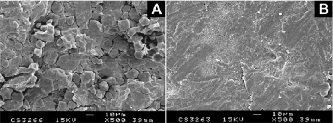
The coated tablets had a transparent glossy continuous film, as confirmed by SEM (Figure 2B). The surface of the film, which was firmly attached to the tablet, was regular, and pores were not visible, unlike the surface of uncoated tab-lets (Figure 2A). The thickness of the film during the coating process was evaluated from micrographs of the cross-sectional area of tablets and was found to be 27.1 ± 2.9 µm on the final tablets. That film corresponded to a coating weight of 2.8 mg/cm2. The mean disintegration time of tablets was 2.3 ± 0.6 minutes, while that for uncoated tab-lets was 1.4 ± 0.7 minutes. At an equivalent coating weight, the dissolution profile of quinidine sulfate from tablets coated with the pseudolatex of Eudragit E was comparable to that of the organic solution of Eudragit E (Figure 3). As expected for an aqueous suspension, the coating made of pseudolatex showed a slightly faster release in the early period than it did later. This demonstrates the value of pseudolatex of Eudragit E as a coating formulation. Fi-nally, coated tablets were evaluated with regard to dura-tion of taste-masking time. In both cases, no bitterness perception of quinidine sulfate was detected over a period of 15 minutes. The average time of taste masking was 22.8 ± 5.4 minutes for the pseudolatex coating and 24.5 ± 4.2 minutes for the organic coating, respectively.
CONCLUSION
Through the use of a solvent partially miscible with water in the emulsification-diffusion method, it has been possible to prepare pseudolatexes of Eudragit RL, RS, and E with-out any additive. The stabilizing parameters of the emul-sion are the low interfacial tenemul-sion, the electrical double layer, and the mechanical strength of the adsorbed layer. In addition, the preliminary mutual saturation of the aqueous and organic phases is necessary to reduce the initial ther-modynamic instability of the emulsion.
As expected for pseudolatexes, the particle size of the colloidal dispersions produced here was higher than that of the so-called true latexes obtained by emulsion polymer-ization of acrylic monomers, but there was no detrimental effect on the film-forming properties, as shown for a Eu-dragit E pseudolatex with quinidine sulfate tablets. In con-trast, the main advantage of preparing colloidal dispersions from preformed methacrylic acid copolymers is the absence of surfactant, which is generally present in true latexes.
ACKNOWLEDGMENTS
The authors would like to thank Dr. Sergio Galindo-Rodriguez for helpful discussions.
REFERENCES
1. De Jaeghere F, Doelker E, Gurny R. Nanoparticles. In: Mathiowitz E, ed. The Encyclopedia of Controlled Drug Delivery. New York, NY: Wiley and Sons Inc; 1999:641Y664.
2. Couvreur P, Dubernet C, Puisieux F. Controlled drug delivery with nanoparticles: current possibilities and future trends. Eur J Pharm Biopharm. 1995;41:2Y13.
3. Gurny R, Peppas NA, Harrington DD, Banker GS. Development of biodegradable and injectable latices for controlled release of potent drugs. Drug Dev Ind Pharm. 1981;7:1Y25.
4. Vanderhoff JW, El-Aasser MS, Ugelstad J, inventors. Polymer emulsification process. US patent 4 177 177. December 4, 1979. 5. Quintanar-Guerrero D, Allémann E, Fessi H, Doelker E. Pseudolatex preparation using a novel emulsion-diffusion process involving direct displacement of partially water-miscible solvents by distillation. Int J Pharm. 1999;188:155Y164.
6. Alonso MJ. Nanoparticulate drug carrier technology. In: Cohen S, Bernstein H, eds. Microparticulate Systems for the Delivery of Proteins and Vaccines. New York, NY: Marcel Dekker, Inc; 1996:203Y242. 7. Krause H-J, Schwarz A, Rohdewald P. Polylactic acid nanoparticles, a colloidal drug delivery system for lipophilic drugs. Int J Pharm. 1985;27:145Y155.
8. Bodmeier R, Chen H. Indomethacin polymeric nanosuspensions prepared by microfluidization. J Control Release. 1990;12:223Y233. 9. Leroux J-C, Allémann E, Doelker E, Gurny R. New approach for the preparation of nanoparticles by an emulsification-diffusion method. Eur J Pharm Biopharm. 1995;41:14Y18.
10. Quintanar-Guerrero D, Fessi H, Allémann E, Doelker E. Influence of stabilizing agents and preparative variables on the formation of
Figure 3. Dissolution profiles of quinidine sulfate from uncoated tablets, tablets coated with Eudragit E pseudolatex, and tablets coated with Eudragit E acetonic solution (mean ± SD, n = 6).
poly(D,L-lactic acid) nanoparticles by an emulsification-diffusion technique. Int J Pharm. 1996;143:133Y141.
11. Quintanar-Guerrero D, Allémann E, Doelker E, Fessi H. A mechanistic study of the formation of polymer nanoparticles by the emulsification-diffusion technique. Colloid Polym Sci. 1997;275: 640Y647.
12. Quintanar D, Fessi H, Doelker E, Allémann E, inventors. Préparation de nanocapsules par émulsification/diffusion. French patent 97 09 672. July 24, 1997.
13. Saito Y, Sato T, Anazawa I. Correlation between distribution of oxyethylene chains of nonionic surfactants and stability of cyclohexane droplets. Colloids Surfaces. 1989;40:107Y114.
14. Chernysheva YV, Babak VG, Kildeeva NR, et al. Effect of the type of hydrophobic polymers on the size of nanoparticles obtained by emulsion-solvent evaporation. Mendeleev Commun. 2003;13:65Y68. 15. Lehmann K, Dreher D, Weisbrod W, inventors. Aqueous coating dispersions. US patent 4 737 357. April 12, 1988.
16. Galindo-Rodriguez S, Allémann E, Fessi H, Doelker E. Physicochemical parameters associated with nanoparticle formation in the salting-out, emulsification-diffusion and nanoprecipitation methods. Pharm Res. 2004;21:1428Y1439.
17. Lobo L, Svereika A. Coalescence during emulsification, II: role of small molecule surfactants. J Colloid Interface Sci. 2003;261:498Y507.