CASE REPORT
Exceptional dissemination of perineal tuberculosis up to the right
flank: a tribute to J.P. Nesselrod
’s study on the anatomy of pelvic
lymphatics published in 1936 in the Annals of Surgery
X. Huber&E. Bernasconi&S. Tschuor-Costa&R. Rosso
Accepted: 27 November 2012 / Published online: 19 January 2013
# The Author(s) 2013. This article is published with open access at Springerlink.com
Case
A 56-year-old cachectic male in poor general and hygienic condition collapsed and was subsequently admitted to hos-pital in a severe sepsis. The personal history was negative for preexisting intraabdominal and cardiorespiratory ail-ments. Recent journeys to tropical countries were denied. However, a lengthy stay at a psychiatric clinic due to alcohol abuse several years ago was noteworthy. The patient had been suffering from perineal fistula for years without any medical treatment. At admission, we found a complex of
perianal and gluteal abscesses with involvement of the skin of the right lower back. The abdomen was clinically unsus-picious. The laboratory studies revealed a severe inflamma-tory syndrome measuring a white cell count of 30×109/L, CRP value of 226 mg/L, and slightly elevated procalcito-nine with 0.47μg/L.
A CT scan revealed an extensive subcutaneous abscess on the right flank with involvement of the right iliopsoas muscle (Fig.1), the presacral space, the fossa ischiorectalis with involvement of the external sphincter, and the levator ani (Fig. 2a–c) with subcutaneous fistula in the perianal region and the adjacent right gluteus. However, no internal orifice was found by rigid rectoscopy. An endoanal ultra-sound to investigate the presence of trans- or intersphinc-teric fistula was omitted due to the patient's bad condition. The MRI performed to analyze whether there was an in-volvement of vertebral bodies or an inin-volvement of inter-vertebral disks was not suitable to evaluate the anal sphincter.
There were no particular intraabdominal findings such as significant lymphatic nodes, wall thickening of the bowel, or ascites. All findings were restricted to the extraperitoneal space.
Despite immediate surgical drainage of the subcutaneous abscess on the right flank and in the perianal region as well as antibiotic treatment with piperacilline/tazobactam, the patient's condition showed signs of deterioration such as weight loss, hypotension, and fever. Additionally, upcoming perianal abscesses were surgically evacuated, and the pre-sacral space was partially drained initially by CT-guided puncture and finally emptied by open surgery over an extraperitoneal right–anterior access to the right iliopsoas muscle. The drained perianal wounds did not show any signs of healing and continued to produce serous turbid secretion. Crohn's disease was suspected early, but not con-firmed in an ileocolonoscopy. The microbial culture from
R. Rosso
Department of General Surgery,
Ospedale Regionale di Lugano, Via Tesserete 46, CH-6900 Lugano, Switzerland
E. Bernasconi
Department of Infectious Diseases,
Ospedale Regionale di Lugano, Via Tesserete 46, CH-6900 Lugano, Switzerland
S. Tschuor-Costa Department of Radiology,
Ospedale Regionale di Lugano, Via Tesserete 46, CH-6900 Lugano, Switzerland
X. Huber
Department of Surgery, University Hospital Basel, Spitalstrasse 31,
CH-4031 Basel, Switzerland X. Huber (*)
Department of General Surgery, University Hospital, Spitalstrasse 31,
CH-4031 Basel, Switzerland e-mail: [email protected]
R. Rosso (*)
Department of Surgery, Ospedale Regionale, Via Tesserete 46, CH-6903 Lugano, Switzerland
e-mail: [email protected] Int J Colorectal Dis (2013) 28:723–726 DOI 10.1007/s00384-012-1624-2
the wound secretion demonstrated growth of Streptococcus intermedius, whereas the surgically obtained specimens from the right presacral abscess evolved to be sterile. Sero-logical screening for syphilis and HIV was negative. In a histological sample from the perineal region, tuberculoid granuloma was found, while an interferon gamma release assay (Quantiferon Gold) in the blood was positive. The patient did not suffer from any pulmonary symptoms, but a CT scan of the chest showed reticulonodular infiltrates typ-ically suggestive of pulmonary tuberculosis. Microscopy of the sputum was negative for acid-fast bacilli, but the culture of surgical probes from the perineum and the retroperitoneal space finally became positive for Mycobacterium tubercu-losis about 1 month after admission. The patient was pre-scribed a classic tuberculostatic treatment combining rifampin, isoniazid, pyrazinamide, and ethambutol. We ob-served a rapid healing of the wounds, a significant increase of the patient's body weight, and recovery of the general condition.
Discussion
It is estimated that abdominal tuberculosis accounts for about 1 % of all extrapulmonary manifestations of tubercu-losis (TB) [1, 2]. However, the true rate of abdominal involvement might be underestimated [3,4]. Perianal tuber-culosis is reported to be rare and mainly referred to as chronic fistula formation or ulceration related to pulmonary TB [1, 5]. In one series of anal sepsis, TB was found in about 1 % of all cases [5]. In everyday life, the general surgeon is often confronted with perineal abscesses caused by common bacteria of the intestinal tract. In more devel-oped countries, complex fistula formation is mainly sugges-tive of Crohn's disease that mimics TB in the abdominal cavity as well as in the perianal region [3]. Abdominal TB most often appears with abdominal pain and distension, weight loss, anorexia, and fever. Intestinal bleeding due to ulceration, intestinal perforation, or obstruction are possible complications that urge a surgical intervention. The ileum, the ileocecal region (including mesenteric lymph nodes), and the peritoneum are found to be involved most frequently [6, 7]. Computed tomography findings correlate with the clinical findings: wall thickening of the small bowel, mes-enteric lymphadenopathy, and ascites are usually reported from patients with abdominal TB [3,8].
In our case, an evident involvement of abdominal organs was absent. The patient never had abdominal discomfort, the ileocolonoscopy was normal, and in the CT scan, signs of intestinal or peritoneal involvement were absent.
Anatomically, the extension of the disease was restricted to the right iliopsoas muscle and surrounding presacral tissues, as well as to the perianal region. Although we repeatedly examined the patient clinically, we were not able to find any fistula passing the anal sphincter. However, we
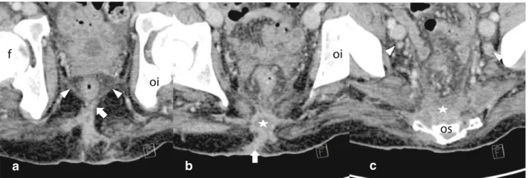
a
b
c
Fig. 2 Coronary reconstruction of the patients' CT scan on the level of the femur (f) and os ischium (oi). Panel a shows a subcutaneous fistula (arrow) reaching the levator ani (triangles). In b, the subcutaneous
abscess (star) is linked over a fistula (arrow) with the skin. In c, the presacral space is filled with abscess (star) whereas bilateral lymph nodes are visible (triangles)
Fig. 1 The patients' CT scan at admission, showing a large subcuta-neous abscess (star) on the flank at the level of the fourth lumbar vertebral body, communicating with the iliopsoas muscle, the para-spinal muscles, and the epidural space (arrow)
were astonished at the bizarre subcutaneous abscess formation on the right flank that was the leading finding at admission to our hospital. An initially suspected spondylodiscitis could not be confirmed by repeated MRI scans. Overall, we interpret the presented case of perianal tuberculosis as a hematogenous dissemination of pulmonary tuberculosis.
Numerous studies have evaluated the practical conse-quences of perianal lymphatic drainage in the context of malignant disease. It is well established that tumors below the linea dentata drain to the inguinal lymph nodes [9–11]. Our patient showed no significant inguinal lymphadenopa-thy, neither in the clinical exam nor in the repeated CT scan.
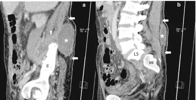
a
b
Fig. 3 Sagittal reconstructions of the patient's CT scan at admission. On a, the subcutaneous abscess (star) and its breakthrough to the iliopsoas muscle (triangle) over the right crista iliaca (il os ilium) are
shown. A thin subcutaneous spur (arrows) delineates potential routes of disease expansion (b) on the level of the lumbar vertebral column (L5 fifth lumbar vertebral body, OS os sacrum)
a
b
Fig. 4 Original figure from Nesselrod's publication in 1936 [12] in
Annals of Surgery showing the perineal and gluteal distribution of lym-phatic vessels after injections of mercury in the skin, respectively. Panel a shows a dorsal view of the sacrococcygeal and superior gluteal region of a
male white fetus at term (original description). Perianal (e), presacral (b), gluteal plexuses (a) and anastomotic vessels (d). Panel b shows a lateral view on the same specimen as in a, showing not only lymphatic vessels above the crista iliaca but also their link (c) to the inguinal zone
But the iliacal axis was certainly involved (Figs.2cand3a). While searching for an explanation for the exceptional ex-tension and localisation of the inflammatory process involv-ing the subcutaneous tissue on the right flank and the right iliopsoas muscle, we came across an anatomic study pub-lished by J.P. Nesselrod in 1936 ([12]; Fig.4).
The eminent proctologist revealed the existence of anas-tomotic lymphatic vessels between the perianal skin and a specific gluteal plexus over the buttocks and further back-wards to the flank above the crista iliaca. Nesselrod con-ducted studies with cadavers of human fetuses at or near term, of newborn infants, as well as few but apparently not useful studies on dogs and humans in vivo. He used differ-ent dyes, whereby the best and depicted results (Fig.4) were achieved with mercury which had been injected in the perianal skin. As shown in Fig. 4a, there is a perianal network linked with another network over the crista iliaca over gluteal and sacrococcygeal plexuses. Over the supra-iliacal plexuses, there is a continuing connection to the inguinal nodes hereby encircling the limb. Nesselrod also describes some minor anastomotic lymphatic vessels that pass the anorectal line and ascend in the columns of Mor-gagni in the ampulla recti. Unfortunately, he does not state whether the iliacal axis was involved upon injection in the perianal plexus.
It is astonishing per se that such an abscess lies subcuta-neously on the right flank. This is rare and requires an explanation. Since it was a case of secondary tuberculosis, it must have been a consequence of hematogenous dissem-ination and not, as it was possible in the case of true intestinal tuberculosis, as a primary infection of the intes-tine. Principally, it could have been a direct seeding into the right flank. It has to be underscored that the most frequent extrapulmonary manifestation of tuberculosis is the lym-phatic form [13]. Therefore, lymphatic networks represent an anatomic space of preferential“homing” for tuberculosis. The sheer presence of lymphatic vessels or plexuses above the crista iliaca made the manner of TB seeding and survival at that precise location more plausible.
Of course, the direction of disease dissemination (break-through (break-through the abdominal flank into or out from the iliopsoas muscle) cannot be definitively cleared. Nesselrod himself reports that perianally injected dyes rise up to the buttocks, but can also descend from the buttocks to the peria-nal region. Consequently, it is a bidirectioperia-nal system. Because of anamnestic hints, one has to assume that the primary focus was a perianal abscess that probably took both routes. In our case, there were minor subcutaneous fistulas forming spurs up to the right gluteus (Fig.3aandb). Internally, CT scan pro-vides clear evidence of the involvement of the puborectalis zone and the iliacal axis (Fig.2). The iliacal axis only could
have been reached over the involvement of the puborectalis zone. What Nesselrod does not show are anastomotic vessels through the abdominal wall. So it seems reasonable to assume that there were two ways of dissemination (an external and an internal one) that completed an anatomic circle and broke a true anatomic barrier by passing the fascia scarpa and con-necting the plexuses above the crista iliaca and the lymphatics of the iliacal axis.
Correlating these old studies with our anatomic findings, we see the clinical relevance of these lymphatic vessels on the buttocks by their concrete involvement.
Acknowledgments The authors thank N. Jonkers for language
correction.
Conflict of interest None.
Open Access This article is distributed under the terms of the Crea-tive Commons Attribution License which permits any use, distribution, and reproduction in any medium, provided the original author(s) and the source are credited.
References
1. Tai WC, Hu TH, Ch L et al (2010) Ano-perianal tuberculosis: 15 years of clinical experiences in Southern Taiwan. Colorectal Dis
12(7 Online):e120–e224
2. Barker JA, Conway AM, Hill J (2011) Supralevator fistula-in-ano
in tuberculosis. Color Dis 13(2):210–214
3. Almadi MA, Ghosh S, Aljebreen AM (2009) Differentiating intes-tinal tuberculosis from Crohn's disease: a diagnostic challenge. Am
J Gastroenterol 104(4):1003–1012
4. Aston NO (1997) Abdominal tuberculosis. World J Surg 21
(5):492–499
5. Kraemer M, Gill SS, Seow-Choen F (2000) Tuberculous anal sepsis: report of clinical features in 20 cases. Dis Colon Rectum
43(11):1589–1591
6. Hassan I, Brilakis ES, Thompson RL et al (2002) Surgical man-agement of abdominal tuberculosis. J Gastrointest Surg 6(6): 862–867
7. Donoghue HD, Holton J (2009) Intestinal tuberculosis. Curr Opin Infect Dis 22(5):490–496
8. Tan KK, Chen K, Sim R (2009) The spectrum of abdominal tuberculosis in a developed country: a single institution's
experi-ence over 7 years. J Gastrointest Surg 13(1):142–147
9. Frost DB, Richards PC, Montague ED et al (1984) Epidermoid
cancer of the anorectum. Cancer 53(6):1285–85
10. Greenali MJ, Quan SH, Stearns MW et al (1985) Epidermoid cancer of the anal margin. Pathologic features, treatment and
clinical results. Am J Surg 149(1):95–101
11. Ryan DP, Compton CC, Mayer RJ (2000) Carcinoma of the anal
canal. N Engl J Med 342(11):792–800
12. Nesselrod JP (1936) An anatomic restudy of the pelvic lymphatics.
Ann Surg 104(5):905–916
13. Mehta JB, Dutt A, Harvill L, Mathews KM (1991) Epidemiology of extrapulmonary tuberculosis. A comparative analysis with pre-AIDS era. Chest 99:1134–1138