Gudrun Feuchtner Robert Goetti Andrè Plass Stephan Baumueller Paul Stolzmann Hans Scheffel Monika Wieser Borut Marincek Hatem Alkadhi Sebastian Leschka Received: 20 November 2009 Revised: 21 February 2010 Accepted: 24 February 2010 Published online: 21 April 2010 # European Society of Radiology 2010
Dual-step prospective ECG-triggered
128-slice dual-source CT for evaluation
of coronary arteries and cardiac function
without heart rate control: a technical note
Abstract: Purpose: To describe pro-spective ECG-triggered dual-source CT dual-step pulsing (pECGdual_step)
for evaluation of coronary arteries and cardiac function. Methods:Fifty-one consecutive patients pre- or post-cardiovascular surgery were exam-ined with adaptive sequential tube current modulated (pECGdual-step)
128-slice dual-source CT without heart rate control (main padding window: 40% RR interval>65bpm/ 70% RR interval<65bpm). Image quality of coronary arteries was graded (4-point scale), and cardiac function was evaluated.Results:Mean HR was 68bpm. Thirty-seven patients were in stable sinus rhythm (SR); 14 had arrhythmia. Image quality of coronary arteries was diagnostic in 804/816 (98%) of segments. The number of non-diagnostic segments was higher in patients with
arrhyth-mia as compared to those in SR (4% vs. 0.5%; p=0.01), and there were fewer segments with excellent image quality (79% vs. 94%; p<0.001) and more segments with impaired image quality (p<0.001 and p=0.002). Global and regional LV function could be evaluated in 41 (80%) and 47 (92%) patients, and valvular func-tion in 48 (94%). In 11/14 of patients with arrhythmia, the second step switched to full mAs, increasing radiation exposure to 8.6mAs (p<0.001). The average radiation dose was 3.8mSv (range, 1.7–7.9) in patients in SR.Conclusion: pECGdual-step128-slice DSCT is
feasible for the evaluation of coronary arteries and cardiac function
without heart rate control in patients in stable sinus rhythm at a low radiation dose.
Keywords Computed tomography . Coronary arteries . Cardiac function . Prospective ECG triggering .
Radiation dose
Introduction
Prospective ECG-triggering (pECG) using sequential (“step-and-shot”) mode [1–7] is an effective imaging technique in coronary computed tomography (CT) angiog-raphy permitting low radiation exposure ranging between 2.5–4.3 mSv [1–7] with radiation dose savings ranging
between 77–79% [5, 6] as compared to conventional retrospective helical ECG gating. However, pECG trigger-ing is restricted to diastolic CT image acquisition; hence, only patients with low (<65–70 bpm) and regular heart rates [1–7] can be examined. Due to the lack of systolic data, the evaluation of cardiac function such as regional and global left ventricular (LV)-function or valvular function is not possible. Left ventricular global [8,9] and regional function
Electronic supplementary material
The online version of this article (doi:10.1007/ s00330-010-1794-7) contains supplementary material, which is available to authorized users.
H. Alkadhi
Cardiac MR-CT-PET group,
Boston Massachussets General Hospital and Harvard Medical School, Boston, USA
G. Feuchtner
:
R. Goetti:
S. Baumueller:
P. Stolzmann:
H. Scheffel:
B. Marincek:
H. Alkadhi:
S. LeschkaInstitute of Diagnostic Radiology, University Hospital Zurich, Zurich, Switzerland
A. Plass
:
M. WieserDivision of Cardiac and Vascular Surgery, University Hospital Zurich, Zurich, Switzerland
G. Feuchtner ())
Department of Radiology II, Innsbruck Medical University, Anichstrasse 35, A-6020, Innsbruck, Austria
e-mail: [email protected] Tel.: +43-512-50481898
analysis is feasible with CT [10–13]. Regional wall motion abnormalities are indicators of ischemic heart disease, and their evaluation, complementary to coronary CTA, can help to diagnose coronary artery disease. Further, native or prosthetic valves may be studied by CT in selected patients for specific reasons [14–17].
A new 128-slice dual-source CT [18–21] system has been introduced recently, providing the high-pitch spiral (“flash”) mode [18–21], which however necessitates a low heat rate of <60 bpm as well [20]. This 128-slice dual-source CT is equipped with a new pECG-triggered module, namely the “dual-step” pulsing (pECGdual-step), encompassing some
novelities for dual-source CT: First, a second pulsing window operating at tube current mAs reduced to 20% can be stretched over the entire cardiac cycle. Second, thefirst major pulsing window (at full tube current mAs) can be set at any arbitrary time point of cardiac cycle, allowing for systolic image acquisition, as recommended in patients with high heart rates [22]. Further, the second pulsing window is triggered by the occurrence of arrhythmia to switch automati-cally to full tube current exposure during the entire cardiac cycle to ensure optimal image quality in patients with irregular heart beats. Finally, temporal resolution of the 128-slice CT scanner is improved to 75 ms by using 0.28-s gantry rotation. These technical features could allow for evaluation of coronary arteries irrespective of heart rate, including a comprehensive evaluation of regional and global LV function. Therefore, the aim of this feasibility study was to explore the new prospective “step-and-shot” ECG-trig-gered “dual-step” (pECGdual-step) mode using
128-dual-source CT in consecutive patients without heart rate control, before or after cardiac or cardiovascular surgery, regarding image quality of coronary arteries and comple-mentary evaluation of cardiac and valvular function.
Materials and methods
Study population
Fifty-one consecutive patients (Table 1) who underwent clinically indicated CT angiography before or after cardiac or cardiovascular surgery were examined between June 2009 and October 2009.
Patients with elevated heart rates and arrhythmia were not excluded. No additional premedication for heart rate control was added to the patients’ baseline medications.
General exclusion criteria for contrast-enhanced CT were nephropathy (defined as a serum creatinine level above 150 μmol/l) and known hypersensitivity to iodine-containing contrast media.
CT data acquisition and post-processing
All examinations were performed using a second-gener-ation dual-source CT system (Somatom Definition Flash,
Siemens Healthcare, Forchheim, Germany). In each patient, 80 ml of iopromide (Ultravist 370, 370 mg/ml, Bayer Schering Pharma, Berlin, Germany) was injected at a flow rate of 5 ml/s followed by 60 ml saline solution. Contrast agent application was controlled by bolus tracking in the ascending aorta (signal attenuation thresh-old 100 HU). The delay after reaching the threshthresh-old to the initiation of the CT scan was 10 s. Patients were advised to hold their breath during the scan. Scanning parameters were: slice collimation 2×128×0.6 mm by means of a z-flying focal spot, gantry rotation time 280 ms, and tube potential 100 kV or 120 kV based on a body mass index (BMI) cutoff of 25 kg/m2[23,24]. Attenuation-based tube current modulation was used with a reference tube current-time product set at 320 mAs per rotation. A cranio-caudal scan direction was chosen, and the start phase for image acquisition was selected manually at the level of the pulmonary artery bifurcation and stopped at the base of the heart. The full tube current window was selected at 40% of the R-R interval if the heart rate was >65 bpm and at 70% if the heart rate was <65 bpm. The second tube current window, reduced to 20% of the nominal output, was selected from 10–90% of the R-R interval.
Images were reconstructed with a slice thickness of 0.75 mm and an increment of 0.5 mm, using a medium smooth tissue convolution kernel (B26f) at 5% increments throughout the cardiac cycle (10–90% of RR interval).
All images were anonymized and transferred to an external workstation (Multi-Modality Workplace, Siemens Healthcare, Forchheim, Germany). The phase with best image quality of coronary arteries was selected for readout of coronary arteries. First, the image quality of the coronary
Table 1 Study population (51 patients)
pECGdual-step
n=51
Age (years) 61±12 (range, 30–83)
Gender
Males (n, %) 34 (66.6%)
Heart rate (bpm) 67.2±9.9 (range, 51–98) >65 bpm: 40% of RR (n, %) 25 (49%) <65 bpm: 70% of RR (n, %) 26 (51%) Heart rhythm Sinus rhythm (n, %) 47 (92%) Without PVC 37 (72.5%) With PVC 10 (20%) Atrialfibrillation (n, %) 4 (8%) Clinical status
Before cardiovascular surgery 24 (47%)
Aortic valve 6
Mitral valve 4
CABG 5
Misc. (e.g., EAP; combined aortic valve/root; Ross procedure, etc.)
9 After cardiovascular surgery 27 (53%)
Mechanic prosthetic 13
Bioprosthetics 6
CABG 3a
Misc. (e.g., EAP) 6
BMI (kg/m2) 25±4.6 (range, 16.7–39.1) aThere was one patient after combined prosthetic and CABG surgery
arteries was evaluated on a per-segment basis (modified AHA 16-segment classification) using interactive oblique multiplanar reformations by two experienced independent observers with 6 years of experience in cardiac CT (G.F.) and 1 year of experience (R.G.) on a 4-point scale: 1, excellent (no artifacts); 2, good (minor artifacts); 3, mediocre (artifacts but still diagnostic); 4, poor (severe artifacts rendering image quality non-diagnostic). A consensus read-out was appended in case of disagreement, and the consensus results were taken forfinal data analysis.
Second, global left ventricular function was measured with dedicated software (Syngo Circulation, Siemens) allowing for computation of left ventricular end systolic volume (ESV), end diastolic volume (EDV), stoke volume (SV) and the ejection fraction (EF). Specific reasons for insufficient evaluations were noted. Then, 4D cine image series were reviewed using the cine mode on Circulation (Syngo, Siemens) on three planes (four-, three- and two-chamber views). The evaluation of regional wall motion abnormalities (WMA) was scored as“assessable” or “non-assessable.” Valvular function was reviewed as well, during both systole and diastole, and the aortic valve (native or prosthetic) leaflets were scored as “assessable” or“non-assessable” on cross-sectional short axis views of the valve and aortic root as derived from left coronal and left sagittal planes through the aortic root. Specific reasons for non-assessability (e.g., type of artifacts) were noted. For all evaluations, consensus reading was performed. The effective radiation dose delivered at thoracic CT was calculated applying a method proposed by the European Working Group for Guidelines on Quality Criteria for CT [25] using the dose-length product (DLP) and a con-version coefficient for the chest of 0.017 mSv/[mGy × cm]. The DLP was obtained from an electronic protocol that summarized the individual radiation exposure param-eters of each CT acquisition.
Statistical analyses
All statistical analyses were performed by commercially available software (SPSS, release 17, SPSS Inc., Chicago, IL). Continuous variables were expressed as means ± stand-ard deviations or as medians and ranges. Categorical variables were expressed as frequencies (counts) or percentages.
Inter-observer agreement for qualitative image quality ratings was evaluated using Cohen’s kappa statistics. A κ value greater than 0.81 was interpreted as excellent agreement, values of 0.61–0.80 were interpreted as good, values of 0.41–0.60 as moderate, values of 0.21–0.40 as fair and values less than 0.20 as poor agreement.
The image quality scores between patients in regular sinus rhythm and those with arrhythmia were tested separately (for each score) for differences with the chi-square test or Fisher's exact test. Differences in radiation dose and heart rate between patients with regular sinus rhythm and arrhythmia were tested with the independent t-test.
Differences in image quality scores between patients in whom coronary arteries were evaluated during systole (at 40% of the R-R interval) or during diastole (at 70% of the RR interval) were tested with the Mann-Whitney U-test. A p-value below 0.05 was considered statistically significant.
Results
A total of 51 patients were successfully examined with the pECGdual-step technique. Table 1 shows patient profiles,
including reason for referral to CT angiography. Mean heart rate was 67 bpm. In 25 (49%) patients, the first pulsing window was set at 70% of the RR interval and in 26 patients (51%) at 40%, respectively. Of the 51 consecutive patients, 37 (73%) had stable sinus rhythm (SR) without arrhythmia, and 14 (27%) patients had arrhythmia [n=4, atrialfibrillation (AF); and n=10, extrasystole].
Coronary arteries
A total of 816 coronary segments were analyzed. Of theses, 733 (89.8%) had excellent image quality (Fig. 1) 56 (7%) segments had good and 15 (1.8%) had mediocre image quality. Twelve of 816 (1.5%) segments were non-diagnostic in 9 patients; of these, 6 had arrhythmia. The main reason for non-diagnostic segments was misregistra-tion artifacts based on “step” artifacts (n=9) leading to missing parts of the artery (n=4), or severely hampered image quality (n=5) in combination with blurring and streak artifacts, or motion blurring artifacts (n=3, of those in one segment combined with high image noise). Interobserver agreement was excellent with K=0.84.
If dividing patients into two groups (SR, n=37 and arrhythmia, n=14; Table2), the number of non-diagnostic segments (n=9) was significantly higher in patients with arrhythmia as compared to those without (n=3; 4% vs. 0.5%; p = 0.01), and the number of segments with excellent image quality was significantly lower (79% vs. 94%, p<0.001). The number of segments with impaired image quality (score 2 and 3) was significantly higher in patients with arrhythmia (11.6% vs. 5.1% and 5.4 vs. 0.5%; p<0.001 and p=0.002), respectively.
There was no difference in image quality scores between patients in which coronary arteries were eval-uated at 40% and 70% of the RR interval (mean score 1.18±0.24 and 1.12±0.32; p=0.301).
Global LV function, regional WMA and valvular function Quantification of LV function was successful and chamber segmentation regarded as accurate in 42/51 (80%) of patients. Mean LV-EDV was 134 ml, mean LV-ESV was 64.8 ml, mean SV was 69.2 and mean EF 52.9%. In eight patients, low LV contrast attenuation and/or high image
Table 2 Performance of pECGdual-stepmodule in 51 patients: CT image quality (IQ) of coronary arteries, cardiac function and radiation
dose. Comparison of patients in stable sinus rhythm (SR) with those having arrhythmia
Patients in stable SR Patients with arrhythmia (AF, PVC) p-value
37 patients 14 patients
IQ coronary arteries (N=592 segments) (N=224 segments) Diagnostic Score 1 556 (94%) 177 (79%) P<0.001c Score 2 30 (5.1%) 26 (11.6%) P=0.002b Score 3 3 (0.5%) 12 (5.4%) P<0.001 Non-diagnostic Score 4 3/592 (0.5%) 9/224 (4%) P=0.01a Cardiac function Global LV function 41/51 (80%) Regional LV function 47/51 (92%) Valvular function 48/51 (94%) Native 31/32 (96%) Prosthetics 17/19 (89%)c
Second step: tube current modulation at 20% mAs
n (%) 37/37 (100%) 3/ 14 (21%) <0.001
Second step at 20% mAs? Yes: 40 patients No: 11 patients
Radiation dose (mSv) 3.8±1.6 8.6±3.1 <0.001
Heart rate (bpm) 66.2±8 70±15 0.194
BMI 26±5 25.4±5 0.708
Effective radiation dose (mSv) 3.8±2 7.9±3 <0.001
4.9±3 mSv (range, 1.7–14) a
Fisher’s exact test;bchi-square IQ score 1 (excellent), score 2 (good) and score 3 (mediocre) were regarded as diagnostic, whereas score 4 was considered non-diagnostic. AF = atrialfibrillation; PVC = premature ventricular contraction; LV = left ventricle; BMI = body mass index; n = counts; bpm = beats per minute; mSv = milli-Sievert
cBoth non-evaluable prosthetic valves were miniroot bioprosthetics (same type)
Fig. 1 a Coronary arteries in pECGdual-stepmode: 38 YOF in sinus
rhythm (heart rate, 68 bpm) before redo aortic valve surgery. Excellent image quality of coronary arteries: note right coronary artery (RCA) permitting preoperative rule out of coronary stenosis. Thefirst pulsing window was set at 40% of the RR interval. Beta-blockers were contraindicated because of aortic stenosis. Radiation dose was 2.23 mSv. b Valvular function: Combined moderate aortic stenosis (left panel, early systole: arrows denote aortic valve orifice area, “fish mouth” opening of bicuspid valve) and severe regurgitation (right panel, end diastole: note incomplete closure of
thickened cusps). Late recurrence of valvular dysfunction after congential aortic stenosis surgery. The corresponding movie shows opening of bicuspid valve and closure with incompetent cusps. The systolic image (left) was derived from 4D cine loops at 15% of RR interval and the diastolic image (right) at 70%. c pECGdual-step
module; ECG during scan: 1=first pulsing window at 40% of RR interval (100% mAs) (arrow). 2=second pulsing window from 10– 90% of the RR interval covering the entire cardiac cycle (tube current reduced to 20% of full mAs)
noise, and in one patient stair-step artifacts led to insufficient chamber segmentation results.
On 4D cine images, regional WMA analysis was assessable in 47/51 (92%) patients. Reasons for non-assessability were: low LV contrast attenuation (n=2), stair-step artifacts (n=1) and high image noise (n=1) in the second step phase (diastole), respectively.
Valvular function was assessable in 48/51 patients (94%) during both systole and diastole (Fig. 1b, c). One native valve was non-assessable because of stair-step artifacts caused by arrhythmia during both phases. There were 19 prosthetic valves, of which 17 could be evaluated, but in 2 miniroot bioprosthetic valves, the leaflets were not adequately delineated because of high image noise during the second pulsing phase (n=1 systole, n= 1 diastole). However, valve morphology could be evaluated in the counterpart phase of the cardiac cycle at 100% mAs exposure. In two further native valves, image noise during the second step pulsing window phase of the cardiac cycle (either systole or diastole) had excessive image noise hampering the evaluation.
pECGdual-steptechnical performance evaluation
The second step switched from 20% up to 100% tube current mAs in 11/14 (79%) of patients with arrhythmia (AF, n=4
and SR with extrasystole in n=7) (Fig. 2). In these 11 patients, the radiation dose was significantly (p<0.001) higher with 8.6 mAs±3.1 compared to those patients in whom the“dual step” worked sufficiently (mean, 3.8 mSv± 1.7) (Table2).
In all 37 patients without arrhythmia, the pECGdual-step
mode pulsed the second window at reduced tube current mAs.
CT data were acquired within 3 heart beats (9 patients), within 4 heart beats (32 patients), within 5 steps (8 patients) or 6 steps (2 patients). Mean scan time was 6.6 s±1.7 s (3.0– 10.5 s), and the time of each pulsing window was 0.56±0.4 s (0.49–1.14 s). Mean dose-length product (DLP) was 284.5 mGy*cm. Mean CDTIvol was 24.4 mGy. Effective
radiation dose was 4.9±3 mSv (range, 1.7–14).
Discussion
Our study shows that the new pECGdual-step module in 128-slice DSCT allows for reliable evaluation of coronary arteries and cardiac function in patients with regular heart rates without arrhythmia at a low radiation dose of 3.7 mSv.
The first advantage of the pECGdual-step module compared to conventional “step-and-shoot” protocols is
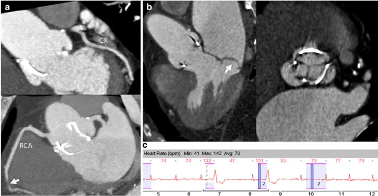
Fig. 2 pECGdual-stepin a patient with arrhythmia (atrialfibrillation
and extrasystole). a Coronary arteries: CX (upper panel) with excellent image quality. RCA (lower panel) had one non-diagnostic coronary segment (arrow, mid segment 2) because of an artifact. Radiation exposure was 6.9 mSv. b Valvular function: Mitral valve prolapse of posterior cusp during systole (arrow, left panel) on three-chamber view and bioprosthetic aortic valve (right panel) on cross-sectional view during diastole. The systolic image (left) was
created from 4D cine loops at 15% of the RR interval and the diastolic image (right) at 70% of the RR interval. c pECGdual-step
ECG during scan: The second pulsing step (2) skipped the second beat (133 bpm) and was operating on full 100% mAs at the next two beats (131 bpm and 73 bpm) instead of reduced tube current to ensure diagnostic image quality of coronary arteries. Note atrial fibrillation and multiple ventricular extrasystoles
the possibility of its application irrespective of the height of the heart rate. To date, the use of prospective ECG triggering has been recommended in low heart rates <65– 70 bpm [1–7, 30] only because the image acquisition (“padding”) window had to be placed at diastole. The pECGdual-stepmodule allows for a placing of the mainfirst
padding window at any arbitrary time point of the cardiac cycle, including the systole. Hence, we explored the use of 40% of the RR interval in patients with a baseline heart
rate of more than 65 bpm. Our data confirm good
diagnostic image quality of coronary arteries during the end-systolic phase in patients with a regular heart rate, which is in line with reports from retrospective ECG gating [22]. At higher heart rates, the diastole shortens more compared to systole; thus, the end systole is a more appropriate phase for image acquisition. Additional medication with beta-blockers to lower to the heart rate before performing the CT scan can be avoided. In clinical practice, some patients may not respond to beta-blockers or the use of beta-blockers is contraindicated, such as in patients with severe aortic stenosis or LV dysfunction. However, we observed a significantly worse image quality of coronary segments in patients with arrhythmia, both in
those with AF and those with extrasystole, which is in line with previous studies [1–7]. Therefore, the pECGdual-step
mode should be used in patients with stable sinus rhythm only.
Second, by establishing a second image acquisition window (second step) with the tube current reduced to 20%, similar to the ECG tube current modulation technique (“ECG pulsing”) [26] during the entire cardiac cycle, full functional CT data sets can be acquired. These data sets allow for calculation of global and regional left ventricular function, and evaluation of cardiac valves on 4D cine movies. Evaluation of wall motion abnormalities [9, 10] is feasible with CT, which may improve the accuracy of coronary CT angiography [13]. The evalua-tion of cardiac valves [14–17] for indications such as assessement of valvular morphology [15], aortic valve area measurement [14], planning of minimally invasive and percutaneous surgery [16] or prosthetic valve (Fig.3) dysfunction [17] is possible. In our study, only in a minority of patients could LV function not be assessed either because of low contrast attenuation in the left ventricle resulting in insufficient left ventricular volume segmentation, which could have been avoided by using a
Fig. 3 Movie 2. Evaluation of mechanical prosthetic valve. a Volume-rendered 3D images of mechanic prosthetic St. Jude bileaflet valve. Diastole (left), closed. Systole (right), open. b Multiplanar reformations (MPR). Three-chamber view (left) during diastole shows normal closed mechanic valves, and cross-sectional view
(right) during systole shows normal opening. Note higher image noise during systole (right) because of tube current modulation during the second step, which did not hamper evaluation of prosthetic valve function (see movie2)
higher volume of contrast agent, or in the presence of stair-step artifacts caused by arrhythmia. High image noise at the second step was problematic in one patient only.
Valvular function could be assessed in the majority of patients, despite merging both native and prosthetic valves. The only problem was one specific prosthetic valve type, a miniroot bioprosthesis in two patients, in which the leaflets were not well delineated because of high image noise during the second pulsing phase at reduced tube current (either systole or diastole depending on the selected phase), but still morphology of leaflets could be assessed at the contrary phase of the cardiac cycle.
However, the pECGdual-stepshould be used in patients
with regular sinus rhythm only, in the absence of arrhythmia such as premature beats, because the main reason for limited image quality was arrhythmia. In patients with arrhythmia, the second pulsing step switched into full 100% mAs exposure to ensure coronary image quality, leading to a significant increase in radiation dose, which with 8.6 mSv was however still lower than in retrospectively ECG-gated spiral scanning using 64-slice CT with 14–16 mSv and 64-slice DSCT [27–29] because of differences in image acquisition, e.g., that the amount of overranging and overscanning is reduced by using the pECGdual-stepmode.
Sequential pECG-triggering using 128-slice DSCT [30] has recently shown to have a radiation exposure of 3.6 mSv when being restricted to beta-blocker medication and low heart rate of 61 bpm. The mean radiation dose in our study was similar and only slightly higher despite adding a second pulsing window at reduced tube current mAs. However, in contrast to Andres et al. [30], applying afixed setting of 120 kV, we used a radiation dose-saving body weight-adapted tube voltage protocol (if <25 kg/m2 BMI: 100 kV and if >25 kg/m2BMI: 120 kV [23,24]).
Average radiation dose in our study using the pECGdual-step
module was within the upper range of previous studies exploring“single-shot” pECG triggering on 64-slice CT with 2.5–4.3 mSv [1–7], but higher compared to the high-pitch spiral mode using 128-slice DSCT with∼1 mSv [19–21].
Study limitations
First, we acknowledge that this was an explorative study design, since there are no reports about the pECGdual-step
module so far. Therefore, we did not compare pECG dual-step with a reference standard method. Further con
firma-tory and comparative head-to-head studies with retro-spective ECG gating in spiral mode are needed.
Second, we did not perform heart rate control by using beta-blocker medication-. Possibly the performance of the pECGdual-stepmodule, e.g., for wall motion abnormalities or
valvular function, might be improved if the heart rate is lowered with beta-blockers. However, one needs to keep in mind that for accurate estimation of global LV function, beta-blocker medication is not recommended because negative inotropic effects lead to a bias. Moreover, beta-blockers might be contraindicated in patients with aortic valve stenosis or other comorbidities before or after cardiovascular surgery.
Third, we explored a selected patient population (pre-and post-cardiac surgery setting) that certainly has a higher prevalence of arrhythmia and high heart rates compared to patients usually referred to coronary CTA (e. g., those with intermediate risk of coronary artery disease).
Conclusion
1. Using the adaptive sequential pECGdual-step mode on
128-slice DSCT is feasible for coronary artery and complementary cardiac function evaluation at low radiation exposure.
2. We suggest its application in patients in stable sinus rhythm without arrhythmia irrespective of the height of heart rate.
3. Our study suggests that placing the padding window at end systole (e.g., 40% of cardiac cycle) in patients with high heart rates of >65 bpm and at end diastole (70% of cardiac cycle) in patients with low heart rates <65 bpm provides diagnostic image quality of the coronary arteries.
References
1. Maruyama T, Takada M, Hasuike T, Yoshikawa A, Namimatsu E, Yoshizumi T (2008) Radiation dose reduction and coronary assessability of prospective electrocardiogram-gated computed tomography coronary angiography: comparison with retrospective electrocardiogram-gated helical scan. J Am Coll Cardiol 52:1450–1455
2. Arnoldi E, Johnson TR, Rist C, Wintersperger BJ, Sommer WH, Becker A, Becker CR, Reiser MF, Nikolaou K (2009) Adequate image quality with reduced radiation dose in prospectively triggered coronary CTA compared with retrospective techniques. Eur Radiol 19:2147–2155
3. Earls JP, Berman EL, Urban BA, Curry CA, Lane JL, Jennings RS, McCulloch CC, Hsieh J, Londt JH (2008) Prospectively gated transverse coronary CT angiography versus retrospectively gated helical technique: improved image quality and reduced radiation dose. Radiology 246:742–753
4. Hirai N, Horiguchi J, Fujioka C, Kiguchi M, Yamamoto H, Matsuura N, Kitagawa T, Teragawa H, Kohno N, Ito K (2008) Prospective versus
retrospective ECG-gated 64-detector coronary CT angiography: assessment of image quality, stenosis, and radiation dose. Radiology 248:424–430
5. Shuman WP, Branch KR, May JM, Mitsumori LM, Lockhart DW, Dubinsky TJ, Warren BH, Caldwell JH (2008) Prospective versus retrospective ECG gating for 64-detector CT of the coronary arteries: comparison of image quality and patient radiation dose. Radiology 248:431–437
6. Scheffel H, Alkadhi H, Leschka S, Plass A, Desbiolles L, Guber I, Krauss T, Gruenenfelder J, Genoni M, Luescher TF, Marincek B, Stolzmann P (2008) Low-dose CT coronary angiography in the step-and-shoot mode: diagnostic performance. Heart 94:1132–1137 7. Stolzmann P, Leschka S, Scheffel H,
Krauss T, Desbiolles L, Plass A, Genoni M, Flohr TG, Wildermuth S, Marincek B, Alkadhi H (2008) Dual-source CT in step-and-shoot mode: noninvasive coronary angiography with low radiation dose. Radiology 249:71–80 8. Dewey M, Müller M, Eddicks S,
Schnapauff D, Teige F, Rutsch W, Borges AC, Hamm B (2006) Evaluation of global and regional left ventricular function with 16-slice computed tomography, biplane
cineventriculography, and two-dimensional transthoracic
echocardiography: comparison with magnetic resonance imaging. J Am Coll Cardiol 48:2034–2044
9. Sarwar A, Shapiro MD, Nasir K, Nieman K, Nomura CH, Brady TJ, Cury RC (2009) Evaluating global and regional left ventricular function in patients with reperfused acute myocardial infarction by 64-slice multidetector CT: a comparison to magnetic resonance imaging. J Cardiovasc Comput Tomogr 3:170–177 10. Cury RC, Nieman K, Shapiro MD, Butler J, Nomura CH, Ferencik M, Hoffmann U, Abbara S, Jassal DS, Yasuda T, Gold HK, Jang IK, Brady TJ (2008) Comprehensive assessment of myocardial perfusion defects, regional wall motion, and left ventricular function by using 64-section multidetector CT. Radiology 248:466–475 11. Kristensen TS, Kofoed KF, Møller DV,
Ersbøll M, Kühl T, von der Recke P, Køber L, Nielsen MB, Kelbæk H (2009) Quantitative assessment of left ventricular systolic wall thickening using multidetector computed tomography. Eur J Radiol 72:92–97 12. Ko SM, Kim YJ, Park JH, Choi NM
(2009) Assessment of left ventricular ejection fraction and regional wall motion with 64-slice multidetector CT: a comparison with two-dimensional transthoracic echocardiography. Br J Radiol. doi:10.1259/bjr/38829806
13. Lüders F, Fischbach R, Seifarth H, Wessling J, Heindel W, Juergens KU (2009) Dual-source computed tomography: effect on regional and global left ventricular function assessment compared to magnetic resonance imaging. Rofo 181:962–969 14. Feuchtner GM, Dichtl W, Friedrich GJ,
Frick M, Alber H, Schachner T, Bonatti J, Mallouhi A, Frede T, Pachinger O, zur Nedden D, Müller S (2006) Multislice computed tomography for detection of patients with aortic valve stenosis and quantification of severity. J Am Coll Cardiol 47:1410–1417 15. Alkadhi H, Desbiolles L, Stolzmann P,
Leschka S, Scheffel H, Plass A, Schertler T, Trindade PT, Genoni M, Cattin P, Marincek B, Frauenfelder T (2009) Mitral annular shape, size, and motion in normals and in patients with cardiomyopathy: evaluation with computed tomography. Invest Radiol 44:218–225
16. Leipsic J, Wood D, Manders D, Nietlispach F, Masson JB, Mayo J, Al-Bugami S, Webb JG (2009) The evolving role of MDCT in transcatheter aortic valve replacement: a radiologists' perspective. AJR Am J Roentgenol 193: W214–W219
17. Tsai IC, Lin YK, Chang Y, Fu YC, Wang CC, Hsieh SR, Wei HJ, Tsai HW, Jan SL, Wang KY, Chen MC, Chen CC (2009) Correctness of multi-detector-row computed tomography for diagnosing mechanical prosthetic heart valve disorders using operativefindings as a gold standard. Eur Radiol 19:857–867 18. Ertel D, Lell MM, Harig F, Flohr T,
Schmidt B, Kalender WA (2009) Cardiac spiral dual-source CT with high pitch: a feasibility study. Eur Radiol 19:2357–2362
19. Achenbach S, Marwan M, Schepis T, Pflederer T, Bruder H, Allmendinger T, Petersilka M, Anders K, Lell M, Kuettner A, Ropers D, Daniel WG, Flohr T (2009) High-pitch spiral acquisition: a new scan mode for coronary CT angiography. J Cardiovasc Comput Tomogr 3:117–121
20. Leschka S, Stolzmann P, Desbiolles L, Baumueller S, Goetti R, Schertler T, Scheffel H, Plass A, Falk V, Feuchtner G, Marincek B, Alkadhi H (2009) Diagnostic accuracy of high-pitch dual-source CT for the assessment of coronary stenoses:first experience. Eur Radiol 19:2896–2903
21. Lell M, Marwan M, Schepis T, Pflederer T, Anders K, Flohr T, Allmendinger T, Kalender W, Ertel D, Thierfelder C, Kuettner A, Ropers D, Daniel WG, Achenbach S (2009) Prospectively ECG-triggered high-pitch spiral acquisition for coronary CT angiography using dual-source CT: technique and initial experience. Eur Radiol. doi:10.1007/s00330-009-1558-4
22. Seifarth H, Wienbeck S, Püsken M, Juergens KU, Maintz D, Vahlhaus C, Heindel W, Fischbach R (2007) Systole better phase: Optimal systolic and diastolic reconstruction windows for coronary CT angiography using dual-source CT. AJR Am J Roentgenol 189:1317–1323
23. Alkadhi H, Stolzmann P, Scheffel H, Desbiolles L, Baumüller S, Plass A, Genoni M, Marincek B, Leschka S (2008) Radiation dose of cardiac dual-source CT: the effect of tailoring the protocol to patient-specific parameters. Eur J Radiol 68:385–391
24. Feuchtner GM, Jodocy D, Klauser A, Haberfellner B, Aglan I, Spoeck A, Hiehs S, Soegner P, Jaschke W (2009) Radiation dose reduction by using 100-kV tube voltage in cardiac 64-slice computed tomography: A comparative study. Eur J Radiol. doi:10.1016/j. ejrad.2009.07.012
25. Menzel H, Schibilla H, Teunen D (2000) European guidelines on quality criteria for computed tomography, Publication no. EUR 16262 EN. Luxembourg: European Commission 26. Jakobs TF, Becker CR, Ohnesorge B,
Flohr T, Suess C, Schoepf UJ, Reiser MF (2002) Multislice helical CT of the heart with retrospective ECG gating: reduction of radiation exposure by ECG-controlled tube current
modulation. Eur Radiol 12:1081–1086 27. Hausleiter J, Meyer T, Hermann F,
Hadamitzky M, Krebs M, Gerber TC, McCollough C, Martinoff S, Kastrati A, Schömig A, Achenbach S (2009) Estimated radiation dose associated with cardiac CT angiography. JAMA 301:500–507
28. Hermann F, Martinoff S, Meyert T et al (2008) Reduction of radiation dose estimates in cardiac 64-slice CT angiography in patients after coronary artery bypass graft surgery. Invest Radiol 43:253–260
29. Leschka S, Stolzmann P, Schmid FT, Scheffel H, Stinn B, Marincek B, Alkadhi H, Wildermuth S (2008) Low kilovoltage cardiac dual-source CT: attenuation, noise, and radiation dose. Eur Radiol 18:1809–1817
30. Anders K, Baum U, Gauss S, Kuefner MA, Achenbach S, Kuettner A, Daniel WG, Uder M, Ropers D (2009) Initial experience with prospectively triggered, sequential CT coronary angiography on a 128-slice scanner. Rofo 181:332–338