ORIGINAL ARTICLE
Are radiogallium-labelled DOTA-conjugated somatostatin
analogues superior to those labelled with other radiometals?
P. Antunes&M. Ginj&H. Zhang&B. Waser& R. P. Baum&J. C. Reubi&H. Maecke
Received: 13 July 2006 / Accepted: 5 November 2006 / Published online: 16 January 2007 # Springer-Verlag 2007
Abstract
Purpose Gallium-68 is a metallic positron emitter with a half-life of 68 min that is ideal for the in vivo use of small molecules, such as [68Ga-DOTA,Tyr3]octreotide, in the diagnostic imaging of somatostatin receptor-positive tumours. In preclinical studies it has shown a striking superiority over its111In-labelled congener. The purpose of this study was to evaluate whether third-generation somato-statin-based, radiogallium-labelled peptides show the same superiority.
Methods Peptides were synthesised on solid phase. The receptor affinity was determined by in vitro receptor autoradiography. The internalisation rate was studied in AR4-2J and hsst-HEK-transfected cell lines. The pharma-cokinetics was studied in a rat xenograft tumour model, AR4-2J.
Results All peptides showed high affinities on hsst2, with the highest affinity for the GaIII-complexed peptides. On hsst3 the situation was reversed, with a trend towards lower affinity of the GaIII peptides. A significantly increased internalisation rate was found in sst2-expressing cells for all
67
Ga-labelled peptides. Internalisation into HEK-sst3 was
usually faster for the 111In-labelled peptides. No internal-isation was found into sst5. Biodistribution studies employ-ing [67Ga-DOTA,1-Nal3]octreotide in comparison to [111In-DOTA,1-Nal3]octreotide and [67Ga-DOTA,Tyr3] octreotide showed a significantly higher and receptor-mediated uptake of the two 67Ga-labelled peptides in the tumour and somatostatin receptor-positive tissues. A patient study illustrated the potential advantage of a broad receptor subtype profile radiopeptide over a high-affinity sst2-selective radiopeptide.
Conclusion This study demonstrates that 67/68 Ga-DOTA-octapeptides show distinctly better preclinical, pharmaco-logical performances than the 111In-labelled peptides, especially on sst2-expressing cells and the corresponding animal models. They may be excellent candidates for further development for clinical studies.
Keywords Somatostatin receptors . Gallium-68 . Indium-111 . Radiopeptides . Imaging
Introduction
Peptide receptors with high overexpression on a variety of human tumours are a focus of research in radiopharmacy and nuclear oncology. The prototype is the somatostatin receptor, which has been found to be overexpressed not only on neuroendocrine tumours but also on renal cell carcinoma, small cell lung cancer, breast cancer, prostate cancer and malignant lymphoma [1]. Somatostatin ana-logues have been radiolabelled with diagnostic radionu-clides for imaging and tumour localisation as well as with particle emitters for targeted radionuclide therapy. Labelling has been performed with the γ-emitters 99mTc [2–4],111In [5,6] and67Ga [7], with the positron emitters 11C [8],18F DOI 10.1007/s00259-006-0317-x
P. Antunes
:
M. Ginj:
H. Zhang:
H. Maecke (*)Division of Radiological Chemistry, University Hospital Basel, Petersgraben 4,
4031 Basel, Switzerland e-mail: hmaecke@uhbs.ch B. Waser
:
J. C. ReubiInstitute of Pathology, University of Bern, Bern, Switzerland
R. P. Baum
Department of Nuclear Medicine/PETCT-Center, Zentralklinik Bad Berka,
[9] and 64Cu [10], and with particle emitters such as 90Y [11–14], 177Lu [15] and 213Bi [16]. [111 In-DTPA]-octreo-tide (Octreoscan) is the first registered radiopepIn-DTPA]-octreo-tide and currently the most sensitive method for staging of neuro-endocrine tumours [17].
Currently, the metallic positron emitter68Ga is of great interest [18] because of its suitable radiophysical properties; its positron yield is high, with 89% of all disintegrations. Its half-life of 68 min matches the pharmacokinetics of many peptides and other small molecules owing to a fast blood clearance, quick diffusion and target local-isation. In addition, and of major importance, is the fact that it can be produced from a 68Ge/68Ga generator. Especially attractive is the long half-life of 270.8 days of the parent 68Ge, which allows use of the generator for up to 1 year or even longer. A version of this generator is now commercially available, and this is strongly motivat-ing radiopharmacists to develop new radiogallium-based radiopharmaceuticals.
We have been interested in designing peptide–chelator conjugates for labelling with 67,68Ga, in particular those based on somatostatin analogues. We used and compared different chelators such as desferrioxamine [7], NOTAGA (1,4,7-triazacyclononane,1-glutaric acid, 4,7-acetic acid) [19] and DOTA (1,4,7,10-tetraazacyclododecane,1,4,7,10-tetraacetic acid) [20, 21]. Interestingly, when DOTA was coupled to the octapeptide [Tyr3]octreotide, the resulting [67Ga-DOTA,Tyr3]octreotide ([67Ga-DOTA]-TOC) showed not only about a fivefold increased affinity to the somatostatin receptor subtype 2 (sst2) but also a 2.5-fold increased tumour uptake compared with [111 In-DOTA]-TOC in a mouse xenograft model (AR4-2J). In addition, the kidney uptake of [67Ga-DOTA]-TOC was distinctly decreased compared with that of [111In-DOTA]-TOC or [90Y-DOTA]-TOC [20, 21]. These very promising preclin-ical data prompted several research groups to study [68 Ga-DOTA]-TOC in patients with somatostatin receptor-positive tumours [22–27]. The Bad Berka group has almost 2 years’
experience employing 68Ga-DOTA-octapeptides in a vari-ety of somatostatin receptor-positive tumour patients, mainly gastroenteropancreatic (GEP) tumours [28, 29]. In addition, first promising results have been reported [30,31] using [68Ga-DOTA,1-Nal3]octreotide ([68Ga-DOTA]-NOC) receptor PET/CT in patients before and after peptide receptor radionuclide therapy (PRRT). Furthermore, [68Ga-DOTA,Tyr3,Thr8]octreotide ([68Ga-DOTA]-TATE) was recently used in a patient with paraganglioma [32].
The main purpose of this work was to study the hypothesis that the influence of radiogallium on pharma-cological parameters such as receptor binding affinity, rate of internalisation, tumour uptake etc. may be operative in new DOTA-octapeptides (“third-generation somatostatin analogues”). The latter differ from DOTA-TOC by virtue
of an improved somatostatin receptor subtype profile [33,
34]. In PET these new radiopharmaceuticals may have the potential to target a wider range of tumours and to detect more lesions in an individual patient. Therefore we determined the affinity profiles with regard to hsst1–5 of
68/nat
Ga-DOTA-octapeptides and compared them with those of111/natIn-DOTA-octapeptides (or in some cases 177/nat Lu-and 90/natY-DOTA-octapeptides). We also studied the rate of internalisation into sst2,3,5-expressing cell lines and the biodistribution in a rat tumour model (AR4-2J). In addition, we present first PET/CT studies using [68Ga-DOTA]-NOC in comparison to [68Ga-DOTA]-TATE.
Materials and methods
All chemicals, including Fmoc(9-fluorenylmethoxycar-bonyl)-protected amino acids, were obtained from com-mercial sources and used without further purification. Tritylchloride-resin was obtained from PepChem (Tübin-gen, Germany).67GaCl3and 111InCl3were from
Mallinck-rodt Medical (Petten, The Netherlands). The prochelator DOTA(tBu)3was synthesised according to Heppeler et al.
[20]. The reactive side chains of the amino acids were masked with one of the following groups: Cys, acetamido-methyl; Lys, t-butoxycarbonyl; Thr, t-butyl; Trp, t-butox-ycarbonyl. Analytical reversed-phase high-performance liquid chromatography (RP-HPLC) was carried out on a Hewlett Packard 1050 HPLC system equipped with a multiwavelength detector and a flow-through Berthold LB506C1 γ-detector. Preparative HPLC was done on a Bischof HPLC-system (Metrohm AG, Switzerland) with HPLC-pumps 2250 and a Lambda 1010 UV-detector. CC250/4 Nucleosil 120-3C18 columns from Macherey-Nagel were used for analytical HPLC and a VP250/21 Nucleosil 200-5C15 column for preparative HPLC. The gradient systems consisted of mixtures of acetonitrile and water with 0.1% trifluoroacetic acid. Quantitative γ-count-ing was performed on a COBRA 5003 γ-system well counter from Packard Instrument Company (Switzerland).
Electrospray ionisation-mass spectrometry (ESI-MS) was carried out with a Finnigan SSQ 7000 spectrometer (Bremen, Germany).
Determination of lipophilicity
The octanol–water partition coefficients were determined using the shake flask method. Both solvents (aqueous and octanol) were presaturated with each other by shaking them in contact for hours. To a solution of 100 nmol/l radio-labelled peptide in 500μl PBS (pH 7.4), 500 μl of octanol was added (n=5). The mixtures were vigorously shaken for 1 h to reach equilibrium. After equilibration, the mixtures
were centrifuged (10 min at 2,000 rpm) to achieve good separation. The activity concentrations in 100-μl samples of both the aqueous and the organic phase were measured in a γ-counter. The partition coefficient (log D) was calculated from the formula:
log D¼ log 10
counts in octanol layer=counts in aqueous layer
ð Þ
Synthesis
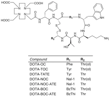
The peptide–chelator conjugates were synthesised by standard Fmoc-solid phase synthesis [34] on tritylchloride resin (substitution 0.8 mmol/g) on a Rink Engineering peptide synthesiser Switch 24 (RinkCombichem Technolo-gies, Bubendorf, Switzerland) according to the general procedure described previously [33], affording compounds included in Fig. 1, which were characterised by ESI-MS and RP-HPLC.
Formation of metal complexes for preclinical studies The DOTA-octapeptides were complexed with InCl3
(anhydrous), Ga(NO3)3·5 H2O Lu(NO3)3·9 H2O and Y
(NO3)3·5 H2O as described by Wild et al. [33]. The ligands
were labelled with natural and radioactive indium or gallium (111/natIn, 67/natGa) according to Ginj et al. [34] and obtained in >99% radiochemical purity at specific activities of >37 GBq/μmol peptide. For internalisation experiments, the DOTA-peptides were labelled to a specific activity of about 37 GBq/μmol peptide and then excess InCl3(anhydrous) or Ga(NO3)3·5 H2O was added, followed
by SepPak purification, to afford structurally characterised homogeneous peptide ligands.
Synthesis of [68Ga-DOTA]-NOC for clinical study
For the68Ga labelling, 30 mCi68Ge/68Ge generators based on TiO2phase (Cyclotron Co., Obninsk, Russia) were used
to obtain 500–750 MBq of68
Ga. The generator eluate was preconcentrated and purified from potential metallic impu-rities on a micro-chromatography cation exchange column (50W-X8, <400 mesh, Bio-Rad AG, Munich, Germany). The activities were eluted with 98% acetone/0.05 mol/ l HCl (400 μl). This was added to 4 ml H2O containing
10 nmol DOTA-NOC and heated at 100°C for 10 min, leading to a radiochemical yield >95%. The mixture was subject to purification using Sep-Pak C18 cartridge, which was washed with 5 ml H2O, followed by 0.5 ml of ethanol
diluted in 5 ml of isotonic saline. After sterility filtration, quality control was performed using HPLC and thin-layer chromatography. [68Ga-DOTA]-NOC was obtained with specific activities of about 15 MBq/μg peptide.
Determination of somatostatin receptor affinity profiles CHO-K1 and CCL39 cells stably expressing human sst1–5 (hsst1–5) were grown as described previously [35]. All culture reagents were supplied by GIBCO/BRL and Life Technologies (Grand Island, NY). Cell membrane pellets were prepared and receptor autoradiography was performed on pellet sections (mounted on microscope slides), as described in detail previously [35]. For each of the tested compounds, complete displacement experiments were performed with the universal somatostatin radioligand [125I]-[Leu8,D-Trp22,Trp25]-somatostatin-28 using increas-ing concentrations of the MetalloIII-DOTA-peptide ranging from 0.1 to 1,000 nmol/l. Somatostatin-28 was run in parallel as control using the same increasing concentrations. IC50
values were calculated after quantification of the data using a computer-assisted image processing system. Tissue standards (autoradiographic [125I] microscales, Amersham, UK) con-taining known amounts of isotopes, cross-calibrated to tissue-equivalent ligand concentrations, were used for quan-tification [35]. The concentrations of the peptide solutions were measured by UV spectroscopy (ɛNOC-ATE, 280 nm=
9,855 cm−1mol−1dm3,ɛBOC-ATE, 280 nm=7,570 cm−1mol−1
dm3,ɛTOC, 280 nm>=6,849 cm−1mol−1dm3,ɛNOC,280 nm>=
9,850 cm−1mol−1dm3).
Cell culture, radioligand internalisation and cellular retention studies
The AR4-2J cell line was maintained by serial passage in mono-layers in Dulbecco’s Modified Eagle’s Medium
N H H N HN O O HN H N N H R2 O O NH2 NH O S S HO O N N N N COOH HOOC HOOC R1 R1 Phe Tyr Tyr Nal-1 Nal-1 BzThi BzThi R2 Thr(ol) Thr(ol) Thr Thr(ol) Thr Thr(ol) Thr Compound DOTA-OC DOTA-TOC DOTA-TATE DOTA-NOC DOTA-NOC-ATE DOTA-BOC DOTA-BOC-ATE
(DMEM), supplemented with 10% fetal bovine serum, amino acids, vitamins and penicillin–streptomycin, in a humidified 5% CO2 atmosphere at 37°C. Human
embry-onic kidney (HEK) 293 cells stably expressing rat sst2, sst3 and sst5 receptors (a gift from Dr. S. Schulz, Magdeburg, Germany) [36] were grown in DMEM supplemented with 10% fetal bovine serum, penicillin–streptomycin and G418 (500μg/ml) in a humidified 5% CO2atmosphere at 37°C.
Cell numbers were counted under the microscope with a Neubauer counting chamber. For all cell experiments, the cells were seeded at a density of 0.8–1.1 million cells/well in six-well plates and incubated overnight with internal-isation buffer to obtain a good cell adherence. The loss of cells during the internalisation experiments was below 10%. When different radiolabelled peptides were compared in cell experiments, the same cell suspension-containing plates were used. Furthermore, the internalisation rate was linearly corrected to 1 million cells/well.
Medium was removed from the six-well plates and cells were washed once with 2 ml of internalisation buffer (DMEM, 1% fetal bovine serum, amino acids and vitamins, pH 7.4). Furthermore, 1.5 ml internalisation buffer was added to each well and incubated at 37°C for about 1 h. Thereafter approximately 500,000 cpm or 0.02 MBq/well
67
Ga/GaIII- and 111In/InIII-labelled peptides (2.5 pmol/well) to a final concentration of 1.67 nmol/l were added to the medium and the cells were incubated in triplicate at 37°C for the indicated time periods. To determine non-specific membrane binding and internalisation, cells were incubated with the radioligand in the presence of 1 μmol/l [InIII -DOTA]-NOC. Cellular uptake was stopped by removing the medium from the cells and by washing twice with 1 ml ice-cold PBS. An acid wash for 10 min with a glycine buffer pH 2.8 on ice was also performed twice. This procedure was performed to distinguish between mem-brane-bound (acid-releasable) and internalised (acid-resis-tant) radioligand. Finally, the cells were treated with 1 mol/l NaOH. The culture medium, the receptor-bound and the internalised fraction were measured radiometrically in a γ-counter (Packard, Cobra II).
For cellular retention studies, HEK-sst2 cells (1 million) were incubated with 2.5 pmol/well (1.67 nmol/l) [111In/ InIII]- or [67Ga/GaIII]-labelled DOTA-NOC, DOTA-BOC or DOTA-TOC for 120 min, respectively, then the medium was removed and the wells were washed twice with 1 ml ice-cold PBS. In each experiment an acid wash for 10 min on ice with a glycine buffer of pH 2.8 was performed twice to remove the receptor-bound ligand. Cells were then incubated again at 37°C with fresh internalisation buffer (DMEM containing 1% fetal bovine serum, pH 7.4). After different time points the external medium was removed for quantification of radioactivity in aγ-counter and replaced with fresh 37°C medium. The cells were solubilised in
1 mol/l NaOH and removed, and the internalised radioac-tivity was quantified in a γ-counter. The externalised fraction was expressed as percentage of the total internal-ised amount per 1 million cells.
Biodistribution studies in tumour-bearing rats
Animals were kept, treated and cared for in compliance with the guidelines of the Swiss regulations (approval #789). Five-week-old male Lewis rats were implanted subcutane-ously with 10–12 million AR4-2J cells freshly suspended in sterile PBS. Fourteen days after inoculation, the rats showed solid palpable tumour masses (tumour weight 0.4–0.7 g) and were used for the experiments. Rats were injected under ether anaesthesia with 2–3 MBq of 0.34 nmol (0.5 μg total peptide mass) [111In-DOTA]-NOC, [67Ga-DOTA]-NOC or [67Ga-DOTA]-TOC, in 0.05 ml NaCl solution 0.9% into the femoral vein. At 4 h and 24 h after injection rats were sacrificed under ether anaesthesia. Organs and blood were collected and the radioactivity in these samples was determined using aγ-counter.
In order to determine the non-specific uptake of the radiopeptides, rats were injected with 50 μg [InIII -DOTA]-NOC in 0.05 ml NaCl solution 0.9% as a co-injection with the radioligand.
Patient study
Whole-body PET/CT scan was performed on a Siemens biograph duo (Siemens, Germany). The 65-year-old male patient was operated on owing to a neuroendocrine pancreatic carcinoma (immunohistochemical staining posi-tive for chromogranin and CD56) 6 months before the PET/CT study by left pancreatectomy, splenectomy and resection of a single liver metastasis in the left lobe (segment SII). Postoperative treatment with octreotide (Sandostatin LAR 20 mg/month) was started and stopped 4 weeks before the first PET/CT study.
Statistical methods
To compare differences between the radiopeptides, Stu-dent’s t test was used. P values <0.05 were considered significant.
Results
The peptides conjugated to DOTA presented in Fig.1were synthesised by solid phase synthesis [33, 35]. Their complexes were characterised by analytical HPLC and ESI-MS. Radioligands were obtained at specific activities of >37 GBq/μmol and a radiochemical purity of >97%.
Receptor binding and affinity profiles
Table1summarises the IC50values of the radiopeptides as
their InIII- and GaIII-complexed versions for the human somatostatin receptors 1–5 (hsst1–hsst5), including data published previously [35]. As a reference peptide, natural somatostatin-28 is included. The values were obtained by performing complete competition experiments with the universal radioligand [125I][Leu8, D-Trp22, Tyr25 ]somato-statin-28. In some cases the values for the LuIII- and/or YIII-complexed peptides were included along with the GaIII-peptides because all IC50 values, including those for
somatostatin-28, were determined in the same assay. All metallopeptides bind with high affinity to hsst2 in the low nanomolar range, with a significantly higher binding affinity of the GaIII-complexed DOTA-peptides compared to the other metallopeptides (InIII-, LuIII-, YIII -complexed versions): differences range between two- and eight-fold, depending on the assay. This holds for the peptides included in Fig. 1 [octreotide, [Tyr3]octreotide (TOC), [1-Nal3,Thr8]octreotide (NOC-ATE), [Tyr3,Thr8] octreotide (TATE), [1-Nal3]octreotide (NOC) and [BzTh3] octreotide (BOC)]. On hsst3 the IC50 values for the GaIII
-DOTA-octapeptides were higher (affinity was lower) than on hsst2. In addition and conversely to the hsst2 situation, the binding affinity of GaIII-complexed peptides was lower than that of the other metallopeptides. On hsst4 the affinity is very low for most of the peptides, with a few exceptions.
Again, a tendency is seen for the GaIII-peptides to have a somewhat higher affinity. On hsst5 all new metallopeptides show good affinity, again with a tendency towards higher affinity of the GaIII-complexed peptides with the exception of [GaIII-DOTA]-NOC-ATE.
Internalisation studies in AR4-2J, HEK-sst2 and HEK-sst3 cells
Figure 2 shows a typical example of the time-dependent internalisation of [68Ga-DOTA]-NOC and [111 In-DOTA]-NOC into the AR4-2J cell line. Table 2 summarises the internalised percentage of several other octapeptides cou-pled to DOTA and labelled with67Ga and111In at 4 h in the three cell lines HEK-sst2, HEK-sst3 and AR4-2J. After 4 h, 23.9% ±1.5% (2.5 pmol/106 cells) of [111In-DOTA]-NOC was internalised into AR4-2J cells, while the corresponding value for [67Ga-DOTA]-NOC was 41.1%±0.6%. In HEK-sst2 cells the respective values were 25%±1.5% and 50%± 2%. Corresponding values for [111In-DOTA]-TOC and [67Ga-DOTA]-TOC in AR4-2J cells were 11.5%±0.7% and 16.5% ±1.0%, respectively, whereas in the HEK-sst2 cells they were 16%±0.5% and 35%±1.0%, respectively. The specific uptake in HEK-sst3 cells was lower for all radiopeptides. For this receptor the 111In-labelled peptides internalised significantly better than the67Ga-labelled ones, with the exception of [111In-DOTA]-NOC/[67 Ga-DOTA]-NOC, which internalised at an equal rate. No internalisation Table 1 Affinity profiles of DOTA-octapeptides (IC50) for hsst1–5 receptors
Compound hsst1 hsst2 hsst3 hsst4 hsst5 Somatostatin-28 3.8±0.3 (10) 2.5±0.3 (11) 5.7±0.6 (10) 4.2±0.3 (11) 3.7±0.4 (11) Ga-DOTA-NOC >10,000 (3) 1.9±0.4 (3) 40.0±5.8 (3) 260±74 (3) 7.2±1.6 (3) In-DOTA-NOC >10,000 (3) 2.9±0.1 (3)b 8.0±2.0 (3)b 227±18 (3) 11.2±3.5 (3) Lu-DOTA-NOC >10,000 (3) 3.4±0.4 (3)b 12.0±3.3 (3)b 747±47 (3)b 14.0±3.5 (3)b In-DOTA-BOC >1,000 (2) 4.4±0.4 (3)b 6.8±0.3 (3)b ND 10.5±1.5 (3)b Lu-DOTA-BOC >1,000 (2) 4.0±0.4 (3)b 6.3±0.2 (3)b 591±88 (2) 6.5±0.1 (3)b Ga-DOTA-BOC 700±300 (2) 1.7±0.2(3) 10.5±0.5 (3) ND 4.4±1.2 (3) Y-DOTA-NOC-ATE >1,000 (2) 4.2±2.0 (3) 47±1 (3) ND 12±1 (3)b Lu-DOTA-NOC-ATE >1,000 (2) 3.6±0.3 (3)b 30±2 (3) ND 15±1 (3)b Ga-DOTA-NOC-ATE >1,000 (2) 2.6±0.3 (3) 113±80 (2) 53±30 (2) 25±4 (3) Y-DOTA-BOC-ATE >1,000 (2) 2.9±0.3 (3)b 23±1 (3) ND 7.8±2.0 (3) Ga-DOTA-BOC-ATE >1,000 (2) 2.0±0.2 (3) 33±23 (2) 35±24 (2) 19.5±13.0 (2) Somatostatin-28a 5.2±0.3 (19) 2.7±0.3 (19) 7.7±0.9 (15) 5.6±0.4 (19) 4.0±0.3 (19) Ga-DOTA-TOCa >10,000 2.5±0.5 613±140 >1,000 73±21 Y-DOTA-TOCa >10,000 11.0±1.7b 389±135 >10,000 114±29 Ga-DOTA-OCa >10,000 7.3±1.9 120±45 >1,000 60±14 Y-DOTA-OCa >10,000 20±2b 27±8b >10,000 57±22 Ga-DOTA-TATEa >10,000 0.20±0.04 >1,000 300±140 377±18 Y-DOTA-TATEa >10,000 1.6±0.4b >1,000 523±239 187±50b
IC50values are in nmol/l (mean±SEM). Number of independent studies is given in parentheses. Somatostatin-28 was used as control
ND not determined
a
Data including the control peptide somatostatin-28 are from Reubi et al. [35]
b
was found for [111In-DOTA]-TOC or [67Ga-DOTA]-TOC in HEK-sst3 cells.
Efflux was studied with HEK-sst2 cells, which were allowed to internalise the radioligands for 2 h. The percentage of externalised radiopeptide at 4 h was 30± 2% for [67Ga-DOTA]-NOC, 30±5% for [67 Ga-DOTA]-TOC, 25±3% for [111In-DOTA]-NOC and 25±4% for [111In-DOTA]-BOC.
Biodistribution in AR4-2J tumour-bearing rats
The 4-h and 24-h uptake values of [67Ga-DOTA]-NOC and [111In-DOTA]-NOC in Lewis rats bearing the AR4-2J rat
pancreatic tumour in sst-positive organs such as the pancreas, adrenals, pituitary and stomach, in the tumour and in other tissues are shown in Table 3, in comparison with the values for [67Ga-DOTA]-TOC. The three radio-peptides showed rapid blood clearance with very low levels of radioactivity remaining in blood at 4 h: 0.02% ±0.00% IA/g for [111In-DOTA]-NOC (% IA/g is the percentage of injected activity per gram tissue), 0.03%±0.01% IA/g for [67Ga-DOTA]-NOC and 0.07% ±0.006% IA/g for [67 Ga-DOTA]-TOC. At 24 h the blood activity was down to 0.01% IA/g for all three radiopeptides. The uptake in the tumour was high at 4 h and receptor mediated, as shown by a separate blocking experiment: co-injection of 100 μg DOTA-NOC resulted in >92% blockage of tumour uptake. [67Ga-DOTA]-NOC accumulated specifically and signifi-cantly higher in the tumour than did [111In-DOTA]-NOC at 4 h and 24 h. Higher uptake of [67Ga-DOTA]-NOC was also found in most other somatostatin receptor-positive organs such as the adrenals, the bowel and the pituitary. The specificity of uptake in these organs was again demonstrated by blocking of the organs with excess cold peptide. Co-injection of 100 μg DOTA-NOC efficiently blocked (by >93%) the uptake in all of these organs except the bowel, where the blocking was by 81%. No blocking was found for the liver, the kidneys or the spleen.
The liver uptake of [67Ga-DOTA]-NOC was relatively high (0.95% ±0.09% IA/g at 4 h) and significantly higher than that of [111In-DOTA]-NOC (0.21%±0.04% IA/g). The lowest liver uptake at 4 h was found for [67 Ga-DOTA]-TOC (0.09% ±0.01% IA/g). The kidney uptake was lowest for [67Ga-DOTA]-NOC (0.95% ± 0.18% IA/g at 4 h; 0.79% ±0.06% IA/g at 24 h), followed by [111 In-DOTA]-NOC (1.37%±0.21% IA/g at 4 h, 1.27%±0.1% IA/g at
0 50 100 150 200 250 -5 0 5 10 15 20 25 30 35 40 45 % Spec if ic I n te rnalise d I A /mio cells
Internalisation Time (min)
Fig. 2 Comparison of the rate of internalisation of [111 In-DOTA]-NOC (Δ) and [67Ga-DOTA]-NOC (∎) into AR4-2J cells. Values and
standard deviations are the result of three independent experiments with triplicates in each experiment and are expressed as specific internalisation (percentage of activity added to 1 million cells at 1.67 nmol/l concentration, 37°C)
Table 2 Internalisation of [67Ga]- or [111In]-labelled peptides into AR4-2J, HEK-sst2 and HEK-sst3 cells after 4-h incubation at 37°Ca
Radiopeptide % internalised in AR4-2J cells % internalised in HEK-sst2 cells % internalised in HEK-sst3 cells [67Ga-DOTA]-TOC 16.50±1.0 35.0±1.0 <0.1 [111In-DOTA]-TOC 11.50±0.7 16.0±0.5 <0.1 [67Ga-DOTA]-TATE 33.7±1.3 45.4±0.7 <0.6 [111In-DOTA]-TATE 21.0±2.3b ND ND [67Ga-DOTA]-NOC 41.1±0.6 50.0±2.0 14.3±0.5 [111In-DOTA]-NOC 23.9±1.5 25.0±1.5 14.0±1.0 [67Ga-DOTA]-NOC-ATE 28.3±1.6 50.0±3.0 2.4±0.15 [111In-DOTA]-NOC-ATE 25.1±1.3 29.0±2.0 13.6±0.8 [67Ga-DOTA]-BOC 26.5±1.3 55.9±1.2 18.5±1.1 [111In-DOTA]-BOC 17.2±1.9 38.7±0.9 24.3±0.9 [67Ga-DOTA]-BOC-ATE 23.7±0.6 56.0±0.5 4.1±0.5 [111In-DOTA]-BOC-ATE 17.8±0.8 51.1±0.95 20.1±0.5 ND not determined a
Specific internalisation (% activity added to 1 million cells at 1.67 nmol/l concentration) and result of three independent experiments with triplicates in each experiment; Student’s t test yielded significant differences (p values <0.02) for the comparison of all [67Ga-DOTA]- vs [111 In-DOTA]-peptides on sst2 (AR4-2J and HEK-sst2) and HEK-sst3 with the exception of [67Ga-DOTA]-NOC vs [111In-DOTA]-NOC in HEK-sst3
b
24 h) and [67Ga-DOTA]-TOC (1.55%±0.25% IA/g at 4 h; 1.37% ±0.18% IA/g at 24 h).
Owing to the fast blood clearance, the tumour-to-blood ratio was very high for all three radiopeptides. At 4 h it was 142 for [67Ga-DOTA]-NOC, 148 for [111In-DOTA]-NOC and 68 for [67Ga-DOTA]-TOC. The ratios increased to 402, 265 and 270, respectively, at 24 h. The tumour-to-kidney ratio at 4 h was 4.5 for [67Ga-DOTA]-NOC, 2.2 for [111 In-DOTA]-NOC and 3.1 for [67Ga-DOTA]-TOC. The ratio increased somewhat for [67Ga-DOTA]-NOC at 24 h (to 5.1) but decreased for the two other peptides. Whereas the tumour-to-liver ratio was very high for [67Ga-DOTA]-TOC (53.2 at 4 h and 27 at 24 h), it was 14.1 at 4 h and 17.7 at 24 h, respectively, for [111In-DOTA]-NOC and 4.5 at 4 h and 4.3 at 24 h for [67Ga-DOTA]-NOC.
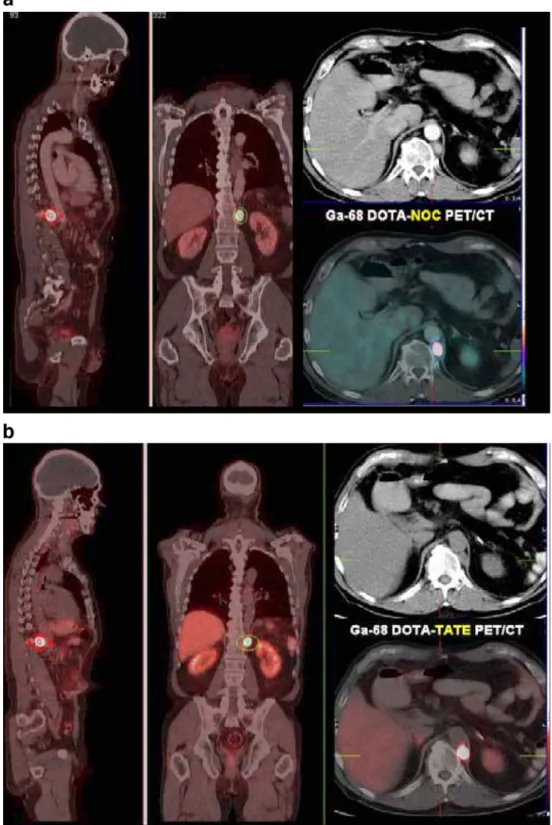
A clinical case report comparing [68Ga-DOTA]-NOC with [68Ga-DOTA]-TATE
In Fig. 3 an example is given of a patient with a neuroendocrine pancreatic carcinoma. The patient was scanned with [68Ga-DOTA]-NOC (Fig. 3a), [68 Ga-DOTA]-TATE (Fig. 3b) and [18F]fluorodeoxyglucose (FDG) (Fig. 3d). Eighty-five MBq [68Ga-DOTA]-NOC was injected. The scan, performed at 140 min p.i., revealed very intense uptake (SUVmax 152) in a left retrocrural
lymph node metastasis (Fig. 3a) as well as in a
para-pancreatic lymph node (SUV 9.2) and in a very small lesion near the processus uncinatus of the pancreas (SUV 6.8). In addition, a small (10 mm) liver metastasis in the right inferior liver segment (S VI) was detected (SUV 11.6) (Fig. 3c), which had also been described in an MRI study. The second PET/CT was performed 3 weeks later after i.v. injection of 130 MBq of [68Ga-DOTA]-TATE, starting the acquisition 85 min p.i. Again, very high uptake (SUVmax 103) was seen in the retrocrural metastasis
(Fig. 3b), but no other lesions were detectable (Fig.3c). There was no increased glucose metabolism (normal [18F]FDG PET/CT) in any of the lesions shown by the receptor PET/CT studies (Fig.3d).
Discussion
The use of 68Ga in nuclear oncology is becoming of increasing interest as it is a generator-produced, easily available positron emitter.68Ga has a physical half-life of 68 min, which is compatible with the pharmacokinetics of most radiopharmaceuticals of low molecular weight such as antibody fragments, peptides, aptamers, oligonucleo-tides, etc. [37, 38]. 68Ga decays to 89% by positron emission and to 11% via electron capture. The parent isotope68Ge has a very long half-life of 270.8 days, which allows routine manufacture and shipment, while the Table 3 Biodistribution in AR4-2J tumour-bearing rats and tissue ratios at 4 h and 24 h p.i. of [111In-DOTA]-NOC, [67Ga-DOTA]-NOC and [67Ga-DOTA]-TOC
Site [111In-DOTA]-NOC [67Ga-DOTA]-NOC [67Ga-DOTA]-TOC
4 h 4 h blockeda 24 h 4 h 4 h blockeda 24 h 4 h 4 h blockeda 24 h
Blood 0.02±0.00 0.03±0.01 0.01±0.00 0.03±0.01 0.02±0.01 0.01±0.00 0.07±0.006** 0.06±0.00 0.01±0.005 Tumour 2.96±0.48 0.24±0.01 2.65±0.41 4.24±0.37* 0.30±0.00 4.02±0.32* 4.6±1.18** 0.28±0.00 2.70±0.17** Kidneys 1.37±0.21 1.83±0.02 1.27±0.10 0.95±0.18 0.91±0.13 0.79±0.06* 1.55±0.25** 1.64±0.018 1.37±0.18** Adrenals 8.24±1.16 0.55±0.07 1.02±0.17 14.14±1.46* 0.32±0.05 6.46±0.60* 10.90±0.35** 0.16±0.00 6.35±0.64 Pancreas 7.94±0.71 0.16±0.03 4.04±0.29 7.13±0.63 0.34±0.04 3.15±0.62 13.32±0.78** 0.299±0.00 6.14±1.04** Spleen 0.09±0.01 0.09±0.03 0.09±0.02 0.12±0.02 0.13±0.03 0.13±0.04 0.08±0.01** 0.088±0.00 0.14±0.11 Stomach 1.64±0.19 0.05±0.02 1.08±0.11 1.99±0.23 0.07±0.01 0.91±0.05 1.76±0.11 0.06±0.00 0.912± 0.22 Bowel 0.36±0.26 0.07±0.01 0.27±0.15 0.26±0.04 0.04±0.01 0.79±0.06* 0.23±0.02 0.04±0.00 0.18±0.006** Liver 0.21±0.04 0.22±0.02 0.15±0.01 0.95±0.09* 1.04±0.11 0.93±0.05* 0.09±0.01** 0.12±0.00 0.10±0.07** Lung 0.06±0.01 0.05±0.001 0.05±0.02 0.13±0.02* 0.12±0.01 0.13±0.03* 0.09±0.01** 0.08±0.00 0.11±0.10 Heart 0.01±0.00 0.02±0.004 0.01±0.00 0.05±0.00 0.04±0.01 0.04±0.01* 0.03±0.01** 0.02±0.00 0.015±0.003** Pituitary 6.21±0.73 0.18±0.05 5.44±0.79 11.08±1.41* 0.20±0.03 6.00±0.66 10.33±1.90 0.21±0.00 6.65±0.06 Tumour to tissue ratios
Tumour/blood 148.0 265.0 142.0 402.0 68.5 270.0
Tumour/kidney 2.2 2.1 4.5 5.1 3.1 1.97
Tumour/liver 14.1 17.7 4.5 4.3 53.2 27.0
Values are the mean of % IA/g ±SD for groups of three or four animals IA injected activity
*,** Significant differences (p<0.05): *between [111In-DOTA]-NOC and [67Ga-DOTA]-NOC; **between [67Ga-DOTA]-NOC and [67Ga-DOTA]-TOC
a
chemical properties of Ge(IV) and Ga(III) are sufficiently different to allow several different methods of efficient separation.
A particularly fascinating group of 68Ga-based radio-pharmaceuticals are chelator-modified regulatory peptides that, owing to their small size, have ideal pharmacokinetics compatible with the short-lived 68Ga. Particularly
somato-statin derivatives such as [natGa-DOTA]-TOC and [nat Ga-DOTA]-TATE show outstanding binding affinity to sst2 that is a factor of 4–8 higher compared with the corresponding yttrium or indium derivatives [5, 20, 35]. The higher binding affinity resulted in significantly higher tumour uptake in a tumour-bearing mouse model [20,21]. These positive preclinical results have prompted several
Fig. 3 Comparison between
68
Ga-DOTA-NOC and68 Ga-DOTA-TATE in the same patient (with a metastatic neu-roendocrine pancreatic tumour), along with an [18F]FDG PET/ CT scan. The first whole-body PET/CT study (Siemens biog-raph duo) was performed after i.v. injection of 85 MBq68
Ga-DOTA-NOC and revealed very intense uptake (SUVmaxof 152)
in a left retrocrural lymph node metastasis (A. In addition, a liver metastasis (about 10 mm in diameter on MRI) in the right inferior liver segment (S VI) (SUV 11.6, C: a) and a very small parapancreatic lymph node metastasis (C: c) were detected. The second PET/CT scan was performed 3 weeks later after i.v. injection of 130 MBq of68Ga-DOTA-TATE. Again, very high uptake (SUVmax103) was seen in the
retrocrural metastasis (B), but no other lesions were detectable (C: b,d). There was no in-creased glucose metabolism (normal18F-FDG PET/CT, D) in any of the lesions shown by receptor PET/CT
clinical groups to use [68Ga-DOTA]-TOC and [68 Ga-DOTA]-TATE in patient studies [22–27,32].
We have recently reported that [111In-DOTA]-NOC may be a promising candidate for somatostatin receptor-positive tumour targeting because it recognises three somatostatin receptor subtypes with high affinity and may therefore target a broader range of tumours. Indeed, some preliminary results suggest that this new radiopeptide, if labelled with
68
Ga, locates more metastases than the second-generation radiopeptides [30, 39–41], indicating that these new peptides are also pharmacologically superior compared with their111In congeners. We designed a study to support these early clinical observations with firm preclinical pharmacological data using the 68Ga congener,67Ga.
A general trend was seen in regard to the binding affinity and the affinity profile of the GaIIIvs MIII-DOTA-peptides Fig. 3 (continued)
(MIII = InIII, LuIII, YIII). On hsst2, the binding affinities were significantly higher for the gallium-labelled peptides than for the MIII-DOTA-peptides (p<0.0125). The differ-ence was not significant for [GaIII-DOTA]-NOC-ATE vs [YIII-DOTA]-NOC-ATE (p>=0.124) but otherwise the GaIIIderivatives had a two- to eightfold higher affinity.
On hsst3 there was a tendency in the opposite direction: the affinities of the GaIII-complexed peptides were two- to fivefold lower. On hsst5 no clear tendency was obvious. None of the peptides has affinity to sst1 but surprisingly some emerging hsst4 affinity can be seen, especially when complexed with GaIII. We do not yet know which structural features are responsible for the latter.
A more striking advantage of the 67Ga-DOTA-peptides becomes evident upon studying the rate of internalisation into different cell lines. In sst2-expressing cells all 67 Ga-labelled peptides showed high and specific internalisation with a significantly higher rate compared with the 111In congeners. The difference between the two radionuclides became more striking when the internalisation was studied in HEK-sst2 cells, which express a higher number of receptors compared with AR4-2J cells.
In an earlier paper, Froidevaux et al. found no statistically significant difference between [67 Ga-DOTA]-TOC and [111In-DOTA]-TOC in AR4-2J cells [21]. The present data, however, showed a significant difference between the two radiopeptides. The internalisation into HEK-sst3 was comparatively low with no uptake of [67Ga/111In-DOTA]-TOC, as expected, and paralleling the very low binding affinity of these two peptides to sst3. In contrast to the sst2 data, the general tendency was for the
111
In-labelled peptides to show more efficient internal-isation into sst3, with the exception of [67Ga-DOTA]- and
111
In-DOTA]-NOC, which internalised at an equal rate, not corresponding to the fivefold difference in binding affinity. None of the radiopeptides internalises into sst5, which most likely is an intrinsic property of this receptor [42]. These in vitro pharmacological data indicate that [67 Ga-DOTA]-BOC is the most promising radiopeptide to develop. However, as it showed some instability during labelling, we decided to study [67Ga-DOTA]-NOC in our tumour model. The analysis of the biodistribution at 4 and 24 h showed a significantly higher tumour uptake of [67 Ga-DOTA]-NOC compared with [111In-DOTA]-NOC. We hypothesise that the more rapid internalisation kinetics measured in vitro contributes to the increased in vivo uptake. We recently found that in a group of six somatostatin-based octapeptides labelled with 99mTc and
111
In, a good correlation indeed existed between tumour and pancreas uptake in vivo and the rate of internalisation into the AR4-2J cell line in vitro [4]. The uptake in other somatostatin receptor-positive tissues, such as the adrenals, the stomach and the bowel, follows this trend.
The potential of an imaging probe depends, among other parameters, on the target-to-non-target ratios, especially the tumour-to-blood ratio. All three radiopeptides showed a very high tumour-to-blood ratio, [67Ga-DOTA]-NOC hav-ing the highest among them. In addition, the tumour-to-kidney ratio of 4.5 at 4 h and 5.1 at 24 h is, to the best of our knowledge, the highest of any radiometal-labelled peptide reported so far. On the other hand, the tumour-to-liver ratio was clearly lowest for [67Ga-DOTA]-NOC. This result was due to the almost fivefold higher liver uptake of [67Ga-DOTA]-NOC compared with that of [111 In-DOTA]-NOC, which is somewhat surprising considering the much lower log D value (higher hydrophilicity; see Table 4) of [67Ga-DOTA]-NOC compared with [111In-DOTA]-NOC. It is currently unclear whether active mechanisms including organic anion transporters are involved in the increased liver uptake.
An interesting finding from this study is that the radiometal indeed influences the pharmacological proper-ties of the radiopeptides; this earlier finding [21,35] can be extended to the new generation of somatostatin-based radiopeptides. Labelling with 67/68Ga results in peptides that are superior to those labelled with111In,90Y or177Lu. We have explained this difference by a difference in the coordination number and geometry of the various radio-metal complexes [20]. The model peptide GaIII-DOTA-D -PheNH2showed a cis-pseudo-octahedral geometry without
the coordination of the amide linkage and the corre-sponding trans carboxy methyl group, whereas the YIII complex is octacoordinate including the amide carboxy oxygen and the carboxymethyl group. The complex geometry is a somewhat distorted square antiprism [20]. The non-involved amide linkage of the GaIII complex consequently offers more flexibility, the free arm acting as a spacer between peptide and chelate. This may lead to the higher binding affinity on hsst2 whereas the free carboxyl-ate group may favour kidney excretion.
In summary, these data suggest that most, if not all,
68
Ga-labelled second- and third-generation somatostatin-based radiopeptides show superior properties in imaging somatostatin receptor-positive tumours compared with first-generation somatostatin-based radiopeptides. This is due to superior pharmacological properties on hsst2, which is the most densely and frequently expressed receptor subtype. Despite some loss in hsst3 and hsst5 affinity, the
radio-Table 4 Log D at pH=7.4 of radiolabelled conjugates
Compound name Log D (pH=7.4)
[67Ga-DOTA]-NOC −2.88±0.12
gallium-labelled peptides still show a broad hsst profile. Given the higher sensitivity of the PET technique, [68 Ga-DOTA]-NOC and [68Ga-DOTA]-BOC should also be further developed into new PET tracers for extended clinical studies.
The intra-individual comparison of [68Ga-DOTA]-NOC and [68Ga-DOTA]-TATE (Fig. 3a–c) in a patient with
metastases of a neuroendocrine pancreatic carcinoma demonstrated that the broader somatostatin receptor sub-type profile of [68Ga-DOTA]-NOC (sst2, 3 and 5 affinity) and internalisation may be of clinical relevance as a significantly higher uptake of this radiopeptide was found (SUVmax 152) compared with the high-affinity but
sst2-selective radiopeptide [68Ga-DOTA]-TATE (SUVmax 103).
In addition, very small lesions were detected when using [68Ga-DOTA]-NOC as compared to [68Ga-DOTA]-TATE. Along with the 68Ge/68Ga generator, these tracers may be very cost effective, sensitive and readily available imaging agents. Furthermore, they cause low radiation doses to patients compared with existing tracers and their 111 In-labelled congeners [31]. Along with 64Cu-labelled radio-pharmaceuticals [43–45], 68Ga-labelled compounds are a fast-growing area of research which benefits from the availability of a commercial generator.
Acknowledgements P. Antunes acknowledges the PhD Fellowship of the Fundação para a Ciência e Tecnologia (Ref. SFRH/BD/3136/ 2000). In addition, P. Antunes, M. Ginj, M. Walter and H. Maecke acknowledge the support from the Swiss National Science Foundation project No. 3100A0-100390, BBW project No C00.0091, and the network of excellence, European Molecular Imaging Laboratories (EMIL). The support provided by Novartis Pharma in respect of ESI-MS analysis is gratefully acknowledged. We thank Dr. S. Schulz for the sst3-transfected human embryonic kidney 293 cells. The authors thank K. Hinni and S. Tschumi for biological technical assistance. This work was performed within the COST B12 Action.
References
1. Reubi JC. Peptide receptors as molecular targets for cancer diagnosis and therapy. Endocr Rev 2003;24:389–427.
2. Maina T, Nock B, Nikolopoulou A, Sotiriou P, Loudos G, Maintas D, et al. [99mTc]Demotate, a new 99mTc-based [Tyr3]octreotate analogue for the detection of somatostatin receptor-positive tumours: synthesis and preclinical results. Eur J Nucl Med Mol Imaging 2002;29:742–53.
3. Decristoforo C, Mather SJ, Cholewinski W, Donnemiller E, Riccabona G, Moncayo R. 99mTc-EDDA/HYNIC-TOC: a new
99m
Tc-labelled radiopharmaceutical for imaging somatostatin receptor-positive tumours; first clinical results and intra-patient comparison with111In-labelled octreotide derivatives. Eur J Nucl
Med 2000;27:1318–25.
4. Storch D, Behe M, Walter MA, Chen J, Powell P, Mikolajczak R, et al. Evaluation of [99mTc/EDDA/HYNIC0]octreotide derivatives
compared with [111In-DOTA0,Tyr3, Thr8]octreotide and [111
In-DTPA0]octreotide: does tumor or pancreas uptake correlate with
the rate of internalization? J Nucl Med 2005;46:1561–9.
5. de Jong M, Bakker WH, Krenning EP, Breeman WA, van der Pluijm ME, Bernard BF, et al. Yttrium-90 and indium-111 labelling, receptor binding and biodistribution of [DOTA0,D -Phe1,Tyr3]octreotide, a promising somatostatin analogue for radionuclide therapy. Eur J Nucl Med 1997;24:368–71.
6. Krenning EP, Kwekkeboom DJ, Bakker WH, Breeman WA, Kooij PP, Oei HY, et al. Somatostatin receptor scintigraphy with [111In-DTPA-D-Phe1]- and [123I-Tyr3]-octreotide: the Rotter-dam experience with more than 1000 patients. Eur J Nucl Med 1993;20:716–31.
7. Smith-Jones PM, Stolz B, Bruns C, Albert R, Reist HW, Fridrich R, et al. Gallium-67/gallium-68-[DFO]-octreotide—a potential radiopharmaceutical for PET imaging of somatostatin receptor-positive tumors: synthesis and radiolabeling in vitro and prelim-inary in vivo studies. J Nucl Med 1994;35:317–25.
8. Henriksen G, Schottelius M, Poethko T, Hauser A, Wolf I, Schwaiger M, et al. Proof of principle for the use of11C-labelled peptides in tumour diagnosis with PET. Eur J Nucl Med Mol Imaging 2004;31:1653–7.
9. Wester H-J, Schottelius M, Scheidhauer K, Meisetschläger G, Herz M, Rau F, et al. PET imaging of somatostatin receptors: design, synthesis and preclinical evaluation of a novel 18 F-labelled, carbohydrated analogue of octreotide. Eur J Nucl Med Mol Imaging 2002;30:117–22.
10. Sprague JE, Peng Y, Sun X, Weisman GR, Wong EH, Achilefu S, et al. Preparation and biological evaluation of copper-64-labeled Tyr3-octreotate using a cross-bridged macrocyclic chelator. Clin
Cancer Res 2004;10:8674–82.
11. Waldherr C, Pless M, Maecke H, Schumacher T, Crazzolara A, Nitzsche E, et al. Tumor response and clincical benefit in neuroendocrine tumors after 7.4 GBq 90Y-DOTATOC. J Nucl
Med 2002;43:610–6.
12. Otte A, Mueller-Brand J, Dellas S, Nitzsche E, Herrmann R, Maecke H. Yttrium-90-labelled somatostatin-analogue for cancer treatment. Lancet 1998;351:417–8.
13. Kwekkeboom DJ, Mueller-Brand J, Paganelli G, Anthony LB, Pauwels S, Kvols LK, et al. Overview of results of peptide receptor radionuclide therapy with 3 radiolabeled somatostatin analogs. J Nucl Med 2005;46:62S–66S.
14. Bodei L, Cremonesi M, Zoboli S, Grana C, Bartolomei M, Rocca P, et al. Receptor-mediated radionuclide therapy with 90 Y-DOTATOC in association with amino acid infusion: a phase I study. Eur J Nucl Med 2003;30:207–16.
15. de Jong M, Breeman WA, Bernard BF, Bakker WH, Schaar M, van Gameren A, et al. [177Lu-DOTA0,Tyr3]octreotate for
somato-statin receptor-targeted radionuclide therapy. Int J Cancer 2001;92:628–33.
16. Norenberg JP, Krenning BJ, Konings IR, Kusewitt DF, Nayak TK, Anderson TL, et al. 213Bi-[DOTA0, Tyr3]octreotide peptide receptor radionuclide therapy of pancreatic tumors in a preclinical animal model. Clin Cancer Res 2006;12:897–903.
17. Jensen RT. Carcinoid and pancreatic endocrine tumors: recent advances in molecular pathogenesis, localization, and treatment. Curr Opin Oncol 2000;12:368–77.
18. Maecke HR, Hofmann M, Haberkorn U.68Ga-labeled peptides in tumor imaging. J Nucl Med 2005;46:172S–8S.
19. Eisenwiener KP, Prata MI, Buschmann I, Zhang HW, Santos AC, Wenger S, et al. NODAGATOC, a new chelator-coupled somato-statin analogue labeled with [67/68Ga] and [111In] for SPECT, PET, and targeted therapeutic applications of somatostatin receptor (hsst2) expressing tumors. Bioconjug Chem 2002;13:530–41. 20. Heppeler A, Froidevaux S, Mäcke HR, Jermann E, Béhé M,
Powell P, et al. Radiometal-labelled macrocyclic chelator-derivatised somatostatin analogue with superb tumour-targeting properties and potential for receptor-mediated internal radiother-apy. Chemistry A European Journal 1999;5:1016–23.
21. Froidevaux S, Eberle AN, Christe M, Sumanovski L, Heppeler A, Schmitt JS, et al. Neuroendocrine tumor targeting: study of novel gallium-labeled somatostatin radiopeptides in a rat pancreatic tumor model. Int J Cancer 2002;98:930–7.
22. Hofmann M, Maecke H, Börner A, Weckesser E, Schöffski P, Oei M, et al. Biokinetics and imaging with the somatostatin receptor PET radioligand 68Ga-DOTATOC: preliminary data. Eur J Nucl Med 2001;28:1751–7.
23. Kowalski J, Henze M, Schuhmacher J, Maecke HR, Hofmann M, Haberkorn U. Evaluation of positron emission tomography imaging using [68Ga]-DOTA-D Phe1-Tyr3-octreotide in
compari-son to [111In]-DTPAOC SPECT. First results in patients with
neuroendocrine tumors. Mol Imaging Biol 2003;5:42–8. 24. Henze M, Dimitrakopoulou-Strauss A, Milker-Zabel S,
Schuhmacher J, Strauss LG, Doll J, et al. Characterization of
68
Ga-DOTA-D-Phe1-Tyr3-octreotide kinetics in patients with meningiomas. J Nucl Med 2005;46:763–9.
25. Henze M, Schuhmacher J, Dimitrakopoulou-Strauss A, Strauss LG, Maecke HR, Eisenhut M, et al. Exceptional increase in somatostatin receptor expression in pancreatic neuroendocrine tumour, visualised with68Ga-DOTATOC PET. Eur J Nucl Med Mol Imaging 2004;31:466.
26. Henze M, Schumacher T, Hipp P, Kowalski J, Becker D, Doll J, et al. PET imaging of somatostatin receptors using [68Ga]DOTA-D -Phe1-Tyr3-octreotide: first results in patients with meningiomas. J Nucl Med 2001;42:1053–6.
27. Dimitrakopoulou-Strauss A, Georgoulias V, Eisenhut M, Herth F, Koukouraki S, Macke HR, et al. Quantitative assessment of SSTR2 expression in patients with non-small cell lung cancer using68Ga-DOTATOC PET and comparison with18F-FDG PET.
Eur J Nucl Med Mol Imaging 2006;33:823–30.
28. Baum R, Niesen A, Leonhardi J, Wortmann R, Mueller D, Roesch F. Receptor PET/CT imaging of neuroendocrine tumours using the Ga-68 labelled, high affinity somatostatin analogue DOTA-1-Nal3 octreotide (DOTA-NOC): clinical results in 327 patients. Eur J Nucl Med Mol Imaging 2005;32 Suppl 1:S54–5. 29. Roesch F, Zhernosekov K, Filosofov D, Jahn M, Jennewein M,
Baum R, et al. Processing of Ge-68/Ga-68 generator eluates for labeling of biomolecules via bifunctional chelators. J Nucl Med 2006;47 Suppl 1:162P.
30. Baum R, Schmücking M, Wortmann R, Müller M, Zhernosekov K, Rösch F. Receptor PET/CT using the Ga-68 labelled somatostatin analog DOTA-1-Nal3-octreotide (DOTA-NOC): clin-ical experience in 140 patients. Nuklearmedizin 2005;44:A57. 31. Rufini V, Calcagni ML, Baum RP. Imaging of neuroendocrine
tumors. Semin Nucl Med 2006;36:228–47.
32. Win Z, Rahman L, Murrell J, Todd J, Al-Nahhas A. The possible role of 68Ga-DOTATATE PET in malignant abdominal
para-ganglioma. Eur J Nucl Med Mol Imaging 2006;33:506.
33. Wild D, Schmitt JS, Ginj M, Maecke HR, Bernard BF, Krenning E, et al. DOTA-NOC, a high-affinity ligand of somatostatin receptor subtypes 2, 3 and 5 for labelling with various radio-metals. Eur J Nucl Med Mol Imaging 2003;30:1338–47. 34. Ginj M, Chen J, Walter MA, Eltschinger V, Reubi JC, Maecke
HR. Preclinical evaluation of new and highly potent analogues of octreotide for predictive imaging and targeted radiotherapy. Clin Cancer Res 2005;11:1136–45.
35. Reubi JC, Schar JC, Waser B, Wenger S, Heppeler A, Schmitt JS, et al. Affinity profiles for human somatostatin receptor subtypes SST1–SST5 of somatostatin radiotracers selected for scintigraphic and radiotherapeutic use. Eur J Nucl Med 2000;27: 273–82.
36. Tulipano G, Stumm R, Pfeiffer M, Kreienkamp HJ, Hollt V, Schulz S. Differentialβ-arrestin trafficking and endosomal sorting of somatostatin receptor subtypes. J Biol Chem 2004;279:21374– 82.
37. Smith-Jones PM, Solit DB, Akhurst T, Afroze F, Rosen N, Larson SM. Imaging the pharmacodynamics of HER2 degradation in response to Hsp90 inhibitors. Nat Biotechnol 2004;22:701–6. 38. Smith-Jones PM, Solit D, Afroze F, Rosen N, Larson SM. Early
tumor response to Hsp90 therapy using HER2 PET: comparison with18F-FDG PET. J Nucl Med 2006;47:793–6.
39. Wild D, Maecke HR, Waser B, Reubi JC, Ginj M, Rasch H, et al.
68
Ga-DOTANOC: a first compound for PET imaging with high affinity for somatostatin receptor subtypes 2 and 5. Eur J Nucl Med Mol Imaging 2004;32:724.
40. Decristoforo C, von Guggenberg E, Haubner R, Rupprich M, Schwarz S, Virgolini I. Radiolabeling of DOTA-derivatised peptides with 68Ga via a direct approach—optimization and
routine clinical application. Nuklearmedizin 2005;44:A191–2. 41. Hofmann M, Oei M, Boerner AR, Maecke H, Geworski L,
Knapp WH, et al. Comparison of 68-DOTATOC and Ga-68-DOTANOC for radiopeptide PET. Nuklearmedizin 2005;44: A58.
42. Cescato R, Schulz S, Waser B, Eltschinger V, Rivier JE, Wester HJ, et al. Internalization of sst2, sst3, and sst5 receptors: effects of somatostatin agonists and antagonists. J Nucl Med 2006;47: 502–11.
43. Smith SV. Molecular imaging with copper-64. J Inorg Biochem 2004;98:1874–901.
44. Rossin R, Pan D, Qi K, Turner JL, Sun X, Wooley KL, et al.
64
Cu-labeled folate-conjugated shell cross-linked nanoparticles for tumor imaging and radiotherapy: synthesis, radiolabeling, and biologic evaluation. J Nucl Med 2005;46:1210–8.
45. Boswell CA, Sun X, Niu W, Weisman GR, Wong EH, Rheingold AL, et al. Comparative in vivo stability of copper-64-labeled cross-bridged and conventional tetraazamacrocyclic complexes. J Med Chem 2004;47:1465–74.
![Table 1 summarises the IC 50 values of the radiopeptides as their In III - and Ga III -complexed versions for the human somatostatin receptors 1–5 (hsst1–hsst5), including data published previously [35]](https://thumb-eu.123doks.com/thumbv2/123doknet/14841077.625093/5.892.75.825.650.1003/summarises-radiopeptides-complexed-somatostatin-receptors-including-published-previously.webp)
![Table 2 Internalisation of [ 67 Ga]- or [ 111 In]-labelled peptides into AR4-2J, HEK-sst2 and HEK-sst3 cells after 4-h incubation at 37°C a](https://thumb-eu.123doks.com/thumbv2/123doknet/14841077.625093/6.892.78.817.739.996/table-internalisation-labelled-peptides-hek-hek-cells-incubation.webp)