HAL Id: hal-02124331
https://hal.archives-ouvertes.fr/hal-02124331
Submitted on 10 Jul 2020
HAL is a multi-disciplinary open access
archive for the deposit and dissemination of sci-entific research documents, whether they are pub-lished or not. The documents may come from teaching and research institutions in France or abroad, or from public or private research centers.
L’archive ouverte pluridisciplinaire HAL, est destinée au dépôt et à la diffusion de documents scientifiques de niveau recherche, publiés ou non, émanant des établissements d’enseignement et de recherche français ou étrangers, des laboratoires publics ou privés.
silver-containing glasses
Yannick Petit, Chang-Yyun Park, Jin-Myoung Mok, Evgeniya Smetanina,
Benoit Chimier, Guillaume Duchateau, Thierry Cardinal, Lionel Canioni,
Seun-Han Park
To cite this version:
Yannick Petit, Chang-Yyun Park, Jin-Myoung Mok, Evgeniya Smetanina, Benoit Chimier, et al.. Ul-trashort laser induced spatial redistribution of silver species and nano-patterning of etching selectivity in silver-containing glasses. Optics Express, Optical Society of America - OSA Publishing, 2019, 27 (10), pp.13675-13680. �10.1364/OE.27.013675�. �hal-02124331�
Ultrashort laser induced spatial redistribution
of silver species and nano-patterning of
etching selectivity in silver-containing glasses
Y. P
ETIT,
1,2,5C.-H. P
ARK,
2,3J.-M. M
OK,
1,3E. S
METANINA,
2,4B. C
HIMIER,
2G. D
UCHATEAU,
2T. C
ARDINAL,
1L. C
ANIONI,
2ANDS.-H. P
ARK3,61University of Bordeaux, CNRS, ICMCB, UMR 5026, 87 avenue du Dr. A. Schweitzer, 33608 Pessac
cedex, France
2University of Bordeaux, CNRS, CEA, CELIA, UMR 5107, 351 Cours de la Liberation, 33405 Talence
Cedex, France
3Institute of Physics and Applied Physics, Yonsei University, Seoul 03722, Korea 4Department of Physics, University of Gothenburg, SE-412 96 Goteborg, Sweden 5yannick.petit@u-bordeaux.fr
6shpark@yonsei.ac.kr
Abstract: Femtosecond laser-induced spatial redistribution of silver species (ions, clusters,
and hole centers) in a silver-containing phosphate glass is investigated by correlative means of near-field scanning optical microscopy (NSOM) images, numerical simulations, chemical micro-probe analysis, and nanoscale spatial profiles after soft etching. In particular, we found that the chemical etching selectivity for nanoscale patterning is strongly dependent upon the irradiation of femtosecond laser due to the spatial redistribution of silver species within the affected area. These results strongly indicate that controlling the distribution of silver species by femtosecond laser irradiation may open new routes for surface nanoscale chemical and/or spatial patterning for the fabrication of 2D surface photonic crystals.
© 2019 Optical Society of America under the terms of the OSA Open Access Publishing Agreement
1. Introduction
Femtosecond (fs) laser modification technology of materials has made tremendous progress over the past decade, to access a large panel of applications that require special optical, physical and/or chemical properties associated with integrated bulk or surface functionalities [1]. While considering laser-induced element redistribution, most of the reported literature deals with processes in thermal regime with temperature increase above glass fusion [2,3]. Additional potentialities rely on the innovative synthesis of prepared materials with photosensitive agents [4]. In this framework, controlling the photosensitive agents under laser irradiation in a non-thermal regime, as well as the spatial redistribution and chemical evolution due to activated chemical reactivity, is of prime importance to achieve reliable nano- and micro-scale material changes with high optical quality. Such control and understanding is highly challenging since it involves multi-scale material changes, which requires correlative investigation approaches.
In this work, we present the correlative description of fs laser-induced spatial redistribution of silver species of our photosensitive silver-containing phosphate glasses, based on near-field scanning optical microscopy (NSOM) images, chemical micro-probe analysis, spatial profile after soft etching, and numerical simulations of the silver species redistribution. These results corroborate each other, and have led us to retrieve for the first time the nanoscale spatial profile of soft chemical etching rate of laser-induced modifications.
2. Experimental methods: glass preparation, laser irradiation and analyses
We have developed a silver-containing zinc phosphate glass 55ZnO/40P2O5/4Ag2O/1Ga2O3 (in mol. %), as previously detailed [5]. Direct laser writing (DLW) was carried out with a Yb
#358348 https://doi.org/10.1364/OE.27.013675
fs oscillator (A with a micros Fig. 1 probe and im DLW was translating the and laser irrad perpendicular
NSOM ex like light inje shown in Fig. interest. The control of a s frequency of single channe injection resu optical fiber t achieve reson the output of t 3. Results a During NSOM showed no su truly flat surf Indeed, by inj nm after a lo distribution o cross section Similarly to c distribution o excitation and While inje filter (665 nm signal and a w position of th decrease) insi image had als fluorescence e clusters is ext (Figs. 2(b) an non-resonant Amplitude Sys cope objective 1. NSOM setup in tip (100 nm aper maging (40 × , 0.60 s performed to e glass sample diance was 5-1 rly to these fluo xperiments wer ection was achi . 1. A nano-po
distance betw shear force de 32.768 kHz). el PMT detec ults from the p tip. Two wavel nant and non-re
the tapered opt
and discussio
M experiment urface height m
face at the stru jecting a laser ong-pass filter f fluorescent s along the diam confocal fluore f silver cluster d fluorescence ecting a He-N m, Edmund RG weakly brighter he fluorescent ide the annular so been obtaine excitation of b tremely weak w nd 2(c), red cur evanescent cou stèmes, T-puls e (Mitutoyo, M n illumination mo rture). LD: laser d 0NA)), LPF: long o create a stru along the lase 10 TW/cm2, ty orescent structu re performed o
ieved with a ta sitioning stage ween the tip an
etection system The collection ctor) is a wide point-by-point lengths were c esonant evanes tical fiber tip in
on t, the feedbac modification wh uctures after p diode excitatio (Edmund GG silvers at the su meter of the flu escent imaging rs, the larger f collection from Ne laser source G-665), the NSO r double-ring d ring image f r silver cluster ed while collec both the random while excited a rve) are mostly upling.
e 500, 9.1 MH M Plan Apo NIR
ode, with light inj diode, objective len
pass filter, PMT: ucture with a er beam propag ypically. The ir ures, to make t n the sample in apered optical e allowed for u nd the sample m based on a q n part of the se e field collect scanning of onsidered, at 4 scent coupling nto free-optics ck control on hile scanning a polishing, whi on source at 40 G-435), the hig urface was ob uorescent tube g (not shown h fluorescence pe m planes below at 632.8 nm OM image (Fig distribution (6% from Fig. 2(a) r distribution ( cting without t mly distributed at 632.8 nm [7 y from the 632 Hz, 1030 nm, 3 R, 20 × N.A. 0.
ection from a tap nses OL (focusing photomultiplier de length of 400 gation. Sample rradiated samp them intersect t n illumination fiber probe tip ultrafine lateral e surface was quartz crystal etup (with a lo tion. The spat the surface sa 405 nm and 63 g of the non-ra propagation in
the shear for a laser induced ich excludes a
05 nm and col gh-resolution im
tained (see Fig es is given by F here), NSOM s edestal coming w surface. and collecting g. 2(b)) led to % increase) tha ), and to a da (Fig. 2(c), red the RG-665 lo d Ag+ ions and 7], we thus beli 2.8 nm residua 390 fs FWHM) .4) [5].
pered optical fiber g (20 × , 0.40NA) etector.
0 μm by longi e velocity was ple was then re the glass interf mode [6], whe p (100 nm ape l scanning of r stabilized by tuning fork (r ow NA objecti tial resolution ample with the 32.8 nm, to res adiating light f n the glass sub rce positioning d structure, ind any topologica llecting light a mage of the tr g. 2(a)). The h Fig. 2(c) (gree shows the clea g from the con g light with a l a non-zero ba at typically ma arker distributi curve). Simila ng-pass filter: d the 3D localiz ieve that meas al background, ), focused r ) itudinally 100 μm/s e-polished face. ere point-erture), as regions of feedback resonance ive and a n of light e tapered spectively field from strate. g system dicating a l artifact. above 435 ransverse horizontal en curve). ar annular ncomitant long-pass ckground atches the ion (17% ar NSOM since the zed silver surements , after the
Fig. 2 profile NSOM im the laser-indu counterpart. T (Figs. 2(b) an element (ions typically show samples and migration of irradiation: B enhancement by chemical a depletion of t annular struct reported by distributions c nanoparticles and/or the loc silver clusters distributions interaction at a local index density aspec non-resonant index experie output of the silver element Remarkab based imagin depletion of method appea composition quantitative) the reservoir [9]). Consequ 2. (a,b) NSOM im es.
mage with the H uced silver-me The proposed nd 2(c)) is that i s and clusters) ws a linear c their silver c silver elemen Bellec et al. ha of silver elem analysis from the silver rese tures (same stru
back-scattered could further b [10]; Third, l cal creation of s). Abou Khal of silver clus 632.8 nm with x change and t cts, thus to the NSOM coupli enced by the lig
tapered optic ts (silver ions a bly, the NSOM ng (instead of
silver ions and ars as a uniqu dependences, generally show depletion of si uently, NSOM mages with 405 n He-Ne injection ediated materia interpretation it corresponds ) after DLW, correlation betw content. Secon nts over meso ave shown by ments correlativ electron micro rvoir (about 1 uctures as thos d electron sca be thermally de local index ch new polarizabl
lil et al. recen
sters and asso h silver elemen the 3D distrib e local of silve ng efficiency a ght field in the al fiber tip. Th and clusters), a M experience a electron beam d silver eleme e imaging app while the ws a limited se
ilver ions, but and electron m nm and 633nm l n remarkably g al modificatio of the observ to a direct ima as discussed h ween the refr
nd, several rep oscopic scales high-resolutio vely located at oprobe, Desmo 0-20%) in the se of the presen anning microsc eveloped into 3 hange can resu le chemical bo ntly reported th ociated refract nts is mostly no bution of silve er element con appears as dire e sub-waveleng hus, it directly as shown in Fig at 632.8 nm se m approaches [8 ent redistributio proach since it chemical mic ensitivity of 1-not the creati microprobe app
laser injections; (c
gave access to on, comparativ ved spatial di age of the local
hereafter. Firs ractive index ports evidence (larger than on electron sc the position of oulin et al. ha e center of lase nt study) [9]; M copy that the 3D-localized pl ult from both ounds (as occur
he direct corre tive index cha on-resonant. A er elements ca ncentration (io ectly depending gth regime at t y depicts the s gs. 2(b) and 2(c eems to be th 8–10]) of the on. Although t provides a gr cro-probe ana -2%: Desmoul on of silver cl pear as highly c c) associated line new insight co vely to the UV stribution at 6 l redistribution st, our phosph of non-structu e of the laser 100 nm) dur anning micros f the silver clu ave observed th er-induced silv Marquestaut e created silve lasmonic meta a local density rring for the cr elation betwee ange [11]. Th Any correlation an mostly be r ons and cluster g on the local r the glass surfa spatial redistrib c). he first direct o laser-induced qualitative, th reat sensitivity alysis (althou
lin et al. had s
lusters (see Fig complementary e oncerning V-excited 632.8 nm n of silver hate glass ured bulk r-induced ring laser scopy the usters [8]; he partial ver-based t al. have er cluster llic silver ty change reation of en spatial hus, light n between related to rs). Thus, refractive ace, at the bution of optically-reservoir he NSOM y to local ugh fully seen only g. 3 from y tools.
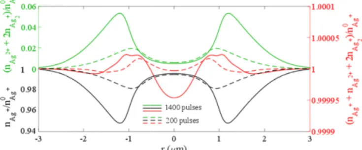
Fig. 3 Npulses cluste
To suppo performed wi silver ion mig and silver-con focal plane r0 including the centers (Ag2+) ions (black c induced clust overall conse central Ag+ d periphery of observed also silver elemen migration and stabilization remarkably m 2(c)), especia a slightly sma possibly corr curve). This description of To go fur considering th the present NS modified area [9]). Spatial e the simulation red curve) in curve). The op at the periph considered so drops by a fac glass. Betwe distribution (F 1.8 times sm chemically-se with non-trivi such etching 3. Numerical mode = 200 or 1400), s ers and hole center
ort the interpre ith our multi-sc gration and pho ntaining glass ( 0 = 2.5 µm). T silver ions (A ). Figure 3 sho curve), the tot ters and holes
rvation of silv depletion due t the irradiated o with the red nt distribution
d the accumu with silver cl matches the 1D
lly for Npulses = aller ring diame responding to corroborates f laser-activated
ther, spatially-he spatial distri SOM data. Fig a revealed afte etching rate dis
n of the associa n very good ag ptimized etchin ery of the las oft etching cond
ctor of 20.6, c en the silver FWHM = 310 maller, respectiv
ensitive materia ial etching sen
rate distributio
eling of the laser-i showing the profil s (red curve) and t
etation of non cale multi-puls otochemistry [ (intensity = 8.8 The full calcul Ag+), the indu ows the normal al silver elem (green curve: ver elements. T to their radial spot by chem curve of silve n (red curve) ulation at the lusters. Such D cross-section = 1400. The sil
eter than that o the non-reson the interpreta d mechanisms -distributed etc ibution of silve gure 4(a) depict er 6 minutes s tributions have ated simulated greement with ng rate profile ser-modified a ditions). At the orresponding t cluster local nm), with cen vely, than tha al modification nsitivity distribu
on (green curv
induced redistribu les of the silver io the overall silver e
n-resonant NS se multi-physic 12], considerin 8 TW/cm2, N pu lated redistribu uced silver clu lized calculated ments (red curv nAg2+ + 2.nAg The Ag+ distri outward diffu mical consump er cluster and confirms both periphery due silver element n of the He-N lver element re of the simulate nant NSOM d ation of the N of silver-conta ching rates hav er elements fro ts the experime soft etching wi e been designe etching proces h the experime shows the nor rea (typically e position of si
to a chemically lization, the e ntral and pedes at of the pristi n highly locate utions. Figure ve) with the mi
ution of silver spec ons (black curve), elements (green cu SOM, numeric cs numerical m ng similar laser ulses = 200 or 1 ution of silver usters (Ag2+) a d silver elemen ve: nAg+ + nA g2+). The nume ibution (black usion, but also ption to create hole distribut h the central e to the chem t distribution Ne injected N edistribution (F ed silver cluster double-ring str NSOM approa aining glasses i ve been invest om micro-probe ental surface m ith dionized w ed (Fig. 4(a), gr ss leads to a su ental surface p rmalized behav corresponding ilver clusters, t y hardened are etching rate s stal values bein ine glass. Thi ed in the center 4(b) provides micro-probe me
cies (8.8 TW/cm2, the induced silver urve). cal investigatio model of laser-r ilaser-rlaser-radiation pa 400, beam rad species was e and the remain nt distributions Ag2+ + 2.nAg2+), erical model o curve) shows o their reducti e the silver clu
tions). Still, th depletion du mical precipita (red curve of SOM experim Fig. 3, red curv rs (Fig. 3, gree ructure (Fig. ach, strengthe in an athermal tigated, by corr e measurement morphology of water (blue cur reen curve) [13 urface profile (
profile (Fig. 4 vior of the pris g to 0.22 nm/ the etching rate ea of the laser-shows a Gaus
ng 2.5 times la s shows a ver r of the laser irr then the comp easurement of t , r ons were -activated arameters dius at the extracted, ning hole s: the Ag+ , and the obeys the both the on at the usters (as he overall ue to ion ation and f Fig. 3) ment (Fig. ve) shows en curve), 2(c), red ening our regime. relatively ts [9] and the laser-rve, from 3], so that (Fig. 4(a), 4(a), blue tine glass /s for the e strongly -modified ssian-like arger and ry strong radiation, parison of the silver
element distribution (blue curve, from [11]) and the present non-resonant NSOM measurement interpreted as the refractive index spatial distribution (red curve), independently revealing the presence of the three same laser-affected areas.
Fig. 4. (a) Experimental topological profile (blue curve) and simulated etching-induced topological profile (red curve) adjusted by optimizing the normalized etching rate distribution (green curve). (b) Silver element distribution by micro-probe measurement (blue curve, from [9]) and NSOM measurement (red curve, from Fig. 3(d)) with respect to the optimized normalized etching rate distribution (green curve).
Indeed, the NSOM measurements of silver element accumulation and cluster creation correlate with a very low etching rate (radius between 1 to 1.8 μm). Moreover, the silver reservoir depletion is independently shown by NSOM and electron micro-probe. Finally, the estimation of a very strong on-axis etching rate (240 nm, FWHM, green curve) corroborates the very narrow on-axis silver depletion (sub-200 nm, FWHM, blue curve), which may even be seen in the on-axis NSOM trace (red curve). Thus, laser-induced index change distributions [11] as well as the spatially-distributed etching rate depend here on the local concentration and nature of silver species [14], and on additional glass matrix rearrangements such as release of molecular oxygen to ensure charge compensation and material stabilization, as reported in thermal poling [15] or for glass irradiations in a thermal regime [16].
4. Conclusion
In conclusion, we have reported for the first time the correlative description of laser-induced silver redistribution in terms of chemical micro-probe, NSOM and numerical modeling. The results significantly strengthen the understanding of material modifications in such glasses in a non-thermal interaction regime. In particular, it has been found that the spatial distribution of species in silver-containing glasses produced by femtosecond laser irradiation has a significant effect on chemical etching selectivity. This should help for future development of nanoscale surface chemical patterning, such as for 2D photonics crystal applications [17].
Funding
This study has been carried out with financial support from the French State, with French National Research Agency (ANR) in the frame of “the investments for the future”
Programme IdEx Bordeaux – LAPHIA (ANR-10-IDEX-03-02), Grand Équipement National pour le Calcul Intensif (GENCI) (A0030506129), and by the BK21 PLUS program through the National Research Foundation (NRF) funded by the Ministry of Education of Korea.
References
1. M. Malinauskas, A. Žukauskas, S. Hasegawa, Y. Hayasaki, V. Mizeikis, R. Buividas, and S. Juodkazis, “Ultrafast laser processing of materials: from science to industry,” Light Sci. Appl. 5(8), e16133 (2016). 2. M. Shimizu, M. Sakakura, S. Kanehira, M. Nishi, Y. Shimotsuma, K. Hirao, and K. Miura, “Formation
mechanism of element distribution in glass under femtosecond laser irradiation,” Opt. Lett. 36(11), 2161–2163 (2011).
3. T. T. Fernandez, M. Sakakura, S. M. Eaton, B. Sotillo, J. Siegel, J. Solis, Y. Shimotsuma, and K. Miura, “Bespoke photonic devices using ultrafast laser driven ion migration in glasses,” K. Miura, Prog. Mater. Sci. 94, 68–113 (2018).
4. A. Royon, Y. Petit, G. Papon, M. Richardson, and L. Canioni, “Femtosecond laser induced photochemistry in materials tailored with photosensitive agents,” Opt. Mater. Express 1(5), 866–882 (2011).
5. K. Bourhis, A. Royon, M. Bellec, J. Choi, A. Fargues, M. Treguer, J.-J. Videau, D. Talaga, M. Richardson, T. Cardinal, and L. Canioni, “Femtosecond laser structuring and optical properties of a silver-containing glass,” J. Non-Cryst. Solids 356(44-49), 2658–2665 (2010).
6. H. Ahn, J. Kim, D.-K. Kim, E. Lee, D.-S. Shin, H. Doh, and S.-H. Park, “High-resolution temporal and spatial photoluminescence measurement of a multiple-quantum-well structure at room temperature,” Proc. SPIE 7214, 721403 (2009).
7. K. Bourhis, A. Royon, G. Papon, M. Bellec, Y. Petit, L. Canioni, M. Dussauze, V. Rodriguez, L. Binet, D. Caurant, M. Treguer, J.-J. Videau, and T. Cardinal, “Formation and thermo-assisted stabilization of luminescent silver clusters in photosensitive glasses,” Mater. Res. Bull. 48(4), 1637–1644 (2013).
8. M. Bellec, A. Royon, B. Bousquet, K. Bourhis, M. Treguer, T. Cardinal, M. Richardson, and L. Canioni, “Beat the diffraction limit in 3D direct laser writing in photosensitive glass,” Opt. Express 17(12), 10304–10318 (2009).
9. J.-C. Desmoulin, Y. Petit, T. Cardinal, M. Dussauze, M. Lahaye, and L. Canioni, “Femtosecond laser structuring of silver-containing glass: silver redistribution, selective etching, and surface topology engineering,” J. Appl. Phys. 118(21), 213104 (2015).
10. N. Marquestaut, Y. Petit, A. Royon, T. Cardinal, and L. Canioni, “Three-Dimensional Silver Nanoparticle Formation Using femtosecond laser irradiation in phosphate glasses: analogy with photography,” Adv. Funct. Mater. 24(37), 5824–5832 (2014).
11. A. Abou Khalil, J.-P. Bérubé, S. Danto, J.-C. Desmoulin, T. Cardinal, Y. Petit, R. Vallée, and L. Canioni, “Direct laser writing of a new type of waveguides in silver containing glasses,” Sci. Rep. 7(1), 11124 (2017). 12. E. Smetanina, B. Chimier, Y. Petit, N. Varkentina, L. Hirsch, E. Fargin, T. Cardinal, L. Canioni, and G.
Duchateau, “Modeling of cluster organization in metal-doped oxide glasses irradiated by a train of femtosecond laser pulses,” Phys. Rev. A (Coll. Park) 93(1), 013846 (2016).
13. B. Da Costa Fernandes, M. Pfiffer, P. Cormont, M. Dussauze, B. Bousquet, E. Fargin, and J. Neauport, “Understanding the effect of wet etching on damage resistance of surface scratches,” Sci. Rep. 8(1), 1337 (2018).
14. Y. Petit, S. Danto, T. Guérineau, A. Abou Khalil, A. Le Camus, E. Fargin, G. Duchateau, J.-P. Bérubé, R. Vallée, Y. Messaddeq, T. Cardinal, and L. Canioni, “On the femtosecond laser-induced photochemistry in silver-containing oxide glasses: mechanisms, related optical and physico-chemical properties, and technological applications,” Adv. Opt. Technol. 7(5), 291–309 (2018).
15. M. Dussauze, V. Rodriguez, A. Lipovskii, M. Petrov, C. Smith, K. Richardson, T. Cardinal, E. Fargin, and E. Kamitsos, “How does thermal poling affect the structure of soda-lime glass?” J. Phys. Chem. C 114(29), 12754– 12759 (2010).
16. N. Varkentina, M. Dussauze, A. Royon, M. Ramme, Y. Petit, and L. Canioni, “High repetition rate femtosecond laser irradiation of fused silica studied by Raman spectroscopy,” Opt. Mater. Express 6(1), 79–89 (2015). 17. N. Ganesh, W. Zhang, P. C. Mathias, E. Chow, J. A. N. T. Soares, V. Malyarchuk, A. D. Smith, and B. T. Cunningham, “Enhanced fluorescence emission from quantum dots on a photonic crystal surface,” Nat. Nanotechnol. 2(8), 515–520 (2007).