HAL Id: hal-00297629
https://hal.archives-ouvertes.fr/hal-00297629
Submitted on 10 Jul 2007
HAL is a multi-disciplinary open access
archive for the deposit and dissemination of
sci-entific research documents, whether they are
pub-lished or not. The documents may come from
teaching and research institutions in France or
abroad, or from public or private research centers.
L’archive ouverte pluridisciplinaire HAL, est
destinée au dépôt et à la diffusion de documents
scientifiques de niveau recherche, publiés ou non,
émanant des établissements d’enseignement et de
recherche français ou étrangers, des laboratoires
publics ou privés.
from culturing experiments
L. J. de Nooijer, G. J. Reichart, A. Dueñas-Bohórquez, M. Wolthers, S. R.
Ernst, P. R. D. Mason, G. J. van der Zwaan
To cite this version:
L. J. de Nooijer, G. J. Reichart, A. Dueñas-Bohórquez, M. Wolthers, S. R. Ernst, et al.. Copper
incorporation in foraminiferal calcite: results from culturing experiments. Biogeosciences, European
Geosciences Union, 2007, 4 (4), pp.493-504. �hal-00297629�
www.biogeosciences.net/4/493/2007/ © Author(s) 2007. This work is licensed under a Creative Commons License.
Biogeosciences
Copper incorporation in foraminiferal calcite: results from
culturing experiments
L. J. de Nooijer1,*, G. J. Reichart1, A. Due ˜nas-Boh´orquez1, M. Wolthers1, S. R. Ernst1, P. R. D. Mason1, and G. J. van der Zwaan1
1Dept. of Earth Sciences, Utrecht University, Budapestlaan 4, 3584 CD Utrecht, The Netherlands
*now at: Institute for Research on Evolution of the Earth, Japan Agency for Marine Science and Technology (JAMSTEC),
2–15 Natsushima-cho, 237-0061, Yokosuka, Japan
Received: 5 March 2007 – Published in Biogeosciences Discuss.: 2 April 2007 Revised: 2 July 2007 – Accepted: 2 July 2007 – Published: 10 July 2007
Abstract. A partition coefficient for copper (DCu) in
foraminiferal calcite has been determined by culturing indi-viduals of two benthic species under controlled laboratory conditions. The partition coefficient of a trace element (TE) is an emperically determined relation between the TE/Ca ra-tio in seawater and the TE/Ca rara-tio in foraminiferal calcite and has been established for many divalent cations. Despite its potential to act as a tracer of human-induced, heavy metal pollution, data is not yet available for copper. Since partition coefficients are usually a function of multiple factors (seawa-ter temperature, pH, salinity, metabolic activity of the organ-ism, etc.), we chose to analyze calcite from specimens cul-tured under controlled laboratory conditions. They were sub-jected to different concentrations of Cu2+ (0.1–20 µmol/l) and constant temperature (10 and 20◦C), seawater salinity and pH. We monitored the growth of new calcite in speci-mens of the temperate, shallow-water foraminifer Ammonia
tepida and in the tropical, symbiont-bearing Heterostegina depressa. Newly formed chambers were analyzed for Cu/Ca
ratios by laser ablation-ICP-MS. The estimated partition co-efficient (0.1–0.4) was constant to within experimental error over a large range of (Cu/Ca)seawater ratios and was
remark-ably similar for both species. Neither did the presence or absence of symbionts affect the DCu, nor did we find a
sig-nificant effect of temperature or salinity on Cu-uptake.
1 Introduction
Trace elements incorporated in foraminiferal calcite tests are widely used in paleoceanography: Mg/Ca ratios are used to reconstruct sea surface (N¨urnberg et al., 1996) and deep-sea
Correspondence to: L. J. de Nooijer
(nooijer@jamstec.go.jp)
temperatures (Rathburn and DeDecker, 1997), Cd and Ba are used to estimate past seawater nutrient levels and alkalinity, respectively (Boyle, 1988; Rosenthal et al., 1997; Lea and Boyle, 1991). These proxies rely on empirically derived par-tition coefficients (DTE) and the dependence of these
coeffi-cients on environmental variables. Temperature is the main controlling factor a DTE in foraminiferal calcite, although
salinity (N¨urnberg et al., 1996) and TE/Ca ratios are also re-ported to affect the DTE(Segev and Erez, 2006).
Although field experiments are useful to determine first order proxy relationships, reliable proxy calibrations should include the contribution of so-called vital effects and sepa-rate the effects of other possible contributing factors. The best way to unravel the contribution of separate variables is through culturing experiments, in which one variable is var-ied and all the others are kept constant. In the case of some divalent cations (e.g. Mg2+and Sr2+: N¨urnberg et al., 1996; Lea et al., 1999), culturing experiments also allow calibra-tion of proxies out of the range of naturally occurring envi-ronmental conditions. This is important for trace elements that are associated with anthropogenic pollution with signif-icantly raised concentrations above natural background lev-els.
Anthropogenic heavy metal pollution is often character-ized by, amongst others, high Cu-concentrations (Borrego et al., 2004; S´ainz and Ruiz, 2006). Foraminifera have been used in several ways to investigate environmental pol-lution as high levels of Cu and other heavy metals poten-tially deform foraminiferal chamber alignment and influence foraminiferal community stucture (Ellison et al., 1986; Samir and El-Din, 2001; Hallock et al., 2003; Armynot du Chˆatelet et al., 2004; Ruiz et al., 2004; Ferraro et al., 2006). How-ever, a number of studies state that test deformations under high heavy metal concentrations occur less often than under
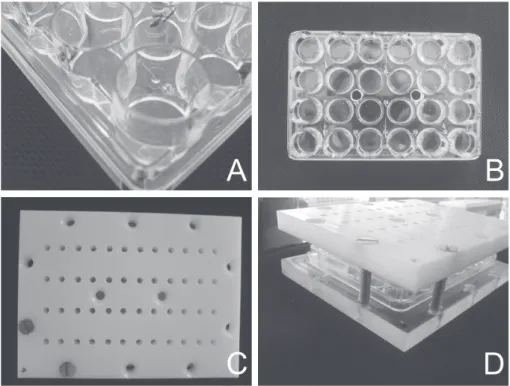
Fig. 1. Design of the experimental set-up. (A): close-up of a culture tray. (B): overview of a culture tray. (C) and (D): culture tray between
plexiglass lids, with in- and outflow openings in upper lid.
medium pollution loads (Alve and Olsgard, 1999; Geslin et al., 2002; Le Cadre and Debenay, 2006). This suggests that reconstructions based on test deformations alone are not ac-curate.
In previous studies, Cu has been one of the most dif-ficult elements to analyze in foraminiferal calcite (Boyle, 1981). However, recent advances in analytical methodol-ogy for trace element determination in foraminiferal cal-cite (Reichart et al., 2003) now enable the calibration of foraminiferal Cu to seawater chemistry for the first time, us-ing cultured benthic foraminifera. Two different intertidal to neritic species (one temperate and one tropical) were cultured to establish possible interspecific differences in the partition coefficient of Cu in foraminiferal calcite.
2 Methods
2.1 Collecting and culturing foraminifera
Two similar culturing experiments were conducted in series. For the first experiment, sediment was collected at an inter-tidal flat in the Dutch Wadden Sea and was kept in the labo-ratory in the dark at 15◦C. Large (>150 µm), living individu-als of Ammonia cf. molecular type T6 (Hayward et al., 2004: further referred to as A. tepida) were transferred to our in-house designed flow-through culture vessels (Fig. 1). Vessels consist of a 24-well culture tray, sandwiched between two Plexiglas plates and cells were connected by silicon tubes,
attached with screws in the upper Plexiglas lid (Fig. 1). A small filter was placed between each cell and tube to pre-vent specimens from moving between cells. Trays were con-nected individually to a 2-liter reservoir with chemically al-tered seawater (see below) and a peristaltic pump was used to circulate seawater through the cells with a speed of 9 ml/h: in this way, six groups of 12 cells were formed, each connected to its own seawater reservoir. Four foraminiferal specimens were placed in each cell. Seawater was enriched with Cu from a stock solution at concentrations of 0, 0.10, 0.20, 0.50, 10 and 20 µmol/l. Calcein (C0875, Sigma-Aldrich, St Louis, USA) was added to the Cu-enriched seawater at a concentra-tion of 5 mg/l.
Calcein is incorporated into biogenic calcite, while ex-isting calcite (i.e. earlier formed chambers) is not affected. Since (incorporated) calcein is fluorescent, foraminiferal chambers that have been built during the time when individu-als were incubated can be recognized (Bernhard et al., 2004). Cells with specimens of Ammonia tepida, contained a thin layer (<0.5 mm) of artificial sediment (Silica, 52–63 µm). Natural seawater from the eastern Mediterranean Sea was ad-justed with MilliQ water to a salinity of 17 to mimic average Wadden Sea salinity. Salinity levels were regularly checked during the experiment with a WTW LF330 conductivity me-ter. All 6*12 cells were kept at a constant temperature of 10◦C for two months: before and after experiments, reser-voirs were sub-sampled and seawater was analyzed by ICP-MS for Cu, Mg and Ca. At the start of the incubation period,
the individuals were fed ∼0.5 mg of autoclaved (20 min at 121◦C) Dunaliella sp. During experiments, the set-up was subjected to the daily sunlight cycle (app. 14 h light/10 h dark).
For experiment 2, trays were replaced and lids rigorously cleaned with HCl, rinsed with MilliQ and re-used to incubate individuals of Heterostegina depressa in seawater with simi-lar Cu-enrichments used in the first experiments. H. depressa is an epibenthic, tropical and symbiont-bearing foraminifer, that was kept in our laboratory under high light intensities (15 W tropical reef lamp; Arcadia, FO15) and after transfer-ring them into the cultutransfer-ring set-up, similar light conditions were maintained in a daily rhythm (14 h light/10 h dark). No sediment was added to the cells, seawater salinity was kept at 35, with a constant temperature of 20◦C. Because of their large size, only two specimens were placed in each cell.
2.2 Temperature and salinity of culture media
For culturing Ammonia tepida, we diluted 35PSU seawa-ter with MilliQ waseawa-ter to mimic inseawa-tertidal ambient conditions with seawater of 17PSU. The dilution decreased both [Ca2+] and [CO2−3 ] and the alkalinity by approximately 50%, re-sulting in a pronounced reduction of the carbonate saturation state (). The temperature maintained during these experi-ments was kept at 10◦C, compared to 20◦C for the
Heteroste-gina depressa experiments, allowing gas exchange with the
atmosphere (ambient pCO2) in both cases. The combined
ef-fect of these changes is a reduction in saturation state from about 5.5 for the H. depressa experiment to about 1.0 for the A. tepida experiment (calculations were performed in CO2sys; Lewis and Wallace, 1998). The lower seawater sat-uration state for the A. tepida cultures was most likely re-sponsible for the fact that newly formed chambers were thin-ner than the pre-experiment chambers (see Results).
2.3 Laser ablation ICP-MS
Newly formed chambers were ablated using an Excimer laser (Lambda Physik) with GeoLas 200Q optics inside a helium atmosphere flushed ablation chamber. Pulse repetition rate was set at 6 Hz, with an energy density at the sample surface of 10 J/cm2. Ablation craters were 60 µm in diameter and ab-lated material was analyzed with respect to time (and hence depth) using a quadrupole ICP-MS instrument (Micromass Platform ICP).
Simultaneous monitoring of Al allowed us to discard the parts of the ablation profiles contaminated by clay minerals from further calculations of elemental concentrations. Since the analytical error increases with shorter ablation time we cleaned all specimens by an incubation of 24 h in 5% NaOCl (Gaffey and Br¨onniman, 1993) before analysis, maximiz-ing the amount of data that could be used for calculatmaximiz-ing (Cu/Ca)calciteratios.
2.4 Calibration strategy
The low calcite saturation state used in the experiment with
Ammonia tepida resulted in formation of new chambers with
thin walls. A similar correlation between test wall thickness and carbonate saturation state has been observed earlier for tests of cultured planktonic foraminifera (Bijma et al., 2002). Unfortunately, these thin chambers break easily during abla-tion when high laser energies are used. Therefore, we ablated
Ammonia tepida with a laser energy of 1 J/cm2, ten times less than the 10 J/cm2 used to ablate newly formed cham-bers of Heterostegina depressa. Analyses were calibrated against NIST glasses 610 and 612, using concentration data of Pearce et al. (1997). Calibrating calcites against glasses is possible because of the relatively matrix independent ab-lation by the Excimer laser (Mason and Kraan, 2002). How-ever, a fluence of <2 J/cm2was close to the ablation thresh-old for glass and calibration was performed instead against matrix matched in-house standards (i.e. pressed calcite pow-der tablets). Calcium was used as an internal standard be-cause (1) the concentration is constant at 40 wt % in calcite and (2) it allows direct comparisons with trace metal to Ca ratios from wet-chemical studies. A collision and reaction cell was used to give improved results by reducing spectral interferences on the minor isotopes of Ca (42Ca, 43Ca and
44Ca: Mason and Kraan, 2002). Good agreement was
ob-served when using both63Cu and65Cu isotopes to calculate Cu concentrations. Relative analytical precision for copper analyses was 15% on average, based on variability during the ablation calculated by GLITTER (New Wave Research, Fremont, CA, USA). This error includes both analytical un-certainties and internal, natural variability in test chemistry.
2.5 Seawater Cu-concentration
The concentration of Cu did not vary considerably in most of our experiments during the experimental period across the range of concentrations used (Table 1).
In the first experiment, all measured Cu-concentrations were lower than the target concentration and most total Cu-concentrations increased during the experiment, resulting in increased seawater Cu/Ca ratios (on average 17%). In exper-iment 2, most Cu-concentrations and all Cu/Ca ratios were higher at the start than at the end of the experiment. Iden-tical procedures and techniques were used before and af-ter subsampling the culture media, making it unlikely that sampling artifacts affected our measurements. Therefore, we used average solution Cu/Ca ratios to estimate the partition coefficient of Cu in foraminiferal calcite and incorporated differences between start and end concentrations for uncer-tainty calculations. Error bars plotted in the different graphs are based on these calculations and largely stem from these changes, which are an order of magnitude larger than the an-alytical uncertainties.
Table 1. Target concentrations of Cu in sea water and measured [Cu]seawaterand (Cu/Ca)seawaterat start and end of both experiments. n.a. =
not available.
Experiment Target [Cu] in µmol/l Measured Cu Cu/Ca at the start of experiment in µmol/l + 1SD at the end of experiment in µmol/l + 1SD at start of experiment ×10−6 + 1SD×10−6 at end of experiment ×10−6 + 1SD×10−6 1. Ammonia 0 0.0843 0.100 ± 0.00584 6.32 ± 5.28 2.91 ± 0.221
tepida 0.10 0.0960 ± 0.0146 n.a. 2.95 ± 0.384 n.a.
0.20 0.197 ± 0.00173 0.256 ± 0.00206 6.00 ± 0.0326 7.34 ± 0.121 0.50 0.332 ± 0.00505 n.a 10.1 ± 0.0143 n.a. 10 12.6 ± 0.293 14.0 ± 0.867 378 ± 4.62 408 ± 6.94 20 17.8 ± 0.489 20.0 ± 3.05 547 ± 13.5 473 ± 20.8 2. Heterostegina 0 0.125 ± 0.00554 0.208 ± 0.0119 1.69 ± 0.0236 3.62 ± 0.343 depressa 0.10 0.744 ± 0.00217 0.211 ± 0.0408 2.32 ± 0.0670 3.74 ± 0.500 0.20 0.242 ± 0.0228 0.368 4.06 ± 0.179 6.71 0.50 1.28 ± 0.0384 1.25 ± 0.0522 20.2 ± 0.0120 23.1 ± 0.711 10 11.2 ± 0.00672 10.4 ± 0.277 172 ± 0.844 212 ± 5.88
20 n.a. n.a. n.a. n.a.
Table 2. Thermodynamic data for calcein (Ueno et al., 1992). L = calceine. a reactions leads to negligible metal binding; bvalues extrapolated by assuming chemical behavior similar to Cu2+(see text for discussion).
Reaction Functional groups Log K
(1) L6−+ H+= HL5− –COOH 11.7 (2) HL5−+ H+= H2L4− –COOH 10.8 (3) H2L4−+ H+= H3L3− –COOH 5.5 (4) H3L3−+ H+= H4L2− –OH 4.2 (5) H4L2−+ H+= H5L− ≡NH+ 2.9 (6) H5L−+ H+= H6L ≡NH+ 2.1 (7) 2Cu2++ L6−= Cu2L2− –COOH 28.9 (8) Cu2++ H2L4−= CuH2L2− ≡NH+ 8.3
(9) Cu2++ H4L2−+ H3L3−= Cu(H4L)(H3L)3− –COOH + –OH 10.4a (10) 2Ca2++ L6−= Ca2L2− –COOH 27.2b (11) Ca2++ H2L4−= CaH2L2− ≡NH+ 6.63
(12) Ca2++ H4L2−+ H3L3−= Ca(H4L)(H3L)3− –COOH + –OH 8.73a,b (13) 2Mg2++ L6−= Mg2L2− –COOH 28.5b (14) Mg2++ H2L4−= MgH2L2− ≡NH+ 7.9
(15) Mg2++ H4L2−+ H3L3−= Mg(H4L)(H3L)3− –COOH + –OH 10.0a,b
2.6 Cu speciation in seawater
In the absence of organic matter, Cu in seawater forms mainly Cu(OH)2and CuCO3, while small amounts of Cu2+
and CuOH−are also present (Zirino and Yamamoto, 1972). In natural seawater, however, usually more than 99.9% of the
Cu is bound to organic compounds (Eriksen et al., 2001), mainly in the colloidal state (Mackey and Zirino, 1994). Foraminifera take up organic particles and seawater by en-docytosis, likely ingesting both free Cu and Cu-ligand com-plexes. The internal routes that organic compounds follow are virtually uninvestigated in foraminifera and therefore, we
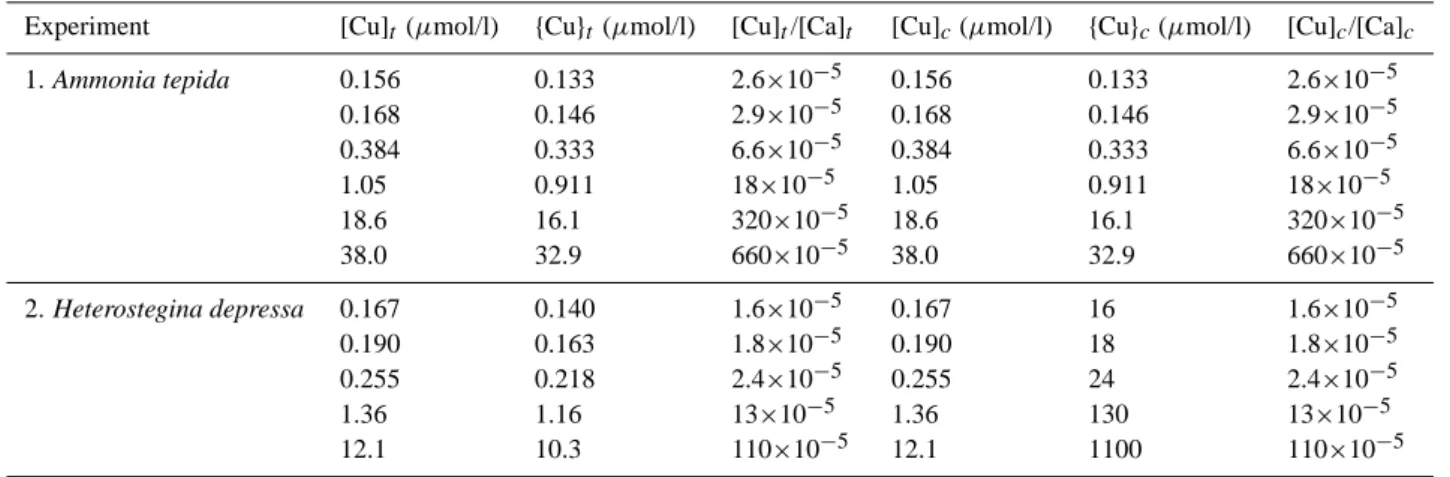
Table 3. Added and free copper concentrations and activities in the experiments. Suffix t = total copper concentration added to the
experi-ment; suffix c = corrected ratios (total Cu or Ca minus calceine-complexed Cu or Ca).
Experiment [Cu]t (µmol/l) {Cu}t(µmol/l) [Cu]t/[Ca]t [Cu]c(µmol/l) {Cu}c(µmol/l) [Cu]c/[Ca]c
1. Ammonia tepida 0.156 0.133 2.6×10⁻⁵ 0.156 0.133 2.6×10⁻⁵ 0.168 0.146 2.9×10⁻⁵ 0.168 0.146 2.9×10⁻⁵ 0.384 0.333 6.6×10⁻⁵ 0.384 0.333 6.6×10⁻⁵ 1.05 0.911 18×10−5 1.05 0.911 18×10−5 18.6 16.1 320×10−5 18.6 16.1 320×10−5 38.0 32.9 660×10−5 38.0 32.9 660×10−5 2. Heterostegina depressa 0.167 0.140 1.6×10⁻⁵ 0.167 16 1.6×10⁻⁵ 0.190 0.163 1.8×10⁻⁵ 0.190 18 1.8×10⁻⁵ 0.255 0.218 2.4×10⁻⁵ 0.255 24 2.4×10⁻⁵ 1.36 1.16 13×10−5 1.36 130 13×10−5 12.1 10.3 110×10−5 12.1 1100 110×10−5
do not know which Cu species are present at the site of cal-cification.
2.7 Modelling Cu speciation
The calcein added in our experiments is a ligand that can bind TE’s and could thus cause concentrations of free Cu to drop. Traditionally, total calcium and TE concentrations in solution are used to calculate partition coefficients for TE’s in calcite. Ideally, activities or effective concentrations, of relevant metals are used to allow application of partition co-efficients in solutions of different compositions (Morse and Bender, 1990).
To correct for Cu binding to calcein, we calculated speci-ation of all abundant cspeci-ations (Cu, Ca and Mg) in our solu-tion. Speciation calculations were performed in PHREEQC 2.8.03 (Parkhurst and Appelo, 1999) with the llnl database and thermodynamic data for calcein listed in Table 2. For calcein complexation with calcium and magnesium, Reac-tions (11) and (14) are reported in the literature. These reac-tions will lead to competition between copper, calcium and magnesium in binding to an amine group on H2L4−, thus
de-creasing calcein-bound Cu. It is, therefore, likely that the behavior of Ca and Mg towards calcein is similar to Cu and similar competition between the three metals occurs in bind-ing accordbind-ing to Reactions (7) and (9) via carboxyl groups (Lu and Allen, 2002). Composition of the solution in the model was either the Cu-enriched seawater with a salinity of 35 or of 17 for the experiment with Ammonia tepida, while both were open to atmospheric CO2.
Cu and Ca-concentrations used to calculate the partition coefficients were those corrected for Cu and Ca complexated with calcein (Table 3).
A
B
100 mμ 100 mμ
Fig. 2. New chambers added by Ammonia tepida (A) and
Heteroste-gina depressa (B), visible by fluorescence of incorporated calcein.
3 Results
3.1 New calcite and survival rates
Specimens that grew new calcite were recognized by fluores-cent, outer chambers (Fig. 2).
None of the individuals of Heterostegina depressa incu-bated at the target Cu-concentration of 20 µmol/l, survived the experimental period. At 10 µmol/l of added Cu, however, several survived of which 1 individual grew new calcite. At lower concentrations, generally more chambers were formed (Table 4). None of the added chambers (n=88) showed ab-normal alignments or deformations.
For Ammonia tepida, the number of successful laser-ablation analyses was significantly lower (3) than the num-ber of added chamnum-bers (34). The limited size (<100 µm) of newly added chambers did not allow multiple analyses of a single chamber. After an unsuccessful attempt to analyze a targeted chamber it was not possible to repeat this mea-surement as the largest part of the carbonate was consumed (Fig. 3).
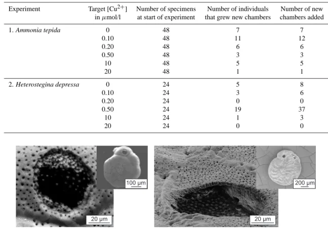
Table 4. Number of individuals at the start of the experiments, number of specimens that formed new calcite and total number of added
chambers.
Experiment Target [Cu2+] Number of specimens Number of individuals Number of new in µmol/l at start of experiment that grew new chambers chambers added
1. Ammonia tepida 0 48 7 7 0.10 48 11 12 0.20 48 6 6 0.50 48 3 3 10 48 5 5 20 48 1 1 2. Heterostegina depressa 0 24 5 8 0.10 24 3 6 0.20 24 0 0 0.50 24 19 37 10 24 1 3 20 24 0 0 20 μm 20 μm 100 μm 200 μm
Fig. 3. Scanning electron microscope image of laser ablation craters in Ammonia (left) and Heterostegina (right). Insets depict the whole
specimen. Scale bar is indicated in the lower right corner only for the magnified image.
3.2 Partition coefficient of Cu – Ammonia tepida
Two ablation profiles were obtained from two specimens of Ammonia tepida that grew new chambers at a low (0.20 µmol/l) Cu-concentration (Fig. 4). Measurements in-dicate that the partition coefficient lies between 0.1 and 0.4. In the right panel of Fig. 4, the same two measurements are depicted at the left end of the graph. Ratios for calcite formed at higher (Cu/Ca)seawater, indicated a partition coefficient
be-tween 0.1 and 0.4.
3.3 Partition coefficient of Cu – Heterostegina depressa
Although individuals of Heterostegina did not survive the highest Cu-levels, we obtained two ratios from specimens that added new chambers at a target concentration of 10 µmol/l. From incubations with lower Cu-concentrations, more specimens were available that grew new chambers that could be analyzed for Cu-concentration (Fig. 5).
4 Discussion
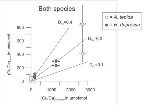
Within the experimental and analytical error both species show a similar (Cu/Ca)seawater to (Cu/Ca)calcite relation,
in-dicating a partition coefficient (DCu) of 0.25±0.15. No
significant difference was observed in copper incorporation between Ammonia tepida and Heterostegina depressa, de-spite large differences in ecology and habitat. Some inter-specimen variation in Cu/Cacalcit e was observed in H.
de-pressa grown at low Cu-concentrations, in which rather large
uncertainties in culture water Cu-concentration (Fig. 5) re-sulted from changes in [Cu2+] over time. Moreover, the al-ternative calibration method used for the thin-walled cham-bers of A. tepida (i.e. using a pressed calcite pellet and a lower ablation energy) increased the analytical uncertainty in the laser ablation-ICP-MS analyses (Fig. 5). Despite these errors, the calculated DCuwas not significantly dependent on
either temperature or salinity.
4.1 Experimental uncertainties
The seawater Cu-concentrations in most culture media in-creased during the incubation period (Table 1), however,
16 12 4 0 8 80 60 40 20 0
(Cu/Ca)sea waterin mol/molμ
(Cu/Ca) in mol/mol calcite μ D =0.2Cu D =0.1Cu 600 3000 2000 0
(Cu/Ca)sea waterin mol/molμ D =0.2Cu D =0.1Cu
Ammonia tepida
D =0.4Cu D =0.4Cu 800 1000 400 200 0Fig. 4. Cu/Ca ratios in Ammonia tepida test carbonate versus Cu/Ca in sea water. Left graph is an enlargement of right one: small circles
indicate the separate [Cu]seawatermeasurements (replicate samples before and after the incubation period), large circles represent the average
Cu/Caseawatervalues. The two measurements in the right panel are from chambers of the same specimen. Lines represent partition coefficients
of 0.1, 0.2 and 0.4.
Both species
= A. tepida = H. depressa (Cu/Ca) in mol/mol calcite μ 600 400 200 0 3000 2000 0(Cu/Ca)sea waterin mol/molμ D =0.2Cu
D =0.1Cu
D =0.4Cu
800
1000
Fig. 5. Plot of Cu/Ca ratios in foraminiferal calcite of the added chamber versus the Cu/Ca of the sea water in which they were incubated.
The two Heterostegina-measurements in the center of the graph represent two chambers of the same specimen. Lines indicate partition coefficients of 0.1, 0.2 and 0.4.
there is no systematical difference in Cu/Caseawaterratios
be-fore and after the two experiments. Therebe-fore, analytical er-rors during ICP-MS or during subsampling of the culture vessels are unlikely to play an important role in this off-set. Sorption of Cu onto organic matter in the culture me-dia or onto the calcite of the foraminifera would have
low-ered the Cu-concentrations in the media, which happened in some cases, and therefore could have contributed to the dif-ferences in Cu/Ca ratios. Alternatively, release of Cu by the culture vessels or trays could have increased seawater Cu-concentrations, although this is unlikely since the equipment used was new or cleaned prior to the experiment. Moreover,
the control media (no Cu added) did not contain consider-able amounts of Cu, indicating that contamination by the materials used played a minor role. The foraminifera them-selves are unlikely to have contained high concentrations of Cu at the start of the experiments since concentrations of this metal are low in natural seawater. Cu-pollution of seawa-ter during subsampling for seawaseawa-ter Cu-analyses may have caused an increase in Cu-concentrations, although care was taken to avoid such contamination. Differences in seawater Cu-concentrations before and after the incubation period are likely to be the result of a combination of the processes men-tioned above.
The use of calcein in our experiments may have affected the uptake of Cu, although it is suggested that the incorpora-tion of trace elements do not seem to be affected by the pres-ence of calcein (Hintz et al., 2004). Measured Mg/Ca ratios in chambers that incorporated calcein were of the same or-der of magnitude as pre-existing chambers of the same spec-imens (data not published). This too suggests that the pres-ence of calcein does not influpres-ence the uptake of trace ele-ments.
The number of new chambers formed by the cultured foraminifera was generally low, especially at high Cu-concentrations (Table 4). The limited production of new cal-cite may indicate that the conditions during the experiments were not optimal for the foraminifera. The accumulation of waste products in the culture vessels, for example, was not monitored and may have changed the seawater chem-istry. Increasing the seawater reservoir size may reduce the effect of such accumulations (see Hintz et al., 2004, 2006a, b). In addition, the micro-environments in which the spec-imens were kept and calcified, may have differed from nat-ural conditions in other ways. The environmental stability in our set-up is not found in intertidal flats, nor was it pos-sible for specimens of Ammonia tepida in these experiments to occupy dysoxic sediment layers, as regularly observed in field studies. Therefore, the results obtained in these experi-ments, may not be applicable for all environments in which the two species potentially calcify. Additional experiments, including anoxia, different pH’s, different light/dark regimes may thus be necessary to investigate the full array of environ-mental conditions under which foraminiferal calcite may be produced. Improving culturing setups and optimizing labo-ratory conditions is important and comparison of our results with those from optimized, future experiments is necessary to establish a more precise DCufor foraminiferal calcite.
4.2 Cu in the calcite lattice
Crystalline CuCO3does not exist, because the most common
coordination of Cu-carbonate complexes is distorted tetrago-nal pyramids or distorted octahedrons (Wells, 1984). These shapes do not allow precipitation of pure CuCO3crystals and
rather Cu2CO3(OH)2(malachite) will form. However,
sorp-tion studies have shown that at the calcite-water interface
these so-called Jahn-Teller distortions can be overcome and a solid solution CuxCa(1−x)CO3 forms (Schlosseler et al.,
1999). It has been proposed that copper in calcite is present in clusters, based on studying the transformation of vaterite to calcite (Nassrallah-Abouka¨ıs et al., 1996, 1998). Recent XAFS work, however, has shown that this mechanism is not applicable to calcite surfaces (Elzinga and Reeder, 2002). This rather unusual complexation behavior would suggest that sorption and subsequent incorporation into the crystal lattice for copper is limited to part of the crystal surface only. This, in turn, would result in a lower partition coefficient for copper than expected based on its ionic radius only.
Contrary to these results, it has been shown that dur-ing inorganic coprecipitation experiments Cu is incorporated in calcite with a distribution coefficient (KCu) of 23 and
constant under a range of Cu-concentrations (Kitano et al., 1980). In the initial stage of calcification, the KCu can
be even higher (40) probably due to the strong affinity of Cu(OH)2 for calcite surfaces (Franklin and Morse, 1982;
Pickering, 1983; Papadopoulos and Rowell, 1989). This in-dicates that the Jahn-Teller distortions are easily overcome during calcification.
Generally, divalent cations with an ionic radius close to Ca (=1.0 ˚A) have a partition coefficient in calcite close to 1. Cd has an ionic radius of 0.95 ˚A (Shannon, 1976) and is in-corporated in both planktonic and benthic species with a D between 1 and 4 (Boyle, 1981, 1988; Havach et al., 2001; Mar´echal-Abram et al., 2004), independent of temperature (Marchitto, 2004). Sr (ionic radius = 1.31 ˚A) is incorpo-rated in foraminiferal calcite with a D of 0.11–0.19, mea-sured in several planktonic genera (Bender et al., 1975) and 0.05–0.25 in Cibicidoides (Elderfield et al., 1996). Coretop studies on Cibicides and Uvigerina show that Ba (1.47 ˚A) is incorporated with a partition coefficient of 0.3–0.4 at 3◦C (Lea and Boyle, 1989). In the planktonic genera
Globoro-talia and Globoquadrina, Ba is incorporated with a D of 0.19
(Lea and Boyle, 1991). Cu has an ionic radius close to Mg (0.73 and 0.72 ˚A, respectively), but the partition coefficient of Mg is much lower (0.1–1×10⁻³; Bender et al., 1975; De-laney et al., 1985) than the measured 0.1–0.4 for Cu (Fig. 7). The large difference between the foraminifer-mediated Cu-incorporation and the inorganic Cu-incorporation of Cu in cal-cite indicates that much energy is spent on removal of Cu at the site of calcification.
4.3 Biological control on DCu
Since magnesium inhibits calcite growth (Berner, 1975; Mucci and Morse, 1983) and high levels of Mg are likely to be present in foraminiferal calcifying reservoirs, it is nec-essary for foraminifera to remove Mg before calcification. It has been suggested that foraminifera actively pump Mg from their calcifying reservoir in order to stimulate CaCO3
precip-itation (Duckworth, 1977; Bentov and Erez, 2006). Usually, Cu is present only in very low concentrations in seawater
and therefore no need exists to actively remove Cu from cal-cifying reservoirs, despite its ability to modify the crystalline structure of calcite. Although under high Cu-concentrations it may be beneficial to remove Cu from calcifying reservoirs, apparently the foraminifera does not do so, as the DCuis
sim-ilar for high and low Cu-concentrations. Alternatively, the concentrations used in our experiments may still be too low to seriously impede CaCO3precipitation.
Another reason for active removal of trace elements from calcifying reservoirs is that these elements are necessary for cellular processes. Since Cu is known to play only minor roles in eukaryotic metabolic processes (Bruland et al., 1991; Sunda and Huntsman, 1995; Chang and Reinfelder, 2000), it is unlikely that the DCuis affected by cellular needs. Organic
compounds may increase Mg contents in foraminiferal cal-cite (Bentov and Erez, 2006). High concentrations of Mg at the primary organic membrane (Hemleben et al., 1986) may explain the observed intra-test variability of Mg/Ca (e.g. Toy-ofuku and Kitazato, 2005). Cu also has a strong affinity for organic compounds (see below), so that the DCumay be
partly determined by the presence of organic compounds in the calcite.
Bresler and Yanko (1995) showed that some benthic, epi-phytic foraminifera have tryptofan-containing proteins that can bind Cu2+ and prevent intracellular Cu-concentrations from becoming harmful. When a significant part of the Cu2+
would have been immobilized by these Cu-binding proteins this would also have lowered the Cu activity in the solution and thus DCu. Since we have not observed such a decrease, it
is unlikely that such molecules play a major role in decreas-ing intracellular Cu-concentrations.
Seawater pH is a potentially important modulator of trace metal uptake (Lea et al., 1999; Zeebe and Sanyal, 2002). To investigate the potential effect we compared species with and without symbionts. In the symbiont-bearing H. depressa the photosynthetic activity of the symbionts changes the local carbonate chemistry because CO2 is taken up and pH
low-ered during light conditions. However, the lack of any sys-tematic offset in Cu/Ca between the H. depressa and A.
tep-ida suggests no significant effect of pH on Cu incorporation.
It may be, however, that difference in pH at the site of calci-fication between and within the species is partly responsible for the observed variation in DCu.
4.4 Test deformation and mortality
A number of studies over the last 20 years have attempted to correlate the number of deformed tests to environmen-tal pollution (Alve, 1991; Elberling et al., 2003; Armynot du Chˆatelet et al., 2004). The empirical correlation be-tween number of deformed tests and for instance heavy metal or hydrocarbon concentration levels was interpreted to signify a causal relationship. Results from such inves-tigations are difficult to apply widely, since deformations in foraminifera take place easily in some groups (e.g.
Mil-iolids and Discorinopsis), while in other taxa (e.g. within the familily Glaberatellidae) deformaties are rarely found (Arnold, 1954). Furthermore, in foraminiferal cultures that were exposed to oil pollution no increased test abnormalities were observed (Ernst et al., 2006). In addition, not a sin-gle deformed chamber alignment was observed in our experi-ments, despite the fact that Cu-concentrations were occasion-ally well above levels found at even the most polluted sites. Although the limited number of observations does not allow a statistical evaluation, our results strongly suggest that high levels of Cu do not cause test deformities. This is also shown by Alve and Olsgard (1999), who found no test deformities in foraminifera living in seawater with high Cu-concentrations. Therefore, we think that relative abundances of deformed tests in fossil samples are not suitable to reconstruct past cop-per concentrations. Most likely other environmental factors, co-varying with environmental trace metal levels must have been responsible for the observed increase in test deformi-ties. The complete absence of deformations in our experi-ments is in high contrast to the low but still detectable lev-els of natural occurring test deformities under environmental pristine conditions. This suggests that the protected envi-ronment of the culture trays may actually have shielded our foraminifera by providing them with a constant temperature, salinity and seawater chemistry in general.
In seawater with the highest concentration of Cu (20 µmol/l), none of the Heterostegina’s survived and only one specimen grew new chambers when cultured at 10 µmol/l. Since the growth or survival of Ammonia
tep-ida did not appear to be hampered by high concentrations
of Cu, we hypothesize that either the symbionts of the trop-ical foraminifera are vulnerable to high Cu-concentrations (Brandt et al., 1986), or that individuals of A. tepida are adapted to cope with (occasional) high levels of heavy met-als.
4.5 Application of Cu/Ca ratios in foraminiferal calcite
In order to quantify pollution levels, heavy metal concen-trations are often analyzed using strong acid extractions and subsequent ICP-MS analyses of bulk sediment. Since heav-ily polluted sites are frequently characterized by high con-centrations of (labile) organic matter, polluted sediments are often anoxic with high levels of sulphate reduction and asso-ciated production of free sulfide. These high sulfide-levels result in immobilization of heavy metals such as Zn, Cu, Cd and Pb, which are precipitated as the highly insoluble minerals PbS, CuS and ZnS, or as co-precipitates in pyrite (Rashid and Leonard, 1973; Saxby, 1973; Huerta-Diaz and Morse, 1992). Because these metals are no longer bioavail-able they do not reflect toxicity of the overlying water to, for instance, benthic biota. Actual analyses of the overlying water itself or organisms living in these waters would give a much more applicable concentration to assess pollution (Nel-son and Donkin, 1985; Bryan and Langston, 1992). This
becomes even more important when at a later stage organic loads decrease and/or the oxygen level increases, (e.g. af-ter improved wastewaaf-ter treatment). Under these conditions lower sedimentary trace metal levels could result in higher actual toxicity as these metals are remobilized by progres-sive re-oxidation of the sediment and escape to the overlying water (Petersen et al., 1997). Monitoring foraminiferal test Cu/Ca ratios could be used to establish the bioavailable frac-tion of Cu and potentially also could record relatively short episodes with elevated bottom water Cu-levels.
Sludge dump sites and associated elevated concentration levels of heavy metals are mostly limited to coastal and estuarine environments. These settings experience consid-erably varying seasonal and even daily temperatures and salinity levels. A significant impact of either temperature or salinity on partition coefficients would, therefore, render foraminiferal trace metal records useless for any reliable re-construction and/or monitoring of such dump sites. Since the obtained DCuis not markedly dependent on either
tempera-ture or salinity, foraminiferal Cu/Ca ratios may be a powerful proxy for the quantitative reconstruction of past heavy metal pollution, even in highly variable environments.
5 Conclusions
Copper is incorporated into foraminiferal calcite with an es-timated partition coefficient of 0.25±0.15 with respect to seawater Cu/Ca values. No effects on the DCu of
species-specific control or of temperature and salinity could be ob-served with the experimental setup used here. Additional ex-periments are needed to better constrain DCuand unravel the
effects of other likely important environmental factors such as temperature, salinity and seawater carbonate chemistry.
Acknowledgements. The authors thank M. Janse from Burger’s
Zoo and W. Renema from Naturalis for providing specimens of
Heterostegina depressa. P. Kleingeld is acknowledged for his help with developing the culturing set-up. The laser ablation measurements were greatly aided by G. Nobbe. E. van Vil-steren and B. van Os provided help with the ICP-MS-analyses and discussion on seawater chemistry. Comments provided by E. Hathorne and two anonymous reviewers improved the text considerably. This research was sponsored by TNO-NITG, Utrecht. Support from the European Science Foundation (ESF) under the EUROCORESProgramme “EuroCLIMATE”, through contract No. ERAS-CT-2003-980409 of the European Commission is acknowledged. NWO is acknowledged for supporting the Dutch partner in “PaleoSalt”.
Edited by: J. Bijma
References
Alve, E.: Benthic foraminifera in sediment across cores reflect-ing heavy metal pollution in Sørfjord, western Norway, J. Foraminifer. Res., 21(1), 1–19, 1991.
Alve, E. and Olsgard, F.: Benthic foraminiferal colonization in ex-periments with copper contaminated sediment, J. Foraminifer. Res., 29(3), 186–195, 1999.
Armynot du Chˆatelet, E., Debenay, J.-P., and Soulard, R.: Foraminiferal proxies for pollution monitoring in moderately polluted harbors, Environ. Pollut., 127, 27–40, 2004.
Arnold, Z. M.: Discorinopsis aguayoi (Bermudez) and
Discorinop-sis vadecens Cushman and Bronnimann: A study of
varia-tion in cultures of living foraminifera, Contr. Cushman Found. Foraminifer. Res., 5(1), 4–13, 1954.
Bender, M. L., Lorens, R. B., and Williams, D. F.: Sodium, magne-sium, and strontium in the tests of planktonic foraminifera, Mi-cropaleontol., 21, 448–459, 1975.
Bentov, S. and Erez, J.: Impact of biomineralization pro-cesses on the Mg content of foraminiferal shells: A biologi-cal perspective, Geochem. Geophys. Geosyst., 7(1), Q01P08, doi:10.1029/2005GC001015, 2006.
Bernhard, J. M., Blanks, J. K., Hintz, C. J., and Chandler, G. T.: Use of fluorescent calcite marker calcein to label foraminiferal tests, J. Foraminifer. Res., 34(2), 96–101, 2004.
Berner, R. A.: The role of magnesium in the crystal growth of cal-cite and aragonite from seawater, Geochim. Cosmochim. Acta, 39(4), 489–504, 1975.
Bijma, J., Honisch, B., and Zeebe, R. E.: Impact of the ocean car-bonate chemistry on living foraminiferal shell weight: Comment on “Carbonate ion concentration in glacial-age deep waters of the Caribbean Sea” by W. S. Broecker and E. Clark., Geochem. Geo-phys. Geosyst., 3(11), 1064, doi:10.1029/2002GC000388, 2002. Borrego, J., L´opez-Gonz´alez, N., and Carro, B.: Geochemical sig-nature as paleoenvironmental markers in Holocene sediments of the Tinto River estuary (Southwestern Spain), Est. Coast. Shelf Sci., 61, 631–641, 2004.
Boyle, E. A.: Cadmium, zinc, copper, and barium in foraminifera tests, Earth Planet. Sci. Lett., 53, 11–35, 1981.
Boyle, E. A.: Cadmium: chemical tracer of deepwater paleoceanog-raphy, Paleoceanogpaleoceanog-raphy, 3, 471–489, 1988.
Brandt, L. E., Sunda, W. G., and Guillard, R. R. L.: Reduction of marine phytoplankton reproduction rates by copper and cad-mium, J. Exp. Mar. Biol. Ecol., 96, 225–250, 1986.
Bresler, V. and Yanko, V.: Chemical ecology: A new approach to the study of living benthic epiphytic foraminifera, J. Foraminifer. Res., 25(3), 267–279, 1995.
Bruland, K. W., Donat, J. R., and Hutchins, D. A.: Interactive in-fluences of bioactive trace metals on biological production in oceanic waters, Limnol. Oceanogr., 36(1), 1555–1577, 1991. Bryan, G. W. and Langston, W. J.: Bioavailability, accumulation
and effects of heavy metals in sediments with special reference to estuaries: A review, Environ. Pollut., 76(2), 89–131, 1992. Chang, S. I. and Reinfelder, J. R.: Bioaccumulation, subcellular
distribution, and trophic transfer of copper in a coastal marine diatom, Environ. Sci. Technol., 34, 4931–4935, 2000.
Delaney, M. L., B´e, A. W. H., and Boyle, E. A.: Li, Sr, Mg, and Na in foraminiferal calcite shells from laboratory culture, sediment traps, and sediment cores, Geochim. Cosmochim. Acta, 49(6), 1327–1341, 1985.
Duckworth, D. L.: Magnesium concentration in the tests of the planktonic foraminifer Globorotalia truncatulinoides, J. Foram. Res., 7(4), 304–312, 1977.
Applying foraminiferal stratigraphy as a biomarker for heavy metal contamination and mining impact in a fiord in west Green-land, Mar. Environ. Res., 55, 235–256, 2003.
Elderfield, H., Bertram, C. J., and Erez, J.: A biomineralization model for the incorporation of trace elements into foraminiferal calcite, Earth Planet. Sci. Lett., 142, 409–423, 1996.
Ellison, R. L., Broome, R., and Oglivie, R.: Foraminiferal response to trace metal contamination in the Patapsco River and Baltimore harbour, Maryland, Mar. Pollut. Bull., 17(9), 419–423, 1986. Elzinga, E. J. and Reeder, R. J.: X-ray absorption spectroscopy
study of Cu2+ and Zn2+ adsorption complexes at the calcite surface: Implications for site-specific metal incorporation prefer-ences during calcite crystal growth, Geochim. Cosmochim. Acta, 66(22), 3943–3954, 2002.
Eriksen, R. S., Mackey, D. J., Van Dam, R., and Nowak, B.: Copper speciation and toxicity in Macquarie Harbour, Tasmania: an in-vestigation using a copper ion selective electrode, Mare Chem., 74, 99–113, 2001.
Ernst, S. R., Morvan, J., Geslin, E., Le Bihan, A., and Jorissen, F. J.: Benthic foraminiferal response to experimentally induced Erika oil pollution, Mar. Micropal., 61, 76–93, 2006.
Ferraro, L., Sprovieri, M., Alberico, I., Lirer, F., Prevedello, L., and Marsella, E.: Benthic foraminifera and heavy metals distribution: A case study from the Naples harbour (Tyrrhenean Sea, Southern Italy), Environ. Pollut., 142, 274–287, 2006.
Franklin, M. L. and Morse, J. W.: The interaction of copper with the surface of calcite, Ocean Sci. Eng., 7, 147–174, 1982. Gaffey, S. J. and Br¨onnimann, C. E.: Effects of bleaching on organic
and mineral phases in biogenic carbonates, Journal of Sediment. Petrol., 63, 752–754, 1993.
Geslin, E., Debenay, J.-P., Duleba, W., and Bonetti, C.: Morpho-logical abnormalities of foraminiferal tests in Brazilian environ-ments: comparison between polluted and non-polluted areas, Mar. Micropaleontol., 45, 151–168, 2002.
Hallock, P., Lidz, B. H., Cockey-Burkhard, E. M., and Donnelly, K. B.: Foraminifera as bioindicators in coral reef assessment and monitoring: the FORAM index, Environ. Monit. and Assess., 83(1–3), 221–238, 2003.
Havach, S. M., Thomas Chandler, G., Wilson-Finelli, A., and Shaw, T. J.: Experimental determination of trace element partition coef-ficients in cultured benthic foraminifera, Geochim. Cosmochim. Acta, 65(8), 1277–1283, 2001.
Hayward, B. W., Holzmann, M., Grenfell, H. R., Pawlowski, J., and Triggs, C. M.: Morphological distinction of molecular types in Ammonia – towards a taxonomic revision of the world’s most commonly misidentified foraminifera, Mar. Micropaleontol., 50, 237–271, 2004.
Hemleben, C. H., Anderson, O. R., Berthold, W., and Spindler, M.: Calcification and chamber formation in Foraminifera- a brief overview, in: Biomineralization in lower plants and animals, Systematics Association Special Volume 30, edited by: Lead-beater, B. S. C. and Riding, R., Clarendon Press, Oxford (for the Systematics Association), p 237–249, 1986.
Hintz, C. J., Chandler, G. T., Bernhard, J. M., McCorkle, D. C., Havach, S. M., Blanks, J. K., and Shaw, T. J.: A physico-chemically constrained seawater culturing system for production of benthic foraminifera, Limnol. Oceanogr-Meth., 2, 160–170, 2004.
Hintz, C. J., Shaw, T. J., Chandler, G. T., Bernhard, J. M.,
Mc-Corkle, D. C., and Blanks, J. K.: Trace/ minor element:calcium ratios in cultured benthic foraminifera. Part I: Inter-species and inter-individual variability, Geochim. Cosmochim. Acta, 70(8), 1952–1963, 2006a.
Hintz, C. J., Shaw, T. J., Bernhard, J. M., Chandler, G. T., Mc-Corkle, D. C., and Blanks, J. K.: Trace/ minor element:calcium ratios in cultured benthic foraminifera. Part II: Ontogenetic vari-ation, Geochim. Cosmochim. Acta, 70(8), 1964–1976, 2006b. Huerta-Diaz, M. A. and Morse, J. W.: Pyritization of trace metals
in anoxic marine sediments, Geochim. Cosmochim. Acta, 56(7), 2681–2702, 1992.
Kitano, Y., Okumura, M., and Idogaki, M.: Abnormal behaviors of copper (II) and zinc ions in parent solution at the early stage of calcite formation, Geochem. J., 14, 167–175, 1980.
Koide, M., Lee, D. S., and Goldberg, E. D.: Metal and transuranic records in mussel shells, byssal threads and tissues, Estuar. Coast. Shelf Sci., 15, 679–695, 1982.
Lea, D. W. and Boyle, E. A.: Ba content of benthic foraminifera controlled by bottom water composition, Nature, 338, 751–753, 1989.
Lea, D. W. and Boyle, E. A.: Ba in planktonic foraminifera, Geochim. Cosmochim. Acta, 55, 3321–3331, 1991.
Lea, D. W., Mashiotta, T. A., and Spero, H. J.: Controls on magne-sium and strontium uptake in planktonic foraminifera determined by live culturing, Geochim. Cosmochim. Acta, 63(16), 2369– 2379, 1999.
Le Cadre, V. and Debenay, J.-P.: Morphological and cytological responses of Ammonia (foraminifera) to copper contamination: Implications for the use of foraminifera as bioindicators of pol-lution, Environ. Pollut., 143, 304–317, 2006.
Lewis, E. and Wallace, D. W. R.: Program Developed for CO2 Sys-tem Calculations, ORNL/CDIAC-105, Carbon Dioxide Informa-tion Analysis Center, Oak Ridge NaInforma-tional Laboratory, U.S. De-partment of Energy, Oak Ridge, Tennessee, 1998.
Lu, Y. F. and Allen, H. E.: Characterization of copper complexa-tion with natural dissolved organic matter (DOM) – link to acidic moieties of DOM and competition by Ca and Mg, Water Res., 36, 5083–5101, 2002.
Mackey, D. J. and Zirino, A.: Comments on trace metal speciation in seawater or do “onions” grow in the sea?, Anal. Chim. Acta, 284, 635–647, 1994.
Mason, P. R. D. and Kraan, W. J.: Attenuation of spectral interfer-ences of during laser ablation inductively coupled plasma mass spectrometry (LA-ICP-MS) using an rf only collision and reac-tion cell, J. Anal. At. Spectrom., 17, 858–867, 2002.
Marchitto, T. M.: Lack of a significant temperature influ-ence on the incorporation of Cd into benthic foraminiferal tests, Geochem. Geophys. Geosyst., 5(10), Q10D11, doi:10.1029/2004GC000753, 2004.
Mar´echal-Abram, N., Debenay, J.-P., Kitazato, H., and Wada, H.: Cadmium partition coefficients of cultured benthic foraminifera
Ammona beccarii, Geochem. J., 38(3), 271–283, 2004.
Morse, J. W. and Bender, M. L.: Partition coefficients in calcite: examination of factors influencing the validity of experimental results and their application to natural systems, Chem. Geol., 82, 265–277, 1990.
Mucci, A.: The solubility of calcite and aragonite in seawater at var-ious salinities, temperatures, and one atmosphere total pressure, Am. J. Sci., 283, 780–799, 1983.
Mucci, A. and Morse, J. W.: The incorporation of Mg2+and Sr2+ into calcite overgrowths: influences of growth rate and solu-tion composisolu-tion, Geochim. Cosmochim. Acta, 47(2), 217–233, 1983.
Nassrallah-Abouka¨ıs, N., Boughriet, A., Fischer, J. C., Wartel, M., Langelin, H. R., and Abouka¨ıs, A.: Electron paramagnetic reso-nance (EPR) study of Cu2+and Mn2+ions interacting as probes with calciumcarbonate during the transformation of vaterite into cubic calcite, J. Chem. Soc. Lond. Faraday Trans., 92, 3211– 3216, 1996.
Nassrallah-Abouka¨ıs, N., Boughriet, A., Laureyns, J., Abouka¨ıs, A., Fischer, J. C., Langelin, H. R., and Wartel, M.: Trans-formations of vaterite into cubic calcite in the presence of Cu(II)species, Chem. Mater., 10, 238–243, 1998.
Nelson, A. and Donkin, P.: Process of bioaccumulation: The im-portance of chemical speciation, Mar. Pol. Bull., 16(4), 164–169, 1985.
N¨urnberg, D., Bijma, J., and Hemleben, C.: Assessing the relia-bility of magnesium in foraminiferal calcite as a proxy for wa-ter mass temperatures, Geochim. Cosmochim. Acta, 60(5), 803– 814, 1996.
Parkhurst, D. L. and Appelo, C. A. J.: User’s guide to PHREEQC (Version 2) – A computer program for speciation, batch-reaction, one-dimensional transport, and inverse geochemical calcula-tions: U.S. Geological Survey Water-Resources Investigations Report, 99-4259, 1999.
Papadopoulos, P. and Rowell, D. L.: The reactions of copper and zinc 5 with calcium carbonate surfaces, J. Soil Sci., 40, 39–48, 1989.
Pearce, N. J. G., Perkins, W. T., Westgate, J. A., Gorton, M. P., Jack-son, S. E., Neil, C. R., and Chenery, S. P.: A compilation of new and published major and trace element data for NIST SRM 610 and NIST SRM 612 glass reference materials, Geost. Newslett., 21(1), 115–144, 1997.
Petersen, W., Willer, E., and Willamowski, C.: Remobilization of trace elements from polluted anoxic sediments after resuspension in oxic waters, Water Air Soil Poll., 99(1–4), 515–522, 1997. Pickering, W. F.: Extraction of copper, lead, zinc and cadmium ions
sorbed on calcium carbonate, Water, Air, Soil Pollut., 20, 299– 309, 1983.
Rashid, M. A. and Leonard, J. D.: Modifications in the solubility and precipitation behavior of various metals as a result of their interaction with sedimentary humic acid, Chem. Geol., 22(2), 89–97, 1973.
Rathburn, A. E. and DeDeckker, P.: Magnesium and strontium com-positions of recent benthic foraminifera from the Coral Sea, Aus-tralia and Prydz Bay, Antarctica, Mar. Micropaleontol., 32, 231– 248, 1997.
Reichart, G. J., Jorissen, F. J., Anschutz, P., and Mason, P. R. D.: Single foraminiferal test chemistry, Geology, 31(4), 335–358, 2003.
Rosenthal, Y., Boyle, E. A., and Slowley, N.: Temperature con-trol on the incorporation of magnesium, strontium, fluorine, and cadmium into benthic foraminiferal shells from Little Bahama Bank: prospects for thermocline paleoceanography, Geochim. Cosmochim. Acta, 61(17), 3633–3643, 1997.
Ruiz, F., Gonz´alez-Regalado, M. L., Borrego, J., Abad, M., and Pend´on, J. G.: Ostracoda and foraminifera as short-term tracers of environmental changes in very polluted areas: the Odiel estu-ary (SW Spain), Environ. Pollut., 129, 49–61, 2004.
S´ainz, A. and Ruiz, F.: Influence of the very polluted inputs of the Tinto-Odiel system on the adjacent littoral sediments of south-western Spain: A statistical approach, Chemosphere, 62, 1612– 1622, 2006.
Samir, A. M. and El-Din, A. B.: Benthic foraminiferal assem-blages and morphological abnormalities as pollution proxies in two Egyptian basins, Mar. Micropaleontol., 41, 193–227, 2001. Saxby, J. D.: Diagenesis of metal-organic complexes in sediments:
formation of metal sulphides from cystine complexes, Chem. Geol., 12(4), 241–248, 1973.
Segev, E. and Erez, J.: Effect of Mg/Ca ratio in seawater on shell composition in shallow benthic foraminifera, Geochem. Geophys. Geosyst., 7(2), Q02P09, doi:10.1029/2005GC000969, 2006.
Shannon, R. D.: Revised effective ionic radii and systematic stud-ies of interatomic distances in halides and chalcogenides, Acta Crystallogr. A, 32, 751–767, 1976.
Sunda, W. G. and Huntsman, S. A.: Regulation of copper concen-tration in the oceanic nutricline by phytoplankton uptake and re-generation cycles, Limnol. Oceanogr., 40(1), 132–137, 1995. Toyofuku, T., Kitazato, H., Kawahata, H., Tsuchiya, M., and
No-hara, M.: Evaluation of Mg/Ca thermometry in foraminifera: Comparison of experimental results and measurements in nature, Paleoceanography, 15(4), 456–464, 2000.
Toyofuku, T. and Kitazato, H.: Micromapping of Mg/Ca val-ues in cultured specimens of the high magnesium benthic foraminifera, Geochem. Geophys. Geosyst., 6(11), Q11P05, doi:10.1029/2005GC000961, 2005.
Ueno, K., Imamura, T., and Cheng, K. L.: Handbook of organic analytical reagents, 2nd ed. CRC Publishers, 1992.
Wells, A. F.: Structural inorganic chemistry, 5th ed, Clarendon, 1984.
Zeebe, R. E. and Sanyal, A.: Comparison of potential strategies of planktonic foraminifera for house building: Mg2+or H+ re-moval?, Geochim. Cosmochim. Acta, 66(7), 1159–1169, 2002. Zeebe, R. E. and Wolf-Gladrow, D.: CO2 in seawater: Equilibrium,
kinetics and isotopes, Elsevier Oceanographic Series, Amster-dam, 2001.
Zirino, A. and Yamamoto, S.: A pH-dependent model for the chem-ical speciation of copper, zinc, cadmium, and lead in seawater, Limnol. Oceanogr., 17(5), 661–671, 1972.
![Table 1. Target concentrations of Cu in sea water and measured [Cu] seawater and (Cu/Ca) seawater at start and end of both experiments](https://thumb-eu.123doks.com/thumbv2/123doknet/14795106.603412/5.892.69.824.146.462/table-target-concentrations-water-measured-seawater-seawater-experiments.webp)