HAL Id: hal-03011084
https://hal.archives-ouvertes.fr/hal-03011084
Submitted on 17 Nov 2020HAL is a multi-disciplinary open access archive for the deposit and dissemination of sci-entific research documents, whether they are pub-lished or not. The documents may come from teaching and research institutions in France or abroad, or from public or private research centers.
L’archive ouverte pluridisciplinaire HAL, est destinée au dépôt et à la diffusion de documents scientifiques de niveau recherche, publiés ou non, émanant des établissements d’enseignement et de recherche français ou étrangers, des laboratoires publics ou privés.
Symbiotic cooperation between freshwater rock-boring
bivalves and microorganisms promotes silicate bioerosion
Damien Daval, François Guyot, Ivan Bolotov, Ilya Vikhrev, Alexander
Kondakov, Artem Lyubas, Andrey Bychkov, Vasily Yapaskurt, Martiane
Cabié, Oleg Pokrovsky
To cite this version:
Damien Daval, François Guyot, Ivan Bolotov, Ilya Vikhrev, Alexander Kondakov, et al.. Symbi-otic cooperation between freshwater rock-boring bivalves and microorganisms promotes silicate bio-erosion. Scientific Reports, Nature Publishing Group, 2020, 10 (1), �10.1038/s41598-020-70265-x�. �hal-03011084�
Symbiotic cooperation between freshwater rock-boring bivalves
1
and microorganisms promotes silicate bioerosion
2
Damien Daval1,*, François Guyot2,3, Ivan N. Bolotov4, Ilya V. Vikhrev4, Alexander V. 3
Kondakov4, Artem A. Lyubas4, Andrey Y. Bychkov5, Vasily O. Yapaskurt5, Martiane Cabié6 4
and Oleg S. Pokrovsky7,8 5
6
1 Université de Strasbourg – CNRS / ENGEES – EOST, Laboratoire d’Hydrologie et de 7
Géochimie de Strasbourg, UMR 7517, Strasbourg, France 8
2 Institut de Minéralogie, de Physique des Matériaux et de Cosmochimie, Museum National 9
d'Histoire Naturelle, Sorbonne-Université, UMR 7590 CNRS, Paris, France 10
3 Institut Universitaire de France 11
4 N. Laverov Federal Center for Integrated Arctic Research, the Ural Branch of the Russian 12
Academy of Sciences, Northern Dvina Emb. 23, 163000 Arkhangelsk, Russia 13
5 Lomonosov Moscow State University, Faculty of Geology, Leninskiye Gory, 1, 119991, 14
Moscow, Russia 15
6 Aix-Marseille Université, CNRS, Centrale Marseille, FSCM, CP2M, Marseille, France 16
7 Geosciences and Environment Toulouse, UMR 5563 CNRS, Toulouse, France 17
8 BIO-GEO-CLIM Laboratory, Tomsk State University, Tomsk, Russia 18
19
* Corresponding author: [email protected]
20 21
Abstract
22
Bioerosion is a process with a high socio-economic impact that contributes to coastal retreat,
23
and likely to increase with climate change. Whereas limestone bioerosion is well explained by
24
a combination of mechanical and chemical pathways, the bioerosion mechanisms of silicates,
25
which are harder and chemically more resistant, remain elusive. Here we investigated the
26
interface between siltstone and freshwater rock-boring bivalves Lignopholas fluminalis
27
(Bivalvia: Pholadidae). Remains of a microbial biofilm were observed only in the poorly
28
consolidated part of the rock within the macroborings created by bivalves. Secondary
Mn-29
bearing minerals identified in the biofilm suggest that microbes promoted silicate rock
30
weathering by dissolving Mn-rich chlorites. Moreover, hard mineral debris found in a biofilm
31
attached to the shells likely contributed to the abrasion of the rock substrate. Thus, beyond the
32
classical view of chemical and/or mechanical action(s) of macroborers, silicate bioerosion may
33
also be facilitated by an unexpected synergistic association between macro- and
34
microorganisms. 35
36
Keywords:
bioerosion; macroborings; silicate weathering; biofilm 37Introduction
38
Bioerosion is a commonplace strategy developed by living organisms, which consists in 39
boring hard substrates of various origins, including biological materials (e.g., wood, shells, and 40
bones)1, mud2, rocks3 and even synthetic materials. Depending on the nature of the substrate 41
and the borer, bioerosion ensures a wide range of metabolic activities and ecosystem services, 42
ranging from nutrition4 to the creation of microhabitats protected from predators for themselves 43
as well as for secondary dwellers5. 44
Gaining knowledge into the occurrence, rates and mechanisms of boring is of fundamental 45
importance for a series of reasons. First and historically, mankind has been confronted with 46
macroborers through the damages caused by shipworms on vessels, wooden wharfs and 47
docks5,6. More broadly, bioerosion has socio-economic impacts whenever manufactured 48
materials are damaged, including plastic, metals and concrete materials such as levees or coastal 49
defences3,6. Second, bioerosion contributes to element recycling, shaping landscapes through 50
the weakening of rocky shorelines and participating to coastal retreat5, for which the current 51
rates are likely to be modified drastically as a result of climate change6. Third, the creation of 52
microhabitats by macroborers such as bivalves is correlated with a significant increase of the 53
abundance of species assemblages, thus partly contributing to local faunal biodiversity7. 54
Finally, fossil records of macro-bioerosion may be used as a biological proxy to estimate the 55
paleo-location of intertidal and shallow subtidal marine environments, marking ancient 56
shorelines8. 57
The mechanisms of rock bioerosion associated to macroborers and especially bivalves have 58
been a source of lively debate for decades, and can be schematically divided into two main 59
pathways. First, rock boring can occur chemically through biocorrosion (also referred to as 60
bioweathering). It is generally admitted that chemical etching is only possible when boring 61
occurs in (at least partly) calcareous substrates5, as most of the common biologically secreted 62
agents by macroborers exhibit a modest impact on the dissolution rate of silicate minerals9. The 63
biologically produced substances that promote the dissolution of carbonates include a variety 64
of lipoproteic components with acidic groups, CO2, or calcium-binding mucoproteins secreted 65
by pallial glands, located along the edge of the mantle of chemically boring bivalves5,10,11. 66
Biocorrosion was then suggested to proceed along grain boundaries first, prior to affect bulk 67
minerals5. Second, rock boring can also occur mechanically through bioabrasion. Several lines 68
of evidence support this mechanism, including (i) the microtexture of the shells, consisting of 69
excavating ridges and rasping structures rendering bivalves comparable to a so-called “living 70
lime”12, (ii) muscular adaptation, as described by Shipway et al.4 regarding the large posterior 71
adductor muscle of the shipworm Lithoredo abatanica (Bivalvia: Teredinidae), (iii) 72
experimental and modeling results of the stimulation of the muscles that control the rotation of 73
the shells of the bivalves, which made it possible to accurately reproduce the shapes of burrows 74
and scrape marks resulting from the boring by the bivalve Barnea candida (Bivalvia: 75
Pholadidae)10. Finally, it is now generally admitted that both mechanical abrasion and chemical 76
etching occur synergistically, especially for living organisms that bore in calcareous rocks, 77
where the substrate is first weakened chemically, while individual grains are subsequently 78
excavated mechanically from the boring5,9. 79
The efficacy of both biocorrosion and bioabrasion is all the more intriguing when it comes 80
to consider boring in silicate rocks such as siltstone8,9, gneiss13, basalts3 or quartzite14, whose 81
hardness is sizably higher than that of the aragonite/calcite shells of the borers. The mechanisms 82
proposed to account for these observations include (i) the possible softening of the rocks, prior 83
to the onset of macroboring, by microendolithic organisms14, capable to create microchannels 84
in silicate materials such as basaltic glass15, (ii) the exploitation of differences in mineral 85
hardness and crystal boundaries in the rock (see 3 and references therein), (iii) a modification 86
of the shell morphology, which may be appropriate to bore into very hard substrates12. 87
However, these mechanisms essentially remain hypothetical, for which direct evidence are most 88
often lacking. 89
The present study was designed to provide new insights into these questions. Recently, 90
ongoing boring in siltstone rocks from a freshwater section of the Kaladan River in Myanmar 91
(Fig. 1) by Lignopholas fluminalis (Bivalvia: Pholadidae) bivalve has been identified8. The 92
boring mechanisms were not revealed in this previous study, but were suggested to be 93
comparable to those used by their marine relatives in softer substrates10, i.e., bioabrasion. Here, 94
we combined analytical scanning electron microscopy (SEM) and focused ion beam (FIB) 95
milling coupled to transmission electron microscopy (TEM) to document the chemical and 96
mineralogical compositions of the host rock and of the shell-rock interface at the micro- to 97
nanometer scale. In addition to the possible bioabrasion mechanisms proposed by Bolotov et 98
al.8, our results suggest that the boring may be microbially-assisted in a symbiotic-like process, 99
where microbes would take advantage of loosening the mineral grains to dissolve minerals 100
cementing the siltstone and containing essential elements for their growth, ultimately resulting 101
in the weakening of the substrate, facilitating further bioerosion by the bivalves. 102
103
Results
104
Distinctive features of the surface of the rock substrate devoid of macroborings 105
At the submillimeter scale, the rock-forming minerals appear homogeneously distributed, 106
devoid of any layering or of specific mineral patches (Fig. 2a, b). The most abundant minerals, 107
i.e., quartz, feldspars and clays are randomly distributed, and heterogeneous clusters could not 108
be evidenced. Statistical analyses of color maps such as shown in Fig. 2b indicate that the 109
surface is covered with ~63% of quartz + clay minerals; ~25% feldspars (note that the 110
proportion of feldspars is most likely overestimated due to possible mistaken assignment of the 111
K and K emission lines in the EDX spectra to K-feldspar instead of illite); ~10% chlorite; 112
3% voids (porosity). These results are in reasonable agreement with the analyses reported by 113
Bolotov et al.8(see Methods). The homogeneous distribution of minerals was also revealed at 114
the submicron scale on the FIB foils that were extracted from the “pristine” (i.e., devoid of 115
macroborings) area (Fig. 2c). Imaging the rock substrate in cross-section further indicates that 116
the grains display a packed and cohesive arrangement, as further suggested by the fact that 117
individual grains did not tend to pop out during the FIB milling procedure (Fig. 2d). 118
119
Distinctive features of the rock surface located in macroborings 120
At the submillimeter scale, the distribution of the rock-forming minerals in the 121
macroborings appears homogeneous and cannot be distinguished from the distribution of 122
minerals in locations devoid of macroborings (Fig. 3a, b). Statistical analyses of color maps 123
such as shown in Fig. 3b indicate that the surface is covered with ~51% of quartz + clay 124
minerals; ~28% feldspars (most likely overestimated, see above); ~13% chlorite; 6% voids 125
(porosity). No specific abrasive imprints could be evidenced in the macroborings, as opposed 126
to other studies that have reported the identification of scrap marks or concentric grooves left 127
in the boreholes, resulting from the rotation of the valves of mechanical borers3,10. It is unlikely 128
that such features would have faded away because of later erosion here, since these observations 129
also apply to macroborings still filled with mollusks. 130
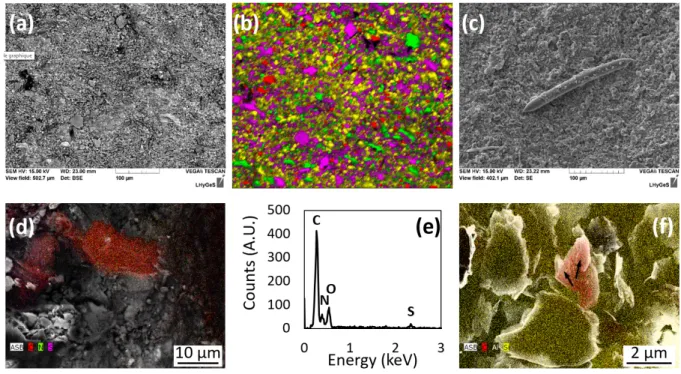
Patches of several µm² of a (C, N, S)-rich organic matrix interpreted as the remains of a 131
complex microbial biofilm were found only in the macroborings (Fig. 3d, e), which also 132
exhibited several bacteriomorph structures approaching the size, shape and chemical 133
composition of bacterial cells (Fig. 3f). Finally, 100-µm long needles were occasionally 134
observed in some of the macroborings (Fig. 3c). These needles are made of pure amorphous 135
silica and resemble in size and shape the megascleres of sponges such as Corvospongilla ultima 136
(Demospongiae: Spongillidae) described in 16. This observation is consistent with the results of 137
Bolotov et al.8, who reported that Corvospongilla ultima was among the nestling species 138
associated with Lignopholas fluminalis’ ecosystem. 139
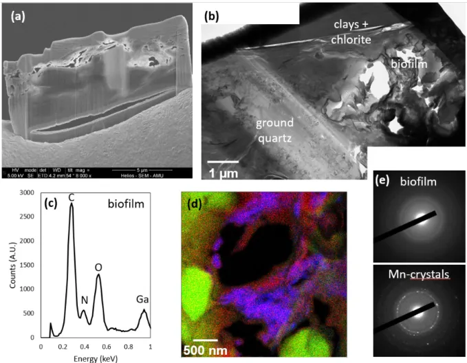
At the submicron-scale, the chemistry and texture of the substrate at the bottom of the two 140
macroborings significantly differ from that described in locations devoid of macroborings. The 141
assemblages were made of low consolidated grains which tended to pop out during the FIB 142
milling procedure (Fig. 4a). Ground silicate grains were observed (Fig. 4b), and µm-size voids 143
filled with an amorphous (C, N)-rich matrix were evidenced in each of the two FIB thin sections 144
(Fig. 4c-e). This matrix was enriched with Mn- and Ca-bearing nanocrystals (Fig. 4d and 4e). 145
However, the values of the interplanar spacing estimated from the electron diffraction patterns 146
were too short (comprised between 0.91 Å and 2.24 Å) to be unambiguously attributed to a 147
given specific mineral or mixture of minerals, such that their exact nature remains unknown. 148
149
Chemical and mineralogical characterizations of the interface between shells and 150
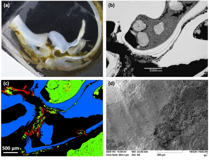
siltstone 151
SEM analyses of the transversal cross-section of the aragonite shell - siltstone interface 152
revealed that the shells of Lignopholas fluminalis were extensively covered with debris of hard 153
minerals from the siltstone, mainly quartz, feldspars and occasionally, accessory minerals such 154
as rutile (Fig. 5a-c, and SI Appendix Figs. S1, S2 and Table S1). Because the hardness of these 155
minerals ranges from 6 to 7, they can easily bore into the phyllosilicates of the siltstone (chlorite 156
and sericite) which have a hardness of 2 to 3. 157
Interestingly, these minerals were not found to be simply deposited on the surface of the 158
shells, but literally fixed to the surface via an organic film enriched in nitrogen (20 wt. %) and 159
sulfur (4 wt. %) (SI Appendix, Fig. S3 and Table S2), which may be interpreted as extracellular 160
polymeric substances (EPS) resulting from microbial activity. The association between 161
carbonate shell, siltstone, and organic matrix with embedded silicate and quartz grains is 162
visualized on integral map of component distribution shown in Fig. 5c. The nitrogen-to-sulfur 163
ratio may reflect the presence of 10% S-containing amino acids such as cysteine in these 164
EPS/proteins. 165
166
Morphological and chemical characterizations of the shells 167
At the µm-scale, it was not possible to identify a specific morphological microtexture of 168
the shell that would have provided the bivalve with an efficient excavation ability (Fig. 5d). 169
Moreover, the extensive coverage of the shells with exogenous minerals considerably 170
complicated the direct observation of fine scale morphology, so that features such as reported 171
by e.g. Fang and Shen12 or Nederlof and Muller10 might have remained obscured. 172
The chemical composition of aragonite that makes up the shells is very homogenous, whilst 173
exhibiting an enrichment in Sr at the extreme point of the shells (SI Appendix, Fig. S4). Total 174
chemical analyses further revealed that the shells of Lignopholas fluminalis were slightly 175
depleted in a number of elements compare to the non-boring species from the same location (SI 176
Appendix, Table S3). The average ratio of element concentration in borers (Lignopholas 177
fluminalis) to non-borers (Scaphula deltae; Bivalvia: Arcidae) demonstrate slight (ca. a factor
178
of 1.2 to 1.5) depletion of borers in Li, Mg, Al, Si, P, K, V, Cr, Mn, Fe, Ni, Zn, Ga, Ge, As, Se, 179
Rb, Cs, Tl, Th. This depletion was not significant because the variations of chemical 180
composition among individual shells were as high as 30%. The average concentrations of Ti, 181
S, Ca, Sr, Cu, Mo and Pb were practically identical (±5%) in both species, whereas Y and REEs 182
were within 10% similar. The concentrations of Cd, Nb, Hf, W and U were a factor of 2.7 to 6 183
higher in non-borers compared to borers, whereas B and Ba were a factor of 4.0 and 1.6 higher 184
in borers compared to non-borers. Given the similarities between the chemical composition of 185
the shells of boring and non-boring species, it can be assumed that the shells of Lignopholas 186
fluminalis do not possess any chemical specificity that might have made them more prone to
187
boring into hard substrata. 188
189
Discussion
190
Overall, the characterizations detailed above point out that the contact between the bottom 191
of the macroborings and the shells of the bivalves likely have represented a hotspot of microbial 192
activity, which was not observed elsewhere at the surface of the siltstone devoid of 193
macroborings. In the next two sections, we discuss how the association between Lignopholas 194
fluminalis and microorganisms may have acted symbiotically to facilitate boring in siltstone.
195 196
A possible strengthening of the mechanical abrasion thanks to microbial EPS 197
As mentioned above, macroborings resulting from bioerosion are most often observed 198
either in calcareous rocks, which are highly sensitive to bioweathering, or in soft substrates 199
such as peat or clays, which are readily drilled through bioabrasion. Here, the siltstone is both 200
chemically much more resistant than carbonates and harder than the substrates commonly 201
subjected to bioabrasion. 202
Bolotov et al.8 have reported that the mean hardness of the siltstone was 62 kgf . mm−2, 203
i.e., twice as much as clayey materials. In comparison, the compilation of Yang et al.17 indicates 204
that the hardness of Bivalvia shell is an order of magnitude lower than that of quartz, and only 205
slightly greater than that of the siltstone, ranging between 110 and 270 kgf . mm−2. In addition, 206
both the structure and hardness of the siltstone were found to be homogeneous, such that it is 207
unlikely that Lignopholas fluminalis took advantage of any local weakness to bore into the rock. 208
Finally, the macroboring walls did not exhibit any marks, as opposed to the experimental results 209
obtained by Nederlof and Muller10 using the piddock Barnea candida, which is a close relative 210
of Lignopholas fluminalis8. However, such scrap marks resulting from the abrasion of the 211
substrate by the denticles of the piddocks were obtained by rotating the shells in a soft materials 212
(wax). The bioabrasion ability of the shells of Barnea candida is thought to be limited to soft 213
substrata such as clays or peat and most likely, they cannot abrade harder substrata such as 214
chalk10. 215
Similarly, we argue that the various features collected here suggest that there is no clear 216
evidence that the direct contact between the shells of Lignopholas fluminalis and the substrate 217
is responsible for the bioabrasion of the siltstone. Instead, single grains excavated from the 218
borehole partly remained trapped at the surface of the shells, embedded into an organic matrix 219
that we interpreted as a biofilm. It can reasonably be assumed that these single grains, which 220
are essentially hard minerals such as quartz and feldspars, acted like abrasive materials that 221
contributed to drill the siltstone through the rotation of the shells. Therefore, the presence of 222
microorganisms in the interfacial region between the substrate and the borers possibly 223
strengthened their boring ability, although at that point, it remains impossible to state whether 224
this interaction is obligatory or facultative. In any case, from a mechanical standpoint, 225
Lignopholas fluminalis bivalves likely took advantage of the biofilm attached to the surface of
226
their shells to increase their boring ability. 227
228
Enhancing the weakening of the rocks through microbially-induced weathering 229
In addition to bioabrasion, some macroborers are also known for their ability to promote 230
bioweathering5. This mechanism of bioerosion is suggested to be limited to calcareous 231
substrates and not significant for substrates such as siltstones, whose rock-forming minerals 232
have a dissolution rate that is between 6 and 8 orders of magnitude lower than that of calcite at 233
circum-neutral pH conditions (according to rate data from 18 for quartz, 19 for albite, 20 for 234
chlorite and 21 for calcite). 235
Notwithstanding, we argue that mass transfer did occur during the process of boring 236
discussed in the present study. We detail below the reasons why we think that this mass transfer 237
cannot result from the abiotic dissolution of the grains by the bulk fluid, and suggest that 238
microorganisms were responsible for the dissolution of the siltstone, which ultimately 239
facilitated the formation of borings by Lignopholas fluminalis. 240
The strongest evidence for mass transfer is the occurrence of secondary Mn-rich crystals 241
found embedded in an organic matrix at the bottom of the macroborings. Because such minerals 242
were not found elsewhere in the rock sample, this finding indicates that the contact region 243
between the bivalves and the siltstone was not simply mechanically eroded, but also chemically 244
weathered. The source of Mn is most likely chlorite, which represents the richest source of Mn 245
among the rock-forming minerals (0.2 to 0.7 wt. % according to quantitative EDX analyses). 246
In addition, the location of the minerals (specifically embedded in the organic matrix) indirectly 247
suggests that microbes were responsible for the dissolution of chlorite. This latter assertion can 248
be further supported by comparing the residence time of a chlorite grain at the bottom of a pit 249
to the time required to dissolve chlorite with a bulk aqueous fluid: 250
First, several studies estimated the lifespan of bivalve piddocks of the family Pholadidae 251
(to which Lignopholas fluminalis belongs) to be on the order of 10 years22. The deepest 252
macroborings that we observed, possibly corresponding to the oldest bivalves, were on the order 253
of 1 cm, leading to a mean erosion rate of Rerosion = 1 mm yr-1.
254
Second, the grain size of the siltstone is comprised between 0.2 µm and 50 µm, with an 255
average value around Ø = 10 µm8. The average time (t) required for a 10-µm grain to be 256
excavated from the bottom of the pit and released to the environment can thus be estimated 257
following: 258
yielding t = 10-2 year. This value indicates that Mn must be efficiently released from chlorite 259
over a time interval as short as 10-2 year (~ 3.7 days) to be incorporated into secondary minerals. 260
Finally, the radial retreat (∆h) of a hypothetical spherical grain of chlorite dissolved over a 261
time interval of 3.7 days, can be calculated using: 262
∆ℎ = 𝑀
𝜌 𝑅 . 𝑡 (2)
where M , ρ, and Rchlorite stand for the molar mass, the density and the dissolution rate of chlorite,
263
respectively. Considering the rate data from Lowson et al.20, the far-from-equilibrium 264
dissolution rate of chlorite at room temperature and circum-neutral pH conditions can be 265
estimated to be on the order of 10-17 mol.cm-2.s-1. Considering a typical value of ρ = 3.0 g.cm-3 266
for chlorite and a molar mass of M = 697 g.mol-1, ∆h is on the order of 0.1 Å, i.e., much less 267
than an atomic monolayer at the chlorite surface. These crude calculations illustrate that Mn 268
mobilization through the dissolution of chlorite with a circum-neutral pH fluid is highly 269
unlikely. Therefore, an alternative mechanism to explain this mass transfer requires the 270
existence of a microenvironment with greater weathering properties, such as that provided by 271
microbial biofilm. 272
Several studies have demonstrated that microenvironments can be generated at the silicate-273
microbe contact23, where the local conditions in terms of pH and saturation state strongly differ 274
from the bulk conditions24,25, with the development of surface biofilms further intensifying this 275
effect through hydraulic decoupling26. Although the large-scale impact of chemical compounds 276
secreted by microbes on silicate weathering rates remains an open and controversial question 277
(e.g. 27-30), several studies showed that chemically aggressive conditions (low pH, high 278
concentration of organic acids) can result in a significant increase of silicate weathering rates, 279
at least locally25,31. Here, an increase of the dissolution rate of chlorite by up to two orders of 280
magnitude would have been required to get an appreciable release of Mn. According to the 281
dissolution rate law developed by Lowson et al.20, such an increase can be reached if the local 282
pH conditions in the vicinity of chlorite are on the order of 3, a value that is fully compatible 283
with pH measured in some microbial biofilms in previous studies24. 284
The microorganisms are the major catalysts of manganese cycling in the natural 285
environment32 and manganese is a micronutrient essential for the development of microbial 286
communities, for which rocks represent the main source33. As such, it might have been targeted 287
by microbes for several reasons, which include Mn oxidation by chemolithoautotrophs32-34 or 288
incorporation as enzyme cofactor35. 289
One can wonder whether (i) the borers specifically targeted areas where microbes were 290
already thriving at the surface of the siltstone and actively dissolving the crystals, or (ii) whether 291
attachment of macroborers was a prerequisite to the establishment of microbial communities 292
dissolving the siltstone. Supporting the first assertion, a few studies have proposed that 293
microborings supposedly attributed to microbial weathering (e.g., 36) might weaken rocky 294
substrates, eventually facilitating the subsequent drilling of microborings by bivalves14. 295
However, all occurrence of silicate microborings that we are aware of dealt with volcanic rocks 296
and more specifically, pre-fissured basalt glass15,36,37. As a matter of fact, our multiscale 297
investigation of the rock substrate did not reveal the presence of any tubular microchannels, 298
and biofilms were not observed anywhere other than in macroborings. As a consequence, we 299
speculate that a nascent bioabrasion of the substrate by the bivalves was required to allow for 300
the establishment of microbial communities and trigger the onset of microbial weathering. 301
Supporting this assertion, freshwater mussels are known to concentrate limiting nutrients such 302
as C, N and P in the benthos and stimulate biofilm growth (38 and references therein). In turn, 303
microbially-induced rock weathering likely contributed to a greater dissolution along grain 304
boundaries, ultimately facilitating grain detachment and rock-boring by Lignopholas fluminalis. 305
Of note, this mechanism would be the biotic equivalent of the abiotic erosion and weathering 306
of limestone39. 307
To conclude, our study sheds new light on the possible mechanisms of silicate bioerosion 308
by macroborers. On the one hand, we suggest that microorganisms likely benefited from the 309
early stages of siltstone drilling by macroborers to thrive at the bottom of macroborings. On the 310
other hand, we provide evidence that microbes contributed to bioerosion by actively dissolving 311
minerals, while hard minerals (quartz and feldspars) trapped in biofilms at the surface of the 312
shells further facilitated the development of macroborings via mechanical abrasion. Therefore, 313
the association between Lignopholas fluminalis and microbes has the main characteristics of 314
what is commonly defined as a symbiotic action. Finally, this finding also raises three main 315
concluding remarks: 316
(i) In addition to the increase in macrofaunal diversity previously reported7, the 317
development of macroborings also likely contributed to an unexpected increase of microbial 318
diversity that remains largely unexplored; 319
(ii) Our study underlines that preventive strategies to mitigate bioerosion might have to 320
target on suppression of bacterial biofilm development in order to achieve effective solutions; 321
(iii) Finally, although the contribution of microbes to silicate weathering at large space and 322
time scales remains unknown and debated, the present study suggests that this impact is far 323
from negligible when coupled to macroborers in what appears as a symbiotic relation. As 324
suggested here, such microbial communities may contain specific microorganisms with 325
efficient weathering-ability, which would be worth investigating to possibly identify efficient 326
bioinspired strategies of silicate weathering, of prime importance for a range of industrial and 327
societal concerns including CO2 sequestration. 328
329
Materials and methods
330
Sample description 331
A detailed description of the sampling site, rock substrate and rock-boring species can be 332
found in Bolotov et al.8. In brief, the samples were collected in a freshwater environment in the 333
middle reaches of Kaladan River: 21.0094°N, 92.9813°E, altitude of 11 m above sea level, 334
Rakhine State, western Myanmar. The samples were deposited in the Russian Museum of 335
Biodiversity Hotspots [RMBH], Federal Center for Integrated Arctic Research of the Ural 336
Branch of the Russian Academy of Sciences, Arkhangelsk, Russia. 337
The rock substrate was classified as a siltstone (primary grain size of 2–62 μm). A mean 338
microindentation hardness (Vickers test) value of the substrate rock is 0.62 GPa with a range 339
of 0.50–0.72 GPa. The main rock-forming minerals consist of quartz (30 wt.%), clay minerals 340
(32-47 wt.%), feldspars (8-15 wt.%), and chlorite (7-9 wt.%). The macroborings from the 341
Kaladan River were identified by Bolotov et al.8 as to correspond to the ichnospecies 342
Gastrochaenolites anauchen Wilson & Palmer, 1998.
343
The freshwater boring species was determined by Bolotov et al.8 as Lignopholas fluminalis 344
on the basis of morphological characters. Phylogenetic analyses conducted in this previous 345
study suggested that Lignopholas fluminalis is related to the marine piddock species Barnea 346
davidi, and the ancestral area reconstruction models suggest that the most recent common
347
ancestor of the Lignopholas + Barnea clade was a marine bivalve. Finally, an assemblage of 348
nestling species was also found in the macroborings, consisting of macroinvertebrates including 349
clams, gastropods, polychaetes and a sponge8. 350
In addition, non-borer bivalves (Scaphula deltae; Arcidae) originated from the same 351
Kaladan River were also investigated in the present study to provide a baseline to distinguish 352
between the chemical compositions of the shells of non-borers species compared to the shells 353
of Lignopholas fluminalis. 354
355
Electron microscopy characterizations of the siltstone substrate 356
The rock sample was first cut with a diamond blade saw (~ 10 cm on a side) across two 357
macroborings of interest (Fig. 1) to facilitate further handling for microscopy observations, 358
which were conducted either with a TESCAN VEGA II SEM equipped with an EDAX 359
PEGASUS energy dispersive X-ray (EDX) spectrometer operated at LHyGeS (Strasbourg, 360
France) or with a Field Effect Gun (FEG) SEM ZEISS ULTRA55 equipped with an EDX 361
system from Bruker operated at IMPMC (Paris, France). Three different locations were selected 362
for detailed nanoscale characterizations (Fig. 1): (i) an area devoid of macroborings; (ii) the 363
bottom of a large macroboring, immediately after removal of the inhabiting bivalve, and finally, 364
(iii) the bottom of a smaller macroboring which did not preserve a bivalve. The first location 365
was selected to document the mineralogical and chemical composition of an area representative 366
of the substrate before boring. Conversely, the bottom of the two macroborings was selected to 367
possibly identify discrepancies between the surface on an abandoned burrow and a macroboring 368
occupied by a bivalve until the collection of the sample. 369
The rock and shell samples were then carbon-coated, and ultrathin electron transparent 370
cross sections were subsequently prepared by FIB milling using the FEI HELIOS 600 371
NANOLAB dual-beam operated at CP2M (Marseille, France) following methods previously 372
described by Daval et al. 40. In brief, FIB Ga+ ion milling was carried out at an ion beam voltage 373
of 30 kV and beam currents ranging from 9 nA to 90 pA for the final steps. Micrometer-thick 374
sections were lifted out in situ using an Omniprobe 200 micromanipulator and transferred to a 375
half copper grid for final ion milling to electron transparency (final thickness of ~100 nm). This 376
milling was performed at a reduced acceleration voltage of 5 kV to reduce beam damage. For 377
the same reasons, the final cleaning steps were then operated at 2 and 1 kV. 378
TEM and scanning transmission electron microscopy (STEM) observations were 379
performed on FIB foils using a 200 kV JEOL 2100F microscope operated at IMPMC (Paris, 380
France) equipped with a field emission gun. EDX spectra were acquired in STEM mode to 381
probe the chemical composition of the imaged materials, with a focused electron beam (1 nm) 382
and a detection limit close to 0.1 wt%. The analyses were conducted on a total of four FIB thin 383
sections (two in the “pristine” area and one in each selected macroboring). 384
385
Microscopic observations and chemical analyses of the shells 386
Independently, detailed analyses of the contact zone between the rock and a bivalve from 387
another macroboring were performed using a combination of SEM-EDX with electron 388
backscatter diffraction (EBSD), following methods previously described by Gabitov et al. 41. 389
In brief, the analyses were conducted with a SEM (JSM-6480LV, JEOL) equipped with an 390
EDX spectrometer (X-Maxn, Oxford Instrument) and an EBSD system (NordlysMax2) operated 391
at Moscow State University, Russia. Polished cut samples were coated with 35 nm of carbon. 392
Analyses were conducted at 20 kV accelerating voltage, 0.7 nA probe current and count rate 393
about 17 kcps (with dead time about 22-25 %) during 100 seconds live time. Program INCA 394
(version 21b, “Oxford Instruments”) with XPP-correction model was used for processing of 395
EDX spectra. Identification of the space group of carbonates was conducted using EBSD. For 396
the analysis of the diffraction patterns and processing the results, software HKL (Oxford 397
Instruments) and Inorganic Crystal Structure Database (ICSD) were used. 398
The elementary compositions of four shells of borers Lignopholas fluminalis and four
399
shells of non-borers Scaphula deltae from the same Kaladan River were compared after acid 400
digestion of organic-free carbonate component of the shells following Bolotov et al. 42. For this, 401
the entire shells were rinsed in MilliQ water and ground in an agate mortar. Acid digestion of 402
the ground shells was performed by treating them in H2O2, HNO3, HNO3+HCl and, finally, 403
HNO3 at 80°C in Teflon containers placed in individual evaporation boxes (class A 100) located 404
inside a clean room (class ISO A 10,000). This allowed dissolving only the carbonate and 405
organic part of the shells without attacking the possible silicate admixtures. The digestion 406
products were evaporated to dryness, redissolved in 10% HNO3 and diluted by a factor of 5000 407
for major and trace elements analysis using an AGILENT 7500CE ICP MS. Three-point 408
calibration against a standard solution of known concentration (1, 10 and 100 ppb) was realized, 409
using indium and rhenium as internal standards to correct for instrumental drift and possible 410
matrix effects. 411
The efficiency of the acid digestion protocol and analysis was checked using the 412
international geostandard for carbonate sediments (CJT-1). The measurement uncertainties 413
basically ranged from 5-10% at 1-1000 µg/L to 20-30% at 0.001 – 0.1 µg/L. In the latter case, 414
elevated uncertainties resulted from the high dilution factor (from 1000 to 5000) of the starting 415
samples. For samples with very low concentrations of trace elements (~ 0.001 µg/L, close to 416
the detection limits), the minimal estimated uncertainty was 30%. 417
418
Acknowledgements
419
The Ministry of Science and Higher Education of Russia, the Ministry of Europe and Foreign 420
Affairs of France (MEAE), and the Ministry of Higher Education, Research and Innovation of 421
France (MESRI) supported I.N.B., I.V.V., A.V.K., and O.S.P. under project No. 422
05.616.21.0114 (unique identification code RFMEFI61619X0114) of the Hubert Curien 423
Partnership (PHC) for the Franco-Russian Cooperation for Science and Technology (PHC 424
Kolmogorov 2019). The collecting of freshwater bivalves and host rocks was performed under 425
the survey permission No. 5/6000/LFR(210/2018) issued by the Ministry of Agriculture, 426
Livestock and Irrigation of Myanmar and the export permission No. 427
NWCD/CITES/9/5666/2018 issued by the Forest Department of the Ministry of Environmental 428
Conservation and Forestry of Myanmar. We thank Gilles Morvan (LHyGeS, France) for 429
preliminary SEM analyses of the rock samples. We are grateful to the late Dr. Tony Whitten 430
(Fauna & Flora International – Asia-Pacific, UK), Mr. Frank Momberg (Director for Program 431
Development and Asia-Pacific Program Director of Fauna & Flora International, UK) and Mr. 432
Mark Grindley (Country Director of Fauna & Flora International – Myanmar Program, 433
Myanmar), Zau Lunn and Nyein Chan (conservation biologists of Fauna & Flora International 434
– Myanmar Program, Myanmar), and the staff of the Department of Fisheries of the Ministry 435
of Agriculture, Livestock and Irrigation of Myanmar for their great help during the field trip. 436
437
Data Availability
438
Data are available from the corresponding author upon request. 439
440
Author contributions
441
D.D., F.G., and O. S. P. designed research and analyzed data. I.N.B. and I.V.V. collected 442
samples of freshwater bivalves and host rocks in Myanmar. A.V.K. and A.A.L. participated in 443
the preparation of bivalve and rock samples for analyses. A.Y.B. and V.O.Y. investigated the 444
shells using SEM-EDX-EBSD. M.C. realized FIB thin sections in the siltstone. F.G. performed 445
SEM and TEM analyses. D.D. wrote the manuscript with inputs from all authors. 446
447
Competing financial interests
448
The authors declare no competing financial interests. 449
450 451
Figures
452
453
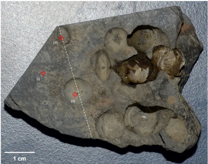
Figure 1. Photograph of a piece of siltstone collected from the middle reaches of the Kaladan 454
River, western Myanmar, showing extensive pitting by Lignopholas fluminalis bivalves. The 455
sample has been cut following the dashed line, and three locations were investigated by electron 456
microscopy, namely (i) an area devoid of macroborings, the bottom of (ii) a large and (iii) a 457
small macroboring, respectively. 458
460
Figure 2. Electron microscope characterizations of the rock sample at location (i), where the 461
surface is devoid of macroborings. (a) BSE image of the surface obtained using SEM, showing 462
the typical assemblage of silicates debris making up the siltstone. (b) Color map obtained from 463
EDX analyses, showing the random distribution of minerals making up the siltstone at the sub-464
mm scale. Green, red, purple and yellow colors represent minerals enriched in Mg (chlorite), 465
Na (feldspars), K (feldspars, clays) and Si (quartz, clays), respectively. Black pixels essentially 466
refer to voids (porosity) in the rock. (c) TEM image of the FIB thin section shown in (d). The 467
grain assemblage appears much more compact than that observed at the bottom of the 468
macroborings (see Fig. 4a, b). 469
470 471
472
Figure 3. Electron microscope characterizations of the rock sample referring to the bottom of 473
the largest macroboring (location (ii) in Fig. 1). (a) BSE image of the surface obtained using 474
SEM, showing the typical assemblage of silicates debris making up the siltstone. (b) Color map 475
obtained from EDX analyses, showing the random distribution of minerals making up the 476
siltstone at the sub-mm scale. Green, red, purple and yellow colors represent minerals enriched 477
in Mg (chlorite), Na (feldspars), K (feldspars, clays) and Si (quartz, clays), respectively. Black 478
pixels refer to voids (porosity) in the rock. (c) SEM image of a needle made of pure amorphous 479
silica and interpreted as a megasclere of Corvospongilla ultima sponge. (d) SEM image of the 480
bottom of the macroboring with superimposed EDX chemical analyses (C, N and S are 481
represented by red, green and purple colors, respectively). (e) EDX spectrum of the C-rich 482
location shown in Fig. 3d. (f) SEM image of the remains of putative bacterial cells (arrows) 483
with superimposed chemical analyses (C, Al and Si are represented by red, black and yellow 484
colors, respectively). 485
486 487
488
Figure 4. TEM characterizations of a FIB thin section excavated from the bottom of a small 489
macroboring (location (iii) in Fig. 1). (a) SEM image of the FIB thin section, showing the poorly 490
consolidated assemblage of minerals in the area that was contacting the bivalve. (b) TEM image 491
of the FIB thin section, showing ground quartz and an area enriched in C and N (labeled 492
“biofilm” on the figure). (c) EDX spectrum of the biofilm. (d) EDX color map of the biofilm 493
area shown in Fig. 4b. Mn, Si and N are represented by blue, green and red colors, respectively. 494
Note the occurrence of nanosized quartz debris. (e) Selected area electron diffraction of the 495
amorphous biofilm and the Mn-bearing crystals. 496
498
Figure 5. Analyses of the cross section of the bottom part of the shells of a bivalve. (a) 499
Photograph of the cross-section imaged in visible light. (b) BSE image of the cross-section of 500
the shells. Aragonite is white, quartz and clays are grey, epoxy is black. The arrow indicates 501
the organic film (see also Fig. S3), which contains debris or hard minerals such as quartz and 502
feldspars. (c) Integral map of organic and mineral distribution at the shell-siltstone interface. 503
The aragonite that makes up the shell is blue, silicate materials are green and the organic film 504
is red. (d) SEM image of the shell of a bivalve. No specific morphological microtexture that 505
could have provided the bivalve with an efficient excavation ability can be seen. 506
507 508
References
509
1 Distel, D. L. et al. Molecular phylogeny of Pholadoidea Lamarck, 1809 supports a single 510
origin for xylotrophy (wood feeding) and xylotrophic bacterial endosymbiosis in 511
Bivalvia. Molecular Phylogenetics and Evolution 61, 245-254, 512
doi:https://doi.org/10.1016/j.ympev.2011.05.019 (2011). 513
2 Distel, D. L. et al. Discovery of chemoautotrophic symbiosis in the giant shipworm 514
<em>Kuphus polythalamia</em> (Bivalvia: Teredinidae) extends wooden-steps 515
theory. Proceedings of the National Academy of Sciences 114, E3652-E3658, 516
doi:10.1073/pnas.1620470114 (2017). 517
3 Santos, A. et al. Extreme habitat adaptation by boring bivalves on volcanically active 518
paleoshores from North Atlantic Macaronesia. Facies 58, 325-338 (2012). 519
4 Shipway, J. R. et al. A rock-boring and rock-ingesting freshwater bivalve (shipworm) 520
from the Philippines. Proceedings of the Royal Society B 286, 20190434 (2019). 521
5 Kleemann, K. Biocorrosion by bivalves. Marine Ecology 17, 145-158 (1996). 522
6 Davidson, T. M., Altieri, A. H., Ruiz, G. M. & Torchin, M. E. Bioerosion in a changing 523
world: a conceptual framework. Ecology letters 21, 422-438 (2018). 524
7 Pinn, E. H., Thompson, R. & Hawkins, S. Piddocks (Mollusca: Bivalvia: Pholadidae) 525
increase topographical complexity and species diversity in the intertidal. Marine 526
Ecology Progress Series 355, 173-182 (2008).
527
8 Bolotov, I. N. et al. Discovery of a silicate rock-boring organism and macrobioerosion 528
in fresh water. Nature communications 9, 2882 (2018). 529
9 Vinn, O. & Toom, U. Borings in phosphatized Cambrian siltstone pebbles, Estonia 530
(Baltica). Geological Magazine 153, 635-642 (2016). 531
10 Nederlof, R. & Muller, M. A biomechanical model of rock drilling in the piddock 532
Barnea candida (Bivalvia; Mollusca). Journal of The Royal Society Interface 9, 2947-533
2958 (2012). 534
11 Dorgan, K. M. The biomechanics of burrowing and boring. Journal of Experimental 535
Biology 218, 176-183 (2015).
536
12 Fang, L.-S. & Shen, P. A living mechanical file: the burrowing mechanism of the coral-537
boring bivalve Lithophaga nigra. Marine Biology 97, 349-354 (1988). 538
13 Rodriguez-Tovar, F. J., Uchman, A. & Puga-Bernabéu, Á. Borings in gneiss boulders 539
in the Miocene (Upper Tortonian) of the Sorbas Basin, SE Spain. Geological Magazine 540
152, 287-297 (2015). 541
14 Johnson, M. E., Wilson, M. A. & Redden, J. A. Borings in quartzite surf boulders from 542
the Upper Cambrian basal Deadwood Formation, Black Hills of South Dakota. Ichnos 543
17, 48-55 (2010). 544
15 Staudigel, H. et al. 3.5 billion years of glass bioalteration: Volcanic rocks as a basis for 545
microbial life? Earth-Science Reviews 89, 156-176 (2008). 546
16 Jakhalekar, S. S. & Ghate, H. V. Taxonomy of freshwater sponges of Maharashtra, 547
India, with illustrated descriptions and notes on ecology and habitats (Porifera: 548
Spongillida: Spongillidae). Zootaxa 4173, 501-529 (2016). 549
17 Yang, W., Zhang, G., Liu, H. & Li, X. Microstructural characterization and hardness 550
behavior of a biological Saxidomus purpuratus shell. Journal of Materials Science & 551
Technology 27, 139-146 (2011).
552
18 Palandri, J. L. & Kharaka, Y. K. A compilation of rate parameters of water-mineral 553
interaction kinetics for application to geochemical modeling. 70 (2004). 554
19 Chou, L. & Wollast, R. Steady-state kinetics and dissolution mechanisms of albite. Am 555
J Sci 285, 963-993 (1985).
20 Lowson, R. T., Comarmond, M. C. J., Rajaratnam, G. & Brown, P. L. The kinetics of 557
the dissolution of chlorite as a function of pH and at 25 degrees C. Geochim Cosmochim 558
Ac 69, 1687-1699, doi:10.1016/j.gca.2004.09.028 (2005).
559
21 Brantley, S. L. & Olsen, A. A. in Treatise on Geochemistry (Second Edition) (ed Karl 560
K. Turekian) 69-113 (Elsevier, 2014). 561
22 Pinn, E. H., Richardson, C., Thompson, R. & Hawkins, S. Burrow morphology, 562
biometry, age and growth of piddocks (Mollusca: Bivalvia: Pholadidae) on the south 563
coast of England. Marine Biology 147, 943-953 (2005). 564
23 Benzerara, K., Yoon, T. H., Menguy, N., Tyliszczak, T. & Brown, G. E. Nanoscale 565
environments associated with bioweathering of a Mg-Fe-pyroxene. Proceedings of the 566
National Academy of Sciences of the United States of America 102, 979-982, doi:DOI
567
10.1073/pnas.0409029102 (2005). 568
24 Barker, W. W., Welch, S. A., Chu, S. & Banfield, J. F. Experimental observations of 569
the effects of bacteria on aluminosilicate weathering. Am Mineral 83, 1551-1563 570
(1998). 571
25 Li, Z., Liu, L., Chen, J. & Teng, H. H. Cellular dissolution at hypha-and spore-mineral 572
interfaces revealing unrecognized mechanisms and scales of fungal weathering. 573
Geology 44, 319-322 (2016).
574
26 Flemming, H.-C. & Wingender, J. The biofilm matrix. Nature reviews microbiology 8, 575
623 (2010). 576
27 Drever, J. & Stillings, L. The role of organic acids in mineral weathering. Colloids Surf 577
A 120, 167-181 (1997).
578
28 Golubev, S. V. & Pokrovsky, O. S. Experimental study of the effect of organic ligands 579
on diopside dissolution kinetics. Chem Geol 235, 377-389 (2006). 580
29 Wild, B., Imfeld, G., Guyot, F. & Daval, D. Early stages of bacterial community 581
adaptation to silicate aging. Geology 46, 555-558, doi:10.1130/g40283.1 (2018). 582
30 Zaharescu, D. G. et al. Ecosystem-bedrock interaction changes nutrient 583
compartmentalization during early oxidative weathering. Scientific Reports 9, 15006, 584
doi:10.1038/s41598-019-51274-x (2019). 585
31 Bonneville, S. et al. Plant-driven fungal weathering: Early stages of mineral alteration 586
at the nanometer scale. Geology 37, 615-618, doi:10.1130/g25699a.1 (2009). 587
32 Gounot, A.-M. Microbial oxidation and reduction of manganese: consequences in 588
groundwater and applications. FEMS Microbiology Reviews 14, 339-349 (1994). 589
33 McLoughlin, N. et al. in Current Developments in Bioerosion 371-396 (Springer, 590
2008). 591
34 Yu, H. & Leadbetter, J. R. Bacterial chemolithoautotrophy via manganese oxidation. 592
Nature 583, 453-458, doi:10.1038/s41586-020-2468-5 (2020).
593
35 Banfield, J. F., Barker, W. W., Welch, S. A. & Taunton, A. Biological impact on mineral 594
dissolution: application of the lichen model to understanding mineral weathering in the 595
rhizosphere. Proceedings of the National Academy of Sciences 96, 3404-3411 (1999). 596
36 Fisk, M. R., Giovannoni, S. J. & Thorseth, I. H. Alteration of Oceanic Volcanic Glass: 597
Textural Evidence of Microbial Activity. Science 281, 978-980, 598
doi:10.1126/science.281.5379.978 (1998). 599
37 Knowles, E., Staudigel, H. & Templeton, A. Geochemical characterization of tubular 600
alteration features in subseafloor basalt glass. Earth Planet Sc Lett 374, 239-250 (2013). 601
38 Nickerson, Z. L., Mortazavi, B. & Atkinson, C. L. Using functional traits to assess the 602
influence of burrowing bivalves on nitrogen-removal in streams. Biogeochemistry, 1-603
19 (2019). 604
39 Emmanuel, S. & Levenson, Y. Limestone weathering rates accelerated by micron-scale 605
grain detachment. Geology 42, 751-754 (2014). 606
40 Daval, D. et al. Dynamics of altered surface layer formation on dissolving silicates. 607
Geochim Cosmochim Ac 209, 51-69, doi:https://doi.org/10.1016/j.gca.2017.04.010
608
(2017). 609
41 Gabitov, R. et al. Elemental uptake by calcite slowly grown from seawater solution: an 610
in-situ study via depth profiling. Frontiers in Earth Science 7, 51 (2019). 611
42 Bolotov, I. N. et al. Trace element composition of freshwater pearl mussels 612
Margaritifera spp. across Eurasia: Testing the effect of species and geographic location. 613
Chem Geol 402, 125-139, doi:https://doi.org/10.1016/j.chemgeo.2015.03.006 (2015).
614 615 616