HAL Id: inserm-02417700
https://www.hal.inserm.fr/inserm-02417700
Submitted on 18 Dec 2019
HAL is a multi-disciplinary open access archive for the deposit and dissemination of sci-entific research documents, whether they are pub-lished or not. The documents may come from teaching and research institutions in France or abroad, or from public or private research centers.
L’archive ouverte pluridisciplinaire HAL, est destinée au dépôt et à la diffusion de documents scientifiques de niveau recherche, publiés ou non, émanant des établissements d’enseignement et de recherche français ou étrangers, des laboratoires publics ou privés.
vitro model of AMD
Mélanie Marie, Pauline Gondouin, Delphine Pagan, Coralie Barrau, Thierry
Villette, José Sahel, Serge Picaud
To cite this version:
Mélanie Marie, Pauline Gondouin, Delphine Pagan, Coralie Barrau, Thierry Villette, et al.. Blue-violet light decreases VEGFa production in an in vitro model of AMD. PLoS ONE, Public Library of Science, 2019, 14 (10), pp.e0223839. �10.1371/journal.pone.0223839�. �inserm-02417700�
Blue-violet light decreases VEGFa production
in an in vitro model of AMD
Me´lanie Marie1, Pauline Gondouin1, Delphine Pagan1, Coralie Barrau2, Thierry Villette2, Jose´ Sahel1,3, Serge Picaud1*
1 Sorbonne Universite´ , INSERM, CNRS, Institut de la Vision, Paris, France, 2 Essilor International R&D, Charenton-le-Pont, France, 3 Department of Ophthalmology, The University of Pittsburgh School of Medicine, Pittsburgh, PA, United States of America
*serge.picaud@inserm.fr
Abstract
Blue light is an identified risk factor for age-related macular degeneration (AMD). The pro-duction of vascular endothelial growth factor (VEGF), leading to neovascularization, is a major complication of the wet form of this disease. We investigated how blue light affects VEGF expression and secretion using A2E-loaded retinal pigment epithelium (RPE) cells, a cell model of AMD. Incubation of RPE cells with A2E resulted in a significant increase in VEGF mRNA and, intracellular and secreted VEGF protein levels, but not mRNA levels of VEGFR1 or VEGFR2. Blue light exposure of A2E-loaded RPE cells resulted in a decrease in VEGF mRNA and protein levels, but an increase in VEGFR1 levels. The toxicity of 440 nm light on A2E-loaded RPE cells was enhanced by VEGF supplementation. Our results suggest that age-related A2E accumulation may result in VEGF synthesis and release. This synthesis of VEGF, which enhances blue light toxicity for the RPE cells, is itself suppressed by blue light. Anti-VEGF therapy may therefore improve RPE survival in AMD.
Introduction
Age related macular degeneration (AMD) is characterized by a loss of central high acuity vision [1]. The dry type of AMD is the most common, accounting for approximately 90% of patients and is characterized by retinal pigment epithelium (RPE) and photoreceptors degen-eration over time in the macular area [2]. The wet type affects approximately 10% of patients and is characterized by abnormal blood-vessel growth [2]. Such blood-vessel growth is mainly attributed to the release of vascular endothelial growth factor (VEGF), as shown by the success of anti-VEGF therapies [3,4]. Anti-VEGF therapies have become a major treatment modality in the daily care of wet AMD to suppress the growth of neovessels, which invade the retina from the underlying choroid, located below the retinal pigment epithelium (RPE) [3,5]. VEGF traps have proven to be highly effective in protecting or even restoring visual function. VEGF, which is secreted by the retinal pigment epithelium [4], is indeed a very potent angiogenic fac-tor in the retina. Although VEGFa binds to both VEGF recepfac-tors VEGFR1 (Flt-1) and
VEGFR2 (KDR/Flk-1), VEGFR2 appears to mediate almost all intracellular signaling pathways in the vascular endothelium [6,7]. Understanding the molecular mechanisms that regulate
a1111111111 a1111111111 a1111111111 a1111111111 a1111111111 OPEN ACCESS
Citation: Marie M, Gondouin P, Pagan D, Barrau C,
Villette T, Sahel J, et al. (2019) Blue-violet light decreases VEGFa production in an in vitro model of AMD. PLoS ONE 14(10): e0223839.https://doi. org/10.1371/journal.pone.0223839
Editor: Olaf Strauß, Eye Hospital, Charite´,
GERMANY
Received: February 18, 2019 Accepted: October 1, 2019 Published: October 23, 2019
Copyright:© 2019 Marie et al. This is an open access article distributed under the terms of the
Creative Commons Attribution License, which permits unrestricted use, distribution, and reproduction in any medium, provided the original author and source are credited.
Data Availability Statement: All relevant data are
within the manuscript.
Funding: This work was financially supported by
research grants from the following: Essilor International, INSERM, Sorbonne Universite´, the LabEx LIFESENSES (ANR-10-LABX-65), as part of the first Investissements d’Avenir program (ANR-11-IDEX-0004-02) and IHU FOReSIGHT (ANR-18-IAHU-0001), both supported by French state funds managed by the Agence Nationale de la Recherche within the Investissements d’Avenir program. The funders had no role in study design, data collection
VEGF production is critical for preventing and controlling the development of this major complication of AMD.
Several risk factors for the development of AMD have already been identified, including age, smoking, genetic factors, and exposure to sunlight [2,8,9]. The role of sunlight exposure has been demonstrated in various epidemiological studies [2,10–15] and is corroborated by the protective role of macular pigments [16,17]. Indeed, within the solar spectrum, blue light is more highly involved in disease induction, consistent with the blue-light filtering capacity of macular pigments [13,18–23]. This severe retinal disease is characterized by the accumulation of lipofuscin within the RPE [24]. RPE cells ultimately degenerate in parallel with the loss of photoreceptors [1]. A2E is one of the retinoid compounds of lipofuscin which can enhance VEGF expression [25,26] and cause RPE degeneration, even in darkness [27]. At low concen-trations, A2E can act as a photosensitizer to induce cellular apoptosis upon blue-light exposure [28–38]. Controversial results on the effect of light on VEGF expression have been reported. In the human ARPE-19 cell line, acute white light (2500 lux for 12 h or 10 mW/cm2for 30 min) or short blue light (430 nm, 1 mW/cm2, for 3 to 7 min) exposure have been shown to enhance VEGF expression [39–41]. In addition, Kerntet al. confirmed that primary human
RPE cells containing lipofuscin exhibited increased VEGFa mRNA levels when exposed to white light for 15–60 min at 350 mW/cm2[42,43]. In contrast, white light (5000 lux 1 h) was shown to downregulate VEGF in the RPE-choroid eye cup, while increasing its expression in the neural retina of albino mice [44].
We recently demonstrated that blue light (415–455 nm) of the solar spectrum reaching the retina is the most toxic spectral band for A2E-loaded primary porcine RPE cells, used as anin vitro model of AMD [45,46]. Here, we examined how blue light affects VEGF levels in AMD. We used thisin vitro model to investigate how blue light affects VEGF mRNA and protein
lev-els and whether VEGF modifies RPE cell survival via the VEGFR2 pathway.
Materials and methods
Cell culture
Porcine eyes were bought at a local slaughterhouse (Etablissements guy Harang, Houdan, France) in agreement with the local regulatory authorities and the slaughterhouse veterinari-ans (agreement FR75105131). This procedure adheres to the European initiative for restricting animal experimentation because not a single animal was killed for our experimentation. Eyes were taken from animals slaughtered daily for food production. Retinal pigment epithelium cells (RPE cells) were extracted as previously described [45]. Three days after seeding in 96-well plates, confluent cells were treated for 6 h with 0, 12.5, or 20μM A2E (Orga-Link, Magny-les-Hameaux, France) in DMEM (Dulbecco’s Modified Eagle Medium, Life Technolo-gies, Carlsbad, CA, USA) without serum. DMSO (Sigma-Aldrich, St Louis, MO, USA) was adjusted to a final concentration of 0.1% for all conditions. After A2E treatment, cells were washed twice with modified DMEM (medium without any photosensitizer, such as phenol red, riboflavin, folic acid, or aromatic amino acids; Life Technologies) and exposed to light.
VEGF supplementation
In experiments designed to investigate the effects of exogenous VEGF on cell viability and apo-ptosis, 10 ng/mL recombinant VEGF (R&D System, Minneapolis, MN, USA) was diluted in modified DMEM and added to the cells 2 h before light exposure. Control cells were treated with the same concentration of the VEGF diluent alone (PBS-0.1% BSA, Sigma-Aldrich). Via-bility and apoptosis were assessed 6 h after the end of light exposure using the ApoLive-Glo™ Multiplex Assay (Promega, Madison, WI, USA) as previously described [45].
and analysis, decision to publish, or preparation of the manuscript. CB and TV received support in the form of a salary from Essilor. The specific roles of these authors are articulated in the ‘author contributions’ section.
Competing interests: I have read the journal’s
policy and the authors of this manuscript have the following competing interests: CB and TV are Essilor employees. SP received fees for participating in a meeting organized by Essilor. SP received a grant from Essilor to work on light toxicity on retinal cells. No competing financial interests exist for PG, and DP. This does not alter our adherence to PLOS ONE policies on sharing data and materials. The authors would like to declare the following patents/patent applications associated with this research: EP2602655 : filtre ophtalmique; EP2602654 : filtre ophtalmique; EP2602653 : Me´thode de la de´termination de la configuration d’un filtre ophtalmique; EP19305328 : filter for eye cone cells protection.
Inhibition of VEGF signaling
The effects of the inhibition of VEGF signaling on cell viability and apoptosis were investigated using a selective inhibitor of VEGFR2 (ZM323881 hydrochloride, Tocris, Bristol, UK) diluted to 1μM in modified DMEM. Control cells were treated with the same concentration of the VEGFR2 inhibitor diluent alone (DMSO). Cells were treated just before light exposure. Viabil-ity and apoptosis were assessed 6 h after the end of light exposure using the ApoLive-Glo™ Multiplex Assay (Promega) as previously described [45].
Light conditions
Cells were exposed to 10 nm-bandwidths of light produced by a specific LED-based optic fibers illumination device as previously described [45]. Physiological light conditions on the retina were mimicked by exposing RPE cells to a normalized light spectrum obtained by apply-ing the ocular media filterapply-ing spectrum onto a referenced solar spectrum (ASTM G173-03, International standard ISO 9845–1, 1992); blue light is partially filtered by the anterior ocular media as a natural protector. Irradiances ranged from 0.11 at 400 nm to a maximum irradiance level of 1.5 mW/cm2obtained for the light band centered at 630 nm [45,46]. Irradiance level, spectral, and uniformity measurements were obtained using a calibrated JAZ spectroradi-ometer (Ocean Optics, Dunedin, USA). For qPCR experiments, cells were exposed to light for 15 h and directly characterized or maintained in darkness for 6 to 24 h prior to characteriza-tion. For ELISA experiments cells were directly characterized after light exposure. For viability and apoptosis measurements, cells were exposed to light for 18 h and maintained in darkness for 6 h as previously described [45].
Enzyme-linked solid phase immunosorbent assay
The Quantikine Human VEGFa Immunoassay (R&D Systems) was used to quantify VEGFa protein levels in cells and the cell culture medium. After 15 h of light exposure, media was col-lected, centrifuged to eliminate cell debris, and the supernatants collected and stored at -80˚C. Cells were then rinsed twice with PBS (Life Technologies) and incubated for 5 min on ice in cell lysis buffer (Cell Signaling Technologies, Danvers, MA, USA) containing an antiprotease cocktail (Roche, Indianapolis, IN, USA). Cell lysates were sonicated, centrifuged, and the supernatants collected and stored at -80˚C. VEGFa protein content in cell lysates and cell cul-ture media was measured by ELISA according to the manufaccul-turer’s instructions. Total pro-tein content was determined for cell lysates using the micro BCA Propro-tein Assay Kit (Thermo Scientific, Rockford, IL, USA).
Real-time PCR
At the end of light exposure or after a 6 to 24 h rest period in darkness, cells were collected and total RNA isolated using the RNeasy Micro Kit (Qiagen, Hilden, Germany). RNA quality and quantity were assessed by spectrophotometry (NanoDrop 2000, Thermo Scientific). cDNA synthesis was performed using SuperScript II (Invitrogen) with random primers (Promega), 10 mM dNTPs (Life Technologies), and 0.1 M DTT (Life Technologies). The mix was incu-bated for 10 min at room temperature, then 50 min at 42˚C, and finally 15 min at 70˚C. Real-time PCR was performed using a StepOne device (Life Technologies) in a mixture containing cDNA, SYBR Green master mix (Life Technologies) and VEGFa forward (5’CACAGGACG GCTTGAAGATG3’) and reverse (5’TCTACCTCCACCATGCCAAG3’) primers, or VEGFR1 forward (5’CTCAACGCCATTCTGACGAG3’) and reverse (5’GGCGTTTGGGGAAAGTTCT T3’) primers, or VEGFR2 forward (5’CAAGAGGATGTTTCGAGCCG3’) and reverse
(5’AAATCCCTCAGCGATGTGGA3’) primers. Data were normalized to that of 18S RNA, which was simultaneously amplified for all samples using 18S forward (5’AGTCGGCATCG TTTATGGTC3’) and reverse (5’CGCGGTTCTATTTTGTTGGT3’) primers. A total of 1 ng of cDNA was sufficient to correctly amplify the VEGF gene, whereas 5 ng of cDNA was necessary for VEGFR1 and VEGFR2. Indeed, VEGFR2 was barely detectable after 6 and 24 h post light exposure, even when using 15 ng of cDNA, and the data could not be properly interpreted because of the low quantity of final amplification products. RT-PCR amplification products were purified and sequenced using the Sanger method and the sequences compared to those of the pig genome. mRNA level ratios were determined using the delta Ct calculation method: ΔCT=CT target gene−CT 18SandΔΔCT=ΔCT sample−ΔCT dark control 0 μM, relative mRNA level to
dark control 0μM = 2 DDCT
.
Statistical analysis
All experiments were repeated at least three times. Data are presented as the mean +/- SEM. Statistical analyses were performed using Statistica software v12 (StatSoft, Tulsa, OK, USA). Two-way ANOVA with repeated measures was used. Bilateral Dunnett post-hoc tests were used to compare variances of all tested groups (for each A2E concentration and each light con-dition) to the dark control groups. Tukey post-hoc tests were used for cell viability and apopto-sis analyses to compare variances of all groups together. Differences between samples were considered to be significant when p < 0.05 (�), p < 0.01 (��), or p < 0.001 (���).
Results
Blue-violet light decrease VEGFa mRNA expression and increase VEGFR1
mRNA expression
We evaluated VEGFa, VEGFR1 and VEGFR2 mRNA levels by qRT-PCR following A2E incu-bation and/or light exposure (Fig 1). All mRNA levels were normalized against the level of the 18S ribosomal subunit mRNA, which was not expected to change during the procedures. RPE cells maintained in darkness showed high levels of VEGFa mRNA relative to those of its recep-tors, VEGFR1 and 2. The average expression ratio to VEGFa reached for VEGFR1 1/142 and 1/1,969 for VEGFR2 (mean delta CtVEGF= 14.24, mean delta CtVEGFR1= 21.39 and mean delta
CtVEGFR2= 25.18), in accordance with the results of a previous study [47]. A2E treatment
increased the expression of VEGFa and VEGFR1. VEGFa mRNA levels increased by two-fold following incubation of RPE cells with A2E, as previously reported by others [26,48], although this increase disappeared by 39 h after the end of A2E incubation (Fig 1A, 1B and 1C). Simi-larly, VEGFR1 mRNA levels also increased up to four-fold after incubation with A2E, and this increase was stable over time (Fig 1D, 1E and 1F). In contrast, VEGFR2 mRNA levels
remained very low, even in the presence of A2E (Fig 1G).
We then investigated how light affects VEGFa and VEGF receptor expression in RPE cells by exposing them to various wavelengths (400, 440, 480 and 630 nm) after A2E incubation. RPE cells were exposed for 15 h, as we recently found that this exposure period enabled us to investigate the molecular mechanisms prior to apoptosis [46] by limiting light toxicity observed with longer light exposures (18 h) [45]. We measured VEGFa and VEGF receptor mRNA levels in RPE cells immediately after light exposure and at two additional time points to investigate the long-term effect of light exposures. Light exposure alone did not modify VEGFa mRNA levels in the absence of A2E at any of the post-exposure time points (Fig 1A, 1B and 1C). In contrast, blue light at 400, 440, or 480 nm decreased the A2E-elicited increase in VEGFa mRNA levels, with the largest reduction at the highest A2E concentration (20μM),
reaching 50% at 440 nm immediately after the end of light exposure (Fig 1A, 1B and 1C). VEGFa levels thus returned to the control values measured in the absence of A2E. At later time points after light exposure, differences from the control condition in darkness were steadily disappeared, as the A2E-elicited increase of VEGF expression was also fading away.
We observed a statistically significant increase in VEGFR1 mRNA levels immediately after light exposure at 440, 480, and 630 nm in the presence of 12.5 and 20μM A2E (Fig 1D). We observed the greatest increase of VEGFR1 mRNA levels 6 h after the end of light exposure at 440 nm, reaching a maximum of 8.8-fold for 12.5μM A2E and 12.3-fold for 20 μM A2E (Fig 1E). This increase completely disappeared by 24 h after the end of light exposure for 12.5μM A2E, but VEGFR1 mRNA levels remained elevated relative to dark controls for 20μM A2E, but at lower levels (Fig 1F). The increase in VEGFR1 expression demonstrates that the decrease of VEGFa mRNA expression is not only due to the reduced number of viable cells.
Fig 1. VEGFa, VEGFR1 and VEGFR2 mRNA levels following A2E incubation and/or light exposure. Each histogram represents the mRNA levels of the gene in
darkness or following exposure to wavelengths (400, 440, 480 or, 630 nm) in the absence or presence of A2E preloaded at 12.5 or 20μM. These measurements were repeated for three time points: immediately, 6 h, or 24 h after light exposure. Note the increase in gene expression for VEGF and VEGFR1 after A2E incubation in cells maintained in darkness. Blue light exposure at 440 and 480 nm suppressed the increase in VEGF expression, whereas it further increased VEGFR1 expression. Data are expressed as the mean +/- SEM (n = 3). Differences between experimental samples and 0, 12.5, or 20μM dark controls were considered to be significant when p < 0.05 (�), p < 0.01 (��) or p < 0.001 (���).
VEGFR2 mRNA levels only increased in the absence of A2E at the 400-nm wavelength exposure but this effect was not maintained in the presence of A2E (Fig 1G). Incubation with A2E resulted in a decrease of VEGFR2 mRNA levels under blue light (440 and 480 nm), but only at the lowest A2E concentration (12.5μM) (Fig 1G). Data from later time points were not used due to the very low levels of VEGFR2 mRNA. The gene expression data for VEGFR2 must be interpreted with caution, as VEGFR2 expression was barely detectable, even after increasing the amount of starting material.
Blue-violet light decreases the intracellular concentration of VEGFa
After observing modulation of VEGFa mRNA levels by A2E and light exposure, we examined whether VEGFa protein synthesis was also modified. We evaluated VEGFa protein concentra-tions in cell lysates by ELISA immediately after light exposure (Fig 2). VEGFa protein concen-trations varied between 1.76 and 4.49 pg/μg proteins for all conditions tested (Fig 2B). Light alone did not affect the intracellular content of VEGFa protein in the absence of A2E at any of the tested wavelengths (Fig 2A, 2B and 2C). Incubation of RPE cells with A2E (20μM) induced a two-fold increase in the intracellular content of VEGFa protein, consistent with the previ-ously observed increase in VEGFa mRNA levels. Light exposure (440, 480 nm) decreased VEGFa protein levels, except for the illumination bands at 400 and 630 nm, in which VEGFa protein concentrations were in the same range as those measured in A2E-loaded cells main-tained in darkness. As a consequence, VEGFa protein concentrations returned to levels mea-sured in the absence of A2E treatment following blue light exposure (440 or 480 nm). Thus, blue light highly counteracts the increase in intracellular VEGFa protein content triggered by A2E incubation.
Blue-violet light decreased VEGFa protein secretion
We next investigated the release of VEGFa protein by RPE cells upon light exposure by mea-suring its concentration in the cell-culture medium by ELISA at the end of light exposure. Cells were washed before light exposure with fresh VEGF-free medium containing neither serum nor added growth factors. Thus, the measured VEGFa protein concentration in the cul-ture medium corresponded to the protein released during the exposure of RPE cells to light. VEGFa protein concentrations varied from 3,347 to 6,107 pg/mL for all conditions tested. RPE cells maintained in darkness for 15 h secreted a high amount of VEGFa, approximately 4,000 pg/mL (Fig 2D). Light exposure alone did not modify VEGFa protein concentrations in the extracellular medium in the absence of A2E (Fig 2D and 2E). In contrast, incubation with A2E significantly increased VEGFa protein release, consistent with the enhanced intracellular levels of VEGFa in A2E-loaded cells. Again, blue light (440 and 480 nm) significantly reduced the release of VEGFa protein into the cell-culture medium, which returned to the levels measured in the absence of A2E (Fig 2D and 2E). The increase of VEGFa protein levels upon A2E incu-bation was statistically significant, as well as its suppression by blue light (440 and 480 nm) (Fig 2D and 2E). These results highlight the association between intracellular VEGFa protein content and VEGFa protein release, suggesting that reduced VEGFa protein release is related to the reduced protein synthesis under blue light stimulation. Finally, our measurements at the mRNA and protein levels were consistent, indicating that the observed changes in protein secretion were directly related to regulation of the mRNA levels, which were increased by A2E in darkness and suppressed by blue-violet light. Unlike for the normalized mRNA levels, it cannot be fully excluded that the reduction in VEGF protein levels was not at least partially due to reduced cell viability.
Effects of VEGFa on cell viability and apoptosis in A2E-loaded RPE cells
exposed to blue-violet light
VEGFa is neuroprotective for different retinal cells [49,50]. It seemed thus possible that the high content of VEGFa in the cell culture medium could act as a pro-survival factor in A2E loaded cells. Therefore, we tested this possibility by incubating A2E-loaded RPE cells with 10 ng/mL recombinant VEGFa protein for 2 h before and during 18 h of light exposure. This con-centration of VEGFa was used based on the VEGF concon-centrations measured by ELISA in the RPE cell culture medium (Fig 2) to have the same initial VEGF protein concentration in the medium for all experimental conditions. This concentration was also found to be neuroprotec-tive in a previous study on retinal ganglion cells [49]. We assessed viability and apoptosis with the Apolive Glo AssayTM(Promega) 6 h after light exposure. In the absence of A2E, VEGFa only significantly increased RPE cell viability at 400 nm but there was no detectable effect on apoptosis (Fig 3). In the absence of added VEGFa, blue light (400, 430,or 440 nm) decreased
Fig 2. Blue-violet light suppression of the A2E-elicited increase in intracellular and secreted VEGFa protein levels. VEGFa protein contents in RPE cell
lysates (A), after their normalization for each sample to its protein content (B), and after a second normalization with respect to the value measured in the dark control without A2E (C). VEGFa protein levels released in the culture media (D), and after their normalization with respect to the value measured in the dark control without A2E (E). Note that A2E increased both the levels of VEGFa in the cells and the secreted VEGFa whereas blue-violet light (440 nm) suppressed these increases in VEGFa proteins levels. These protein levels were measured by ELISA following 15 h of light exposure with or without pre-incubation in 20μM A2E. Data are expressed as mean +/- SEM (n = 4) for intracellular content and as mean +/- SEM (n = 8) for secreted VEGFa. Differences between experimental samples were considered to be significant when p < 0.05 (�), p < 0.01 (��) or p < 0.001 (���). Stars on the 20μM columns represent significant differences with the corresponding 0μM averaged value.
cell viability and induced up to a 5.5-fold increase in apoptosis/viability of A2E-loaded RPE cells relative to dark controls (Fig 3), as previously described by our group [45]. Addition of VEGF to the incubation medium resulted in a statistically significant enhancement in the loss of cell viability at 440 and 630 nm in A2E-loaded RPE cells (Fig 3A). This decrease in cell via-bility correlated with an increase in apoptosis at 440 nm (Fig 3B), which was significant after normalization to the rate of viability (Fig 3C). Differences in VEGF sensitivity observed between 430 and 440 nm could be related to the different ranges of toxicity at these two wave-lengths. Indeed, in the absence of VEGF, 430 nm was already much more toxic than 440 nm on A2E-loaded RPE cells and thus could prohibit a further increase in toxicity upon addition of VEGF. VEGF was not protective to A2E-loaded RPE cells exposed to light but further increased light-induced toxicity at 440 nm.
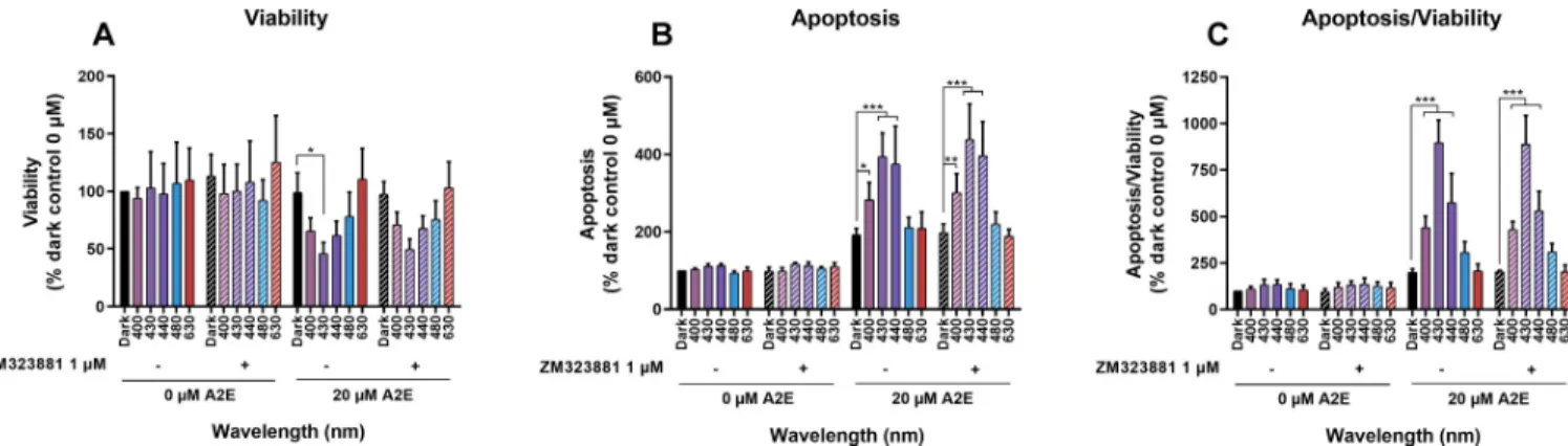
A2E-loaded RPE cells synthesized and released VEGF into the extracellular medium at a concentration that enhanced blue-light toxicity at 440 nm (Fig 3). We investigated whether this secreted VEGF contributes to A2E-induced phototoxicity by measuring cell viability and apoptosis in the presence of a selective inhibitor of VEGFR2 (ZM323881 hydrochloride) to block the main exogenous VEGFa signaling pathway in RPE cells [51]. A VEGFR inhibitor concentration of 1μM was chosen to exert selective efficacy on VEGFR2, while avoiding inter-action with VEGFR1, PDGFRb, FGFR1, EGFR, or erbB2, described at higher concentrations [51,52]. Thus, the VEGFR1 inhibitor can block any autocrine and paracrine effect of VEGFa release by RPE cells in the culture medium. RPE cells were loaded with A2E at 0 or 20μM, washed, and then incubated with the inhibitor in fresh medium just before starting the 18-h light exposure. We assessed viability and apoptosis 6 h after light exposure (Fig 4A, 4B and 4C). Addition of the inhibitor did not modify RPE cell viability or apoptosis under the experi-mental conditions tested, consistent with the low VEGFR2 expression in RPE cells (Fig 1). Under our experimental conditions, this autocrine pathway of VEGFa mediated VEGFR2 acti-vation was not critical in inducing A2E-loaded RPE cell degeneration under light exposure.
Discussion
VEGF plays an important role in the development of the wet form of age-related macular degen-eration. Indeed, excessive release of this factor by RPE cells induces rapid neovascularization
Fig 3. Effects of VEGFa supplementation on cell viability and apoptosis in A2E-loaded RPE cells exposed to light. RPE cells were loaded with 0 or 20μM A2E and then the media supplemented with 10 ng/mL recombinant VEGFa before being exposed to light for 18 h. Viability (A) and apoptosis (B) were assessed with the Apolive Glo AssayTM after light exposure following a 6 h rest period in darkness. The number of apoptotic cells was normalized to cell viability to define the density of apoptotic cells in the remaining viable cells at the end of the experiment (C). In the absence of A2E, VEGFa supplementation increased RPE cell viability in the dark and at 400 nm. VEGFa supplementation of A2E-loaded RPE cells decreased viability at 440 and 630 nm. VEGFa supplementation on RPE cells without A2E did not affect apoptosis upon light exposure. By contrast, VEGFa supplementation on A2E-loaded RPE cells increased apoptosis at 440 nm. Data are expressed as the mean +/- SEM (n = 3). Differences between samples were considered to be significant when p < 0.05 (�), p < 0.01 (��) or p < 0.001 (���).
under the retina. The growth of abnormal vessels in the subretinal space, with a porous wall, causes disruption of the epithelial barrier and leakage of fluids under the retina, which is partially responsible for vision loss. Among the various structures of the eye, the retina and, more specifi-cally RPE cells contain the largest amounts of VEGF protein [47]. Indeed, RPE cells are responsi-ble for the production and secretion of several growth factors, including VEGF, required for all other structures of the eye. Despite their high VEGF content, RPE cells express only low levels of the VEGF receptors, VEGFR1 and VEGFR2, with expression of VEGFR2 being less than that of VEGFR1 [47]. We confirmed the high levels of VEGF mRNA expression and protein synthesis, whereas the level of VEGFR1 mRNA was low and that of VEGFR2 barely detectable. We showed that A2E increased VEGF mRNA levels and VEGF protein synthesis. This result, obtained with primary porcine RPE cells, is consistent with those reported for the ARPE-19 cell line [26,48]. This A2E-induced increase in VEGF synthesis was attributed to the sustained activation of reti-noic acid receptors (RAR) in both the ARPE-19 cell line and Y79 retinoblastoma cells andin vivo [25,26,53]. Our results are in accordance with these studies, showing that incubation of RPE cells with A2E in the dark increased VEGF synthesis and secretion following a dose-depen-dent increase in mRNA expression. Our results confirm the A2E-induced potentiation of VEGF synthesis and release, suggesting that A2E accumulation could thus stimulate the neovasculariza-tion observed in wet AMD.
Light, more specifically blue light, has been shown to be a cumulative risk factor for the development of AMD [2]. We are all exposed to this environmental factor in a cumulative manner during our lifetime. It is thus of great importance to understand how it can influence the development of AMD in particular the VEGF-related complications of the wet form of this disease. VEGF expression was reported to increase in several studies in which oxidative stress was induced in ARPE-19 cells by the addition of chemical compounds, such as H2O2or
etha-nol [54,55]. Light-elicited oxidative stress especially in the presence of A2E, may thus also increase VEGF expression. High H2O2production has indeed been demonstrated in
A2E-loaded cells upon blue light exposure [46,56]. Furthermore, this light-elicited oxidative stress was found to induce a translocation of mitochondria to the perinuclear area [46,56], which has been shown to enhance the expression of hypoxia-sensitive genes, such as VEGF, in other cell systems [57]. Others have already reported that bright white light reduces cell survival but increases VEGF synthesis in human primary RPE cells [43]. However, the A2E content in
Fig 4. Effects of VEGFR2 inhibition on viability and apoptosis of A2E-loaded RPE cells exposed to light. RPE cells were loaded with 0 or 20μM A2E and treated with ZM323881 hydrochloride (1μM), a selective inhibitor of VEGFR2 activity, before being exposed to light for 18 h light exposure. Viability (A) and apoptosis (B) were evaluated 6 h after the end of light exposure with the Apolive Glo AssayTM. The number of apoptotic cells was normalized to cell viability to define the density of apoptotic cells in the remaining viable cells at the end of the experiment (C). VEGFR2 inhibitor did not modify RPE cell viability or apoptosis under any conditions tested. Data are expressed as the mean +/- SEM (n = 4). Differences between samples were considered to be significant when p < 0.05 (�), p < 0.01 (��) or p < 0.001 (���).
these cells is unknown. Also, an un-tinted intraocular lens (IOL) produced a greater increase in VEGF expression than a blue-light filtering IOLin vitro showing that blue light further
increases VEGF synthesis [42]. Similarly, white light increased VEGF synthesis in A2E-loaded ARPE-19 cells and this effect was attenuated by a blue-light filtering IOL, suggesting potentia-tion of VEGF synthesis by blue light stimulapotentia-tion [40,58]. Surprisingly, we obtained the oppo-site results; blue light (400, 440, or 480 nm) reduced VEGF mRNA expression and protein synthesis of A2E-loaded primary porcine RPE cells. This difference may be due to the different cell types used in the studies: the ARPE-19 cell line or primary human or porcine RPE cells. The human primary RPE cells were not incubated with A2E, in contrast to our experimental conditions and those under which the ARPE-19 cell line was used [40,58]. The main differ-ence may indeed lie in the light exposure conditions, because the other groups mainly used bright white light and blue light filtering-IOLs to assess the specific effect of blue light [40,58]. In contrast, we used specific exposure to 10 nm bandwidths, such that it would be difficult to anticipate the summed response of a white light spectrum. In addition, we did not apply UV light, in contrast to other studies that used white light, possibly demonstrating an effect of UV-absorbing IOL [40]. Several studies on A2E-loaded ARPE-19 cells also restricted their illumi-nation to a specific bandwidth (430 nm), but the illumiillumi-nation periods were very short (3–7 min) in most cases [41], whereas we used moderate irradiance and long-duration illumination (15-18h). Our irradiance levels were indeed moderate (below 1.5 mW/cm2) and lower than those of other studies (8–10 mW/cm2) [40,41]. Finally, we used wavelength irradiances that were normalized to the sunlight spectrum that reaches the retina for each wavelength band to mimic thein vivo conditions of the natural protection of the crystalline lens as closely as
possi-ble. Although blue light has been considered to further enhance the risk of vascular complica-tions in other cell models, our study suggests that it may instead reduce VEGF synthesis and release by A2E-loaded RPE cells when exposed to moderate irradiance normalized to the sun-light spectrum with a long duration exposure.
VEGF has been described to act as an autocrine survival factor in an RPE cell line (APRE-19) under H2O2-induced oxidative stress via the autocrine VEGFR2 activation pathway [59].
Here, we observed a two-fold increase in VEGF synthesis upon incubation with A2E, as have others [26]. Because blue light (415–455 nm) exposure induces substantial H2O2production in
A2E-loaded RPE cells leading to RPE cell apoptosis [45,46,56], we examined whether VEGF can act as a survival factor for A2E-loaded cells exposed to blue light. Surprisingly, addition of VEGF to the culture medium (10 ng/mL) increased blue-light toxicity at 440 nm in A2E-loaded RPE cells. These results are consistent with thein vivo prevention of light damage to
the RPE by anti-VEGF strategies [44]. VEGFa binds to both VEGF receptors VEGFR1 (Flt-1) and VEGFR2 (KDR/Flk-1). VEGFR2 appears to mediate almost all intracellular signaling pathways in the vascular endothelium [6,7]. However, we showed that RPE cells exhibit much lower levels of VEGFR2 mRNA than VEGFR1 mRNA. VEGFR1 mRNA levels were substan-tially increased by blue light (440 nm) exposure in A2E-loaded RPE cells. Although, a VEGFR2 inhibitor did not show any effect on cell survival due to the secreted VEGF in our culture medium, it does not exclude that the VEGFR2 signaling pathway is responsible for the VEGF enhancement of blue light toxicity on A2E-loaded RPE cells. In AMD, the accumulation of A2E may thus lead to VEGF synthesis and release by RPE cells, such that the VEGF released into the extracellular medium may not only induce angiogenesis but also increase blue light toxicity. As a consequence, VEGF therapy could thus prevent angiogenesis and protect RPE cells from light damage. Interestingly, blue light may naturally mimic such anti-VEGF therapy in RPE cells by increasing VEGFR1 expression and decreasing VEGF expression. VEGFR1 can indeed trap free VEGF as do the therapeutic molecules in all anti-VEGF therapies [60,61].
However, this anti-angiogenic effect acts in parallel with the very high risk of blue-light damage.
Conclusion
Anti-VEGF therapies have become a major treatment modality in the daily care of wet AMD and diabetic retinopathy to suppress the growth of neovessels and/or macular edema [5]. The photosensitizer A2E can upregulate both VEGF mRNA and protein levels in RPE cells by acti-vating retinoic receptors [26]. A2E photosensitization by blue light induce oxidative stress and RPE cell death in anin vitro model of AMD [45,46]. Here, we showed that blue light exposure of A2E-loaded RPE cells for 15 h at moderate irradiance, normalized to sunlight irradiance reaching the retina, can also down regulate VEGF synthesis while up-regulating VEGFR1 mRNA expression. The upregulation of VEGFR1 may act as a VEGF-trap or as a protective mechanism against oxidative stress [60–62]. Surprisingly, we found that supplementation with VEGF, a well-known pro-survival factor, can further increase blue light toxicity to A2E-loaded RPE cells. The conversion of VEGF pro-survival signaling into cell apoptosis has already been described in endothelial cells [63,64]. This suggests that anti-VEGF therapy may not only resolve vascular complications, but may also limit the VEGF-elicited enhancement of blue-vio-let light toxicity on A2E-loaded RPE cells.
Acknowledgments
The authors thank the cell culture and sequencing platforms at the Institut de la Vision.
Author Contributions
Conceptualization: Me´lanie Marie, Serge Picaud. Formal analysis: Me´lanie Marie, Coralie Barrau. Funding acquisition: Jose´ Sahel, Serge Picaud.
Investigation: Me´lanie Marie, Pauline Gondouin, Delphine Pagan. Project administration: Me´lanie Marie, Serge Picaud.
Visualization: Me´lanie Marie.
Writing – original draft: Me´lanie Marie, Serge Picaud.
Writing – review & editing: Pauline Gondouin, Delphine Pagan, Coralie Barrau, Thierry
Vill-ette, Jose´ Sahel.
References
1. Bhutto I, Lutty G. Understanding age-related macular degeneration (AMD): relationships between the photoreceptor/retinal pigment epithelium/Bruch’s membrane/choriocapillaris complex. Mol Aspects Med. 2012; 33(4):295–317.https://doi.org/10.1016/j.mam.2012.04.005PMID:22542780
2. Sui GY, Liu GC, Liu GY, Gao YY, Deng Y, Wang WY, et al. Is sunlight exposure a risk factor for age-related macular degeneration? A systematic review and meta-analysis. Br J Ophthalmol. 2013; 97 (4):389–394.https://doi.org/10.1136/bjophthalmol-2012-302281PMID:23143904
3. Miller JW. VEGF: From Discovery to Therapy: The Champalimaud Award Lecture. Transl Vis Sci Tech-nol. 2016; 5(2):9.https://doi.org/10.1167/tvst.5.2.9PMID:26981331
4. Penn JS, Madan A, Caldwell RB, Bartoli M, Caldwell RW, Hartnett ME. Vascular endothelial growth fac-tor in eye disease. Prog Retin Eye Res. 2008; 27(4):331–371.https://doi.org/10.1016/j.preteyeres. 2008.05.001PMID:18653375
5. Miller JW. Beyond VEGF-The Weisenfeld Lecture. Invest Ophthalmol Vis Sci. 2016; 57(15):6911– 6918.https://doi.org/10.1167/iovs.16-21201PMID:28027565
6. Stuttfeld E, Ballmer-Hofer K. Structure and function of VEGF receptors. IUBMB Life. 2009; 61(9):915– 922.https://doi.org/10.1002/iub.234PMID:19658168
7. Holmes DI, Zachary IC. Vascular endothelial growth factor regulates stanniocalcin-1 expression via neuropilin-1-dependent regulation of KDR and synergism with fibroblast growth factor-2. Cell Signal. 2008; 20(3):569–579.https://doi.org/10.1016/j.cellsig.2007.11.009PMID:18164591
8. Schick T, Ersoy L, Lechanteur YT, Saksens NT, Hoyng CB, den Hollander AI, et al. History of Sunlight Exposure Is a Risk Factor for Age-Related Macular Degeneration. Retina. 2016; 36(4):787–790.https:// doi.org/10.1097/IAE.0000000000000756PMID:26441265
9. Lambert NG, ElShelmani H, Singh MK, Mansergh FC, Wride MA, Padilla M, et al. Risk factors and bio-markers of age-related macular degeneration. Prog Retin Eye Res. 2016; 54:64–102.https://doi.org/ 10.1016/j.preteyeres.2016.04.003PMID:27156982
10. Augood CA, Vingerling JR, de Jong PT, Chakravarthy U, Seland J, Soubrane G, et al. Prevalence of age-related maculopathy in older Europeans: the European Eye Study (EUREYE). Arch Ophthalmol. 2006; 124(4):529–535.https://doi.org/10.1001/archopht.124.4.529PMID:16606879
11. Cruickshanks KJ, Klein R, Klein BE, Nondahl DM. Sunlight and the 5-year incidence of early age-related maculopathy: the beaver dam eye study. Arch Ophthalmol. 2001; 119(2):246–250. PMID:11176987
12. Young RW. Sunlight and age-related eye disease. J Natl Med Assoc. 1992; 84(4):353–358. PMID:
1507250
13. Taylor HR, West S, Munoz B, Rosenthal FS, Bressler SB, Bressler NM. The long-term effects of visible light on the eye. Arch Ophthalmol. 1992; 110(1):99–104.https://doi.org/10.1001/archopht.1992. 01080130101035PMID:1731731
14. Fletcher AE, Bentham GC, Agnew M, Young IS, Augood C, Chakravarthy U, et al. Sunlight exposure, antioxidants, and age-related macular degeneration. Arch Ophthalmol. 2008; 126(10):1396–1403.
https://doi.org/10.1001/archopht.126.10.1396PMID:18852418
15. Butt AL, Lee ET, Klein R, Russell D, Ogola G, Warn A, et al. Prevalence and risks factors of age-related macular degeneration in Oklahoma Indians: the Vision Keepers Study. Ophthalmology. 2011; 118 (7):1380–1385.https://doi.org/10.1016/j.ophtha.2010.11.007PMID:21310490
16. Loane E, Kelliher C, Beatty S, Nolan JM. The rationale and evidence base for a protective role of macu-lar pigment in age-related maculopathy. Br J Ophthalmol. 2008; 92(9):1163–1168.https://doi.org/10. 1136/bjo.2007.135566PMID:18669545
17. Snodderly DM. Evidence for protection against age-related macular degeneration by carotenoids and antioxidant vitamins. Am J Clin Nutr. 1995; 62(6 Suppl):1448S–1461S.
18. Mainster MA, Turner PL. Blue-blocking IOLs decrease photoreception without providing significant photoprotection. Surv Ophthalmol. 2010; 55(3):272–289.https://doi.org/10.1016/j.survophthal.2009.07. 006PMID:19883931
19. Kijlstra A, Tian Y, Kelly ER, Berendschot TT. Lutein: more than just a filter for blue light. Prog Retin Eye Res. 2012; 31(4):303–315.https://doi.org/10.1016/j.preteyeres.2012.03.002PMID:22465791
20. Jia YP, Sun L, Yu HS, Liang LP, Li W, Ding H, et al. The Pharmacological Effects of Lutein and Zeaxan-thin on Visual Disorders and Cognition Diseases. Molecules. 2017; 22(4).
21. Lima VC, Rosen RB, Farah M. Macular pigment in retinal health and disease. Int J Retina Vitreous. 2016; 2:19.https://doi.org/10.1186/s40942-016-0044-9PMID:27847637
22. Loskutova E, Nolan J, Howard A, Beatty S. Macular pigment and its contribution to vision. Nutrients. 2013; 5(6):1962–1969.https://doi.org/10.3390/nu5061962PMID:23760061
23. Junghans A, Sies H, Stahl W. Macular pigments lutein and zeaxanthin as blue light filters studied in lipo-somes. Arch Biochem Biophys. 2001; 391(2):160–164.https://doi.org/10.1006/abbi.2001.2411PMID:
11437346
24. Sparrow JR, Boulton M. RPE lipofuscin and its role in retinal pathobiology. Exp Eye Res. 2005; 80 (5):595–606.https://doi.org/10.1016/j.exer.2005.01.007PMID:15862166
25. Iriyama A, Inoue Y, Takahashi H, Tamaki Y, Jang WD, Yanagi Y. A2E, a component of lipofuscin, is pro-angiogenic in vivo. J Cell Physiol. 2009; 220(2):469–475.https://doi.org/10.1002/jcp.21792PMID:
19418485
26. Iriyama A, Fujiki R, Inoue Y, Takahashi H, Tamaki Y, Takezawa S, et al. A2E, a pigment of the lipofuscin of retinal pigment epithelial cells, is an endogenous ligand for retinoic acid receptor. J Biol Chem. 2008; 283(18):11947–11953.https://doi.org/10.1074/jbc.M708989200PMID:18326047
27. Suter M, Reme C, Grimm C, Wenzel A, Jaattela M, Esser P, et al. Age-related macular degeneration. The lipofusion component N-retinyl-N-retinylidene ethanolamine detaches proapoptotic proteins from mitochondria and induces apoptosis in mammalian retinal pigment epithelial cells. J Biol Chem. 2000; 275(50):39625–39630.https://doi.org/10.1074/jbc.M007049200PMID:11006290
28. Boulton M, Rozanowska M, Rozanowski B. Retinal photodamage. J Photochem Photobiol B. 2001; 64 (2–3):144–161.https://doi.org/10.1016/s1011-1344(01)00227-5PMID:11744401
29. Rozanowska M, Pawlak A, Rozanowski B, Skumatz C, Zareba M, Boulton ME, et al. Age-related changes in the photoreactivity of retinal lipofuscin granules: role of chloroform-insoluble components. Invest Ophthalmol Vis Sci. 2004; 45(4):1052–1060.https://doi.org/10.1167/iovs.03-0277PMID:
15037568
30. Wihlmark U, Wrigstad A, Roberg K, Nilsson SE, Brunk UT. Lipofuscin accumulation in cultured retinal pigment epithelial cells causes enhanced sensitivity to blue light irradiation. Free Radic Biol Med. 1997; 22(7):1229–1234.https://doi.org/10.1016/s0891-5849(96)00555-2PMID:9098097
31. Schutt F, Davies S, Kopitz J, Holz FG, Boulton ME. Photodamage to human RPE cells by A2-E, a reti-noid component of lipofuscin. Invest Ophthalmol Vis Sci. 2000; 41(8):2303–2308. PMID:10892877
32. Sparrow JR, Nakanishi K, Parish CA. The lipofuscin fluorophore A2E mediates blue light-induced dam-age to retinal pigmented epithelial cells. Invest Ophthalmol Vis Sci. 2000; 41(7):1981–1989. PMID:
10845625
33. Davies S, Elliott MH, Floor E, Truscott TG, Zareba M, Sarna T, et al. Photocytotoxicity of lipofuscin in human retinal pigment epithelial cells. Free Radic Biol Med. 2001; 31(2):256–265.https://doi.org/10. 1016/s0891-5849(01)00582-2PMID:11440838
34. Sparrow JR, Cai B. Blue light-induced apoptosis of A2E-containing RPE: involvement of caspase-3 and protection by Bcl-2. Invest Ophthalmol Vis Sci. 2001; 42(6):1356–1362. PMID:11328751
35. Nilsson SE, Sundelin SP, Wihlmark U, Brunk UT. Aging of cultured retinal pigment epithelial cells: oxi-dative reactions, lipofuscin formation and blue light damage. Doc Ophthalmol. 2003; 106(1):13–16.
https://doi.org/10.1023/a:1022419606629PMID:12675480
36. Sparrow JR, Cai B, Fishkin N, Jang YP, Krane S, Vollmer HR, et al. A2E, a fluorophore of RPE lipofus-cin: can it cause RPE degeneration? Adv Exp Med Biol. 2003; 533:205–211.https://doi.org/10.1007/ 978-1-4615-0067-4_26PMID:15180266
37. Westlund BS, Cai B, Zhou J, Sparrow JR. Involvement of c-Abl, p53 and the MAP kinase JNK in the cell death program initiated in A2E-laden ARPE-19 cells by exposure to blue light. Apoptosis. 2009; 14 (1):31–41.https://doi.org/10.1007/s10495-008-0285-7PMID:19052872
38. van der Burght BW, Hansen M, Olsen J, Zhou J, Wu Y, Nissen MH, et al. Early changes in gene expres-sion induced by blue light irradiation of A2E-laden retinal pigment epithelial cells. Acta Ophthalmol. 2013; 91(7):e537–545.https://doi.org/10.1111/aos.12146PMID:23742627
39. Liu Y, Song X, Zhang D, Zhou F, Wang D, Wei Y, et al. Blueberry anthocyanins: protection against age-ing and light-induced damage in retinal pigment epithelial cells. Br J Nutr. 2012; 108(1):16–27.https:// doi.org/10.1017/S000711451100523XPMID:22018225
40. Hui S, Yi L, Fengling QL. Effects of light exposure and use of intraocular lens on retinal pigment epithe-lial cells in vitro. Photochem Photobiol. 2009; 85(4):966–969.https://doi.org/10.1111/j.1751-1097.2008. 00506.xPMID:19192204
41. Zhou J, Cai B, Jang YP, Pachydaki S, Schmidt AM, Sparrow JR. Mechanisms for the induction of HNE-MDA- and AGE-adducts, RAGE and VEGF in retinal pigment epithelial cells. Exp Eye Res. 2005; 80 (4):567–580.https://doi.org/10.1016/j.exer.2004.11.009PMID:15781285
42. Kernt M, Neubauer AS, Liegl R, Eibl KH, Alge CS, Lackerbauer CA, et al. Cytoprotective effects of a blue light-filtering intraocular lens on human retinal pigment epithelium by reducing phototoxic effects on vascular endothelial growth factor-alpha, Bax, and Bcl-2 expression. J Cataract Refract Surg. 2009; 35(2):354–362.https://doi.org/10.1016/j.jcrs.2008.10.052PMID:19185255
43. Kernt M, Neubauer AS, Liegl RG, Hirneiss C, Alge CS, Wolf A, et al. Sorafenib prevents human retinal pigment epithelium cells from light-induced overexpression of VEGF, PDGF and PlGF. Br J Ophthal-mol. 2010; 94(11):1533–1539.https://doi.org/10.1136/bjo.2010.182162PMID:20962354
44. Cachafeiro M, Bemelmans AP, Samardzija M, Afanasieva T, Pournaras JA, Grimm C, et al. Hyperacti-vation of retina by light in mice leads to photoreceptor cell death mediated by VEGF and retinal pigment epithelium permeability. Cell Death Dis. 2013; 4:e781.https://doi.org/10.1038/cddis.2013.303PMID:
23990021
45. Arnault E, Barrau C, Nanteau C, Gondouin P, Bigot K, Vienot F, et al. Phototoxic action spectrum on a retinal pigment epithelium model of age-related macular degeneration exposed to sunlight normalized conditions. PLoS One. 2013; 8(8):e71398.https://doi.org/10.1371/journal.pone.0071398PMID:
24058402
46. Marie M, Bigot K, Angebault C, Barrau C, Gondouin P, Pagan D, et al. Light action spectrum on oxida-tive stress and mitochondrial damage in A2E-loaded retinal pigment epithelium cells. Cell Death Dis. 2018; 9(3):287.https://doi.org/10.1038/s41419-018-0331-5PMID:29459695
47. Kim I, Ryan AM, Rohan R, Amano S, Agular S, Miller JW, et al. Constitutive expression of VEGF, VEGFR-1, and VEGFR-2 in normal eyes. Invest Ophthalmol Vis Sci. 1999; 40(9):2115–2121. PMID:
10440268
48. Zhang J, Bai Y, Huang L, Qi Y, Zhang Q, Li S, et al. Protective effect of autophagy on human retinal pig-ment epithelial cells against lipofuscin fluorophore A2E: implications for age-related macular degenera-tion. Cell Death Dis. 2015; 6:e1972.https://doi.org/10.1038/cddis.2015.330PMID:26561782
49. Froger N, Matonti F, Roubeix C, Forster V, Ivkovic I, Brunel N, et al. VEGF is an autocrine/paracrine neuroprotective factor for injured retinal ganglion neurons. Scientific Reports. 2019;In press. 50. Saint-Geniez M, Maharaj AS, Walshe TE, Tucker BA, Sekiyama E, Kurihara T, et al. Endogenous
VEGF is required for visual function: evidence for a survival role on muller cells and photoreceptors. PLoS One. 2008; 3(11):e3554.https://doi.org/10.1371/journal.pone.0003554PMID:18978936
51. Endo A, Fukuhara S, Masuda M, Ohmori T, Mochizuki N. Selective inhibition of vascular endothelial growth factor receptor-2 (VEGFR-2) identifies a central role for VEGFR-2 in human aortic endothelial cell responses to VEGF. J Recept Signal Transduct Res. 2003; 23(2–3):239–254.https://doi.org/10. 1081/RRS-120025567PMID:14626450
52. Whittles CE, Pocock TM, Wedge SR, Kendrew J, Hennequin LF, Harper SJ, et al. ZM323881, a novel inhibitor of vascular endothelial growth factor-receptor-2 tyrosine kinase activity. Microcirculation. 2002; 9(6):513–522.https://doi.org/10.1038/sj.mn.7800164PMID:12483548
53. Akiyama H, Tanaka T, Doi H, Kanai H, Maeno T, Itakura H, et al. Visible light exposure induces VEGF gene expression through activation of retinoic acid receptor-alpha in retinoblastoma Y79 cells. Am J Physiol Cell Physiol. 2005; 288(4):C913–920.https://doi.org/10.1152/ajpcell.00116.2004PMID:
15613498
54. Atienzar-Aroca S, Flores-Bellver M, Serrano-Heras G, Martinez-Gil N, Barcia JM, Aparicio S, et al. Oxi-dative stress in retinal pigment epithelium cells increases exosome secretion and promotes angiogene-sis in endothelial cells. J Cell Mol Med. 2016; 20(8):1457–1466.https://doi.org/10.1111/jcmm.12834
PMID:26999719
55. Marazita MC, Dugour A, Marquioni-Ramella MD, Figueroa JM, Suburo AM. Oxidative stress-induced premature senescence dysregulates VEGF and CFH expression in retinal pigment epithelial cells: Implications for Age-related Macular Degeneration. Redox Biol. 2016; 7:78–87.https://doi.org/10.1016/ j.redox.2015.11.011PMID:26654980
56. Marie M. IOVS. 2015;56:ARVO E-Abstract 4256.
57. Murphy MP. Modulating mitochondrial intracellular location as a redox signal. Sci Signal. 2012; 5(242): pe39.https://doi.org/10.1126/scisignal.2003386PMID:22990116
58. Yanagi Y, Inoue Y, Iriyama A, Jang WD. Effects of yellow intraocular lenses on light-induced upregula-tion of vascular endothelial growth factor. J Cataract Refract Surg. 2006; 32(9):1540–1544.https://doi. org/10.1016/j.jcrs.2006.04.012PMID:16931269
59. Byeon SH, Lee SC, Choi SH, Lee HK, Lee JH, Chu YK, et al. Vascular endothelial growth factor as an autocrine survival factor for retinal pigment epithelial cells under oxidative stress via the VEGF-R2/ PI3K/Akt. Invest Ophthalmol Vis Sci. 2010; 51(2):1190–1197.https://doi.org/10.1167/iovs.09-4144
PMID:19834034
60. Shih SC, Ju M, Liu N, Smith LE. Selective stimulation of VEGFR-1 prevents oxygen-induced retinal vas-cular degeneration in retinopathy of prematurity. J Clin Invest. 2003; 112(1):50–57.https://doi.org/10. 1172/JCI17808PMID:12840058
61. Rahimi N. Vascular endothelial growth factor receptors: molecular mechanisms of activation and thera-peutic potentials. Exp Eye Res. 2006; 83(5):1005–1016.https://doi.org/10.1016/j.exer.2006.03.019
PMID:16713597
62. Rahimi N. VEGFR-1 and VEGFR-2: two non-identical twins with a unique physiognomy. Front Biosci. 2006; 11:818–829.https://doi.org/10.2741/1839PMID:16146773
63. Ferrari G, Pintucci G, Seghezzi G, Hyman K, Galloway AC, Mignatti P. VEGF, a prosurvival factor, acts in concert with TGF-beta1 to induce endothelial cell apoptosis. Proc Natl Acad Sci U S A. 2006; 103 (46):17260–17265.https://doi.org/10.1073/pnas.0605556103PMID:17088559
64. Ferrari G, Cook BD, Terushkin V, Pintucci G, Mignatti P. Transforming growth factor-beta 1 (TGF-beta1) induces angiogenesis through vascular endothelial growth factor (VEGF)-mediated apoptosis. J Cell Physiol. 2009; 219(2):449–458.https://doi.org/10.1002/jcp.21706PMID:19180561