Controlling Nanostructures of Globular Protein-Polymer
Block Copolymers in Bulk Solutions and in Thin Films
by
Dongsook Chang
B.S. Chemical and Biological Engineering, Seoul National University, 2006 M.S. Interdisciplinary Program in Bioengineering, Seoul National University, 2008
Submitted to the Department of Chemical Engineering in partial fulfillment of the requirements for the degree of
Doctor of Philosophy in Chemical Engineering at the
MASSACHUSETTS INSTITUTE OF TECHNOLOGY December 2015
tF
C 2015 Massachusetts Institute of Technology. All rights reserved.
Signature of Author: Certified by: Accepted by: MASSACHUSETTS INSTITUTE OF TECHNOLOGY
Signature redacted
Department of Chemical EngineeringDecember, 2015
Signature redacted
Bradley D. Olsen Associate Professor of Chemical Engineering Thesis Supervisor
Signature redacted
Richard D. Braatz Professor of Chemical Engineering Chairman, Departmental Committee on Graduate Students
Controlling Nanostructures of Globular Protein-Polymer Block
Copolymers in Bulk Solutions and in Thin Films
Dongsook Chang
Submitted to the Department of Chemical Engineering on December 4, 2015 in partial fulfillment of the requirements of the degree of
Doctor of Philosophy in Chemical Engineering
Abstract
The self-assembly of globular protein-polymer diblock copolymers represents a promising technology for protein nanopatterning. The self-assembled materials have a high density of proteins and internal nanostructures that serve as continuous transport pathways for substrates, products, cofactors, and/or charges. The polymer block can act as a protective matrix for the protein, improving its stability and longevity in materials. The self-assembly of protein-polymer diblock copolymers is substantially different from that of traditional synthetic diblock copolymers due to the globular and rigid shape, heterogeneous composition, and anisotropic interactions of proteins. This thesis focuses on the control of nanostructures in self-assembled materials with a goal to gain a better understanding of the governing principles in self-assembly.
This thesis presents experimental studies on the effect of modulated interactions between protein and polymers on the self-assembly of globular protein-polymer block copolymers. Bioconjugates composed of a model red fluorescent protein, mCherry, and a synthetic homopolymer with different chemical moieties are synthesized. Modulated interactions between protein and polymer by introducing polymer blocks with different hydrogen bonding capabilities change order-disorder transition concentrations in solution and the type of nanostructures formed. Bioconjugates with a weakly segregating polymer block are found to form a double gyroid structure with Ia~d symmetry, as opposed to perforated lamellae of bioconjugates with a strongly segregating polymer block. Common phase behaviors are also revealed, including the order of lyotropic order-order transitions and a re-entrant disordering behavior at high concentrations. Birefringence of the disordered solutions with increasing protein fraction suggests the formation of a nematic liquid crystalline phase arising from protein interactions. Self-assembly of protein-zwitterionic polymer bioconjugates shows that electrostatic segregation of mCherry constitutes one of the major driving forces for microphase separation. Nanostructures of the conjugates are further controlled by changing solvent selectivity.
Important considerations in preparing bioconjugate thin films are also presented and discussed. Surface effects as well as kinetics such as solvent evaporation rate and film coating speed are shown to have a large impact on the long-range order of self-assembled nanostructures.
Thesis supervisor: Bradely D. Olsen, Paul M. Cook Career Development Associate Professor of Chemical Engineering
Acknowledgements
First and foremost, I am very grateful for my thesis adviser, Professor Bradley Olsen. I was able to learn much from him, and I am grateful for the opportunity to work on many exciting projects while growing to be a better researcher under his mentorship. I also sincerely thank my thesis committee Professor Robert Cohen, Professor Hadley Sikes, and Professor Alfredo Alexander-Katz for their helpful advice and guidance.
I sincerely thank all Olsen group members who were also very critical for success of my graduate study. I very much enjoyed working in truly collaborative environment in Olsen group. I especially thank many earlier Olsen group members including Carla Thomas, Matthew Glassman, Mitchell Wang, Charlotte Stuart-Sloan, Christopher Lam, Shengchang Tang, Yin Fan, Michelle Sing, and Dr. Minkyu Kim for all their help with experiments, discussion, and being truly supportive colleagues. I also greatly enjoyed working with Dr. Guokui Qin, Dr. Allie Obermeyer, Aaron Huang, and Reginald Avery, as well as many newer group members. I appreciate help from all ISN staff members as well as Lionel Moh for their helpful advice on various materials characterizations. I also would like to thank Professor Paula Hammond, Zhiyong Poon, Amanda Engler, Dan Bonner, and Dr. Xiaoyong Zhao for all their support with my initial endeavor at MIT on a nanoparticle drug delivery project and for many fond memories.
I could not have achieved any of these without support of my family -I send my best wishes to my mother, little sister Seulji, and my father.
Table of Contents
A cknow ledgem ents...6
List of Schem es... 9
List of Tables ... 9
List of Figures ... 10
Chapter 1. Introduction and Background... 13
1.1 M otivation ... 13
1.2 Traditional Block Copolym er Self-A ssem bly... 16
1.3 Self-A ssem bly of Globular Protein-Polym er Bioconjugates ... 19
1.4 Thesis Overview ... 20
1.5 References ... 22
Chapter 2. Experim ental M ethods ... 27
2.1 Polym er Synthesis ... 27
2.2 Characterization of Synthetic Hom opolym ers ... 44
2.3 Bioconjugate Synthesis and Purification... 51
2.4 N anostructure Characterization in Bulk ... 60
2.5 Bioconjugate Thin Film Preparation and Characterization... 63
2.6 References ... 70
Chapter 3. Effect of Polymer Chemistry on Globular Protein-Polymer Block copolymer Self-assem bly...72
3.1 Abstract ... 72
3.2 Introduction ... 73
3.3 Experim ental M ethods ... 75
3.4 Results and D iscussion... 84
3.5 Conclusion... 108
3.6 References ... 109
Chapter 4. Kinetic Effects on Self-Assembly of Protein-Polymer Bioconjugates in Thin Films Prepared by Flow Coating ... 114
4.1 Abstract ... 114
4.2 M aterials and M ethods ... 114
4.3 Results and D iscussion... 118
4.4 Supplem entary Inform ation... 132
4.5 References ... 137
Chapter 5. Self-Assembly of Protein-Zwitterionic Polymer Bioconjugates into Nanostructured M aterials ... 141
5.1 A bstract ... 141
5.2 Introduction ... 142
5.7 References ... 171
Chapter 6. Conclusion... 175
6.1 Thesis Sum m ary ... 175
6.2 Outlook... 178
Appendix A . List of Abbreviations... 180
Appendix B. List of Buffers and the Components Used for Synthesis and Purification of m CherryS 131 C and Bioconjugates ... 182
List of Schemes
Scheme 2-1. Synthesis of precursors of chain transfer agents. (a) EMP synthesis and (b) Imide synthesis.
Scheme 2-2. Synthesis of two chain transfer agents Scheme 2-3. Synthesis of MEEA monomer
Scheme 3-1. Synthesis of poly(hydroxypropyl acrylate) (PHPA) and poly(oligoethyleneglycol acrylate) (POEGA) by RAFT polymerization.
Scheme 5-1. Synthesis of mCherry-b-PDMAPS bioconjugates.
List of Tables
Table 2-1. Grade, purity, and source of solvents used in this thesis. Table 2-2. Purity and source of chemicals used in this thesis.
Table 2-3. Comparison of cloud point values measured and reported in literature for PHPA. Table 2-4. Parts list for flow coater.
Table 3-1. Molecular Properties of Protein-Polymer Conjugates.
Table 3-2. Thermal transition temperatures observed via DPLS and turbidimetry. Table 3-3. Scattering length densities (SLDs) of molecules.
Table 4-1. Elemental composition of mCherryS 131 C, POEGA, and bioconjugate used in this study.
Table 5-1. Molecular properties of mCherry-PDMAPS bioconjugates.
List of Figures
Figure 2-1. TLC and 'H-NMR spectrum of purified EMP.
Figure 2-2. 'H-NMR spectrum of exo-3a,4,7,7a-tetrahydro-2-(3-hydroxypropyl)-4,7-epoxy-14-isoindole-1,3(2H)-dione (Imide) in CDCl3.
Figure 2-3. TLC analysis of fractions collected during column chromatography for EMP-imide Figure 2-4. 1H-NMR spectrum of EMP-imide in CDCl
3.
Figure 2-5. 'H-NMR spectrum of CPP-imide in CDCl3. Figure 2-6. CPP-imide purification.
Figure 2-7. MEEA monomer purification
Figure 2-8. 'H-NMR spectrum of MEEA monomer in CDCl3 solution.
Figure 2-9. GPC traces of PHPA and POEGA before and after deprotection. Figure 2-10. GPC traces of PDMAPS before and after deprotection.
Figure 2-11. 'H-NMR spectra showing efficiency of deprotection reactions of PDMAPS. Figure 2-12. Plot of differential refractive index of (a) PHPA and POEGA, and (b) PDMAPS. Figure 2-13. A transmittance (%T) measured with heating and cooling for (a) PHPA with M" =
27,220 g/mol, (b) POEGA with M, = 26,080 g/mol and (c) PDMAPS with M, = 32,880 g/mol. Figure 2-14. UV-vis absorption spectrum of PDMAPS and the determination of molar attenuation
coefficient (s).
Figure 2-15. PDMAPS (Mn 67,360, PDI 1.05 1) powder in various mixtures of organic solvents. Figure 2-16. DSC curves of PHPA, POEGA, and PDMAPS homopolymers.
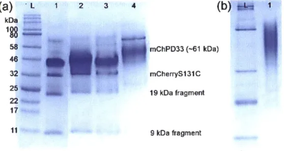
Figure 2-17. SDS-PAGE gel showing fractions collected from mCherryS131C nickel affinity chromatography.
Figure 2-18. UV-vis spectrum of purified mCherryS131C in 20 mM Tris-Cl pH 8.0 buffer. Figure 2-19. SDS-PAGE gels showing purification of mCherry-PHPA 27K bioconjugate. Figure 2-20. SDS-PAGE gels showing purification of mCherry-POEGA 26K bioconjugate Figure 2-21. Representative FPLC spectrum for a purification of mCherry-PDMAPS bioconjugate. Figure 2-22. mCherry-b-PDMAPS 33K bioconjugate purification protein gels.
Figure 2-23. Circular dichroism (CD) spectra of mCherry and of PHPA, mCherry-POEGA, and mCherry-PDMAPS bioconjugates.
Figure 2-24. Flow coater set up.
Figure 2-25. Detailed design of a humidity chamber. Figure 2-26. Typical annealing chamber set up.
Figure 2-27. GISAXS patterns demonstrating preservation of nanostructures up to 5 months. Figure 2-28. Representative X-ray reflectivity scan of a bioconjugate film.
Figure 3-1. GPC traces of (a) PHPA and (b) POEGA homopolymers with three different molar masses used in the study.
Figure 3-2. 'H-NMR spectrum of deprotected PHPA with Mn 27,220 g/mol in CDCl3 solution.
Figure 3-3. 'H-NMR spectrum of deprotected POEGA with Mn 26,080 g/mol in CDCl3 solution.
Figure 3-4. Native protein gel of mCherry-b-PHPA and mCherry-b-POEGA bioconjugates. Figure 3-5. Circular dichroism (CD) spectrum of mCherry and activity study.
Figure 3-6. Phase diagrams of mCherry-b-PHPA and mCherry-b-POEGA in aqueous solutions as a function of concentration and temperature.
Figure 3-7. Order-disorder transition concentration (CODT) values for the three types of
bioconjugates versus polymer volume fraction.
Figure 3-9. SAXS curves of solid state samples in comparison to 80 wt.% solution samples for mChPH18 and mChPOE18.
Figure 3-10. SAXS curves of mCherry-b-PHPA and mCherry-b-POEGA solutions at 5'C. Figure 3-11. SAXS curves of mCherry-b-PHPA and mCherry-b-POEGA solutions at 30'C and
35'C, respectively.
Figure 3-12. Bicontinuous cubic phase formed by mChPH27 and mChPOE26.
Figure 3-13. Representative SAXS curves for perforated lamellar (PL) phase of 70 wt.% bioconjugate solutions measured at 350C.
Figure 3-14. Small-angle neutron scattering absolute intensities of mChPH27 and mChPOE26 in different D20/H20 blend compositions at T = I 00C.
Figure 3-15. Small-angle neutron scattering peak intensities and curve fits for mChPH27 and
mChPOE26 in different D20/H20 blend compositions at 10 'C.
Figure 4-1. mCherry-b-poly(oligoethylene glycol acrylate) (POEGA) 26 kDa bioconjugate used in the study.
Figure 4-2. Surface segregation of bioconjugate in thin films prepared under ambient conditions analyzed by XPS.
Figure 4-3. AFM phase images and GISAXS patterns showing nanostructures of films prepared at low humidity (<RH25).
Figure 4-4. AFM phase images and corresponding GISAXS patterns of mCherry-POEGA 26kDa bioconjugate films prepared at high relative humidity (RH 80).
Figure 4-5. AFM phase images and GISAXS patterns prepared at different coating speed. Figure 4-6. Fluorescence activity of mCherry-POEGA 26 kDa bioconjugate films.
Figure 4-7. SDS-PAGE gel showing the molecular weight and purity of bioconjugate used in this study.
Figure 4-8. XPS spectra confirming the presence of PEO brushes on Si surface.
Figure 4-9. N/C ratio of mCherry-POEGA 26 kDa bioconjugate film surface prepared under different relative humidity conditions measured by XPS.
Figure 4-10. An AFM height image showing islands and holes structure.
Figure 4-11. GISAXS patterns showing comparison of as-cast and annealed films. Figure 4-12. Fluorescence emission spectra of films prepared at <RH25 and RH80. Figure 5-1. The phase behaviour of mCherry-b-PDMAPS bioconjugate in pure water.
Figure 5-2. Order-disorder transition concentrations (CODT) of mCherry-polymer bioconjugates
with different polymer block chemistry plotted as a function of polymer volume fraction. Figure 5-3. Temperature dependence of 50 wt.% solutions shown by SAXS and DPLS.
Figure 5-4. SAXS curves of 50 wt.% solution of mChPD33 conjugate in the presence of various concentrations of sodium chloride.
Figure 5-5. SAXS curves of 50 wt.% solution of mChPD33 and mChPD76 conjugates in the presence of ammonium sulfate.
Figure 5-6. H1-NMR spectrum of CPP-imide in CDCl3 solution. Figure 5-7. Characterization of PDMAPS homopolymers by GPC.
Figure 5-8. 'H-NMR spectrum of PDMAPS with Mn 32,880 g/mol dissolved in D20.
Figure 5-9. Thermal transition of PDMAPS homopolymers and corresponding bioconjugates. Figure 5-10. Native gel of purified mChPD33 and mChPD77.
Figure 5-13. A plot of calculated X-ray scattering contrast (ASLD) and ASLD2 as water
distribution changes.
Figure 5-14. Peak positions at the maximum intensity as a function of salt concentrations of mChPD33 bioconjugate at 50 wt.%.
Figure 5-15. SAXS curves of mChPD33 solutions in the presence of 250 mM NaCl.
Figure 5-16. The Debye lengths in bioconjugate solutions in the presence of either NaCl or (NH4)2SO4.
Chapter 1. Introduction and Background
Section 1.1 is adapted from a book chapter Protein Nanopatterning by Christopher N. Lam,
Dongsook Chang, and Bradly D. Olsen in Carbon Nanomaterials for Biomedical Applications,
M. Zhang et al. (eds.), Springer International Publishing Switzerland 2016.
1.1 Motivation
Enzymes have evolved to catalyze reactions that are beneficial to the survival of various organisms. Their ability to catalyze unique reactions that cannot be easily accessed using a synthetic route, high specificity towards a target molecule, regio- and stereospecificity which is crucial for production of pharmaceuticals and fine chemicals, and the mild reaction conditions-mild temperature, pressure, and environmental friendly solvent, water-are advantageous in addressing multiple industrial challenges. Enzymes can be used as molecular sensors-for
example, glucose"2 and neurotoxin sensors3-5-and as a critical component in the enzyme-linked
immunosorbent assay (ELISA). Enzymes have also proven to be beneficial for synthesizing drug compounds in the pharmaceutical industry,6 and they have found utility in numerous industrial applications as biocatalysts for laundry and dishwasher detergents,7 industrial synthetic chemistry, 8-10 and the food industry." Biocatalytic devices for carbon sequestration,12 carbon
dioxide reduction, 13-15 and hydrogen production""-8 have also been proposed utilizing enzymes.
Biofuel cells have attracted significant attention, offering the possibility of consuming biomass as fuel and eliminating the need for both transition metal catalysts and electrolyte membranes due to the high specificity of the enzymatic reactions on both electrodes.9,20 Beyond
materials have also found utility in developing surfaces for studies of cell growth and cell-surface interactions and for biomedical applications including tissue engineering.24,25
The function of biosensors, biocatalysts, bioelectronics, and biomedical materials can be improved through control over the position, orientation, and density of proteins presented within a bioactive material. Because proteins are fairly large compared to transition metal catalysts, it is often desirable to achieve a high density of proteins localized within a small area, improving catalytic rate in biocatalysts or signal to noise in sensors. The orientation of the protein and transport through thin film or bulk materials must be carefully controlled to enable efficient transport of substrates, products, cofactors, or charges through the device, thus requiring control of protein arrangement in nanometer scale. Furthermore, processing methods must not be deleterious to the native protein structure; ideally, the nanostructured material will provide an environment that enhances the stability and activity of the protein. Finally, practical applications require that the cost of patterning the material be commensurate with the value of the eventual application.
Two broad approaches, a top-down and bottom-up, are typically employed in order to prepare materials that meet as many of the above mentioned engineering criteria as possible. In the top-down approach, a surface is lithographically patterned and proteins are directed to selective regions of a surface. A variety of conventional and unconventional lithographic techniques including photolithography,2 6-30 electron beam lithography,31-34 dip-pen
nanolithography,35,36 nanografting,37-39 and nanocontact printing40~42 are used for protein
nanopatterning, but with different resolution, speed and cost. Selective adsorption or reaction of proteins is primarily achieved by physical adsorption, covalent conjugation, or bioaffinity interactions.43,44 In the bottom-up approach, templates may be constructed into which protein is
inserted or onto which protein is reacted/absorbed, where the structure of the template directs the location of the protein. The templates are typically composed of lipid or amphiphilic block copolymers. While lithographic processes can form patterns of arbitrary shape (not necessarily periodic) with resolution that often approaches a single protein molecule, patterning is limited to two dimensions and the resultant protein density per unit area is relatively low. Methods using lipids or diblock copolymers to template proteins can achieve relatively high protein density with low self-assembly cost compared to top-down approaches, but the majority of the material is typically template polymer, reducing the total functional density of protein.
Our group has recently developed a new method of nanopatterning proteins by self-assembling protein-polymer diblock copolymers into a bulk nanostructured material.45,46 This method meets the multiple requirements for a successful catalytic device. The self-assembled materials have a very high density of proteins; typically 30-60 wt.% of total mass is made of proteins depending on the size of proteins and polymers. Despite the high density, proteins remain accessible to substrates, products, cofactors, or charges due to continuous transport pathways generated by nanostructures. Protein orientation along the domain interfaces is controlled also by nanostructure formation. The stability and longevity of proteins are improved in the materials due to the presence of the protective polymer blocks, and the cost of patterning is relatively low compared to top-down approaches.
It is very interesting that a diblock copolymer consisting of a globular protein and a synthetic polymer undergoes microphase separation to form a variety of nanostructures, resembling the microphase separation of a diblock copolymer made of two synthetic homopolymers. While a great deal of knowledge is available about the self-assembly of
section 1.2, it is easy to see that the self-assembly principles learned from traditional synthetic block copolymers cannot be directly applied to the self-assembly of protein-polymer bioconjugates. Introducing a protein molecule with a globular shape, anisotropic interactions, and heterogeneous composition in replacement of a polymer chain with random coil conformation is expected to affect the optimal molecular packing geometry in equilibrium. Also, due to the limited availability of thermal annealing owing to a thermally unstable protein block, understanding kinetic processing pathways is critical in obtaining a good control of nanostructures of final solid state materials. As nanostructures in the self-assembled materials are governed by complex thermodynamics and kinetics due to the nature of proteins, a thorough investigation of the self-assembly behavior is required. This thesis is motivated by the intriguing question about how the nanostructures are controlled in the protein-polymer diblock copolymer materials.
1.2 Traditional Block Copolymer Self-Assembly
Diblock copolymers composed of two synthetic homopolymers undergo microphase separation, creating domains with nanometer length scales.47,48 The ability of block copolymers form regular nanometer scale patterns49 make them attractive candidates for the applications such
as fuel cells,5 0',51 batteries,5 2,3 and solar cells.5 4,55 Similar to the phase separation of two
immiscible liquids such as oil and water resulting from unfavorable energy of mixing, two distinct homopolymer blocks in a block copolymer undergo phase separation due to their chemical dissimilarity. However, as the blocks are covalently connected, the size of phase separation is limited by entropic penalty imposed by chain stretching; thus resulting in microphase separation with a nanoscale domain size. As the entropy gain from mixing of
polymers is much smaller than that of small molecules, even a small difference between two distinct monomers is sufficient to lead to microphase separation. The segregation strength is represented by a product of Flory Huggins interaction parameter,
x,
and the degree of polymerization, N. As a result of interplay between enthalpy and entropy, block copolymers form several classical phases including a lamellar phase, hexagonally packed cylindrical phase, BCC cubic phase, and double gyroid phase with Ia 3 d symmetry. Size and structure of nanodomains can be tuned by changing types of monomers (X), block copolymer composition(fA), polymer size (N), and molecular architecture.
The phase behavior of block copolymers in solution is more complex compared to the melt phase behavior due to the distribution of the second component - a solvent - throughout the domains and interfaces. When a neat block copolymer is diluted in a solvent, it forms nanostructures due to the repulsion that drives microphase separation in melt, although the repulsion is now modulated by solvent molecules. The solution eventually becomes disordered above the critical concentration of a solvent, which is often referred to as an order-disorder transition concentration (CODT). Quality of solvent for each block determines interactions
between blocks, and the selectivity of a solvent affects swelling of chains which in turn affects a relative volume fraction between two blocks. For a nonselective good solvent, the dilution approximation assumes that solvent molecules are uniformly distributed throughout the block copolymer structure, and an added neutral solvent screens unfavorable interactions between different blocks effectively lowering the x parameters between two blocks such that Xeff =
However, substantial deviation from dilution approximation has been observed for prediction of order-disorder transitions (ODT).19-6163 , Blocks are asymmetrically swollen in a selective solvent which is a good solvent for one block but it a poor solvent for the other block. Changing block copolymer concentration in a selective solvent can be qualitatively viewed as traversing a diagonal trajectory in a melt phase diagram.60,61 Also, segregation strength can be increased or decreased depending on the degree of selectivity such that a weakly selective solvent lowers the segregation strength while a strongly selective solvent can increases the segregation strength.60,61 For a nonselective poor solvent, the solution undergoes macrophase separation into a water-rich region and a block-copolymer-rich region.
Thin film geometry introduces two additional energy considerations; surface energetics and spatial confinement effect govern the phase behavior in addition to the factors governing the bulk self-assembly.49,64,61 Interactions of block copolymers with two surfaces, either symmetric or asymmetric, determine segregation of blocks on each surface so that the block with lower interfacial energy will preferentially wet the surface. As a result, parallel orientation of nanodomains to the surface is often energetically favored over perpendicular orientation, which results in highly oriented grains as opposed to randomly oriented grains in bulk materials. In order to promote perpendicular ordering of nanodomains which is motivated by applications such as ultrafiltration membrane,66-68 substrates are modified with random copolymer brushes to create a neutral surface.69-71 It has been also shown that fast evaporation of solvent promotes perpendicularly ordered cylinders.72-74 Incommensurability between block copolymer domain spacings and film thickness often results in rich morphological behavior including islands and holes structures,75 76 or perpendicular orientation of nanodomains when surfaces are not strongly preferential.69
1.3 Self-Assembly of Globular Protein-Polymer Bioconjugates
The main differences between proteins and synthetic homopolymers originate from the heterogeneous composition of proteins made of 20 different amino acids building blocks. The diverse amino acids in a protein with different hydrophobicity, polarity, and acidity result in secondary and tertiary structures of protein in aqueous solutions through hydrophobic, hydrogen bonding, and electrostatic interactions. As a result, most functional proteins (as opposed to structural proteins such as collagen, elastin, or keratin) assume a globular shape. The dense and rigid nature of proteins is dramatically different than polymers with random coil conformation. Proteins cannot overlap with other proteins or swell in solution as polymers which change the conformation depending on the solvent quality and highly overlap with other chains in concentrated solution or melt. Anisotropic surface patches of globular proteins that lead to crystallization are also different from isotropic interactions of random coil polymers. In the self-assembly perspective, while the Flory-Huggins interaction parameter (X) of two distinct blocks is defined based on the dissimilarity of two monomers, the interactions between protein and polymer are much more complex.
Our group has demonstrated the self-assembly of protein-polymer diblock copolymer into nanostructured materials using a model system of a conjugate of a red fluorescent protein, mCherry, and a synthetic biocompatible polymer block, poly(N-isopropylacrylamide) (PNIPAM).4 5 mCherry is selected for its well-established expression and purification methods,
high yield, robust structure, and relatively simple activity measurement using spectroscopy. In addition, application of fluorescent proteins as a biolasing component is recently reported.77 78 PNIPAM is a widely used biocompatible polymer with the lower critical solution temperature
been shown that mCherry forms a bilayer structure in a lamellar nanostructure to minimize the interfacial area at a large polymer coil fraction ($PNIPAM = 0.70). A more recent study shows
multiple important differences between the phase behavior of protein-diblock copolymers and that of traditional coil-coil block copolymers; a lamellar region in the phase diagram is highly stretched to large coil fractions, no spherical cubic phase is observed, no cylindrical phase made of mCherry core is formed, and a re-entrant behavior at high concentration is observed.7 9 Kinetics in processing the final solid materials are found to have a large impact on the final nanostructures due to kinetic aspects of solvent annealing process.80 Kinetic effects on the
processing of bioconjugate thin films are explored in detail in Chapter 4.
Equilibrium nanostructures in concentrated solution provided further insight into the thermodynamics governing the self-assembly of the bioconjugates.81 mCherry-b-PNIPAM bioconjugates are found to undergo order-disorder transitions at concentrations between 30-40 wt.% in water. Multiple lyotropic and thermotropic phase transitions are observed as a function of concentration and temperature. The order-disorder transition concentrations (CODT) are lowest
for the symmetric bioconjugates with a coil fraction (<PNIPAM) near 0.5, which suggests that the solvent-mediated net repulsive interactions are responsible for the microphase separation of protein-polymer diblock copolymers. This study directly motivated the current thesis to explore the effect of polymer block chemistry (Chapter 3) and the effect of electrostatic interactions on the self-assembly (Chapter 5).
1.4 Thesis Overview
This thesis describes studies on understanding the governing principles of the self-assembly of protein-polymer bioconjugates in concentrated solutions (Chapter 3, 5) and in thin films
(Chapter 4). Experimental methods about how polymerizations, bioconjugate preparation, and nanostructure characterization are conducted are described in detail in Chapter 2. Chapter 3 studies the effect of polymer block chemistry as a structure-directing element on the phase behavior of bioconjugates in concentrated solution. The results show that changing protein-polymer interactions by changing protein-polymer block chemistry affects many aspects of phase behavior in solution. Higher order-disorder transition concentrations as well as formation of double gyroid nanostructure suggest the weaker segregation strengths of mCherry-b-PHPA and mCherry-b-POEGA bioconjugates compared to the previously studied mCherry-b-PNIPAM conjugates. The knowledge on the solution phase behavior of mCherry-b-POEGA is translated to the thin film self-assembly, which is presented in Chapter 4. Due to the limited annealing conditions available for protein-polymer conjugates as well as decreased activity of protein after annealing, understanding of nanostructures of as-cast films are critical. The study presents multiple important kinetic parameters in processing bioconjugate thin films including relative humidity which impacts solvent evaporation rate and flow coating speed. High relative humidity and high flow coating speed are found to result in films with longer range order. Chapter 5
describes the effect of electrostatic interaction and reduced charge asymmetry on the self-assembly of protein-polymer bioconjugates, in the context of understanding the nature of repulsive interactions of protein-polymer bioconjugates. Bioconjugates composed of mCherry and a zwitterionic polymer block are synthesized. The bioconjugates display weaker segregation strength compared to all previously studied mCherry-polymer conjugates, demonstrated by high
CODT, extremely narrow ordered region in the phase diagram, and no nematic crystalline ordering
be critical in the self-assembly, and the ability to control nanostructures in solution by modulating protein-protein interactions by adding kosmotropic salt is demonstrated.
1.5 References
(1) Heller, A. Electrical Connection of Enzyme Redox Centers to Electrodes. J. Phys. Chem.
1992, 96, 3579-3587.
(2) Yehezkeli, 0.; Yan, Y.-M.; Baravik, I.; Tel-Vered, R.; Willner, I. Integrated Oligoaniline-Cross-Linked Composites of Au Nanoparticles/Glucose Oxidase Electrodes: A Generic
Paradigm for Electrically Contacted Enzyme Systems. Chem. - A Eur. J. 2009, 15, 2674-2679.
(3) Drevon, G. F.; Russell, A. J. Irreversible Immobilization of Diisopropylfluorophosphatase
in Polyurethane Polymers. Biomacromolecules 2000, 1, 571-576.
(4) Russell, A. J.; Berberich, J. A.; Drevon, G. F.; Koepsel, R. R. Biomaterials for Mediation of Chemical and Biological Warfare Agents. Annu. Rev. Biomed. Eng. 2003, 5, 1-27.
(5) Simonian, A. L.; Grimsley, J. K.; Flounders, A. W.; Schoeniger, J. S.; Cheng, T.-C.; DeFrank, J. J.; Wild, J. R. Enzyme-Based Biosensor for the Direct Detection of
Fluorine-Containing Organophosphates. Anal. Chim. Acta 2001, 442, 15-23.
(6) Patel, R. N. Synthesis of Chiral Pharmaceutical Intermediates by Biocatalysis. Coord
Chem. Rev. 2008, 252, 659-701.
(7) Hasan, F.; Shah, A. A.; Hameed, A. Industrial Applications of Microbial Lipases. Enzyme
Microb. Technol. 2006, 39, 235-25 1.
(8) Schmid, A.; Dordick, J. S.; Hauer, B.; Kiener, A.; Wubbolts, M.; Witholt, B. Industrial Biocatalysis Today and Tomorrow. Nature 2001, 409, 258-268.
(9) Schoemaker, H. E.; Mink, D.; Wubbolts, M. G. Dispelling the Myths--Biocatalysis in Industrial Synthesis. Science (80-.). 2003, 299, 1694-1697.
(10) Straathof, A. J. J.; Panke, S.; Schmid, A. The Production of Fine Chemicals by
Biotransformations. Curr. Opin. Biotechnol. 2002, 13, 548-556.
(11) Kirk, 0.; Borchert, T. V.; Fuglsang, C. C. Industrial Enzyme Applications. Curr. Opin.
Biotechnol. 2002, 13, 345-3 5 1.
(12) Favre, N.; Christ, M. L.; Pierre, A. C. Biocatalytic Capture of C02 with Carbonic Anhydrase and Its Transformation to Solid Carbonate. J. Mol. Catal. B Enzym. 2009, 60,
163-170.
(13) Reda, T.; Plugge, C. M.; Abram, N. J.; Hirst, J. Reversible Interconversion of Carbon Dioxide and Formate by an Electroactive Enzyme. Proc. Natl. Acad Sci. U. S. A. 2008,
105, 10654-10658.
(14) Parkinson, B. A.; Weaver, P. F. Photoelectrochemical Pumping of Enzymatic Co2 Reduction. Nature 1984, 309, 148-149.
(15) Kuwabata, S.; Tsuda, R.; Yoneyama, H. Electrochemical Conversion of Carbon Dioxide to Methanol with the Assistance of Formate Dehydrogenase and Methanol Dehydrogenase
(16) Karyakin, A. A.; Morozov, S. V; Karyakina, E. E.; Varfolomeyev, S. D.; Zorin, N. A.; Cosnier, S. Hydrogen Fuel Electrode Based on Bioelectrocatalysis by the Enzyme
Hydrogenase. Electrochem. commun. 2002, 4, 417-420.
(17) Hambourger, M.; Gervaldo, M.; Svedruzic, D.; King, P. W.; Gust, D.; Ghirardi, M.; Moore, A. L.; Moore, T. A. [FeFe]-Hydrogenase-Catalyzed H2 Production in a Photoelectrochemical Biofuel Cell. J. Am. Chem. Soc. 2008, 130, 2015-2022.
(18) Krassen, H.; Schwarze, A.; Friedrich, B.; Ataka, K.; Lenz, 0.; Heberle, J. Photosynthetic Hydrogen Production by a Hybrid Complex of Photosystem I and [NiFe]-Hydrogenase. ACS Nano 2009, 3, 405 5-4061.
(19) Minteer, S. D.; Liaw, B. Y.; Cooney, M. J. Enzyme-Based Biofuel Cells. Curr. Opin.
Biotechnol. 2007, 18, 228-234.
(20) Leech, D.; Kavanagh, P.; Schuhmann, W. Enzymatic Fuel Cells: Recent Progress.
Electrochim. Acta 2012, 84, 223-234.
(21) Cogdell, R. J.; Gall, A.; Kohler, J. The Architecture and Function of the Light-Harvesting Apparatus of Purple Bacteria: From Single Molecules to in Vivo Membranes.
Q.
Rev.Biophys. 2006, 39, 227-324.
(22) Das, R.; Kiley, P. J.; Segal, M.; Norville, J.; Yu, A. A.; Wang, L.; Trammell, S. A.; Reddick, L. E.; Kumar, R.; Stellacci, F.; et al. Integration of Photosynthetic Protein Molecular Complexes in Solid-State Electronic Devices. Nano Lett. 2004, 4, 1079-1083. (23) Choi, J.-W. W.; Fujihira, M. Molecular-Scale Biophotodiode Consisting of a Green
Fluorescent Protein/cytochrome c Self-Assembled Heterolayer. Appl. Phys. Lett. 2004, 84, 2187-2189.
(24) Falconnet, D.; Csucs, G.; Michelle Grandin, H.; Textor, M. Surface Engineering Approaches to Micropattern Surfaces for Cell-Based Assays. Biomaterials 2006, 27, 3044-3063.
(25) V6rds, J.; Blilttler, T.; Textor, M. Bioactive Patterns at the 100-Nm Scale Produced Using
Multifunctional Physisorbed Monolayers. Mrs Bull. 2005, 30, 202-206.
(26) Mooney, J. F.; Hunt, A. J.; McIntosh, J. R.; Liberko, C. A.; Walba, D. M.; Rogers, C. T. Patterning of Functional Antibodies and Other Proteins by Photolithography of Silane
Monolayers. Proc. Natl. Acad. Sci. 1996, 93, 12287-12291.
(27) Flounders, A. W.; Brandon, D. L.; Bates, A. H. Patterning of Immobilized Antibody Layers via Photolithography and Oxygen Plasma Exposure. Biosens. Bioelectron. 1997,
12, 447-456.
(28) Sorribas, H.; Padeste, C.; Tiefenauer, L. Photolithographic Generation of Protein Micropatterns for Neuron Culture Applications. Biomaterials 2002, 23, 893-900.
(29) Shin, D.-S.; Lee, K.-N.; Jang, K.-H.; Kim, J.-K.; Chung, W.-J.; Kim, Y.-K.; Lee, Y.-S. Protein Patterning by Maskless Photolithography on Hydrophilic Polymer-Grafted Surface.
Biosens. Bioelectron. 2003, 19, 485-494.
(30) Petrou, P. S.; Chatzichristidi, M.; Douvas, A. M.; Argitis, P.; Misiakos, K.; Kakabakos, S. E. A Biomolecule Friendly Photolithographic Process for Fabrication of Protein Microarrays on Polymeric Films Coated on Silicon Chips. Biosens. Bioelectron. 2007, 22, 1994-2002.
(32) Lussi, J. W.; Tang, C.; Kuenzi, P. A.; Staufer, U.; Csucs, G.; Voros, J.; Danuser, G.;
Hubbell, J. A.; Textor, M. Selective Molecular Assembly Patterning at the Nanoscale: A Novel Platform for Producing Protein Patterns by Electron-Beam Lithography on
SiO2/indium Tin Oxide-Coated Glass Substrates. Nanotechnology 2005, 16, 1781-1786.
(33) Pallandre, A.; De Meersman, B.; Blondeau, F.; Nysten, B.; Jonas, A. M. Tuning the Orientation of an Antigen by Adsorption onto Nanostriped Templates. J. Am. Chem. Soc.
2005, 127, 4320-4325.
(34) Kolodziej, C. M.; Maynard, H. D. Electron-Beam Lithography for Patterning Biomolecules at the Micron and Nanometer Scale. Chem. Mater. 2012, 24, 774-780. (35) Lee, K. B.; Park, S. J.; Mirkin, C. A.; Smith, J. C.; Mrksich, M. Protein Nanoarrays
Generated by Dip-Pen Nanolithography. Science (80-.). 2002, 295, 1702-1705.
(36) Ginger, D. S.; Zhang, H.; Mirkin, C. A. The Evolution of Dip-Pen Nanolithography.
Angew. Chemie-International Ed. 2004, 43, 30-45.
(37) Case, M. A.; McLendon, G. L.; Hu, Y.; Vanderlick, T. K.; Scoles, G. Using Nanografting
to Achieve Directed Assembly of de Novo Designed Metalloproteins on Gold. Nano Lett.
2002, 3, 425-429.
(38) Hu, Y.; Das, A.; Hecht, M. H.; Scoles, G. Nanografting de Novo Proteins onto Gold
Surfaces. Langmuir 2005, 21, 9103-9109.
(39) Bano, F.; Fruk, L.; Sanavio, B.; Glettenberg, M.; Casalis, L.; Niemeyer, C. M.; Scoles, G. Toward Multiprotein Nanoarrays Using Nanografting and DNA Directed Immobilization
of Proteins. Nano Lett. 2009, 9, 2614-2618.
(40) Csucs, G.; Ktnzler, T.; Feldman, K.; Robin, F.; Spencer, N. D. Microcontact Printing of Macromolecules with Submicrometer Resolution by Means of Polyolefin Stamps.
Langmuir 2003, 19, 6104-6109.
(41) Li, H. W.; Muir, B. V. 0.; Fichet, G.; Huck, W. T. S. Nanocontact Printing: A Route to Sub-50-Nm-Scale Chemical and Biological Patterning. Langmuir 2003, 19, 1963-1965.
(42) Ruiz, S. A.; Chen, C. S. Microcontact Printing: A Tool to Pattern. Soft Matter 2007, 3,
168-177.
(43) Rusmini, F.; Zhong, Z. Y.; Feijen, J. Protein Immobilization Strategies for Protein
Biochips. Biomacromolecules 2007, 8, 1775-1789.
(44) Sassolas, A.; Blum, L. J.; Leca-Bouvier, B. D. Immobilization Strategies to Develop Enzymatic Biosensors. Biotechnol. Adv. 2012, 30, 489-511.
(45) Thomas, C. S.; Glassman, M. J.; Olsen, B. D. Solid-State Nanostructured Materials from Self-Assembly of a Globular Protein-Polymer Diblock Copolymer. In ACS Nano;
American Chemical Society, 2011; Vol. 5, pp. 5697-5707.
(46) Olsen, B. D. Self-Assembly of Globular-Protein-Containing Block Copolymers.
Macromol. Chem. Phys. 2013, 214, 1659-1668.
(47) Bates, F. S. Polymer-Polymer Phase Behavior. Science 1991, 251, 898-905.
(48) Bates, F. S.; Fredrickson, G. H. Block Copolymers-Designer Soft Materials. Phys.
Today 1999, 52, 32-38.
(49) Segalman, R. A. Patterning with Block Copolymer Thin Films. Mater. Sci. Eng.
R-Reports 2005, 48, 191-226.
(50) Meier-Haack, J.; Taeger, A.; Vogel, C.; Schlenstedt, K.; Lenk, W.; Lehmann, D. Membranes from Sulfonated Block Copolymers for Use in Fuel Cells. Sep. Purif Technol.
(51) Elabd, Y. A.; Hickner, M. A. Block Copolymers for Fuel Cells. Macromolecules 2011, 44,
1-11.
(52) Young, W. S.; Kuan, W. F.; Epps, T. H. Block Copolymer Electrolytes for Rechargeable
Lithium Batteries. J. Polym. Sci. Part B Polym. Phys. 2014, 52, 1-16.
(53) Orilall, M. C.; Wiesner, U. Block Copolymer Based Composition and Morphology
Control in Nanostructured Hybrid Materials for Energy Conversion and Storage: Solar
Cells, Batteries, and Fuel Cells. Chem. Soc. Rev. 2011, 40, 520-535.
(54) Sun, S.-S. Design of a Block Copolymer Solar Cell. Sol. Energy Mater. Sol. Cells 2003, 79, 257-264.
(55) Crossland, E. J. W.; Nedelcu, M.; Ducati, C.; Ludwigs, S.; Hillmyer, M. A.; Steiner, U.;
Snaith, H. J. Block Copolymer Morphologies in Dye-Sensitized Solar Cells: Probing the Photovoltaic Structure-Function Relation. Nano Lett. 2009, 9, 2813-2819.
(56) Helfand, E.; Tagami, Y. Theory of the Interface between Immiscible Polymers. II. J.
Chem. Phys. 1972, 56, 3592-3601.
(57) Whitmore, M. D.; Noolandi, J. Self-Consistent Theory of Block Copolymer Blends:
Neutral Solvent. J. Chem. Phys. 1990, 93, 2946.
(58) Hashimoto, T.; Shibayama, M.; Hiromichi, K.; Kawai, H. Ordered Structure in Block Polymer Solutions. 4. Scaling Rules on Size of Fluctuations with Block Molecular Weight, Concentration, and Temperature in Segregation and Homogeneous Regimes.
Macromolecules 1983, 16, 1093-1101.
(59) Sakurai, S.; Hashimoto, T.; Fetters, L. J. Thermoreversible Cylinder-Sphere Transition of Polystyrene-Block-Polyisoprene Diblock Copolymers in Dioctyl Phthalate Solutions.
Macromolecules 1996, 29, 740-747.
(60) Hanley, K. J.; Lodge, T. P. Effect of Dilution on a Block Copolymer in the Complex
Phase Window. J. Polym. Sci. Part B Polym. Phys. 1998, 36, 3101-3113.
(61) Hanley, K. J.; Lodge, T. P.; Huang, C. I. Phase Behavior of a Block Copolymer in Solvents of Varying Selectivity. Macromolecules 2000, 33, 5918-5931.
(62) Naughton, J. R.; Matsen, M. W. Limitations of the Dilution Approximation for Concentrated Block Copolymer/solvent Mixtures. Macromolecules 2002, 35, 5688-5696. (63) Lodge, T. P.; Pan, C.; Jin, X.; Liu, Z.; Zhao, J.; Maurer, W. W.; Bates, F. S. Failure of the
Dilution Approximation in Block Copolymer Solutions. J. Polym. Sci. Part B Polym. Phys.
1995, 33, 2289-2293.
(64) Fasolka, M. J.; Mayes, A. M. Block Copolymer Thin Films: Physics and Applications.
Annu. Rev. Mater. Res. 2001, 31, 323-355.
(65) Albert, J. N. L.; Epps, T. H. Self-Assembly of Block Copolymer Thin Films. Mater.
Today 2010, 13, 24-33.
(66) Phillip, W. A.; Rzayev, J.; Hillmyer, M. A.; Cussler, E. L. Gas and Water Liquid Transport through Nanoporous Block Copolymer Membranes. J. Memb. Sci. 2006, 286,
144-152.
(67) Phillip, W. A.; O'Neill, B.; Rodwogin, M.; Hillmyer, M. A.; Cussler, E. L. Self-Assembled Block Copolymer Thin Films as Water Filtration Membranes. ACS Appl.
Mater. Interfaces 2010, 2, 847-853.
(69) Kellogg, G.; Walton, D.; Mayes, A.; Lambooy, P.; Russell, T.; Gallagher, P.; Satija, S.
Observed Surface Energy Effects in Confined Diblock Copolymers. Phys. Rev. Lett. 1996,
76, 2503-2506.
(70) Huang, E.; Rockford, L.; Russell, T. P.; Hawker, C. J. Nanodomain Control in Copolymer Thin Films. Nature 1998, 395, 757-758.
(71) Huang, E.; Russell, T. P.; Harrison, C.; Chaikin, P. M.; Register, R. A.; Hawker, C. J.;
Mays, J. Using Surface Active Random Copolymers To Control the Domain Orientation
in Diblock Copolymer Thin Films. Macromolecules 1998, 31, 7641-7650.
(72) Turturro, A.; Gattiglia, E.; Vacca, P.; Viola, G. T. Free Surface Morphology of Block
Copolymers: 1. Styrene-Butadiene Diblock Copolymers. Polymer (Guildf). 1995, 36, 3987-3996.
(73) Kim, G.; Libera, M. Morphological Development in Solvent-Cast Polystyrene -Polybutadiene - Polystyrene ( SBS ) Triblock Copolymer Thin Films. Macromolecules
1998, 31, 2569-2577.
(74) Phillip, W. A.; Hillmyer, M. A.; Cussler, E. L. Cylinder Orientation Mechanism in Block
Copolymer Thin Films upon Solvent Evaporation. Macromolecules 2010, 43, 7763-7770. (75) Coulon, G.; Collin, B.; Ausserre, D.; Chatenay, D.; Russell, T. P. Islands and Holes on the
Free Surface of Thin Diblock Copolymer Films. I. Characteristics of Formation and
Growth. J. Phys. 1990, 51, 2801-2811.
(76) Smith, A. P.; Douglas, J. F.; Meredith, J. C.; Amis, E. J.; Karim, A. Combinatorial Study
of Surface Pattern Formation in Thin Block Copolymer Films. Phys. Rev. Lett. 2001, 87,
015503.
(77) Jonas, A.; Kiraz, A. In Vitro and in Vivo Biolasing of Fluorescent Proteins Suspended in
Liquid Microdroplet Cavities. Lab Chip 2014, 3093-3 100.
(78) Gather, M. C.; Yun, S. H. Bio-Optimized Energy Transfer in Densely Packed Fluorescent Protein Enables near-Maximal Luminescence and Solid-State Lasers. Nat. Commun. 2014,
5, 5722.
(79) Thomas, C. S.; Olsen, B. D. Coil Fraction-Dependent Phase Behaviour of a Model
Globular Protein-polymer Diblock Copolymer. Soft Matter 2014, 10, 3093-3102.
(80) Thomas, C. S.; Xu, L.; Olsen, B. D. Kinetically Controlled Nanostructure Formation in Self-Assembled Globular Protein-Polymer Diblock Copolymers. Biomacromolecules
2012, 13, 2781-2792.
(81) Lam, C. N.; Olsen, B. D. Phase Transitions in Concentrated Solution Self-Assembly of Globular Protein-polymer Block Copolymers. Soft Matter 2013, 9, 2393.
Chapter 2. Experimental Methods
2.1 Polymer Synthesis
Materials.
All solvents and chemicals used in this thesis are listed in Table 2-1 and 2-2, respectively.
Table 2-1. Grade, purity, and source of solvents used in this thesis.
Solvent CAS Number Grade and Purity Supplier
Acetone 67-64-1 ACS grade ( 99.5%) Macron Fine ChemicalsTM
Diethyl ether 60-29-7 ACS grade ( 99.0%) J.T.Baker
Dichloromethane 75-09-2 ACS grade ( 99.5%) Macron Fine ChemicalsTM Hexane 110-54-3 ACS grade ( 98.5%) Macron Fine Chemicals TM Ethyl acetate 141-78-6 ACS grade ( 99.5%) Macron Fine Chemicals TM
Dioxane 123-91-1 ACS grade ( 99.0%) Sigma-Aldrich
Trifluoroethanol 75-89-8 ReagentPlus* (> 99%) Sigma-Aldrich Chloroform 67-66-3 ACS grade ( 99.8%) Macron Fine Chemicals TM
Chloroform-d 865-49-6 D, 99.8% Lambridge Isotope
Deuterium oxide 7789-20-0 D, 99.8%, sterility tested Cambridge Isotope
_____________________________________________ LaboratoriesInc.
Table 2-2. Purity and source of chemicals used in this thesis.
Chemical CAS Purity Supplier
_______________________ NumberPuiySple
Ethanethiol 75-08-1 97% Aldrich
Aliquat* 336 63393-96-4 N/A Aldrich
Carbon disulfide (CS2) 75-15-0 > 99.9% Aich
exo-3a,4,7,7a-tetrahydro-2-(3-hydroxypropyl)-4,7-epoxy- 14- 6118-51-0 N/A Aldrich
isoindole-1,3(2H)-dione
N,N'-dicyclohexylcarbodiimide 538-75-0 99%
Aldrich (DCC)
95%
2-hydroxypropyl acrylate (mixture 25584-83-2 200 ppm hydroquinone
Sigma-of isomers) monomethyl ether as Aldrich
inhibitor
Aluminum oxide (activated, basic, 1344-28-1 N/A (Pore size 58A, pH
Sigma-Brockmann I) 9.5 0.5 in H20) Aldrich
Azobisisobutyronitrile (AIBN) 78-67-1 98% Aldrich
Trimethylamine 121-44-8 > 99% Aich
2-(2-methoxyethoxy)ethanol 111-77-3 ReagentPlus, >99.0%i > 97%
Acryloyl chloride 814-68-6 (-400 ppm phenothiazine Sim
as stabilizer)
Anhydrous Na2SO4 7757-82-6 ACS grade Mheona Fin
98%
2-methoxyethyl acrylate 3121-61-7 (50-100 ppm MEHQ as Aldrich
inhibitor)
2,2'-Azobis[2-(2-imidazolin-2-
Wako
yl)propane]dihydrochloride (VA- 27776-21-2 N/A chemicals
044) chem__as
3-[N-(2-methacroyloyethyl)-N,N-dimethylammonio]propane 3637-26-1 97% Aldrich
sulfonate (DMAPS)
Hydroquinone (HQ) 123-31-9 99% Alfa Aesar
Butylated hydroxytoluene (BHT) 128-37-0 > 99.0% Aldrich
(a) SH + CS2 + (b) + NaOH Aliquat336 on 0 0 H NS HO. Oy 0 EMP 0 0 Imide
Scheme 2-1. Synthesis of precursors of chain transfer agents. (a) EMP synthesis and (b) Imide synthesis.
Synthesis of 2-ethylsulfanylthiocarbonyl sulfanyl-2-methylpropionic acid (EMP). Synthesis
of EMP was conducted according to a previous work (Scheme 2-la)1 which was adapted from a
2-dodecylsulfanylthiocarbonylsulfanyl-2-methyl propionic acid (DMP) synthesis, a similar compound with a longer alkyl chain.2 Typically, 7.21 mL (6.21 g) ethanethiol, 73.3 mL acetone, 1.13 mL Aliquat* 336 were combined and cooled in ice bath and under nitrogen stream. 9 mL of
50% NaOH aqueous solution was added to the reaction mixture over 10 minutes. 6.03 mL CS2
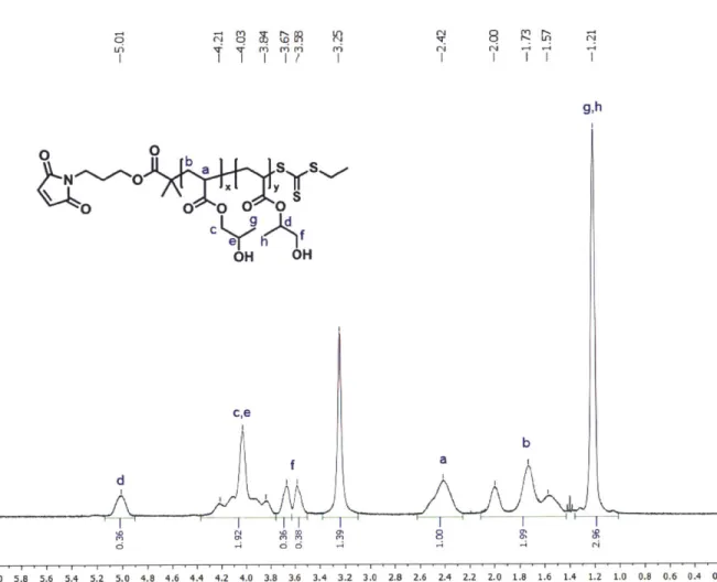
and 12.64 mL acetone were combined in a dropping funnel and added to the reaction mixture over 30 minutes. 80 mL of the 50% NaOH solution was added over 20 minutes and the reaction mixture was stirred overnight. Next day, 200 mL of distilled water and 80 mL of concentrated HCl were added to the solution. Crude product was extracted three times in 100 mL of diethyl ether and solvent was removed by rotary evaporation. The crude product was purified over silica gel column (typically 400 mL of silica was used). First yellow fraction was washed using 1 L hexane which was discarded afterwards. As much as the second streaky fraction with orange/brown color was collected using a 1:1 mixture of hexane and diethyl ether as a mobile phase. The third fraction was discarded. Solvent was removed by rotary evaporation and the product was purified further by distillation. Impurity was collected as the first fraction up to 135 'C 190 mTorr with bright yellow color. The pure EMP was collected as the second fraction up to 157 'C 120 mTorr. The final orange-colored product was analyzed using thin-layer chromatography (TLC) and 1H-NMR (Figure 2-1).
W) C-3 ; M U I ) &) 2.0-bb S 1.5-1.0 z 0.5- C CHCI3 312 1.83 5.32 3.00 Chemical Shift (ppm)
Figure 2-1. 1H-NMR spectrum of EMP in deuterated chloroform (CDCl3), and TLC result of
purified EMP showing purity after distillation. The TLC was run in a 1:1 mixture of hexane and diethyl ether. The second lane of the TLC plate represents the same sample as the first lane but twice more concentrated.
Synthesis of exo-3a,4,7,7a-tetrahydro-2-(3-hydroxypropyl)-4,7-epoxy-I 4-isoindole-1,3(2
H)-dione (Imide). Synthesis of Imide was performed following a previously published method' (Scheme 2-1b) which was adapted from a literature.3 500 mL methanol was added to a 1 L round-bottom flask containing 9 g of exo-3,6-epoxy-1,2,3,6-tetrahydrophthalic anhydride. 4.08 g of 3-amino-1-propanol was added dropwise to the solution while stirring over approximately 30 minutes, after which the solution became clear. The reaction mixture was submerged in an oil bath heated at 56'C and stirred for 3-4 days. The flask was removed from the oil bath, and the
solvent was removed by rotary evaporation to obtain clear yellow oil. 200 mL of dichloromethane was added to the oil, and washed three times with 200 mL saturated sodium chloride solution. The collected organic layer was dried over anhydrous sodium sulfate which was removed by filtration. Dichloromethane was removed by rotary evaporation, resulting in a white solid (yield 20%).
0) C4 CD W ( I C-5C1) 0 ) . (N0-U
140624 Imide T6.001.001 r.esp DCO WRfU1 O I
b e b f a |O N, OH 1.0 4 0 CDC13 z C 0.5-a 0-2.01 2.00 2,10203 2.00 212 LA W. LI W U WA Chemical Shift (ppm)
Figure 2-2. 'H-NMR spectrum of exo-3a,4,7,7a-tetrahydro-2-(3-hydroxypropyl)-4,7-epoxy-14-isoindole-1,3(2H)-dione (Imide) in CDC3.
(a)
HO ><SkS.N. +
I
.. NAOH DCC,CDMAP0 N0 0 DCM 00 0I
EMP Imide EMP-amide
(b)
+ DCC, DMAP N N
000
CPP Imide CPP-imide
Scheme 2-2. Synthesis of two chain transfer agents used for reversible addition-fragmentation chain-transfer (RAFT) polymerization in this thesis. (a) EMP-imide synthesis from EMP and Imide, and (b) CPP-imide RAFT agent from CPP and Imide, both through a DCC coupling reaction.
Synthesis of EMP-imide. Synthesis of EMP-imide shown in Scheme 2-2a was conducted following the previously published protocol.1 2.10 g of EMP (9.35 mmol, 1 equiv.) and 2.28 g of Imide (10.21 mmol, 1.1 equiv.) were dissolved in 85 mL of dichloromethane. 113 mg of DMAP (1.01 mmol, 0.1 equilv.) was added to the reaction mixture, and 2.30 g of DCC (11.15 mmol, 1.2 equiv.) was added. The mixture was stirred overnight at room temperature. The solution was filtered to remove urea, and the solvent was removed by rotary evaporation. The crude mixture was purified using silica gel column chromatography using a 1:1 mixture of hexane and ethyl acetate as the mobile phase. Fractions containing the product with Rf - 0.4
were collected based on TLC analysis (Figure 2-3). A majority of the solvent was removed by rotary evaporation, and the solid was further dried in a vacuum oven at room temperature overnight. Purity of product was analyzed using 1H-NMR (Figure 2-4). 2.55 g of yellow solid was obtained after drying (yield: 64 %) and stored at -20 0C in the dark until it was used.
Figure 2-3. TLC analysis of fractions collected during column chromatography for EMP-imide purification. The mobile phase used for TLC was a 1:1 mixture of hexane and ethyl acetate. The number of fractions are indicated on the top of each TLC plate. From this particular purification, fractions from 9 to 25 were collected.
z 0.8 07 0.6 0.5 0.4 0.3 0.2 0.1 0 13030&MP-kid* r|%@1.001.1r.esp m toinW CE C3 (00) D 0) r- It (N4 0) CD C. Q W, W, VN eN eN "N (N( L4. Tl' " yL4-J11el d b b S 0 N O a d f g h0 200 U h 2.02 d I- (D -4 C-4 () (D~ C4 lb a C U S 2 215 1 71 2 07 2,104-35 2,56 .'-'-I... 4.5 40 35 3.'. . Li i
Synthesis of CPP-imide RAFT agent. 4-cyano-4-(phenylcarbonothioylthio)pentanoic acid (min. 97%) (CPP) was purchased from Strem Chemicals. 1.5 g of CPP (5.37 mmol, 1 equiv.), 1.36 g of Imide (5.91 mmol, 1.1 equiv.) and 67.4 mg of 4-dimethylaminopyridine (DMAP) (0.54 mmol, 0.1 equiv.) were dissolved in 50 mL of dichloromethane. 1.37 g of N,N'-dicyclohexylcarbodiimide (DCC) (6.44 mmol, 1.2 equiv.) was added to the mixture and stirred under nitrogen overnight. The reaction mixture was washed using house distilled water three times and dried under anhydrous sodium sulfate. The solvent was removed by rotary evaporation. The crude product was further purified using a flash chromatography in an Isolera One Biotage system. A KP-Sil 50 g cartridge was used with hexane and ethyl acetate as eluents. After a quick wash step at 30% ethyl acetate for 1 column volume (CV), percent ethyl acetate was linearly increased to 80% over 15 CV, followed by a final wash step at 100% for 4 CV as shown in Figure 2-7a. The fractions containing the correct molar mass compound analyzed by liquid chromatography-mass spectrometry (LC-MS) were collected. LC-MS was performed using a poroshell 120 EC-C 18; 3.0 x 50 mm; 2.7 micron column. Water with 0.1% formic acid was used for solvent A and acetonitrile with 0.1% formic acid for solvent B. A default linear gradient program applied to analyze CPP-imide samples is shown in the table below.
Time (min) A (%) B (%) Flow (mL/min)
0.00 95.0 5.0 5.20 5.0 95.0 0.550 6.00 5.0 95.0 0 6.40 95.0 5.0 8.00 95.0 5.0
Solvent was removed by rotary evaporation, and the red oil was further dried in a vacuum oven at room temperature overnight. Due to the high viscosity of the product, drying process of this
compound was carefully monitored and vacuum level was adjusted as necessary to prevent excessive bubbling. Purity of product was analyzed using 'H-NMR (Figure 2-5) and LC-MS (Figure 2-6). 1.9 g of red oil was obtained after drying (yield: 73 %) and stored at -20 'C in the dark until it was used.
33,32,31,29,30 7,2 M 8,3n tO c' M t- C4 CD c)I) 15 13 5,4 14,25 19,20 r, n n r, .-J O:) C O )- CD) M) r- U U') M) (0 (NM(N qJ 4 0 LC "W W S 2 33 23 21 19 22 31 29 I..
1?ii
2.09 LI 199 U 0 11 7 0 14 ", N -1 1 16 26 15 13 10O -.4 ,1 2 0 6 12I.1
.1k
'iL
2.06 2,08 210 5.01 214 1.37 U U U LI WLJ 80 . 1 .5 7 I .0 Y 6 .5 Chemical Shit (ppm) 3. 5 3.0 2.5 2. 1.5 1.0 051Figure 2-5. 'H-NMR spectrum of CPP-imide in CDCl3. 7 6 5 4 3 2 C (D .N CO E 0 Z
ii
UI
1.85 106 2 04 I I di11
1.
1
1
.1 1 .... .... .... .... III I Vill If'. I I I IN if ImAU (a) 1 21000- 18000-15000. 12000- 9000- 6000- 3000-iii.U 4 , R \ I 1 .
DAWi 0, Sig280,4 Ret0750,1 00 (Mirqng\OgMEF_LC 2014-07-31 16819-3M\CCPP-kimdeT1.D)
(C) U /-vis at 280 nm
~,
L4
2 3 4507
MSD1 TIC, MS Fde (C:\Chem321\Data\MingVag\DEFLC 2014-07-31 16-19-39\DC CPP-mndeT1.D) ES-API, Pos Scam, Frig
1: CPP 2: Crude product 3: Imide 12000000 1000 200 S4 2) 100
TotaIIon Chromatogram (TIC) in positive mode
NI
br* At _ V V
150 200 250 300 350 400 450 500 550 600 650 700 750 800 850
m/z
Figure 2-6. CPP-imide purification. (a) UV-vis spectra collected during chromatography in Biotage. Fractions from 33 to 63 were collected from this particular purification as marked by black arrows. (b) TLC results using 4:6 mixture of hexane and ethyl acetate as the mobile phase
Detection Mode: Lambda-all
UVI (Monitor) 254 nm UV2 (Monitor) 280 nm Ua' 00 40 50 40 30 20 (b) 4 5W0-400 ~3-200 100-Jr
414
2 2 3 4 5 6 7 ri a 485.1 MS positive modefrom the fraction 5.922-6.055 min
0 0 507.1 [M+Na], 0 1 198.1 0, 1 0 2 ffir 7