SCIENTIFIC ARTICLE
Computed tomography of the cervical spine: comparison
of image quality between a standard-dose and a low-dose
protocol using filtered back-projection and iterative
reconstruction
Fabio Becce&Yosr Ben Salah&Francis R. Verdun&
Bruno C. Vande Berg&Frederic E. Lecouvet&
Reto Meuli&Patrick Omoumi
Received: 6 September 2012 / Revised: 15 November 2012 / Accepted: 6 January 2013 / Published online: 29 January 2013 # ISS 2013
Abstract
Objective To compare image quality of a standard-dose (SD) and a low-dose (LD) cervical spine CT protocol using filtered back-projection (FBP) and iterative reconstruction (IR).
Materials and methods Forty patients investigated by cer-vical spine CT were prospectively randomised into two groups: SD (120 kVp, 275 mAs) and LD (120 kVp, 150 mAs), both applying automatic tube current modula-tion. Data were reconstructed using both FBP and sinogram-affirmed IR. Image noise, signal-to-noise (SNR) and contrast-to-noise (CNR) ratios were measured. Two radiol-ogists independently and blindly assessed the following anatomical structures at C3–C4 and C6–C7 levels, using a four-point scale: intervertebral disc, content of neural fo-ramina and dural sac, ligaments, soft tissues and vertebrae.
They subsequently rated overall image quality using a ten-point scale.
Results For both protocols and at each disc level, IR signif-icantly decreased image noise and increased SNR and CNR, compared with FBP. SNR and CNR were statistically equiv-alent in LD-IR and SD-FBP protocols. Regardless of the dose and disc level, the qualitative scores with IR compared with FBP, and with LD-IR compared with SD-FBP, were significantly higher or not statistically different for inter-vertebral discs, neural foramina and ligaments, while signif-icantly lower or not statistically different for soft tissues and vertebrae. The overall image quality scores were significant-ly higher with IR compared with FBP, and with LD-IR compared with SD-FBP.
Conclusion LD-IR cervical spine CT provides better image quality for intervertebral discs, neural foramina and liga-ments, and worse image quality for soft tissues and verte-brae, compared with SD-FBP, while reducing radiation dose by approximately 40 %.
Keywords Cervical spine . Computed tomography . Filtered back-projection . Iterative reconstruction . Image quality . Radiation exposure
Introduction
Computed tomography (CT) of the cervical spine is an alter-native to magnetic resonance imaging (MRI) in the diagnosis of cervical disc herniation and/or spondylosis [1,2]. While MRI is definitely the examination of choice to assess spinal cord abnormalities, CT is more accurate for evaluating
F. Becce (*)
:
R. MeuliDepartment of Diagnostic and Interventional Radiology, Centre Hospitalier Universitaire Vaudois, University of Lausanne, Rue du Bugnon 46,
1011 Lausanne, Switzerland e-mail: [email protected]
F. Becce
:
Y. Ben Salah:
B. C. Vande Berg:
F. E. Lecouvet:
P. OmoumiDepartment of Radiology, Cliniques Universitaires Saint-Luc, Université Catholique Louvain, Avenue Hippocrate 10,
1200 Brussels, Belgium F. R. Verdun
Institute of Radiation Physics, Centre Hospitalier Universitaire Vaudois, University of Lausanne, Rue du Grand-Pré 1,
1007 Lausanne, Switzerland DOI 10.1007/s00256-013-1576-9
osteophytes and other bony changes, particularly in the case of neural foraminal stenosis [1,2]. Owing to both its time- and cost-effectiveness, CT is still used as a first cross-sectional imaging technique in a few countries, especially in Europe [1, 3]. It may further be performed when MRI is contraindicated and/or inconclusive. However, CT of the spine is associated with substantial radiation exposure [4].
Over the past decade, significant advances in CT tech-nology have led to an increase in the number of indications and, consequently, radiation dose delivered to patients by CT examinations [4,5]. Therefore, CT manufacturers pro-gressively developed several tools to manage and/or reduce radiation dose. These include automatic tube current modu-lation, low electronic noise detector systems and, most recently, iterative reconstruction (IR) techniques [5, 6]. Moreover, variation in tube voltage and bismuth thyroid shields also proved to be useful in reducing radiation expo-sure in CT of the neck [7, 8]. Besides, the advent of CT scanners with up to 320 detector rows helped to further reduce radiation dose by using the volumetric mode, which reduces over-ranging and overbeaming effects [9].
Since their implementation in clinical routine, IR methods have been extensively applied in abdominal [10], thoracic [11] and cardiovascular [12,13] CT imaging. These techniques allowed the radiation dose to be substantially reduced, while maintaining nearly constant diagnostic image quality. One feature of the musculoskeletal system is its varied composition of both soft (ligament, muscle, tendon, fat) and dense (bone) tissues. To our knowledge, the impact of IR on the conspicuity of these different anatomical structures, each evaluated sepa-rately, has not been assessed so far.
Thus, the aim of our study was to quantitatively and qualitatively compare the overall image quality and the conspicuity of different anatomical structures between a standard-dose and a low-dose cervical spine CT protocol using both filtered back-projection (FBP) and IR. We main-ly focused on the comparison between the standard-dose FBP and low-dose IR protocol.
Materials and methods
This single-centre prospective study was approved by the institutional ethics committee and all patients gave written informed consent.
Patients
From October to November 2011, 45 consecutive patients with chronic neck pain and/or cervical radiculopathy were investigated by unenhanced CT of the cervical spine in our radiology department. Three patients were excluded ow-ing to substantial metallic artefacts caused by surgical
material, while two refused to take part in the study. Thus, the final study population consisted of 40 patients (28 women, 12 men; mean age 53.1 years, range 18– 80), who were randomly assigned to one of the follow-ing two groups: standard-dose CT (n=20; 14 women, 6 men; mean age 51.6 years, range 25–80), or low-dose CT (n=20; 14 women, 6 men; mean age 54.5 years, range 18–77). As an indicator of the patient’s morpho-type, the anteroposterior (AP) diameter of the neck was measured parallel to the intervertebral disc at the C6–C7 level on the lateral CT scout view, and measurements of the two groups were compared.
CT protocol
All unenhanced CT examinations were performed on a 40-detector row CT scanner (Somatom Definition AS; Siemens Healthcare, Forchheim, Germany). Patients were positioned supine, with the head first on the CT table. Data acquisition was obtained from the C3 to T1 vertebrae, using the follow-ing parameters: tube voltage, 120 kVp; reference tube cur-rent–time product, 275 or 150 mAs in the standard-dose or low-dose protocol respectively; effective tube current–time product, 178–305 or 120–201 mAs, respectively (by apply-ing Care Dose 4D; Siemens Healthcare); detector configu-ration, 40×0.6 mm; pitch, 0.8; gantry rotation time, 1 s. The low-dose settings were inspired by the lowest-dose proto-cols reported in the literature for cervical spine CT [14,15], as well as by our prior experience on the subject (personal unpublished data).
The CT raw data were reconstructed by using both a conventional FBP and an IR (sinogram-affirmed iterative reconstruction, SAFIRE; Siemens Healthcare) algorithm in the standard-dose (Fig.1) and the low-dose (Fig.2) proto-col. SAFIRE is a recently introduced second-generation IR technique, whose process of image reconstruction has been presented in detail elsewhere [12,13]. As recommended by the manufacturer and used in other clinical studies [12,13], we applied a medium strength level of IR (i.e. SAFIRE 3) in this study. The following image reconstruction parameters were used: field-of-view (FOV), 12×12 cm; section thick-ness/increment, 0.75/0.75 mm; soft tissue (B41s for FBP and I41s for IR algorithms respectively) and bone (B70h and I70h respectively) convolution kernels. The overall image reconstruction time was approximately 20 and 60 s for the FBP and SAFIRE 3 examinations respectively. Noise power spectrum analysis
In order to characterise the spatial frequency bandwidth trans-ferred with FBP (B41s convolution kernel) and SAFIRE 3 (I41s convolution kernel), the noise power spectrum (NPS) of CT images of a 20-cm diameter homogeneous phantom filled
with water were calculated. For both reconstruction algo-rithms, 100 images were acquired using a similar FOV as for clinical CT examinations with a volume CT dose index (CTDIvol) set to 10 mGy. The NPS analysis was then per-formed using four ROIs of 64×64 pixels extracted from each reconstructed image. Each ROI overlapped its direct neigh-bours by 16 pixels in both horizontal and vertical directions. Radiation exposure estimations
The CTDIvol(expressed in the 16-cm diameter CTDI phan-tom) and the dose–length product (DLP) were automatically generated by the CT unit and archived. The effective dose (ED) was estimated by multiplying the DLP by the appro-priate conversion coefficient (0.0051 mSv × mGy−1× cm−1 for an adult, neck region and 120 kV [16]).
Image analysis
Two musculoskeletal radiologists (observers 1 and 2, with 7 and 4 years of experience in spine imaging respectively, working in two different institutions) independently reviewed
all CT examinations on a picture archiving and communica-tion system (PACS) workstacommunica-tion (Carestream Client version 11.3; Carestream Health, Rochester, NY, USA). They were both blinded to CT parameters and image reconstruction algorithms, and examinations were displayed in a random order.
Quantitative analysis
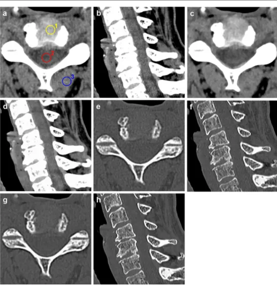
All measurements were performed by observer 2. Three region-of-interests (ROI) of 50 mm2 each were placed in the most homogeneous area of the intervertebral disc, the spinal cord, and the posterior paraspinal muscles (i.e. multi-fidus or semispinalis capitis) on axial CT images at both C3–C4 and C6–C7 levels (Fig.1a). In order to limit partial volume effects, care was taken to avoid other structures present on adjacent sections. Furthermore, the ROI were directly copied and pasted from FBP to SAFIRE 3 images to be exactly in the same position. Measurements were repeated on two consecutive sections and CT numbers (CTn, i.e. Hounsfield unit, HU) averaged. Image noise, defined as the standard deviation (SD) of the mean CTn
Fig. 1 A 50-year-old woman with chronic neck pain, who belongs to the standard-dose group. Axial (C3–C4 level) and sagittal-reformatted
unenhanced CT images (window level/width, 60/300) of the cervical spine, reconstructed with a, b, e, f filtered back-projection (FBP) and c, d, g, h sinogram-affirmed iterative reconstruction (SAFIRE, strength level 3), using soft tissue (a–d) and bone convolution kernels (e–h). a Three region-of-interests (ROI) of 50 mm2each are drawn in the intervertebral disc (1), the spinal cord (2) and posterior paraspinal muscles (3). Note the change in conspicuity of the different anatomical structures with iterative reconstruction (IR), compared with FBP
within a ROI, was measured in the posterior paraspinal muscles. The signal-to-noise (SNR) and contrast-to-noise (CNR) ratios were calculated as follows:
– SNR = mean CTn/SD, within the ROI placed in the posterior paraspinal muscles
– CNR = Δ mean CTn/[(Σ SD)/2], within the ROI
placed in the intervertebral disc and the spinal cord
Qualitative (semi-quantitative) analysis
Before starting the analysis, both observers were instructed on the image grading system with five test cases that were not included in the study. All CT images were displayed with the window level/width set to 60/300 for the soft tissues and 400/2,000 for bone respectively. The conspicuity of the following anatomical structures was assessed using a four-point scale (4 = excellent; 3 = good; 2 = moderate; 1 = poor), at both C3–C4 and C6–C7 levels: the intervertebral disc, the content of neural foramina (i.e. the spinal nerve roots and ganglia) and of the dural sac (i.e. the spinal cord),
the flavum and posterior longitudinal ligaments, the subcu-taneous tissue and muscles (including intermuscular spaces), and the trabecular bone of vertebrae. In addition, the presence of photon starvation (streak) artefacts was evaluated using a four-point scale (4 = no artefact; 3 = minor artefacts; 2 = moderate artefacts, not reducing diagnostic acceptability; 1 = major artefacts, unacceptable). Finally, the overall image quality (in terms of diagnostic acceptability) was rated using a ten-point scale (10 = excellent to 1 = poor). This image grading system was inspired by the Eu-ropean guidelines on quality criteria for computed tomogra-phy [17].
Statistical analysis
All data were processed using a statistical software package (MedCalc version 11.6; MedCalc Software, Mariakerke, Belgium). Patients’ characteristics in both groups were com-pared using the Chi-squared and Student t tests for unpaired samples. Continuous variables (quantitative analysis) were compared using the Student t test for unpaired samples (all variables were distributed normally according to the
Fig. 2 A 77-year-old man with right C4 radiculopathy, who belongs to the low-dose group. Axial (C3–C4 level) and sagittal-reformatted unenhanced CT images (window level/width, 60/300) of the cervical spine, reconstructed with a, b, e, f filtered back-projection (FBP) and c, d, g, h sinogram-affirmed iterative reconstruction (SAFIRE, strength level 3), using a–d soft tissue and e– h bone convolution kernels. Note the change in conspicuity of the different anatomical structures with IR, compared with FBP
Kolmogorov–Smirnov test). Ordinal variables (qualitative analysis) were compared using the Mann–Whitney U test and its extension, the Kruskal–Wallis test. Given that a non-statistically significant difference in a Student t test is not a proof of equivalence [18], an equivalence test was further performed to compare the standard-dose FBP and low-dose IR protocols. Interobserver agreement was assessed by cal-culating weighted kappa coefficients (with linear weight-ing), and interpreted as follows: ≤0 = poor, 0.01–0.20 = slight, 0.21–0.40 = fair, 0.41–0.60 = moderate, 0.61–0.80 = substantial, and≥0.81 = almost perfect agreement. A signif-icance level of p≤0.05 was considered for all tests.
Results Patient groups
There was no significant difference in gender (p=0.73), age (p=0.54) and AP diameter of the neck (Δ=0.9 cm, p=0.07) between the standard-dose and low-dose patient groups. Noise power spectrum
As shown in Fig.3, the centroids of the spatial frequency range obtained with FBP and SAFIRE 3 are very close to one another. However, the maximum value of NPS is reached at a slightly lower frequency with SAFIRE 3. Radiation exposure estimations
The mean CTDIv o l, DLP and ED were 39.0 mGy,
473.7 mGy × cm and 2.42 mSv for the standard-dose protocol, compared with 22.9 mGy, 275.3 mGy × cm and 1.40 mSv for the low-dose protocol respectively (Table1). The differences in CTDIvol, DLP and ED were all statisti-cally significant (all p<0.01).
Quantitative analysis
The results of the quantitative analysis are reported in Table 1 and Fig. 4.
For both standard-dose and low-dose protocols, and at the C3–C4 and C6–C7 levels, the application of IR signif-icantly decreased image noise and increased SNR and CNR, compared with FBP (all p≤0.02). Mean noise levels were significantly higher at C6–C7 than C3–C4 for both acquisi-tion protocols and each image reconstrucacquisi-tion algorithm (all p≤0.04).
The low-dose IR and standard-dose FBP protocols were statistically equivalent at each disc level in terms of SNR and CNR (all p≤0.01, with a tolerance interval of ±2). These two protocols were also equivalent in terms of image noise at the C3–C4 level (p=0.02, with a tolerance interval of ±2), but neither a statistical equivalence nor a difference was found at C6–C7 (p=0.11 and 0.60 for difference and equiv-alence tests, respectively, with a tolerance interval of +/− 2). Qualitative (semi-quantitative) analysis
The results of the qualitative analysis are illustrated in Fig. 5. They were comparable for both observers and for each disc level, except as described below.
For both standard-dose and low-dose protocols, the scores obtained with IR for the intervertebral discs, content of the neural foramina, and ligaments were significantly higher or not statistically different compared with FBP. As regards intervertebral discs, the scores with IR were signif-icantly higher than with FBP (all p≤0.01), except for ob-server 2 at C6–C7 level with the low-dose protocol (p= 0.15). As regards the content of neural foramina, no signif-icant difference was found between IR and FBP (all p≥ 0.22), except for observer 1 at both disc levels with the standard-dose protocol (IR significantly better than FBP, all p≤0.02), and at the C3–C4 level with the low-dose protocol (IR significantly better, p=0.03). As regards liga-ments, there was no significant difference between IR and FBP (all p≥0.06), except at the C3–C4 level with the low-dose protocol (IR significantly better, p≥0.02). For these three anatomical structures, the low-dose IR protocol re-ceived significantly higher scores than the standard-dose FBP (all p≤0.05).
Besides, the scores of the standard-dose and low-dose protocols obtained with IR and FBP for the content of the dural sac were comparable (all p≥0.06).
In contrast, the scores obtained with IR for the soft tissues and vertebrae were significantly lower or not statistically different compared with FBP, for both standard-dose and low-dose protocols. As regards soft tissues, the scores with IR were significantly lower than with FBP (all p≤0.04). As regards vertebrae, the scores with IR were also significantly
Fig. 3 Line graph illustrates the noise power spectrum (NPS) with filtered back-projection (FBP) and sinogram-affirmed iterative recon-struction (SAFIRE, strength level 3). SAFIRE 3 drastically reduces image noise while avoiding oversmoothing of the data, compared with FBP.
lower than with FBP (all p≤0.01), except for observer 2 at both disc levels with the low-dose protocol (all p≥0.26), and for observer 1 at the C6–C7 level with the standard-dose protocol (p=0.15). For these two anatomical structures, the low-dose IR protocol received significantly lower scores than the standard-dose FBP (all p≤0.02), except for observ-er 1 at C6–C7 level (p>0.99).
Furthermore, the scores of the standard-dose and low-dose protocols obtained with IR and FBP for photon star-vation artefacts were comparable (all p≥0.53). Regardless
of the protocol, no significant difference in the frequency of those artefacts was found, either at the C3–C4 or the C6–C7 level (all p≥0.12).
Finally, the overall image quality scores obtained with IR were significantly higher than with FBP, for both standard-dose and low-standard-dose protocols (all p<0.01). The low-standard-dose IR protocol received significantly higher scores than the standard-dose FBP (all p<0.01).
Interobserver agreement was substantial for the low-dose IR (κ=0.73) and both standard-dose protocols (κ=0.66 and
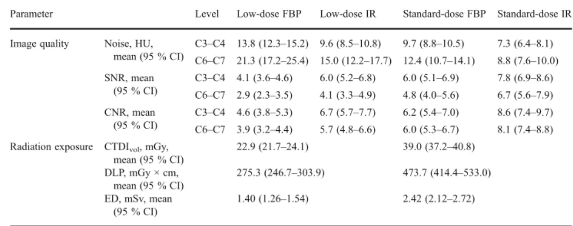
Table 1 Objective image quali-ty and radiation exposure for the standard-dose and low-dose CT protocols using filtered back-projection (FBP) and iterative reconstruction (IR)
SNR signal-to-noise ratio; CNR contrast-to-noise ratio; CTDIvol volume CT dose index; DLP dose–length product; ED effec-tive dose; HU Hounsfield unit; CI confidence interval
Parameter Level Low-dose FBP Low-dose IR Standard-dose FBP Standard-dose IR
Image quality Noise, HU, mean (95 % CI) C3–C4 13.8 (12.3–15.2) 9.6 (8.5–10.8) 9.7 (8.8–10.5) 7.3 (6.4–8.1) C6–C7 21.3 (17.2–25.4) 15.0 (12.2–17.7) 12.4 (10.7–14.1) 8.8 (7.6–10.0) SNR, mean (95 % CI) C3–C4 4.1 (3.6–4.6) 6.0 (5.2–6.8) 6.0 (5.1–6.9) 7.8 (6.9–8.6) C6–C7 2.9 (2.3–3.5) 4.1 (3.3–4.9) 4.8 (4.0–5.6) 6.7 (5.6–7.9) CNR, mean (95 % CI) C3–C4 4.6 (3.8–5.3) 6.7 (5.7–7.7) 6.2 (5.4–7.0) 8.6 (7.4–9.7) C6–C7 3.9 (3.2–4.4) 5.7 (4.8–6.6) 6.0 (5.3–6.7) 8.1 (7.4–8.8) Radiation exposure CTDIvol, mGy,
mean (95 % CI) 22.9 (21.7–24.1) 39.0 (37.2–40.8) DLP, mGy × cm, mean (95 % CI) 275.3 (246.7–303.9) 473.7 (414.4–533.0) ED, mSv, mean (95 % CI) 1.40 (1.26–1.54) 2.42 (2.12–2.72)
Fig. 4 Bar charts illustrate the impact of filtered back-projection (FBP) and iterative reconstruction (IR, strength level 3) on image noise (in Hounsfield units, HU), signal-to-noise (SNR) and contrast-to-noise (CNR) ratios, for standard-dose and low-dose CT protocols, at a C3–C4 and b C6–C7 levels. Error bars represent 95 % confidence intervals
0.79 with IR and FBP respectively), while it was almost perfect for the low-dose FBP protocol (κ=0.81).
Discussion
This patient-based study demonstrates that the application of IR in cervical spine CT enhances image quality both quantitatively and qualitatively. However, its impact depends on the anatomical structure to be analysed. Both observers found that the overall diagnostic image quality was significantly better with IR, compared with FBP (Fig. 5e). When focusing on different anatomical struc-tures, they noted that the conspicuity of the intervertebral discs, the content of the neural foramina, and the ligaments was significantly higher (or not statistically different) with IR (Fig. 5a–d). This finding might be explained by the higher CNR obtained with IR (Table 1, Fig. 4), and is important because all these structures need to be accurately assessed in patients with chronic neck pain and/or cervical radiculopathy [1–3]. In contrast, both soft tissues and ver-tebrae received significantly lower (or not statistically dif-ferent) scores with IR (Fig.5a–d). Therefore, conventional FBP images should still be evaluated when assessing soft tissues and the trabecular bone of the vertebrae.
Radiation exposure related to imaging studies, partic-ularly CT, is a growing concern [19]. Among the tools implemented to reduce radiation dose in CT imaging, IR techniques have recently proved to be effective [5, 6, 10–13]. Compared with traditional FBP algorithms, their main advantage lies in the lower noise level of recon-structed images, without sacrificing spatial resolution. Consequently, both SNR and CNR are improved. Itera-tive reconstruction methods can be used either to en-hance image quality or to reduce radiation dose. Given that current CT technology provides diagnostic image quality for all anatomical regions, most recent studies focused on the latter objective [10–13]. Indeed, signifi-cant dose reductions of up to 66 % were achieved in thoracic and abdominal CT, while preserving acceptable diagnostic image quality [10–13]. To date, only one study has assessed the impact of IR in CT of the musculoskeletal system [20]. Focusing on the lumbar spine, Gervaise et al. demonstrated that a dose reduction of approximately 52 % was achievable by applying adaptive iterative dose reduction (AIDR; Toshiba, Tochigi, Japan), while preserving diagnostic image qual-ity [20]. Similarly, we found that the application of SAFIRE (Siemens Healthcare) in CT of the cervical spine allowed for an average 42 % reduction in
Fig. 5 Bar charts illustrate the impact of FBP and IR (strength level 3) on image quality scores for a–d different anatomical structures and photon starvation artefacts, and e overall diagnostic image quality. a
C3–C4 level, observer (OBS) 1; b C3–C4 level, OBS 2; c C6–C7 level, OBS 1; d C6–C7 level, OBS 2. Error bars represent 95 % confidence intervals
radiation dose (Table 1), while maintaining diagnostic image quality. Indeed, when comparing standard-dose FBP and low-dose IR protocols, we noted that objective image quality was kept almost constant, as the mean noise, SNR and CNR of the two protocols were com-parable (Table 1, Fig. 4). Interestingly, both observers found that overall diagnostic image quality was signifi-cantly better with low-dose IR than with standard-dose FBP (Fig. 5e). Further studies are necessary to deter-mine the highest level of dose reduction, which may depend on the anatomical region to be examined.
In order to standardise the local practices, diagnostic reference levels (DRL) have been established for most CT examinations [21]. In this context, we aimed to compare the standard-dose protocol provided by the manufacturer for cervical spine CT (120 kVp, 275 mAs) with the lowest-dose protocol reported in the literature (120 kVp, 150 mAs) [14,15]. A second-generation IR technique (SAFIRE; Sie-mens Healthcare) was applied to the latter protocol to en-hance its image quality. While we opted rather to reduce tube current–time product, Hoang et al. recently reported that lowering tube voltage (from 120 to 80 kVp) in CT of the neck resulted in a greater than 50 % radiation dose reduc-tion, without impairing subjective image quality [7]. Further studies should focus on optimising both kVp and mAs values. Lowering kVp would indeed allow the radiation dose to be further reduced, with a concomitant increase in contrast and noise, thus maintaining CNR nearly constant.
We acknowledge the following limitations of the study. First, a relatively small number of patients were included in each group. Second, the diagnostic performance of low-dose cervical spine CT with IR was not assessed. However, CT is a recognised imaging modality in the diagnosis of cervical disc herniation and/or spondylosis [1–3]. Moreover, the overall diagnostic image quality was found to be better with IR (Fig. 5e). Further studies with surgical correlation are necessary to assess the diagnostic accuracy of this new technique. Third, we only evaluated the strength level of IR recommended by the manufacturer (i.e. SAFIRE 3). Further studies are needed to determine the optimal strength level of IR.
In conclusion, the application of IR in cervical spine CT decreases image noise, increases SNR and CNR, and enhan-ces the conspicuity of some anatomical structures. Low-dose CT of the cervical spine with IR provides better image quality of the intervertebral discs, the content of neural foramina, and ligaments compared with standard-dose CT with FBP, while reducing radiation dose by approximately 40 %. However, the former protocol provides lower image quality of the soft tissues and vertebrae. Further studies are necessary to determine the optimal strength level of IR, which may depend on the anatomical structure to be analysed.
Acknowledgements The authors thank Julien G. Ott for his help with the noise power spectrum analysis.
Conflict of interest The authors declare that they have no conflict of interest.
Funding or grant None
References
1. Freund M, Sartor K. Degenerative spine disorders in the context of clinical findings. Eur J Radiol. 2006;58:15–26.
2. Douglas-Akinwande AC, Rydberg J, Shah MV, et al. Accuracy of contrast-enhanced MDCT and MRI for identifying the se-verity and cause of neural foraminal stenosis in cervical radi-culopathy: a prospective study. AJR Am J Roentgenol. 2010;194:55–61.
3. Tins B. Technical aspects of CT imaging of the spine. Insights Imaging. 2010;1:349–59.
4. Biswas D, Bible JE, Bohan M, Simpson AK, Whang PG, Grauer JN. Radiation exposure from musculoskeletal computer-ized tomographic scans. J Bone Joint Surg Am. 2009;91:1882– 9.
5. Lee TY, Chhem RK. Impact of new technologies on dose reduction in CT. Eur J Radiol. 2010;76:28–35.
6. Tamm EP, Rong XJ, Cody DD, Ernst RD, Fitzgerald NE, Kundra V. Quality initiatives: CT radiation dose reduction: how to imple-ment change without sacrificing diagnostic quality. Radiographics. 2011;31:1823–32.
7. Hoang JK, Yoshizumi TT, Nguyen G, et al. Variation in tube voltage for adult neck MDCT: effect on radiation dose and image quality. AJR Am J Roentgenol. 2012;198:621–7.
8. Leswick DA, Hunt MM, Webster ST, Fladeland DA. Thyroid shields versus z-axis automatic tube current modulation for dose reduction at neck CT. Radiology. 2008;249:572–80.
9. Gervaise A, Louis M, Batch T, et al. Dose reduction at CT of the lumbar spine using a 320-detector row scanner: initial results. J Radiol. 2010;91:779–85.
10. Singh S, Kalra MK, Hsieh J, et al. Abdominal CT: comparison of adaptive statistical iterative and filtered back projection reconstruc-tion techniques. Radiology. 2010;257:373–83.
11. Pontana F, Pagniez J, Flohr T, et al. Chest computed tomography using iterative reconstruction vs filtered back projection (Part 1): evaluation of image noise reduction in 32 patients. Eur Radiol. 2011;21:627–35.
12. Moscariello A, Takx RA, Schoepf UJ, et al. Coronary CT angiog-raphy: image quality, diagnostic accuracy, and potential for radia-tion dose reducradia-tion using a novel iterative image reconstrucradia-tion technique-comparison with traditional filtered back projection. Eur Radiol. 2011;21:2130–8.
13. Winklehner A, Karlo C, Puippe G, et al. Raw data-based iterative reconstruction in body CTA: evaluation of radiation dose saving potential. Eur Radiol. 2011;21:2521–6.
14. Wirth S, Meindl T, Treitl M, Pfeifer KJ, Reiser M. Comparison of different patient positioning strategies to minimize shoulder girdle artifacts in head and neck CT. Eur Radiol. 2006;16:1757–62. 15. Mulkens TH, Marchal P, Daineffe S, et al. Comparison of low-dose
with standard-dose multidetector CT in cervical spine trauma. AJNR Am J Neuroradiol. 2007;28:1444–50.
16. Deak PD, Smal Y, Kalender WA. Multisection CT protocols: sex-and age-specific conversion factors used to determine effective dose from dose-length product. Radiology. 2010;257:158–66. 17. Bongartz G, Golding SJ, Jurik AG, et al. European guidelines on
www.drs.dk/guidelines/ct/quality/index.htm accessed October 13, 2011.
18. Jones B, Jarvis P, Lewis JA, Ebbutt AF. Trials to assess equiva-lence: the importance of rigorous methods. BMJ. 1996;313:36–9. 19. Brenner DJ, Hall EJ. Computed tomography–an increasing source
of radiation exposure. N Engl J Med. 2007;357:2277–84.
20. Gervaise A, Osemont B, Lecocq S, et al. CT image quality improve-ment using Adaptative Iterative Dose Reduction with wide-volume acquisition on 320-detector CT. Eur Radiol. 2012;22:295–301. 21. Dougeni E, Faulkner K, Panayiotakis G. A review of patient dose
and optimisation methods in adult and paediatric CT scanning. Eur J Radiol. 2012;81:e665–83.