Conformational proofreading of distant 40S
ribosomal subunit maturation events by a
long-range communication mechanism
Valentin Mitterer
1,5,6, Ramtin Shayan
2,6, Sébastien Ferreira-Cerca
3, Guillaume Murat
4, Tanja Enne
1,
Dana Rinaldi
2, Sarah Weigl
1, Hajrija Omanic
1, Pierre-Emmanuel Gleizes
2, Dieter Kressler
4,
Celia Plisson-Chastang
2& Brigitte Pertschy
1Eukaryotic ribosomes are synthesized in a hierarchical process driven by a plethora of assembly factors, but how maturation events at physically distant sites on pre-ribosomes are coordinated is poorly understood. Using functional analyses and cryo-EM, we show that ribosomal protein Rps20 orchestrates communication between two multi-step maturation events across the pre-40S subunit. Our study reveals that during pre-40S maturation, for-mation of essential contacts between Rps20 and Rps3 permits assembly factor Ltv1 to recruit the Hrr25 kinase, thereby promoting Ltv1 phosphorylation. In parallel, a deeply buried Rps20 loop reaches to the opposite pre-40S side, where it stimulates Rio2 ATPase activity. Both cascades converge to thefinal maturation steps releasing Rio2 and phosphorylated Ltv1. We propose that conformational proofreading exerted via Rps20 constitutes a checkpoint per-mitting assembly factor release and progression of pre-40S maturation only after completion of all earlier maturation steps.
https://doi.org/10.1038/s41467-019-10678-z OPEN
1Institute for Molecular Biosciences, University of Graz, Humboldtstrasse 50, 8010 Graz, Austria.2Laboratoire de Biologie Moléculaire Eucaryote, Centre de
Biologie Intégrative, Université de Toulouse, CNRS, UPS, 118 route de Narbonne, 31062 Toulouse Cedex, France.3Biochemistry III– Institute for Biochemistry, Genetics and Microbiology, University of Regensburg, Universitätsstraße 31, 93053 Regensburg, Germany.4Unit of Biochemistry, Department of Biology, University of Fribourg, Chemin du Musée 10, 1700 Fribourg, Switzerland.5Present address: Biochemistry Centre, University of Heidelberg, 69120 Heidelberg,
Germany.6These authors contributed equally: Valentin Mitterer, Ramtin Shayan. Correspondence and requests for materials should be addressed to
S.F.-C. (email:[email protected]) or to D.K. (email:[email protected]) or to C.P.-C. (email:[email protected]) or to B.P. (email:[email protected])
123456789
E
ukaryotic ribosomes consist of a large 60S and a small 40S subunit, each composed of ribosomal RNA (rRNA) and ribosomal proteins (r-proteins). The synthesis of ribosomes is a highly complex process starting with the assembly of rRNA, r-proteins, and ribosome assembly factors (AFs) into pre-ribosomal particles in the nucleolus (reviewed in refs. 1–4). Inyeast, thefirst steps in the synthesis of 40S subunits occur within large precursors termed SSU processomes or 90S particles, in which rRNA folding and processing steps, as well as incorpora-tion of r-proteins and AFs, take place5–9. Endonucleolytic
clea-vage of the pre-rRNA, together with the dissociation of a large number of AFs, then results in the release of a 43S particle, also termed pre-40S particle, which contains a 3′ extended precursor of the mature 18S rRNA (the 20S pre-rRNA), most 40S r-pro-teins, and only a few AFs2,4,10. These particles are exported into the cytoplasm, where further 40S maturation events take place. Finally, conversion of the 20S pre-rRNA into the 18S rRNA by the endonuclease Nob1 results in the release of mature, translation-competent 40S subunits11–13.
Several biochemical studies together with recent cryo-electron microscopy (cryo-EM) structural analyses provided insights into the organization of early cytoplasmic pre-40S particles, thus revealing that AFs Tsr1, Pno1, Nob1, Dim1, and Rio2 are located on the intersubunit side of the pre-40S subunit and that Enp1 and Ltv1 are bound on the solvent-exposed side in the area of the beak structure10,14–22. These early cytoplasmic pre-40S particles
undergo a cascade of maturation events, with the twofirst and presumably rate limiting ones being two different ATP-dependent maturation steps, resulting in dissociation of AFs Rio2 and Ltv114,19,23–26.
Rio2 is an ATPase that is bound at the junction between the head and the body of 40S subunits and its release from pre-40S particles is promoted by ATP hydrolysis14,27. In addition,
Rio2 release was suggested to require and trigger conformational changes necessary for progression of pre-40S particles into sub-sequent maturation steps14,27.
Ltv1 interacts with two 18S rRNA helices (h16 and h41) and several proteins (i.e., Enp1 and r-proteins Rps3 (uS3 according to the new nomenclature of r-proteins28) and Rps20 (uS10)) within
the head domain of pre-40S particles, and its release involves at least two different steps. One is promoted by phosphorylation of three serines in the C-terminal part of Ltv1 by the casein kinase I homolog Hrr2519,23,24. The other step comprises the formation of
contacts of the Rps3 N-domain with Rps20 and rRNA helix h4119,29. It is, however, not known whether these two steps are
interconnected and in which order they occur.
Notably, mutations blocking Ltv1 release also inhibited the release of other AFs from pre-40S particles, including Rio2, indicating that Ltv1 dissociation is a prerequisite for the release of these factors19. However, previous studies on Rio2 demonstrated that a catalytically inactive rio2 mutant, which is impaired in Rio2 release, accumulates Ltv1 on pre-40S particles, indicating that Rio2 catalytic activity and/or its subsequent release is, on the other hand, a prerequisite for Ltv1 release14,30. How Ltv1 release,
taking place on the solvent side of the pre-40S subunit, might be coordinated with Rio2 catalytic activity and/or release, which occurs >50 Å apart on the intersubunit side (distance between the h41 Ltv1 rRNA-binding site, which is later occupied by the Rps3 N-domain, and the h31 Rio2 rRNA-binding site15), has however
remained elusive.
Here we report that Rps20 coordinates Ltv1 and Rio2 release. The largest part of Rps20 is located on the solvent-exposed side of pre-40S particles where it contacts Rps3, thereby promoting Hrr25 recruitment by Ltv1 and subsequent Ltv1 phosphorylation. Twoβ-strands in Rps20 connected by an unstructured loop dive deeply into the (pre-)40S subunit, almost reaching to the
Rio2-binding site. Deletion of this Rps20 loop leads to a reduction of Rio2’s ATPase activity; however, Ltv1 phosphorylation can still occur normally. Vice versa, rps20 mutations preventing Ltv1 phosphorylation still allow Rio2 ATP hydrolysis. In either case, however, final release of both Rio2 and Ltv1 from pre-40S par-ticles is inhibited. We conclude that Rio2 and Ltv1 release are multi-step processes, with the respective final steps occurring in an interdependent manner. Notably, pre-40S particles from an rps20Δloop mutant differ in several structural features from recently published wild-type pre-40S particles. We suggest that sensing of the correct conformations of both maturation sites, exerted by Rps20, provides a quality control checkpoint, which ensures that release of Ltv1 and Rio2 is only triggered once all necessary earlier maturation steps have been completed.
Results
Rps3 N-domain assembly promotes Hrr25 recruitment. Our previous studies suggested that both the phosphorylation of Ltv1 by Hrr25 and the contact formation between the Rps3 N-domain and Rps20 are required for Ltv1 release, but the order of these maturation events was unclear19. To better define the effects of
mutations impairing the establishment of contacts between positively charged amino acids of the Rps3 N-domain (K7 and K10) and negatively charged amino acids of Rps20 (D113 and E115), we analyzed the composition of pre-40S particles isolated from such mutants. We considered the AF Tsr1 as a suitable bait protein for this purification, since it purifies a broad range of pre-40S particles by binding in the nucleus and dissociating at a late cytoplasmic step, after Ltv1 and Rio2 release10,31,32.
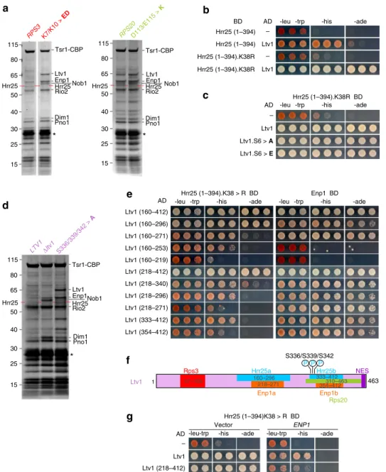
In line with our previous results, several AFs, including Ltv1, accumulated on pre-40S particles isolated from rps3.K7/K10>ED or rps20.D113/E115>K mutants (Fig. 1a). Surprisingly however, one band (indicated by a red dashed line in Fig. 1a) was significantly reduced in rps3.K7/K10>ED and rps20.D113/ E115>K mutants. Analysis by mass spectrometry revealed that this band corresponded to the Hrr25 kinase, which executes phosphorylation of Ltv119,23,24. Hence, formation of the
Rps3-Rps20 contact occurs prior to and is even a pre-requisite for Ltv1 phosphorylation.
We speculated that restructuring of the Rps3 N-domain19
promotes recruitment of Hrr25 by unleashing a crucial binding surface. To better understand how Hrr25 is recruited to pre-40S particles, we performed yeast two-hybrid (Y2H) analyses testing the interaction of Hrr25 with several other AFs contained in pre-40S particles, as well as with some r-proteins of the pre-40S head domain (Supplementary Fig. 1a). Ltv1 showed a robust interac-tion with Hrr25, while no other tested AF interacted with Hrr25 in this assay and only the r-protein Rps15 displayed a weak interaction with Hrr25 (Supplementary Fig. 1a). Since full-length Hrr25 (494 aa) fused to the Gal4 DNA-binding domain displayed some self-activation of the HIS3 reporter gene (Supplementary Fig. 1a, left panel), we performed subsequent analyses with a C-terminally truncated version of Hrr25 (aa 1–394)33, which
showed no self-activation and still interacted with Ltv1, albeit with reduced efficiency (Fig. 1b). Moreover, mutation of the catalytic lysine residue to arginine (K38R mutant) strengthened the interaction between Ltv1 and this truncated (1–394) Hrr25 variant (Fig. 1b and Supplementary Fig. 1a, right panel). The interaction was fully maintained when the three main Hrr25 phosphorylation sites on Ltv1 (S336, S339, and S342) and an additional three proximal serines (S344, S345, and S346) were either exchanged for alanines (Ltv1.S6>A) or glutamates (Ltv1. S6>E), mimicking the unphosphorylated or phosphorylated states of these residues, respectively (Fig. 1c). Accordingly, Hrr25 was present in pre-40S purifications from the ltv1.S336/339/342>A
mutant but absent in pre-40S preparations from a Δltv1 strain (Fig.1d). Together, these results suggest that Ltv1 is necessary to recruit Hrr25 to pre-40S particles and that Ltv1’s phosphorylated serines are only transiently bound by Hrr25 during its catalytic
activity, while the main Hrr25 interaction site lies elsewhere within Ltv1.
The fact that Hrr25 is less efficiently recruited when the contact between the Rps3 N-domain and Rps20 is abolished
b a Hrr25 (1–394) Ltv1 BD AD – Ltv1 – Hrr25 (1–394) Hrr25 (1–394).K38R Hrr25 (1–394).K38R
-leu -trp -his -ade
-leu -trp -his -ade
-leu -trp -his -ade
-his -ade -his -ade
-leu -trp -his -ade Hrr25 (1–394).K38R BD AD – Ltv1 Ltv1.S6 > A Ltv1.S6 > E c d Enp1 BD e Ltv1 (160–412) Ltv1 (160–296) Ltv1 (333–412) Ltv1 (160–271) Ltv1 (218–412) Ltv1 (218–340) Ltv1 (160–253) Ltv1 (218–271) Ltv1 (218–296) Ltv1 (354–412) AD Hrr25 (1–394).K38 > R BD Hrr25 (1–394)K38 > R BD AD – Ltv1 -leu-trp Ltv1 (218–412) Vector ENP1 f g -leu-trp 1 Ltv1 Rps3 463 S336/S339/S342 P P P Rps20 NES Hrr25a Enp1a 354–412 Enp1b 310–463 333–412 Hrr25b 160–296 218–271 57–105 115 80 65 50 40 30 25 15 RPS20D113/E115 > K Tsr1-CBP Rio2 Enp1 Hrr25 Dim1 Pno1 Ltv1 RPS3 K7/K10 > ED Tsr1-CBP Rio2 Enp1 Nob1 Hrr25 Dim1 Pno1 Ltv1 Hrr25 Hrr25 Nob1 115 80 65 50 40 30 25 15 * * * S336/339/342 > A Tsr1-CBP Rio2 Enp1Nob1 Hrr25 Dim1 Pno1 Ltv1 115 80 65 50 40 30 25 15 Hrr25 LTV1Δltv1 Ltv1 (160–219)
Fig. 1 Rps3 N-domain assembly promotes Hrr25 recruitment. a Reduced binding of Hrr25 to particles impaired in Rps3 N-domain/Rps20 contact formation. Tsr1-TAP particles isolated from wild-type cells or from cells expressing the indicatedrps3 or rps20 mutant alleles were analyzed by sodium dodecyl sulfate-polyacrylamide gel electrophoresis (SDS-PAGE) and Coomassie staining. Mass spectrometric (MS) analyses revealed that, while Hrr25 is present on particles isolated from wild-type strains, it is absent or strongly reduced from particles on which the Rps3 N-domain is not assembled (i.e.,rps3. K7/K10>ED andrps20.D113/E115>K). Asterisks indicate the band of the Tobacco etch virus (TEV) protease used to elute pre-40S particles from the IgG beads.b, c Characterization of the interaction between Ltv1 and Hrr25. A C-terminal Hrr25 truncation (Hrr25(1–394)) and a mutant version thereof (Hrr25 (1–394).K38R) fused to the Gal4 DNA-binding domain (BD) was tested for Y2H interaction with Ltv1 (b) and Ltv1 phosphomutants (Ltv1.S6>A and Ltv1. S6>E) (c) fused to the Gal4 activation domain (AD). Cells were spotted in ten-fold serial dilutions on SDC-Leu-Trp, SDC-His-Leu-Trp (-his; growth on this medium indicates a weak interaction), and SDC-Ade-Leu-Trp (-ade; growth on this medium indicates a strong interaction) plates.d Ltv1 is required to recruit Hrr25. Tsr1-TAP particles were isolated fromltv1Δ cells expressing plasmid-borne wild-type LTV1, the ltv1(S336/S339/S342>A) phosphomutant, or harboring an empty plasmid (Δltv1). Eluates were analyzed by SDS-PAGE, Coomassie staining, and MS. The asterisk indicates the TEV protease. e Hrr25 (1–394).K38R (left panel) and Enp1 (right panel) fused to the BD were tested for Y2H interaction with the indicated Ltv1 fragments fused to the AD. f The deduced, minimal binding sites for Hrr25 and Enp1 on Ltv1 are indicated. In addition, the sites for Rps3 binding, Rps20 binding, and phosphorylation by Hrr2519, as well as the nuclear export sequence35, are depicted.g Enp1 overexpression weakens the Hrr25-Ltv1 interaction. Y2H strains expressing Hrr25
(1–394).K38R fused to the BD in combination with the indicated Ltv1 constructs fused to the AD were transformed with empty plasmid (left panel) or with aURA3 plasmid for ENP1 overexpression under control of the ADH1 promoter (right panel). Cells were spotted in ten-fold serial dilutions on SDC-Leu-Trp-Ura (panel: -leu-trp), SDC-His-Leu-Trp-SDC-Leu-Trp-Ura (panel: -his), and SDC-Ade-Leu-Trp-SDC-Leu-Trp-Ura (panel: -ade) plates
suggests that a structural re-arrangement brings Ltv1 into a conformation where it is able to interact with Hrr25. To better understand which part of Ltv1 needs to be exposed to allow for Hrr25 recruitment, we mapped the Hrr25-binding sites on Ltv1. Remarkably, Ltv1 appears to harbor two sites, which can bind Hrr25 independently, and whose minimal fragments either exhibit a robust (aa 160–296) or moderate (aa 333–412) Y2H interaction with Hrr25(1–394).K38R (Fig. 1e and Supplementary Fig. 2). Notably, the second, weaker interaction surface comprises the residues that are phosphorylated by Hrr25 and partially overlaps with the previously mapped Rps20-binding region (Fig.1f, ref.19). Enp1 is another pre-40S
component that directly interacts with Ltv1 via its bystin domain (aa 155–4709, Supplementary Fig. 1b) and was
suggested to be a phosphorylation substrate of Hrr2514,24,34. Strikingly, Ltv1 also contains two independent Enp1-binding sites; both of these minimal Enp1-binding fragments (aa 218–271 and aa 354–412 of Ltv1) display an equally strong Y2H interaction and largely overlap with the Hrr25-binding regions (Fig.1e, f and Supplementary Fig. 2). In line with this, overexpression of Enp1 weakened the Y2H interaction between Ltv1 and Hrr25, indicating that Enp1 and Hrr25 compete for Ltv1 binding (Fig.1g). Therefore, we propose that the binding of Hrr25 helps to physically detach the interaction between Ltv1 and Enp1, thereby loosening the interaction of Ltv1 with pre-40S particles even before its phosphorylation and final dissociation take place.
Cytoplasmic pre-40S maturation steps are functionally linked. Mutations blocking Ltv1 release also inhibited release of other AFs from pre-40S particles, including Rio2, while mutations impairing Rio2 release also accumulated Ltv1 on pre-40S parti-cles, indicating that Ltv1 and Rio2 release are mutually interdependent14,19,30. To confirm that ATP hydrolysis by Rio2 is necessary for Ltv1 release, we made use of an Ltv1-GFP reporter construct that shows a nuclear steady-state localization due to mutation of Ltv1’s nuclear export sequence (NES) (Ltv1-NES3>A-GFP) (Supplementary Fig. 3a, refs.19,35). As we showed
previously, cytoplasmic Ltv1 release defects can be readily iden-tified by means of a cytoplasmic mislocalization of such a reporter construct19. The localization of the reporter construct was
eval-uated in a mutant expressing the catalytically inactive rio2.D253A allele, which is viable but shows reduced Rio2 disassembly from 40S maturation intermediates14. Indeed, in contrast to its
pre-dominantly nuclear localization in wild-type cells, the Ltv1-NES3>A-GFP reporter construct mislocalized to the cytoplasm in rio2.D253A cells (Supplementary Fig. 3b). In addition, the Enp1-GFP fusion protein also accumulated in the cytoplasm in the rio2. D253A mutant, indicating a block of downstream pre-40S maturation steps.
The interdependence of the spatially separated Rio2 and Ltv1 release events prompted us to test for functional connections between mutations affecting Rio2 release and mutations inhibit-ing Ltv1 release. To this end, we tested the ltv1.S336/S339/ S342>A phosphorylation site mutant for genetic interaction with the catalytically inactive rio2.D253A variant, notably revealing a synthetic lethal phenotype (Fig. 2a). In addition, combining the rio2.D253A allele with rps3 or rps20 variants, which are impaired in establishing contacts between Rps3 (K7 and K10) and Rps20 (D113 and E115) or between Rps3 (K8 and R9) and rRNA (h41), resulted in synthetic lethal phenotypes (Fig.2b, c). These results suggest a tight functional connection between Rps3 N-domain assembly, Ltv1 phosphorylation and release, and ATP-hydrolysis-dependent release of Rio2.
A conserved Rps20 loop is essential for pre-40S maturation. While Ltv1, Rps3, and Rps20 are positioned on the solvent-exposed side of the pre-40S head domain, Rio2 is positioned on the subunit-interface side, bridging the head and the body14–18,20–22. We speculated that one or several proteins might physically link these distant sites to allow communication between them. We recognized that two longβ-strands of Rps20 protrude deeply into the interior of the mature 40S subunit and an unstructured loop connecting theseβ-strands reaches almost to the opposite subunit-interface side29. Remarkably, this loop is
in close contact with 18S rRNA helix h31, which also contains one of the rRNA-binding sites (nucleotides 1194–1196) of Rio2 in pre-40S particles, suggesting a cross-talk between the Rps20 loop and Rio2 either by a direct interaction or via their rRNA contact with helix h31 (Fig.2d, refs.15,20). The 11-amino-acid-long Rps20 loop (aa 68–78) contains four charged amino acids (R68, K69, K77, and E74), which might establish interactions with rRNA and/or proteins, and is highly conserved among eukaryotes (Fig.2d).
To address the possibility that the Rps20 loop-region participates in 40S biogenesis, we analyzed the phenotypes resulting from deletion of the loop or from point mutations of the charged residues. Simultaneous substitution of amino acids R68, K69, and E74 to opposite charges (rps20.R68/K69>E/E74K) was lethal, whereas all other tested mutants were viable (Fig.2e, f, Supplementary Fig. 3c, d). However, two of the combined point mutants (rps20.R68/K69>E and rps20.R68/K69>A/E74K), as well as deletion of the Rps20 loop (Δ68–78), caused a pronounced slow-growth phenotype (Fig. 2f, Supplementary Fig. 3c, d). Moreover, in contrast to wild-type cells and the rps20.R68/K69>A mutant, the rps20.R68/K69>E and, to an even greater extent, the rps20Δloop mutant displayed a 40S synthesis defect in polysome analyses, as evidenced by a strong increase of free 60S subunits (Fig.2g). Moreover, presumably as a consequence of the severe imbalance between 40S and 60S subunits, the rps20Δloop mutant also showed significantly reduced polysome levels. Northern blot analyses revealed a reduction of the mature 18S rRNA and a substantial accumulation of its direct precursor, the 20S pre-rRNA, in the rps20.R68/K69>E and the rps20Δloop mutant (Fig.2h). Furthermore,fluorescence in situ hybridization showed that the 20S pre-rRNA accumulated in the cytoplasm, demon-strating that the inhibition of the pre-40S maturation pathway in rps20 loop mutants occurs at the stage of cytoplasmic pre-40S particles (Fig.2i). Since cytoplasmic 20S pre-rRNA accumulation was observed as well upon depletion of Rps2036, we wanted to
exclude the possibility that the observed phenotypes are the consequence of a failure to incorporate Rps20, mimicking depletion of the protein. To this end, we assessed the levels of N-terminally HA-tagged Rps20 variants in purified pre-40S particles by western blotting with anti-HA antibodies. Notably, not only the Rps20Δloop and Rps20.R68/K69>E but also the Rps20.D113/E115>K variant were fully incorporated into pre-40S particles, indicating that the observed phenotypes are specific maturation defects caused by the structural alterations introduced into the Rps20 protein (Supplementary Fig. 4).
The Rps20 loop is genetically linked to 40S maturation. To get insights into whether the Rps20 loop could participate in the crucial steps leading to Ltv1 and Rio2 release, we undertook genetic analyses. Intragenic combination of the rps20Δloop deletion or the rps20.R68/K69>E mutation with the rps20.D113/ E115>K mutation, which prevents binding of Rps20 to the Rps3 N-domain19, abolished cell growth (Fig.3a). In line with this, we
combining the rps20 loop-mutants with mutants in which the Rps3-Rps20 contact was reduced from the Rps3 side (Fig. 3b). Moreover, polysome analyses revealed that the 40S synthesis defect of the rps20.D113/E115>K mutant is strongly enhanced by the R68/K69>A loop-mutation (Fig. 3c). Remarkably, we also found synthetic lethal phenotypes of rps20 loop-mutants, including mutants that display no growth phenotype on their own, with the catalytically inactive rio2.D253A mutant (Fig.3d). Furthermore, rps20 loop-mutants severely enhanced the growth defect of the ltv1.S336/S339/S342>A phosphomutant (Fig. 3e). Taken together, we unraveled a genetic network (schematically depicted in Fig. 3f) exhibiting multifaceted interconnections between AFs and r-proteins and linking the Rps20 loop to cytoplasmic pre-40S restructuring events and Rio2 ATPase activity.
The Rps20 loop is required for release of AFs. To address whether the Rps20 loop participates in release of Rio2 and/or Ltv1, we purified pre-40S particles from rps20Δloop cells using again Tsr1-TAP as bait protein. These analyses revealed that Rio2 strongly accumulated on pre-40S particles isolated from the rps20Δloop mutant (Fig. 3g, Supplementary Fig. 5). In addition, the levels of Ltv1 and of downstream maturation factors like Nob1 and Pno1 increased, suggesting that the loop deletion blocks pre-40S maturation at an early cytoplasmic step prior to Ltv1 and Rio2 disassembly. The strong accumulation of Rio2 further strengthens the hypothesis that the Rps20 loop-region is directly involved in release of the Rio2 ATPase.
Rio2 and the Rps20 loop triggerfinal release of Ltv1. To study the influence of the different mutants on ATP-dependent Ltv1
RPS20
ITS1 DAPI Merge PC
R68/K69 > A R68/K69 > E Δloop i g RPS20 R68/K69 > E Δloop 40S 80S Polysomes 40S 60S 80S Polysomes 40S 60S 80S Polysomes 60S R68/K69 > A 40S 60S 80S Polysomes d LTV1 S336/S339/S342 > A Vector -leu-trp d4 5-FOA-leu d5 RIO2 LTV1 S336/S339/S342 > A Vector rio2 .D253A a b RIO2 RPS20 D113/E115 > K D113/E115 > A D113/E115 > K D113/E115 > A rio2 .D253A RPS3 K8/R9 > A K7/K10 > A RIO2 rio2 .D253A d2 d5 -leu-trp 5-FOA RPS3 K8/R9 > A K7/K10 > A c RPS20 -leu-trp 5-FOA d2 d5 RPS20 Δloop R68/K69 > E YPD d2 R68/K69 > A Δrps20 f RPS20 Δloop R68/K69 > A R68/K69 > E R68/K69 > E/E74K E74K R68/K69 > A/E74K -leu 5-FOA d2 d5 e E74K R68/K69 > A/E74K 20S 33/32S 18S 25S RPS20 R68/K69 > A R68/K69 > E Δ loop h 18S 5.8 25S 32S 35S 18S 5.8 18S 18S 5.8 5.8 5.8 18S 18S 18S 25S 25S 25S 27S 25S 5.8S 20S 18S S20 S3-S20 S3-N S20 Rio2 64 46 61 64 62 82 64 79 82 80 S.cerevisiae H.volcanii C.elegans D.melanogaster H.sapiens S20 loop loop contact 35S ITS1 ITS2
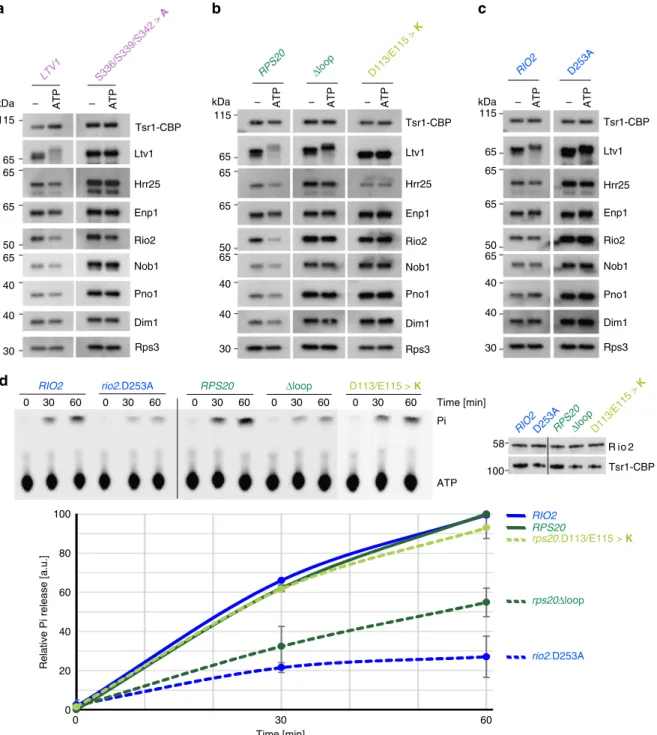
phosphorylation and release of Ltv1 and Rio2, we performed in vitro ATP incubation assays with purified pre-40S subunits. Tsr1-TAP particles were immobilized on IgG-Sepharose beads, incubated in the presence or absence of ATP and, after a subsequent washing step, eluted with Tobacco etch virus (TEV) protease. Notably, we observed an Ltv1 double band already in the untreated wild-type particles, likely corresponding to partial phosphorylation of Ltv1 in a subpopulation of the preparation (Fig.4a, LTV1; Fig.
4b, RPS20; and Fig. 4c, RIO2). After incubation of wild-type par-ticles with ATP, we observed a significant band-shift of both Ltv1 bands due to phosphorylation by Hrr25 (Fig. 4a–c); however, the
phosphorylated form of the protein was largely released from these particles. In addition, also the association of Rio2 with wild-type particles diminished upon ATP incubation.
As expected, Ltv1 was neither phosphorylated nor released upon ATP incubation of particles purified from a strain in which serines 336, 339, and 342 of Ltv1 were exchanged for alanines, preventing phosphorylation of Ltv1, despite the enrichment of Hrr25 in these particles (Fig. 4a). In contrast, Hrr25 levels were reduced in pre-40S particles from the rps20.D113/E115>K mutant, confirming that assembly of the Rps3 N-domain is important for Hrr25 recruitment (Fig.4b). Notably, Ltv1 was not phosphorylated and remained fully attached to these particles, as well as to particles from the rps3.K7/K10>ED mutant (Fig. 4b and Supplementary Fig. 6). As Ltv1 was not phosphorylated at all, despite the presence of residual levels of Hrr25 in pre-40S particles from the rps20. D113/E115>K mutant, we speculate that the low amounts of Hrr25 recruited in these mutants may not be correctly positioned relative to Ltv1 to perform the phosphorylation. Moreover, also Rio2 release was inhibited in these particles (Fig.4b).
ATP-dependent Rio2 release was also blocked in pre-40S particles derived from the rps20Δloop mutant (Fig.4b), as well as the rio2.D253A mutant (Fig.4c). Most remarkably, however, Ltv1 was fully phosphorylated but nevertheless remained associated with these pre-40S particles, together with Hrr25, thus clearly indicating that the Ltv1 release cascade proceeds in rps20Δloop and rio2.D253A mutants until Ltv1 phosphorylation, but final dissociation of Ltv1 and Hrr25 from pre-40S particles is inhibited. Together, the results of this assay suggest the following cascade of events culminating in Ltv1 release: the Rps3 N-domain re-orients and forms its contact with Rps20, thereby providing the prerequisites for Ltv1 phosphorylation by Hrr25. Concomitant repositioning of the Rps20 loop and ATP hydrolysis by Rio2 finally promote dissociation of phosphorylated Ltv1 from pre-40S particles.
Since ATP-dependent Rio2 release was not only inhibited in pre-40S particles from the rio2.D253A mutant (Fig.4c) but also
in both tested rps20 mutants (Fig.4b), we wondered whether all of these mutations prevented ATP hydrolysis by Rio2 or whether other effects were blocking Rio2 release. To address this question, we analyzed Rio2 ATP hydrolysis in pre-40S particles purified from different mutants by single-turnover experiments (Fig.4d). As expected, wild-type particles were able to hydrolyze ATP, while ATP hydrolysis was largely reduced in the catalytic rio2. D253A mutant, confirming that Rio2 is the main protein contributor in the purified pre-40S particles hydrolyzing ATP and releasing the resulting phosphate. Strikingly, ATP hydrolysis was substantially reduced in the rps20Δloop mutant, revealing that the Rps20 loop plays a crucial role in activating the Rio2 ATPase. In contrast, ATP hydrolysis was not reduced in particles from the rps20.D113/E115>K mutant, indicating that Rio2 can hydrolyze ATP but is afterwards kept from dissociating from pre-40S particles (see Fig.4b).
In conclusion, our results indicate a strong cooperativity between Ltv1 and Rio2 release, with both events being multi-step processes: the initial steps (Rps3-Rps20 contact formation, phosphorylation of Ltv1, and ATP hydrolysis by Rio2) can occur independently of the events on the respective other side of the 40S head, while the final steps, resulting in the dissociation of both factors, depend on the successful execution of all earlier steps. Moreover, our data suggest that Rps20, by sensing the correct Rps3 N-domain positioning via its Rps3-interacting residues D113 and E115 as well as the correct Rio2 maturation stage via its loop region, coordinates these distantly occurring release events.
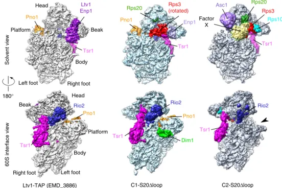
Rps20Δloop pre-40S particles are trapped in distinct states. We speculated that the delay in pre-40S maturation upon Rps20 loop deletion may also be reflected at the structural level and may trap particles in distinct, otherwise potentially short-lived, maturation stages. To determine three-dimensional (3D) structures of pre-40S particles from rps20Δloop mutant cells, purified via Tsr1 as bait, we used cryo-EM and single particle analysis. Initial pro-cessing allowed us to isolate a pool of particles showing a very stable and well-resolved body (consensus 3D structure in Sup-plementary Fig. 7a), whose features appeared similar to those of available pre-40S structures16,20. The head of the particles was
much more blurred, which led us to perform focused 3D classi-fications on this region37. This resulted in separation of pre-40S
particles into two distinct populations, hereafter called C1-S20Δloop and C2-C1-S20Δloop representing 16% and 12%, respec-tively, of the 344,959 purified particles, which were initially included in the single particle analysis scheme (Fig.5and Sup-plementary Fig. 7a). The structures of the C1-S20Δloop and
Fig. 2 A conserved loop in Rps20 promotes cytoplasmic 40S subunit maturation. a–c Crucial cytoplasmic pre-40S maturation steps are functionally interconnected. ARIO2 (rio2Δ) shuffle ltv1Δ strain (a), a RIO2 (rio2Δ) RPS3 (rps3Δ) double shuffle strain (b), and a RIO2 (rio2Δ) RPS20 (rps20Δ) double shuffle strain (c) were transformed with plasmids carrying the indicated wild-type and mutant alleles. Transformants were spotted in ten-fold serial dilutions on SDC-Leu-Trp (-leu-trp) or 5-FOA containing medium (to select for cells that have lost the respectiveURA3 plasmid(s) harboring the wild-type gene(s)) and growth was monitored after incubation at 30 °C for the indicated days.d Rps20 loop reaches through the pre-40S subunit toward Rio2 on the intersubunit side. Rps20 (green), Rps3 N-domain (red), and Rio2 (blue) in the pre-40S structure (PDB 6FAI)20(upper panel). Sequence alignment reveals
conservation of the Rps20 loop between eukaryotes and archaea (lower panel). Arrows point to residues R68, K69, E74, and K77, which were mutated in this study.e, f Rps20 loop is crucial for cell growth. AnRPS20 (rps20Δ) shuffle strain was transformed with the indicated plasmid-based alleles and transformants were spotted in ten-fold serial dilutions on SDC-Leu and SDC+5-FOA plates (e) or, after plasmid shuffling on 5-FOA, on YPD plates (f). Growth was monitored after incubation at 30 °C for the indicated days.g–i Rps20 loop mutants strongly impair 40S subunit synthesis. g Polysome profiles of the indicatedrps20 loop mutants were recorded after centrifugation on 7–45% sucrose gradients. h Northern blot analyses of total RNA extracts from wild-typeRPS20 or the indicated rps20 mutant cells using probes detecting the 20S rRNA, mature 18S rRNA, and 25S rRNA (left panel). Simplified pre-rRNA processing scheme (right panel). All detected (pre-)pre-rRNA species are indicated in bold letters. The binding site of the D/A2probe used to detect the
20S pre-rRNA and precursors thereof is indicated in magenta.i Cells expressing wild-typeRPS20 or the indicated rps20 alleles were analyzed by fluorescence in situ hybridization (FISH) using a Cy3-labeled probe specific to the D/A2 segment of internal transcribed spacer 1 (ITS1; depicted in h). The nucleoplasm was stained with DAPI. PC phase contrast. Scale bar is 5µm
C2-S20Δloop particles could be solved at an average of 3.47 and 3.79 Å, respectively (Supplementary Fig. 7b–d). In both classes, the body is very well resolved (3.0–3.2 Å; Supplementary Fig. 7d), while, as previously observed in pre-40S particle structures16,20,22, the resolution of the head domain is lower (~3.6–6 Å in C1- and C2-“head only,” reconstructed with images where the signal of the body was subtracted; ~5–7 Å in C1-S20Δloop and C2-S20Δloop structures, arising from full particles; Supplementary
Fig. 7b, d), hinting at higher structural dynamics in this region. The high resolution of these 3D reconstructions allowed us to build and refine an atomic model for both classes of particles, based on a published pre-40S structure for the C1-S20Δloop class20, and on the mature 40S subunit for the C2-S20Δloop
class29(Supplementary Table 1).
The C1-S20Δloop class displays body and platform domains globally similar to those of other structures of yeast pre-40S
RPS20 R68/K69 > A D113/E115 > K 40S 80S Polysomes 60S Polysomes 40S 60S 80S f -leu-trp D253A RPS20 Δloop R68/K69 > A R68/K69 > E R68E R68/K69 > A/E74K E74K RIO2 5-FOA d3 d c d3 LTV1 RPS20 R68/K69 > A R68/K69 > E E74K -leu-trp 5-FOA -trp d5 R68/K69 > A/E74K d3 -leu-trp S336/S339/S342 > A 5-FOA -trp d5 e Ltv1 S336 S339 S342 Intersubunit side Solvent-exposed side g R68/K69 > A 40S 60S 80S Polysomes D113/E115 > K 40S 60S 80S Polysomes -leu RPS20 D113/E115 > K Δloop R68/K69 > A R68/K69 > E E74K R68/K69 > A/E74K a b d3
-leu-trp 5-FOA -leu-trp 5-FOA RPS3 K7/K8/R9/K10 > A RPS20 R68/K69 > A R68/K69 > E E74K R68/K69 > A/E74K -leu-trp 5-FOA K7/K10 > ED d3 d5 d3 d5 d3 d5 Ltv1 Rio2 Tsr1-CBP Nob1 Tsr1-TAP RPS20 rps20 Δ loop Pno1 Dim1 Enp1 115 50 65 65 65 40 30 D113/E115 > K 5-FOA Δloop -leu-trp 5-FOA Δloop S20 D113E115 K7K10 D253 S3-N S3-C Rio2 d6 d3 d6 d5
particles (Fig.5, compare C1-S20Δloop with the cryo-EM map of
wild-type Ltv1-TAP purified particles (EMDB 388616)). The main
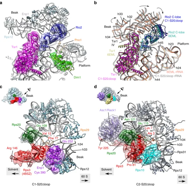
differences are within the head and at the bottom of the platform region. In the beak region, we were able to partlyfit the solvent-exposed domain of Rps20 close to its mature position (Supplementary Fig. 10a, d); however, the cryo-EM maps harbored no clear density for the two β-strands that deeply dive into the pre-40S head. This suggests that Rps20 is not yet fully accommodated and stabilized in its mature position. The overall conformation of Rio2 in C1-S20Δloop particles was similar as in other pre-40S particles16(Fig.6a, b), with the C-lobe domain of
Rio2 adopting an open conformation compared to the ATP-bound form of Archaeoglobus fulgidus Rio238, likely preventing
ATP hydrolysis by Rio2 (Supplementary Fig. 8b).
Notably, however, C1-S20Δloop pre-40S particles displayed several differences to published pre-40S structures on the solvent side of the head domain. We and others previously observed that, before its assembly into pre-40S particles, Rps3 is bound to its dedicated chaperone Yar1 in a conformation in which the C-domain is rotated relative to the N-domain (compared to 40S-bound Rps3)19,39. Strikingly, we observed a density detached
from the head in the vicinity of rRNA helix h34, in which we could unambiguously rigid-bodyfit the X-ray structure of Rps3 in this rotated conformation (Figs. 5 and 6c and Supplementary Fig. 10a). While the Rps3 C-domain protrudes from the pre-40S structure, the Rps3 N-domain is bound to this pre-40S particle but in a different orientation than in mature 40S subunits (Supplementary Fig. 10a, d). As the residues in Rps3 (K7 and K10) and Rps20 (D113 and E115) interacting with each other in 40S subunits are not visible in this structure, it is unclear whether the contact between the Rps3 N-domain and Rps20 has already formed. We could not distinguish Ltv1 in the structure, suggesting that it is very flexible. Enp1 was also more flexible than in other structures, but we could nevertheless rigid-bodyfit its central part (aa 252–390, compared to aa 205–465 resolved in wild-type pre-40S particles16) next to rRNA helix h34 (Fig.6c).
In the platform region, not only the 3′ end of mature 18S rRNA, which is protected by Pno1, but also the first two nucleotides of ITS1 are distinguishable in the C1-S20Δloop cryo-EM map (Supplementary Fig. 8c). Under the platform region on the 60S interface, C1-S20Δloop particles harbor protruding densities; focused classification of this region allowed us to unambiguously fit the alpha-helical C-terminal moiety of Dim1 (amino acids 136–318) into these densities (Figs.5 and 6a and Supplementary Fig. 7a). Dim1 is absent from most previously reported yeast and human pre-40S structures16,20,22and was only
observed in a small sub-population of particles purified from a catalytically inactive Nob1 mutant20. We speculate that C1-S20Δloop particles are trapped at a stage in which Dim1 is
in a more stable conformation. Last but not least, although Tsr1 is in a similar position as observed before, domain IV of the protein40is slightly shifted toward the solvent side, hence pushing the head rRNA closer to its mature position (Fig.6b).
The second structural class, C2-S20Δloop, resembles in many aspects mature 40S subunits (Supplementary Fig. 9). The head of C2-S20Δloop particles possesses densities clearly accommodating the full structures of Rps20, Rps3, and the presumably late assembling Rps10 and Asc1 (Figs.5and6d, and Supplementary Fig. 10b), all in their mature position, whereas neither Ltv1 nor Enp1 are visible in the structure. As Rps10 occupies part of their binding sites, they have either already dissociated completely or, alternatively, have partly detached from the particles and are therefore tooflexible to be visible.
Surprisingly, C2-S20Δloop particles harbor an additional large density protruding from the beak region in front of Rps3, with a position and a shape similar to that of the unidentified factor X in human late cytoplasmic pre-40S particles22(Fig. 5and
Supple-mentary Fig. 9a, yellow density in C2-S20Δloop panels). Our attempts to gain resolution in this region by performing focused classifications were unsuccessful, suggesting that the pre-40S association of this factor X is highly labile. Given that Hrr25 is enriched in Rps20Δloop pre-40S particles (Fig.4b), and factor X is moreover ideally placed on the head close to the binding region of Ltv1, Hrr25 is a good candidate for factor X. We thus used cryo-EM map segmentation to isolate the density from the rest of the EM map, followed by rigid-body docking of the X-ray structure of Hrr25 (PDB 5CZO)33into this density. Indeed, the
central domain of Hrr25 (aa 85–359) could be fitted, although only with a correlation coefficient of ~0.51 (Supplementary Fig. 11), preventing unambiguous assignment of Hrr25 to this density. Nevertheless, these results further support that Hrr25 is a possible candidate for this unidentified factor on our C2-S20Δloop particles.
On the intersubunit side of C2-S20Δloop particles, the upper part of rRNA helix h44 is still detached from the body as typical for pre-40S particles (Supplementary Fig. 9a, b). Moreover, the platform region is completely blurred (and thus averaged out), probably due to a wriggling movement of Rps1, Rps14, Pno1, and 18S rRNA helices h23 and h45 (right panel in Fig. 5, region indicated by an arrow, and Supplementary Fig. 9c). C2-S20Δloop particles also display fragmented densities where Rio2 and Tsr1 are located on C1-S20Δloop and previously described pre-40S particles (Fig. 5 and Supplementary Fig. 9a), suggesting that, although highlyflexible, besides the bait protein also Rio2 is still associated with this otherwise more mature class of pre-40S particles.
To sum up, our cryo-EM analyses revealed that the maturation defects arising in the absence of the Rps20 loop trap pre-40S
Fig. 3 A genetic network between Rps20 loop and 40S maturation mutants. a–c rps20 loop alleles enhance the phenotypes of mutants that impair Rps3 N-domain assembly.a AnRPS20 (rps20Δ) shuffle strain was transformed with the indicated plasmid-based rps20 alleles. Transformants were spotted in ten-fold serial dilutions on SDC-Leu and SDC+5-FOA plates and growth at 30 °C was monitored after 3 and 5 days, respectively. b An RPS20 (rps20Δ) RPS3 (rps3Δ) double shuffle strain was transformed with the indicated plasmid-based RPS20 and RPS3 wild-type and mutant alleles. Transformants were spotted in ten-fold serial dilutions on SDC-Leu-Trp and SDC+5-FOA plates and growth at 30 °C was monitored after 3 and 5 days, respectively. c Polysome profiles of cells expressing the indicated rps20 alleles, revealing an increased 40S synthesis defect when a rps20 loop mutation is combined with a rps20 allele that affects the interaction with the Rps3 N-domain (R68/K69>A D113/E115>K mutant). The panels below the profiles show growth of the respective strains on YPD plates incubated for 2 days at 30 °C.d, e Genetic interactions betweenrps20 loop alleles and mutants of pre-40S assembly factors (AFs). AnRPS20 (rps20Δ) RIO2 (rio2Δ) double shuffle strain (d) and an RPS20 (rps20Δ) shuffle ltv1Δ strain (e) were transformed with the indicated plasmid-based alleles. Transformants were spotted in ten-fold serial dilutions on SDC-Leu-Trp (-leu-trp) or 5-FOA-containing medium and growth at 30 °C was monitored at the indicated days.f Genetic network illustrating the interplay of the Rps20 loop with AFs and r-proteins at distant sites on the pre-40S ribosome. All genetic interactions discovered in this study (Figs.2and3) and in our previous study19are indicated by dashed lines.g The Rps20 loop is
required for AF release from cytoplasmic pre-40S particles. Tsr1-TAP particles were purified from wild-type RPS20 or rps20Δloop yeast strains. Eluates were analyzed by sodium dodecyl sulfate-polyacrylamide gel electrophoresis and western blotting using the indicated specific antibodies
particles in distinct structural states displaying several differences to previously reported structures.
Discussion
In this work, we unraveled the intricate mechanism leading to the release of AFs Ltv1 and Rio2 from pre-40S particles, which is
coordinated by the r-protein Rps20 (Fig. 7). On the solvent-exposed side of pre-40S particles, the Rps3 N-domain re-orients and forms its contact with Rps20. This conformational re-arrangement not only presumably weakens Ltv1’s interaction with pre-40S particles19but also positions Ltv1 in a way that it
can efficiently recruit Hrr25. Owing to competition for over-lapping binding sites, Hrr25 likely (partially) disrupts the – ATP LTV1 Tsr1-CBP Rps3 Rio2 ATP Nob1 Pno1 Dim1 – D113/E115 > K Nob1 Pno1 Dim1 115 Tsr1-CBP Rps3 Rio2 – ATP S336/S339/S342 > A – ATP RIO2 c d – ATP RPS20 ATP – Δloop – ATP Rio2 Tsr1-CBP Nob1 Pno1 Dim1 Rps3 D253A b a Time [min] Pi ATP 0 30 60 0 30 60 0 30 60 0 30 60 0 30 60
RIO2 rio2.D253A RPS20 Δloop D113/E115 > K
Relative Pi release [a.u.]
Time [min] 0 RIO2 RPS20 rps20.D113/E115 > K rps20Δloop rio2.D253A 0 20 40 60 80 100 Hrr25 30 115 115 30 Ltv1 65 Ltv1 65 Enp1 65 Enp1 50 50 65 65 40 40 40 40 Hrr25 65 65 65 Hrr25 65 30 65 Ltv1 Enp1 65 50 65 40 40 Tsr1-CBP R io 2 D113/E115 > K
RIO2D253ARPS20Δloop 58 100 kDa kDa kDa 60 30
Fig. 4 Rio2 ATP hydrolysis stimulated by the Rps20 loop triggers release of Ltv1. a–c In vitro phosphorylated Ltv1 remains trapped on pre-40S particles containing the catalytically inactive Rio2.D253A or the mutant Rps20Δloop protein. Tsr1-TAP particles from cells expressing wild-type or mutant alleles of LTV1 (left panel), RPS20 (middle panel), and RIO2 (right panel) were immobilized on IgG beads. Particles were incubated in the absence (−) or presence (ATP) of ATP, washed, and eluted with TEV protease. Eluates were analyzed by western blotting using the indicated antibodies. The displayed blots within each section (a–c) originate from the same membrane and the same exposures, allowing for direct comparison of the levels of detected proteins. d The Rps20 loop stimulates Rio2 ATPase activity. Relative ATPase activity obtained from purified Tsr1-TAP particles, carrying the indicated RIO2 or RPS20 alleles, was monitored by single-turnover experiment. The input was adjusted to equal amounts of Rio2 (right panel). An exemplary thin layer chromatography showing ATP and released phosphate (Pi) is depicted (upper left panel). Mean values from quantification of the results obtained from two biological and two technical replicates are plotted (lower panel). Error bars represent standard deviations. Source data are provided as a Source Datafile
interaction between Ltv1 and Enp1. Hrr25 then phosphorylates Ltv1 and thereby further destabilizes its association. On the intersubunit side, the Rps20 loop stimulates ATP hydrolysis by Rio2. Our data show that, while mutants lacking the Rps20 loop are viable and cold-sensitive, as are catalytic Rio2 mutants14, the combination of both is lethal. This suggests that the Rps20 loop has, apart from its requirement for ATP hydrolysis, a second role, possibly by establishing the structural context needed for final Rio2 release. Most outstandingly, all the above described maturation events on the solvent-exposed and on the inter-subunit side can take place independently of each other; how-ever, the ultimate release of both Ltv1 and Rio2 is inhibited as soon as the maturation cascade is disrupted on either side. We propose that only the correct positioning of Rps20 on both sides commits the particles to proceed to Ltv1 and Rio2 dissociation. The eventual triggers for release of Rio2 and Ltv1 are not known. However, based on this study and recent data27, we speculate
that Rps20 and/or rRNA re-arrangements are involved. The two AFs may either dissociate at the same time or one after the other. Indeed, there is evidence that Rio2 may dissociate after Ltv131,32.
Comparison of recent cryo-EM structures representing differ-ent yeast and human pre-40S maturation intermediates16,20,22
with our C1- and C2-S20Δloop pre-40S structures (Figs.5and6) can help to position our particles in a structural pre-40S maturation timeline.
Notably, some of the previously reported pre-40S structures16,22 lack densities for Rps3 and Rps20, despite bio-chemical evidence that they are already assembled at this maturation stage (see, for example, refs. 19,21,41). This suggests
highflexibility of the region Rps3 and Rps20 are bound to. Both Rps3 and Rps20 are partly visible in our C1-S20Δloop particles, indicating that they are in a maturation stage where Rps3 and Rps20 have already been partly stabilized. Nevertheless, the Rps20 β-strands are not visible and the Rps3 C-domain sticks out of the pre-40S particles. Moreover, the Rps3 N-domain is in a different
orientation than in mature 40S subunits, supporting our previous suggestion that the Rps3 N-domain is initially assembled in an incorrect orientation19.
The actual residues responsible for the interaction between the Rps3 N-domain and Rps20 are not resolved in our C1-S20Δloop structure; therefore, it remains subject to speculation whether this contact has already formed even though the Rps3 N-domain has not yet moved into itsfinal position or whether the structural re-orientation of the Rps3 N-domain and contact formation with Rps20 are coupled and have not occurred yet in this population of S20Δloop particles. Importantly, however, the properties of pre-40S particles incapable of forming the Rps3-Rps20 contact clearly differ from Rps20Δloop particles (Fig. 4), for example, in the ability to recruit Hrr25. Interestingly, the pre-40S structure from the Ban laboratory20shows a maturation stage where Rps20 and the Rps3 N-domain have already been accommodated in their mature position, while the Rps3 C-domain still sticks out of the particles, suggesting that stable integration of the Rps3 C-domain occurs even later than Rps3 N-domain accommodation. The step-wise incorporation of Rps3 is in line with previously published results showing that both Rps3 and Rps20 initially bind only loosely to pre-40S particles and their stable incorporation occurs at a later stage24,41.
Our cryo-EM structural analysis revealed no altered position-ing of Rio2 in C1-S20Δloop particles. It is possible that the absence of the Rps20 loop leads to changes inflexible regions that are not resolved in the structure. Alternatively, the transition from the open conformation of Rio2 to the closed, ATP hydro-lysis competent state, which is likely impeded in Rps20Δloop particles, might quickly trigger Rio2 ATP hydrolysis followed by its dissociation, a process that presumably is kinetically too fast to be captured by a static method like cryo-EM.
Our biochemical results strongly suggest that there is a com-munication between the Rps20 loop and Rio2. We suggest two possible scenarios how this could occur: (1) Both the Rps20 loop and Rio2 directly interact with 18S rRNA helix 3120,29; therefore, C1-S20Δloop Solvent view 60S interface view Ltv1-TAP (EMD_3886) Dim1 Rps20 Rps3 Rps10 Factor X Enp1 Pno1 Asc1 Tsr1 Rio2 Tsr1 Rio2 Rio2 Ltv1 Enp1 Rps20 Pno1 Rps3 (rotated) Pno1 Pno1 Tsr1 Tsr1 Tsr1 Tsr1 Beak Platform
Left foot Right foot
Head Body Beak Platform Left foot Right foot Head Body 180° C2-S20Δloop
Fig. 5 Cryo-electron microscopic (cryo-EM) analysis of Rps20Δloop pre-40S particles. Surface views of cryo-EM maps of Ltv1-purified wild-type pre-40S particles16(left panel) and the Rps20Δloop pre-40S particles (central panel: C1-S20Δloop particles; right panel: C2-S20Δloop particles). Assembly factors
and r-proteins of interest have been segmented and colored; the arrowhead next to the C2-S20Δloop map indicates missing density in the platform region compared to the other maps
communication between the Rps20 loop and Rio2 may occur via h31. Notably, a recent study revealed that Rio2 ATPase activity is inhibited by RNA27. Hence, re-arrangements in, or dependent on
the Rps20 loop may reposition inhibitory rRNA elements, thereby relieving the inhibition of Rio2. (2) An alternative possibility would be a direct interaction between the Rps20 loop and Rio2. Indeed, Rio2 also contains an unstructuredflexible loop, which has not been resolved in any of the so far published X-ray or cryo-EM structures. However, the last two resolved amino acids, lysine 129 and serine 145, are positioned close enough to the Rps20 loop20that such a contact would theoretically be possible. Since, however, neither the Rio2 flexible loop nor rRNA helix h31 are visible in our C1-S20Δloop structure due to flexibility, our structure does not provide further evidence for either model.
It is puzzling that, despite the massive effects of Rps20 loop deletion on pre-40S maturation, rps20Δloop cells are viable. This suggests that at least some pre-40S particles can mature into translation-competent 40S subunits. We hypothesize that the second structural class we observed, C2-S20Δloop, comprising ~12% of the particles in the purification, could represent particles that have escaped the blockade posed by the absence of the Rps20 loop. Alternatively, they might represent particles trapped in a dead end due to the inability to release the remaining AFs like Rio2 and Hrr25. In C2-S20Δloop particles, Rps3 and Rps20 are already in their mature position and Rps10 is assembled, while Ltv1 and Enp1 have either partly dissociated or completely left the particle. Moreover, Rio2 and Tsr1 seem to be only loosely attached to these particles. Interestingly, these particles contain a Enp1 Rps3 (4BSZ) Rps20 h33 Val 12 Phe 77 Rps12 Rps29 Glu 109 Glu 252 Cys 390 Arg 146 60 S Solvent Beak h34 Tsr1 Dim1 Rio2 Rps12 Pno1 Platform Beak Tsr1 C1-S20Δloop Rio2 C-lobe 6EML Rio2 C-lobe C1-S20Δloop C1-S20Δloop rRNA 6EML rRNA Tsr1 6EML h33 h32 Platform Beak h24 h23 h45 Enp1 Tsr1 IV a b h44 Rps3 Rps20 Rps10 Asc1/Rack1 h33 Rps31 Rps12 Rps29 Asn 121 Glu 115 Lys 7 Pro 81 Ala 3 Tyr 225 Beak Beak 60 S Solvent Beak h34 h34 C1-S20Δloop C2-S20Δloop d c
Fig. 6 Structural details of C1-S20Δloop and C2-S20Δloop pre-40S particles. a Close-up of the atomic model of C1-S20Δloop, as seen from the 60S interface; segmented cryo-electron microscopic (cryo-EM) densities corresponding to Tsr1, Rio2, and Dim1 are shown in magenta, blue, and lime green, respectively. rRNA is in gray, and other r-proteins in pale blue.b Same view of the C1-S20Δloop model as in a, superimposed to the model of pre-40S particles purified with Ltv1 as bait (PDB 6EML)16. Spatial alignment was realized using the Matchmaker option in Chimera, using C1-S20Δloop rRNA as
reference. For more clarity, only Tsr1, Rio2, and rRNA of both models are displayed. Color codes are indicated on the panel. Arrows indicate the rotation of the head of C1-S20Δloop particles compared to the PDB 6EML model. c, d Close-up of the heads of C1-S20Δloop (c) and C2-S20Δloop pre-40S particles (d), as seen from the top of the head/beak of the particles (top-left insets indicate direction of view). Segmented cryo-EM densities corresponding to Rps20, Rps3, Enp1, Asc1, and Rps10 are represented in green, red, purple, pale violet, and turquoise, respectively. rRNA is in gray, and other r-proteins in pale blue
factor X, which was previously also observed in a late inter-mediate in a series of human pre-40S structures representing different maturation stages22. The factor might be Hrr25, which is
enriched in Rps20Δloop pre-40S particles purified via Tsr1-TAP (Fig.4b); the presence of Hrr25 in such late particles is, however, unexpected and needs to be verified in future studies, especially since Hrr25 appears to require Ltv1 for its efficient binding to pre-40S particles (Fig.1), while it is unclear whether Ltv1 is still present in the C2-S20Δloop subpopulation.
Although cryo-EM analyses gave important insights into early cytoplasmic pre-40S maturation, it has to be mentioned that the picture obtained from these structural investigations remains incomplete. Indeed, no published structures cover the totality of all distinct particles present in the given bait purification, and often, to reach near-atomic resolutions, a majority of particles has to be omitted from 3D reconstructions because of theirflexibility or ill-resolved features. In our case, as frequently in cryo-EM studies, our high-resolution maps included ~30% of the purified pre-40S particles. Furthermore, despite the great progress in the field gained in the last few months, cryo-EM analyses have only provided snapshots describing structurally stable states of purified macromolecules. None of the so far published pre-ribosomal structures were able to fully temporally or causally resolve interdependent, multi-step maturation cascades as we have revealed here by a combination of genetic, biochemical, and structural approaches. Thus our study exemplifies that only by combining structural knowledge with in-depth functional ana-lyses it is possible to unravel individual steps of the pre-ribosomal maturation pathway at a mechanistic level.
Several hundred maturation factors participate in ribosome biogenesis, and it has to be an immense logistic challenge to coordinate their action. A consecutive order in which the point of action of each factor is precisely defined would be highly ineffi-cient, as every single factor would need to communicate with many different, remote factors. Communication across the ribo-somal subunit at certain checkpoints, as performed by Rps20 in
the Rio2/Ltv1 release pathway, is a much more sophisticated way to coordinate the action of ribosome AFs. Interestingly, additional examples lead to the proposal that communication across nascent ribosomal subunits may play a role in coordinating ribosome biogenesis (see, for example, refs.27,42,43); however, the molecular
mechanisms underlying these processes remain subject to future investigations. Moreover, it is likely that similar long-distance communication mechanisms, as described in our study, are also used to ensure proper assembly and functional regulation of other large ribonucleoprotein complexes.
Methods
Yeast strains and plasmids. Saccharomyces cerevisiae strains used in this study are listed in Supplementary Table 2 and are, unless otherwise specified, derived from W303. Strains were constructed utilizing established gene disruption, C-terminal tagging, and tetrad dissection methods. For Y2H analyses, the reporter strain PJ69-4A (Supplementary Table 2) was used. Plasmids used in this study are listed in Supplementary Table 3 and were constructed according to standard DNA cloning techniques and verified by sequencing.
Plasmid shuffle assays. Shuffle strains were constructed by knocking out an essential gene in a diploid yeast strain, transformation with a URA3 plasmid containing the respective wild-type gene, and sporulation to generate haploids harboring the gene knockout and the complementing URA3 plasmid. These shuffle strains were transformed with LEU2 or TRP1 plasmids carrying different alleles of the gene of interest and analyzed on 5-FOA-containing plates to select for cells that have lost the URA3 plasmid carrying the wild-type gene, allowing to evaluate the phenotype caused by the allele on the LEU2 or TRP1 plasmid. Double shuffle strains contained knockouts of two essential genes complemented by two URA3 plasmids with wild-type genes, and they were generated by crossing of two different shuffle strains with opposing mating types and subsequent sporulation and iden-tification of haploids containing both knockouts and both URA3 plasmids with the corresponding wild-type genes. Double shuffle strains were transformed with combinations of LEU2 and TRP1 plasmids carrying different alleles of the two genes of interest. They were also analyzed on 5-FOA-containing plates to select for cells that have lost both URA3 plasmids.
Y2H interaction analyses. Plasmids expressing the bait proteins, fused to the GAL4 DNA-binding domain, and the prey proteins, fused to the GAL4 activation domain, were co-transformed into the reporter strain PJ69-4A. Y2H interactions were documented by spotting representative transformants in ten-fold serial
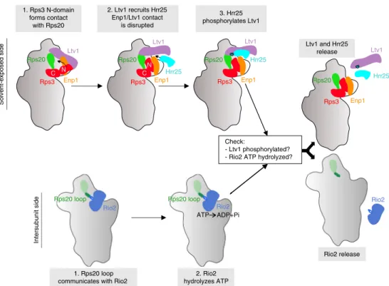
1. Rps3 N-domain forms contact with Rps20 2. Ltv1 recruits Hrr25 Enp1/Ltv1 contact is disrupted 1. Rps20 loop communicates with Rio2
2. Rio2 hydrolyzes ATP Rio2 release Ltv1 and Hrr25 release Solvent-exposed side Intersubunit side Rps3 Rps20 Enp1 Ltv1 Rps3 Rps20 Enp1 Ltv1 Hrr25 Rps3 Rps20 Enp1 Ltv1 Hrr25 C N Rps3 Rps20 Enp1 Ltv1 Hrr25 C N Rps20 loop Rio2 Rio2 Rps20 loop ATP ADP+Pi Rio2 3. Hrr25 phosphorylates Ltv1 Check: - Ltv1 phosphorylated? - Rio2 ATP hydrolyzed?
Fig. 7 Model of Rps20-mediated coordination of cytoplasmic pre-40S maturation events. Maturation events on the solvent-exposed and intersubunit side of pre-40S subunits converge into afinal maturation event leading to the release of Ltv1 and Rio2 (see “Discussion”)
dilution steps on synthetic dextrose complete (SDC)-Trp-Leu, SDC-Trp-Leu-His (HIS3 reporter), and Trp-Leu-Ade (ADE2 reporter) plates. Growth on SDC-Trp-Leu-His plates is indicative of a weak interaction, whereas only relatively strong interactions permit growth on SDC-Trp-Leu-Ade plates.
Affinity purification of pre-40S particles. Pre-40S particles were purified using the AF Tsr1 as bait protein. Cells expressing C-terminally TAP-tagged Tsr1 (Tsr1-TAP) were grown in 2 L YPD medium at 30 °C and harvested at an OD600of ~1.8.
TSR1-TAP strains expressing mutant variants of Ltv1 from LEU2 plasmids were grown in 4 L SDC medium lacking leucine and harvested at an OD600of ~0.9. Cell
pellets were resuspended in lysis buffer containing 50 mM TRIS pH 7.5, 100 mM NaCl, 1.5 mM MgCl2, 0.075% NP-40, 1 mM DTT, and 1× FY protease inhibitor
cocktail (Serva). Cells were broken in the presence of glass beads by shaking in a bead mill (B. Braun homogenizer 853022) cooled with CO2. Lysates were cleared by
centrifugation at 4 °C for 10 and 30 min at 5000 and 14,000 rpm, respectively. Lysates were incubated with IgG-beads (GE Healthcare) on a rotating wheel at 4 °C for 75 min. Beads were washed with 5 mL lysis buffer w/o protease inhibitors, subsequently transferred into Mobicol columns (Mobitec) and washed with 10 mL lysis buffer. After resuspension in lysis buffer, pre-40S particles were eluted by incubation with TEV protease at room temperature (RT) on a rotating wheel for 90 min. Eluates were analyzed by sodium dodecyl sulfate-polyacrylamide gel electrophoresis (SDS-PAGE) on 4–12% polyacrylamide gels (Invitrogen) and Coomassie staining or western blotting with the indicated antibodies.
In vitro phosphorylation assay with pre-40S particles. After IgG incubation and subsequent washing steps, bound Tsr1-TAP particles derived from different mutant strains were divided into two Mobicol columns and incubated on a rotating wheel, in the presence or absence of 1 mM ATP, for 30 min at 4 °C with buffer containing 50 mM TRIS pH 7.5, 100 mM NaCl, 5 mM MgCl2, 0.075% NP-40, and
1 mM DTT. After washing with 2 mL buffer to remove ATP, TEV-cleavage was performed as described above and eluates were analyzed by SDS-PAGE with 4–12% polyacrylamide gels (Invitrogen) and western blotting.
ATPase activity measurements on purified pre-40S particles. The corre-sponding yeast cells (1 L) were grown in YPD supplemented with 20 mg/L adenine at 30 °C to an OD600of 1.5 and harvested by centrifugation. Cell pellets were
resuspended in a buffer containing 50 mM Tris-HCl, pH 7.5, 100 mM NaCl, 10 mM MgCl2, and 5% glycerol and lysed using a Precellys cell disruptor (Bertin
instruments) in 15-ml vials containing 5 mL zirconia beads (Ø 100 µm—Roth). Lysis settings were: 6 × 30 s at 6000 rpm with 30 s pause in between; cell disruptor pre-cooled down with liquid nitrogen. Cell lysates were clarified by centrifugation (5 min at 4000 rpm, 4 °C). IgG-beads (GE Healthcare) were added to the resulting clarified supernatants and incubated for 90 min at 4 °C under mild agitation. The immobilized pre-40S particles werefirst extensively washed in batch (40 beads volumes) and then on a gravity-flow chromatography column (60 beads volumes). TEV protease cleavage was performed overnight (O/N) at 4 °C in the presence of ribonuclease inhibitor. The resulting TEV eluates were stored at 4 °C until used for ATPase assays. The levels of co-purified Rio2 present in the eluates was estimated by western blot analyses and accordingly adjusted in order to use similar amounts of Rio2 in the single turnover assay. For single turnover analysis, 5% of the Rio2-adjusted eluates werefirst incubated 10 min at 30 °C and then mixed with an equal volume of a pre-warmedγ32P-labeled ATP (Hartmann Analytic 6000 Ci/mmol)/
ATP mix (750 nCi ofγ32P-labeled ATP; ATP end concentration 200 nM). Aliquots (5%) were collected and stopped at the indicated time points by addition of 18 volumes of perchloric acid (1 M) followed by addition of 6 volumes of potassium acetate (8 M) and stored in liquid nitrogen. Reactions were centrifuged at 14,000 rpm for 10 min at RT. Aliquots (1.6%) of each time point were loaded on Polygram Cel 300 PEI (Macherey-Nagel) thin layer chromatography (TLC) plates and developed with 350 mM KH2PO4buffer for 45–60 min and dried14,30. TLC plates
were exposed to a phosphorimager screen and signals were acquired on a Typhoon FLA-9500 (GE Healthcare). Single-turnover experiment quantifications (Image J) and standard deviations were derived from at least two biological replicates (two different purifications) and two technical replicates (two single turnover experi-ments per purification). Values are expressed relative to the amounts of Piliberated
in one wild-type condition after 60 min (set to 100%).
Western blotting. Western blot analysis was performed using the following anti-bodies: anti-Rps3 antibody (1:30,000; provided by Matthias Seedorf), anti-Nob1 antibody (1:5000; provided by David Tollervey), anti-Ltv1 antibody (1:8000; provided by Katrin Karbstein), Enp1 antibody (1:4000; provided by Katrin Karbstein), anti-Tsr1 antibody (1:4000; provided by Katrin Karbstein), anti-Dim1 antibody (1:4000; provided by Katrin Karbstein), anti-Pno1 antibody (1:2000; provided by Katrin Karbstein), anti-Rio2-antibody (1:5000; provided by Katrin Karbstein), anti-Hrr25 antibody (1:5000; provided by Wolfgang Zachariae), anti-CBP antibody (1:5000; Merck-Millipore, Cat.Nr. 07-482), secondary anti-rabbit horseradish peroxidase-conjugated antibody (1:15000; Sigma-Aldrich, Cat.Nr. A7058), horseradish-peroxidase-conjugated anti-HA antibody (1:5000; Roche, Cat.Nr. 12013819001). Uncropped scans of the most important blots are provided as a Source Data File.
Polysome profile analyses. Cells expressing wild-type RPS20 or the indicated rps20 mutants were grown at 25 °C in 80 mL of YPD medium to logarithmic growth phase (OD600of ~0.6). In all, 100μg/mL cycloheximide was added to the
cultures, and after incubation for 5 min on ice, cells were pelleted and washed with lysis buffer (10 mM TRIS, pH 7.5, 100 mM NaCl, 30 mM MgCl2, 100μg/mL
cycloheximide). After resuspension in lysis buffer and cell lysis with glass beads, 6 A260units of the cell extracts were loaded onto 7–45% sucrose gradients (sucrose
dissolved in 50 mM TRIS pH 7.5, 50 mM NaCl, 10 mM MgCl2) and centrifuged at
180,000 × g for 2 h 45 min at 4 °C. Gradients were analyzed using a UA-6 system (Teledyne ISCO) by continuously monitoring at A254.
Northern blot analyses. Cells expressing wild-type RPS20 or the indicated rps20 mutants were grown in YPD medium at 25 °C to logarithmic growth phase. Cells were resuspended in lysis buffer containing 10 mM TRIS pH 7.5, 10 mM EDTA, and 0.5% SDS and lysed by vortexing in the presence of glass beads. RNA was extracted in two subsequent steps with phenol:chloroform:isoamyl alcohol (25:24:1) and once with chloroform:isoamyl alcohol (24:1). RNA was precipitated by addition of 1/10 volume of 3 M sodium acetate (pH 5.2) and 2.5 volumes of 100% ethanol and, after drying, dissolved in nuclease-free water. Three µg of the isolated RNA was denatured at 65 °C, separated on 1.5% agarose gels (containing 20 mM 3-(N-morpholino)propanesulfonic acid (MOPS), 5 mM sodium acetate, 1 mM EDTA, 0.75% formaldehyde, pH 7.0) and transferred O/N onto Hybond N+ nylon membranes (GE Healthcare) by capillary transfer. Hybridization with32P
5′-radiolabeled oligonucleotides (probe D/A2(20S): 5′-GACTCTCCATCTCTTGTCT
TCTTG-3′, probe 18S: 5′-GCATGGCTTAATCTTTGAGAC-3′, probe 25S: 5′-CT CCGCTTATTGATATGC-3′) was performed O/N at 42 °C in buffer containing 0.5 M Na2HPO4, pH 7.2, 7% SDS, and 1 mM EDTA. After three subsequent
washing steps with buffer containing 40 mM Na2HPO4, pH 7.2, 1% SDS, signals
were detected by exposing X-rayfilms.
Fluorescence in situ hybridization,fluorescence microscopy. For fluorescence in situ hybridization44, yeast cells were grown in 50 mL YPD at 25 °C to
loga-rithmic growth phase. Cells werefixed by addition of formaldehyde (4% final concentration) for 15 min at RT. Cells were collected by centrifugation, resus-pended in 5 ml of 0.1 M KPO4(pH 6.4)/4% formaldehyde, and incubated for
60 min. Subsequently, cells were washed twice with 0.1 M KPO4(pH 6.4), and once
with wash buffer (0.1 M KPO4(pH 6.4), 1.2 M sorbitol). The pellet was
resus-pended in 1 mL wash buffer containing 500 µg/mL zymolyase 100 T (amsbio) for ~30 min at 30 °C, followed by one wash with wash buffer. The spheroblasts were resuspended in ~2-fold pellet volume, applied to adhesive 10-well microscopic slides (Thermo Scientific), and incubated for 10 min. Nonadhered cells were removed by aspiration and washed with 2× SSC (0.3 M NaCl, 0.03 M Na-citrate). Approximately 0.4 pmoles/µL of a Cy3-labeled ITS1-specific probe (5′-Cy3-ATGC TCTTGCCAAAACAAAAAAATCCATTTTCAAAATTATTAAATTTCTT-3′) in 12 µL of hybridization buffer (50% formamide, 10% dextran sulfate, 4× SSC, 0.02% polyvinyl pyrrolidone, 0.02% bovine serum albumin, 0.02% Ficoll-400, 125 µg/mL tRNA, 500 µg/mL of denatured salmon sperm DNA) were applied to each well. The hybridizations were incubated O/N at 37 °C in a humid chamber and subsequently washed at RT for 10 min in 2× SSC, 10 min in 1× SSC, and 10 min in 0.5× SSC. Slides were incubated with 0.5× SSC containing 25 µg/mL DAPI for 5 min and washed two times 10 min in 0.5× SSC. Slides were mounted with Mowiol (Sigma).
Cells were examined byfluorescence microscopy on a Zeiss Axioskop microscope. Live yeast cells expressing GFP fusion proteins were imaged by fluorescence microscopy using a Zeiss Axioskop microscope.
Pre-40S particle purification for cryo-EM. Mutant rps20Δloop cells were grown in 10 L YPD medium at 30 °C to an OD600of ~1.8. Pre-40S particles were purified
via Tsr1-TAP as described above (see section:“Affinity purification of pre-40S particles”) but using a lysis buffer containing 5 mM MgCl2. After affinity
pur-ification, eluted Rps20Δloop pre-40S particles were deposited on a 10–30% sucrose gradient in lysis buffer. Eleven-mL-containing gradient tubes were subjected to ultracentrifugation on a Beckman Coulter Optima L-100 XP Ultracentrifuge, using a SW41 rotor (3 h 20 min, 38,000 rpm, 4 °C). Gradient fractions were subsequently scanned at A254and collected using a Fraction Collector system (Foxy R1, Teledyne
Isco). Fractions of interest were pooled and sucrose was removed byfive successive series of concentrating/washing with lysis buffer on Vivacon 2 centrifugal devices (Sartorius).
Grid preparation and cryo-EM image acquisition. Cryo-EM grids were prepared and systematically checked at METI, the Toulouse cryo-EM facility. In all, 3.5 µL of purified pre-40S particles (with RNA concentrations ranging from 35 to 45 ng/µL as estimated by NanoDrop measurements) were deposited onto freshly glow-discharged holey carbon grids (QUANTIFOIL®R2/1, 300 mesh with a 2-nm continuous layer of carbon on top). Grids were plunge-frozen using a Leica EM-GP automat; temperature and humidity level of the loading chamber were maintained at 20 °C and 95%, respectively. Excess of solution was blotted with a Whatmanfilter paper no.1 for 1.7–1.9 s and grids were immediately plunged into liquid ethane (−183 °C).
Cryo-EM images of Tsr1-TAP Rps20Δloop pre-40S particles were recorded on the CM01 beamline at ESRF, Grenoble. The CM01 Titan Krios electron microscope