HAL Id: hal-03184647
https://hal.archives-ouvertes.fr/hal-03184647
Submitted on 29 Mar 2021HAL is a multi-disciplinary open access
archive for the deposit and dissemination of sci-entific research documents, whether they are pub-lished or not. The documents may come from teaching and research institutions in France or abroad, or from public or private research centers.
L’archive ouverte pluridisciplinaire HAL, est destinée au dépôt et à la diffusion de documents scientifiques de niveau recherche, publiés ou non, émanant des établissements d’enseignement et de recherche français ou étrangers, des laboratoires publics ou privés.
Investigation of the [Cp*Mo(PMe 3 ) 3 H] n + ( n = 0,
1) Redox Pair: Dynamic Processes on Very Different
Time Scales
Miguel Baya, Pavel Dub, Jennifer Houghton, Jean-Claude Daran, Natalia
Belkova, Elena S. Shubina, Lina Epstein, Agusti Lledos, Rinaldo Poli
To cite this version:
Miguel Baya, Pavel Dub, Jennifer Houghton, Jean-Claude Daran, Natalia Belkova, et al.. Inves-tigation of the [Cp*Mo(PMe 3 ) 3 H] n + ( n = 0, 1) Redox Pair: Dynamic Processes on Very Different Time Scales. Inorganic Chemistry, American Chemical Society, 2009, 48 (1), pp.209-220. �10.1021/ic801676n�. �hal-03184647�
Investigation of the [Cp*Mo(PMe
3)
3H]
n+(n = 0, 1) redox pair: dynamic
processes on very different time scales
Miguel Baya,a,b† Pavel A. Dub,a,c,d Jennifer Houghton,a Jean-Claude Daran,a,d Natalia V.
Belkova,c Elena S. Shubina*,c Lina M. Epstein,c Agustí Lledós*,b Rinaldo Poli*a,d,e
a CNRS; LCC (Laboratoire de Chimie de Coordination); 205, route de Narbonne, F-31077 Toulouse,
France; Fax: (+) 33-561553003; E-mail: poli@lcc-toulouse.fr
b Departament de Química, Edifici Cn, Universitat Autónoma de Barcelona, 08193 Bellaterra, Spain
c Nesmeyanov Institute of Organoelement Compounds, Russian Academy of Sciences, Vavilov Street
28, 119991 Moscow, Russia d
Université de Toulouse; UPS, INP; F-31077 Toulouse, France e
Institut Universitaire de France, 103, bd Saint-Michel, 75005 Paris, France
Summary
Compound [Cp*Mo(PMe3)3H] (1) is reversibly oxidized at E1/2 = -1.40 V vs. ferrocene in
MeCN. Its oxidation with Cp2FePF6 yields thermally stable [Cp*Mo(PMe3)3H]PF6 (2), which
has been isolated and characterized by IR and EPR spectroscopy and by single-crystal X-ray diffraction. The 1H and 31P NMR spectra of 1 show two types of PMe3 ligands in a 1:2 ratio at
low temperature, but only one average signal at room temperature, with activation parameters of H‡ = 11.7(3) kcal mol-1 and S‡ = -3(1) e.u. for the exchange process. Although only one species is evidenced by NMR for 1 and by EPR for 2, the solution IR spectra of each complex show two bands in the (Mo-H) region (1: major at 1794 cm-1 and minor at ca. 1730 cm-1; 2: ca. 1800 and 1770 cm-1 with approximately equal intensity), the position and relative intensity
being little dependent on solvent. A thorough DFT investigation suggests that these are different rotamers involving different relative orientations of the Cp* ring and the PMe3
ligands in these complexes. This ring rotation process is very rapid on the NMR and EPR timescale but slow on the IR timescale. The X-ray data and the theoretical calculations suggest the presence of weak Mo-H∙∙∙F interactions in compound 2. The possibility of PMe3
dissociation, as well as other intramolecular rearrangements, for 1 and 2 is excluded by experimental and computational studies. Protonation of 1 yields [Cp*Mo(PMe3)3H2]+ (3)
which also reveals a dynamic process interconverting the two inequivalent H ligands and the three PMe3 ligands (two sets in a 1:2 ratio in the frozen structure) on the NMR timescale
(activation parameters of H‡ = 9.3(1) kcal/mol and S‡ = -4.1(4) e.u.). A DFT study suggests that this exchange process occurs via a low energy symmetric dihydride intermediate and not through a dihydrogen complex.
Keywords: Molybdenum, Phosphine ligands, Cyclopentadienyl ligands, Paramagnetic
Introduction
Hydride complexes generally feature a closed shell configuration. They are implicated in a variety of catalytic cycles and are also intermediates of C-H oxidative addition processes. Open-shell, paramagnetic versions, on the other hand, have not so far demonstrated broad utility, mainly because of their instability and multitude of decomposition pathways, including deprotonation,1 disproportionation,2 dihydrogen reductive elimination (for complexes
containing at least two hydride ligands),3 atom transfer, etc.4 Yet, fundamental processes such as dihydrogen evolution in Nature appear to take place via open-shell hydride complexes.5-8It is also possible to imagine potential implication in electrocatalytic hydrocarbon oxidation. Therefore, paramagnetic hydride complexes are attracting renewed interest. It has long been known that one-electron oxidation of hydride complexes increases the M-H bond acidity.1
However, this property highly depends on the medium.9, 10 For instance, hydride complexes with hydridic character may be strong acids in water.10
Many oxidized hydride complexes spontaneously decompose by transferring a proton to a base, which may be the 18-electron hydride precursor itself.4 Suppression of this reactivity has been achieved by the use of strong donating ligands possessing, at the same time, a significant steric bulk.11, 12 We have previously shown that the electron-rich hydride complex [CpMo(PMe3)3H] is reversibly oxidized to yield unstable 17-electron [CpMo(PMe3)3H]+,13
which decomposes by deprotonation and disproportionation at room temperature. We have therefore turned our attention to the bulkier and electron-richer Cp* analogue, [Cp*MoH(PMe3)3], 1.14 In this contribution, we will show that this is indeed leading to a
more stable one-electron oxidation product, [Cp*MoH(PMe3)3]+, 2. We present here the
synthesis and characterization of this material, including detailed spectroscopic studies, and a computational investigation designed to elucidate the nature of a second product that has been detected in solution for both 1 and 2.
Results and Discussion
(a) Synthesis and characterization of [Cp*Mo(PMe3)3H]+PF6-, 2.
A cyclic voltammetric study of 1 shows a reversible oxidation process (Ep = 64 mV; cf. 67 mV for ferrocene under the same conditions) at E1/2 = -1.40 V vs. ferrocene in MeCN
(-1.35 V in THF). This should be compared with the reversible oxidation of the analogous Cp compound at E1/2 = -1.46 V vs. ferrocene in MeCN. These low potentials make these
complexes stronger reductant than Cp2Co (for which E1/2 = -1.33 V vs. ferrocene in CH2Cl2),
although not as strong as Cp*2Co (E1/2 = -1.94 V).15 We are not aware of other hydride
complexes with such low E1/2 values. Note that the oxidation of 1 is slightly less facile than
that of the Cp analogue. This is an unexpected result, because replacement of Cp with the electron-richer Cp* ligand usually causes a considerable negative shift in the oxidation potential. The reason of this trend reversal is unclear and is still under investigation. In line with our expectations (see Introduction), however, the 1-electron oxidation product 2, generated by oxidation of [Cp*Mo(PMe3)3H] with 1 equiv. of Cp2FePF6 in THF (see equation
1), is thermally more stable than the Cp analogue. It is moderately stable in MeCN at room temperature, decomposing slowly over a period of 24 h and could be crystallized by slow diffusion of either diethyl ether or pentane into a THF solution at –40°C to yield suitable single crystals for an X-ray analysis. In the solid state, the compound does not significantly deteriorate upon standing in air at room temperature, in contrast to the extreme air sensitivity of its precursor 1. A view of the molecule is shown in Figure 1. Relevant bonding parameters are collected in Table 1. The coordination geometry around the Mo atom is that of a typical 4-legged piano stool. The hydride H atom was directly located from difference Fourier maps and freely refined. Although the Mo-H distance of 1.72(6) Å is not very accurately determined, it corresponds closely to the Mo-H distances determined by neutron diffraction
for other Mo hydride compounds: 1.685(3) Å for Cp2MoH2,16 1.69(5) Å for Mo(H)(2
-Me2C=NAr)[NiPr(3,5-C6H3Me2)]2,17 1.712(8), 1.719(9) and 1.719(9) Å for
(1,2,4-C5H2iBu3)Mo(PMe3)2H3,12 and 1.789(7) for Cp*Mo(CO)3H.18
[Cp*Mo(PMe3)3H] + Cp2FePF6 Cp2Fe + [Cp*Mo(PMe3)3H]+PF6- (1)
Figure 1. An ORTEP view of compound 2 (ellipsoids are drawn at the 30% probability level). All hydrogen atoms except the hydride are not shown for clarity.
One interesting structural feature is the relative location of the two ions, with the PF6
-ion facing the cat-ion on the same side as the hydride ligand. Since the oxidat-ion of hydride complexes is known to often induce decomposition by deprotonation,4 this observation suggests that the hydride ligand has become polarized + and that it establishes a Coulombic interaction with the PF6- anion. The H∙∙∙F distances are quite long (3.33, 3.56 and 3.58 Å) for
consideration as hydrogen bonds, but the PF6- ion is prevented from more closely approaching
the hydride ligand by contacts with a number of Cp* and PMe3 H atoms (see Figure S1 in the
Supporting Information; the shortest contacts being F41∙∙∙H11A, 2.80; F44∙∙∙H21D, 2.50; F46∙∙∙H51A, 2.85 Å). For other previously described cationic paramagnetic hydride complexes, for instance [Cp*W(dppe)H3]+PF6-,3 [(1,2,4-C5H2tBu3)Mo(PMe3)2H3]+PF6-,11 and
anion and the hydride ligand(s). The question of the charge on the hydride ligand in this compound has been addressed by computational methods (vide infra).
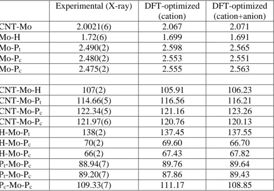
Table 1. Selected bond distances (Å) and angles (°) for compound 2 and comparison with the
DFT-optimized parameters for the cation in the gas phase.a
aThe subscripts in P
c and Pt refer to the position cisoid and transoid to the hydride ligand.
In agreement with the presence of a single unpaired electron, solutions of complex [Cp*Mo(PMe3)3H]+ in THF are EPR active. However, the isotropic spectrum is rather broad
(width = 274G at 273 K) and does not reveal any hyperfine structure, in contrast to the sharp EPR spectrum of the Cp analogue.13 Cooling the sample does not result in any significant sharpening down to the freezing point. A frozen glass spectrum exhibits an essentially cubic tensor and is much sharper (width = 21.3 G at 110K) than the isotropic spectrum, but insufficiently so to reveal any hyperfine coupling. Figures of these spectra are available as supporting information (Figure S2 and S3). Whereas the neutral precursor is unstable in CH2Cl2 solution, leading to hydride transfer to the solvent and formation of
Experimental (X-ray) DFT-optimized (cation) DFT-optimized (cation+anion) CNT-Mo 2.0021(6) 2.067 2.071 Mo-H 1.72(6) 1.699 1.691 Mo-Pt 2.490(2) 2.598 2.565 Mo-Pc 2.480(2) 2.553 2.551 Mo-Pc 2.475(2) 2.555 2.563 CNT-Mo-H 107(2) 105.91 106.23 CNT-Mo-Pt 114.66(5) 116.56 116.21 CNT-Mo-Pc 122.34(5) 121.16 123.26 CNT-Mo-Pc 121.97(6) 120.76 120.13 H-Mo-Pt 138(2) 137.45 137.55 H-Mo-Pc 70(2) 69.60 66.70 H-Mo-Pc 66(2) 67.43 67.82 Pt-Mo-Pc 88.94(7) 89.76 89.64 Pt-Mo-Pc 89.20(7) 87.86 89.43 Pc-Mo-Pc 109.33(7) 111.17 108.85
Cp*Mo(PMe3)3Cl,19 the oxidized complex is stable under the same conditions. This is in line
with the expected hydridicity reduction upon oxidation.
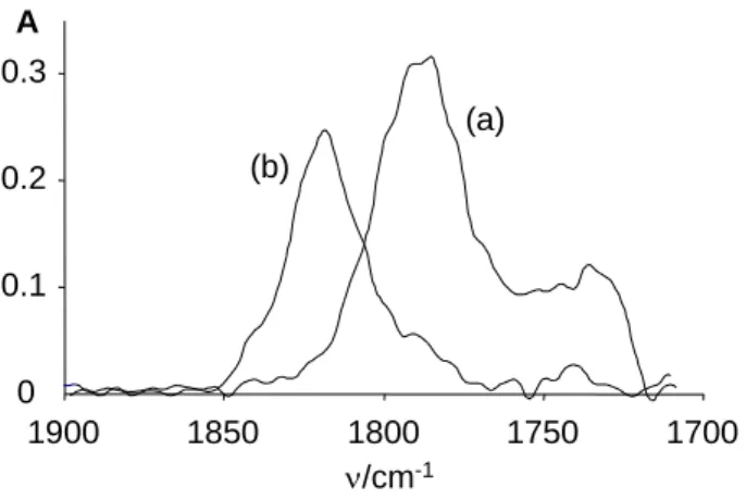
(b) Infrared characterization of complexes 1 and 2.
The ground state properties of the Mo-H bond for the neutral and cationic complexes were probed by infrared spectroscopy in solution (Figure 2). An immediately striking observation is the presence of two bands in each case, whereas only one band is expected for a monohydride complex. A major band around 1794 cm-1 and a minor one at ca. 1730 cm-1 are observed in both n-hexane and THF for 1, whereas two bands of approximately equal intensity at ca. 1800 and 1770 cm-1 are observed in both THF and CH2Cl2 for the cationic
complex 2. The observed blue shift upon oxidation parallels that reported for other redox couples, for instance [CpFeH(dippe)]0/+• (M-H = +28 cm-1) and [Cp*FeH(dippe)]0/+• (M-H
= +14 cm-1).20 The parallel magnetic resonance investigations (NMR for 1 and EPR for 2)
did not show any evidence for the presence of more than one species in solution. However, the timescale of the magnetic resonance techniques is longer than that of infrared spectroscopy, thus they may reveal average resonances of rapidly equilibrating species. The alternative possibility that the second band is due to a Fermi resonance appears discarded, at least for compound 1, by the observation that both bands disappear upon carrying out an H/D exchange at the hydride position. The exchange was quite slow in CD3OD at room
temperature, since the two bands decreased only by a factor of ca. 50% upon stirring overnight, but both decreased by essentially the same factor (Imajor/Iminor = 2.89 initially, 2.57
after overnight stirring and 47% conversion) proving that they belong to two difference hydride species in rapid equilibrium. The corresponding Mo-D stretching vibrations (expected at ca. 1270-1220 cm-1) could not be observed because of their very low relative intensity and because many other bands are present in this region of the spectrum. However, positive evidence for the formation of [Cp*Mo(PMe3)3D] was obtained by 2H-NMR, with the
observation of a quartet resonance at -5.23 (2J
DP = 7.4 Hz) in ca. 30:70 C6D6-CD3OD (cf.
-4.88 with 2JHP = 47.5 Hz in the 1H NMR under the same conditions).
Contrary to the solution infrared spectra, the solid state spectra of 1 and 2 do not clearly reveal a second component. In the spectrum of 1 (Figure 2(a)), the presence of a second low-frequency component is not completely excluded because of the large band width (the band marked with an asterisk centered at 1650 cm-1 results from decomposition, as shown by its irreproducible intensity from different samples and time evolution, due to the extreme air sensitivity of 1), but the spectrum of 2 (Figure 2(b)) quite clearly shows the presence of a single component centered at 1786 cm-1. Note that the asymmetric unit in the crystal structure of 2 contains only one molecule (one cation and one anion). For both compounds, the band observed in the solid state is closest to the higher frequency band observed in solution.
0 0.1 0.2 0.3 0.4 0.5 0.6 1650 1700 1750 1800 1850 1900 A (b) in CH2Cl2 solid 0 0.1 0.2 0.3 0.4 0.5 0.6 1650 1700 1750 1800 1850 1900 0 0.1 0.2 0.3 0.4 0.5 0.6 1650 1700 1750 1800 1850 1900 A (b) in CH2Cl2 solid 0 0.02 0.04 0.06 0.08 0.1 0.12 0.14 0.16 0.18 1550 1600 1650 1700 1750 1800 1850 1900 in THF in hexane solid A (a) * 0 0.02 0.04 0.06 0.08 0.1 0.12 0.14 0.16 0.18 1550 1600 1650 1700 1750 1800 1850 1900 in THF in hexane solid A (a) *
Figure 2. Infrared spectra of: (a) complex Cp*Mo(PMe3)3H (0.08 M, 0.4 mm pathlength) at
room temperature (thinner line: in hexane; thicker line: in THF); (b) complex [Cp*Mo(PMe3)3H]PF6 (0.042 M, 1.2 mm pathlength) in CH2Cl2 at 200 K. The band labelled
The two bands have a smaller intensity difference in 2. Under the assumption of similar extinction coefficients for the Mo-H vibration in the two species, this means that the two species are more equally populated (smaller free energy difference) in 2. The solution IR spectra recorded at different temperatures (supporting Figures S4 and S5) showed a change of the band intensity ratio, this being more evident for 1 where the Boltzmann population difference is greater. For the neutral complex, an estimation of ca. 0.2 kcal/mol as G (Gminor
-Gmajor) can be derived from the I(1800)/I(1730) ratios at the two extreme temperatures: 2.95 at
290 K and 3.5 at 200 K.
The two possibilities envisaged for such equilibrating species are (i) a phosphine ligand dissociation and (ii) a geometric isomerism. In order to probe for the ligand dissociation hypothesis, spectroscopic studies were carried out in the presence of deliberately added PMe3.
The measurement of the intensity ratio of the two IR bands (conditions as in Figure 2, with 10 equiv of PMe3) was prevented by the strong absorption of free PMe3 in the Mo-H stretching
region. Useful information, however, could be obtained by NMR and EPR spectroscopy. For the neutral complex, the 1H and 31P NMR spectra of 1/PMe3 mixtures showed the
superposition of the distinct free ligand and hydride complex resonances, demonstrating that ligand exchange between coordinated and free PMe3 is slow in the NMR timescale. Together
with the absence of the 1H and 31P NMR resonances of free PMe3 for a solution of pure 1, this
observation rules out the presence of a dissociation equilibrium. Concerning the 2/PMe3
solutions, the 1H and 31P NMR spectra showed sharp resonances at the usual diamagnetic position for the free PMe3, unaffected by the presence of the paramagnetic complex. Thus,
ligand exchange is slow on the NMR timescale also for the cationic complex. The parallel EPR investigation shows that the presence of free PMe3 does not alter the linewidth of the
remain with the option of a dynamic process interconverting two different stereoisomers. This possibility was addressed in more detail by DFT calculations (see section d below).
(c) Low-temperature NMR study of compound 1.
Compound 1 has already been described,14, 21 but its NMR investigation was limited to
room temperature, showing a single 31P resonance and a binomial quartet 1H resonance for the hydride signal. This shows that either the three PMe3 ligands occupy symmetry-equivalent
positions or that a rapid dynamic exchange process is occurring. The peculiar IR spectrum observed for compound 1 has encouraged us to carry out a new NMR investigation at variable temperatures.
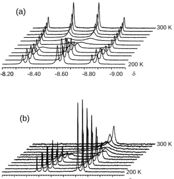
The shape of the hydride resonance in the 1H NMR spectrum changes upon cooling to ultimately yield a doublet of triplets (Figure 3), demonstrating the presence of a dynamic behaviour and two types of inequivalent PMe3 ligands (in a 2:1 ratio) in the ground state
structure. The hydride nucleus has a stronger coupling (J = 77 Hz) with the two equivalent P nuclei and a smaller one (J = 13 Hz) with the unique P nucleus. The two different types of P nuclei are coupled to each other in the low-T 31P{1H} spectrum with a J of 54 Hz. Simulations with SpinWorks22 provided an excellent fit, at each temperature, of both 1H and 31P{1H} spectra with the same mutual exchange rate constant (data in the Supporting
Information). The Eyring analysis of the temperature dependent rate constants provided the activation parameters for the mutual exchange: H‡ = 11.7(3) kcal mol-1; S‡ = -3(1) e.u. The most obvious structure of 1 that fits these data is the four-legged piano stool, identical to that observed by X-ray diffraction for the corresponding cation 2. We were not successful in growing single crystals of compound 1, but assuming that the geometry, particularly the bond angles, is not significantly different than that observed for 2, it is interesting to note that the pseudo-trans P nucleus shows the weaker coupling and the pseudo-cis P nuclei the stronger one, contrary to the typical situation for trans and cis arrangements in square planar and
octahedral complexes. Stronger coupling in four-legged piano stool geometries between pseudo-cis ligands relative to pseudo-trans ligands have also been observed in other compounds, notably CpMo(CO)2(PR3)H (R = Me, Ph, Cy)23-25 and CpOs(PiPr3)H3.26
-9.00 -8.80 -8.60 -8.40 -8.20 - - - --8. 0 16.0 17.0 18.0 19.0 20.0 21.0 22.0 23.0 24.0 25.0 (a) (b) 200 K 300 K 300 K 200 K
Figure 3. Variable temperature 1H (a) and 31P{1H} (b) NMR properties of compound Cp*Mo(PMe3)3H in toluene-d8. Spectra were taken at 10 K intervals, between 200
and 300 K.
The most interesting observation is the absence of any other hydride resonance in the low-temperature spectra. According to the low-temperature IR study (vide supra) a significant amount of the minor isomer (20%) remains present at 200 K, under the assumption that the extinction coefficient for the Mo-H vibration is the same in both species. Since no separate NMR resonance is observed for this isomer, we conclude that the dynamic process that exchanges the major and minor species observed in the IR spectrum is faster than the dynamic process that exchanges the inequivalent PMe3 ligands in the ground state structure.
Furthermore, this dynamic process must maintain the chemical inequivalence of the PMe3
The PMe3 exchange process must take place through a more symmetric, higher energy
intermediate or transition state. In the case of an intermediate, this is not significantly populated in the temperature range used in our study and is therefore unobserved by IR spectroscopy. The most likely candidate is a pseudo-trigonal bipyramidal structure, characterized by trans Cp* and H ligands in the axial positions and equivalent PMe3 ligands
in the pseudo-trigonal plane. As will be shown later in the DFT section, a higher energy local minimum is indeed observed for this geometry. Examples of pseudo-trigonal bipyramidal structures for CpML4 complexes are known,27, 28 and the interconversion mechanism between
these two geometries has been addressed.29
In conclusion, the combined IR and NMR investigation of compound [Cp*Mo(PMe3)3H] demonstrates that there must be at least three structures in solution: the
ground state structure with inequivalent PMe3 ligands in a 2:1 ratio (presumably a 4-legged
piano stool), a symmetric intermediate responsible for the mutual PMe3 exchange process
(presumably a trigonal bipyramid) that is not thermally populated, and a second asymmetric, thermally populated structure (at ca. 0.2 kcal/mol in enthalpy from the ground state) that exchanges rapidly with the ground state structure on the NMR time scale, but not on the IR time scale. The nature of these structures has been explored by the DFT calculations.
(d) DFT study of the ground state structures of 1 and 2 and exchange barriers
Geometry optimizations were run for both neutral and cationic complexes, using systems [(C5R5)Mo(PR’3)3H]n+ (n = 0, 1), with R = R’ = H (A), R = H, R’ = Me (B), and R =
R’ = Me (C, real systems). All optimized geometries, as well as tables of optimized distances and angles, Mo-H stretching frequencies, and a discussion of the structural changes as a function of the chosen model (A, B or C), are provided as supporting information.
The most stable structure was in all cases the typical 4-legged piano stool (PS), independent of the model and of the oxidation state. This agrees with our assignment of the
observed major (Mo-H) band of 1 to the PS isomer. The calculated Mo-H stretching frequencies of 1C-PS and 2C-PS (1794 and 1836 cm-1 respectively) qualitatively reproduce the experimental trend of a blue shift upon oxidation. The optimized distances and angles for
2C-PS compare quite well with those experimentally determined by X-ray diffraction (see
Table 1), attesting to the suitable level of theory used for the calculations. Comparison of the optimized structures of each redox pair shows interesting trends. The Mo-P distances are longer (by 0.06 - 0.15 Å) for the oxidized species, whereas the Mo-CNT (ring centroid) and Mo-H distances show a smaller dependence on the metal oxidation state, which is generally in the opposite direction (shorter in the higher oxidation state). These trends cannot be verified experimentally in the absence of an X-ray structure of compound 1, but were noted recently in the related pair of structurally characterized [(5-1,2,4-C5H2tBu3)Mo(PMe3)2H3]n+ (n = 0, 1)
complexes.12
Calculations were also run for the ion pair, [Cp*Mo(PMe3)3H]+PF6-, in the gas phase, in
order to probe the nature of the cation-anion interaction. The optimized geometric parameters are shown in Table 1. The geometry of the cation is relatively close to that calculated for the isolated cation. The optimization was conducted starting from the relative cation-anion orientation as found in the crystal structure, but the PF6 anion rearranges itself during the
optimization procedure to yield two shorter contacts with the hydride ion (Figure 4). These are significantly shorter than in the crystal, suggesting the presence of an attractive force between the hydride ligand and the fluorine atoms of the PF6- ion. Indeed, the Mulliken
charge calculated for the hydride ligand in 2 is slightly positive (+0.017), whereas it is negative in 1 (-0.106) and also, although to a lesser extent, in the isolated cation of 2 (-0.066). This Mulliken charge changes confirm the effect of oxidation on the metal-hydride bond polarization and underline the importance of the cation-anion interaction for this polarization. In further support of an attractive force between the hydride ligand and the PF6- ion, the
separations (2.819, 3.210 and 3.375 Å). The experimentally observed H∙∙∙F separations in 2 are much longer than the optimized values, possibly because of the effect of neighboring pairs (crystal packing), or because the computational method overestimates the strength of these interactions.
2.657 2.489
Figure 4. Gas-phase optimized geometry of [Cp*Mo(PMe3)3H]PF6. Hydrogen atoms on the
Cp* and PMe3 ligands have been omitted for clarity.
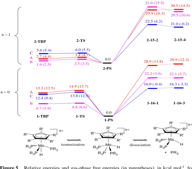
Energy minima corresponding to a pseudo-trigonal bipyramidal geometry (TBP), with the hydride and cyclopentadienyl ligands occupying the apical positions, were also located for all model systems in both oxidation states. The geometry with an apical phosphine ligand is unstable and collapses back to the lower energy minimum. We have also optimized the transition state (TS) leading from the ground state PS geometry to the TBP intermediate. The energies for all models, relative to those of the corresponding PS isomer, are visually summarized in Figure 5. Note that the relative energy for 1C-TS is consistent with the experimentally observed barrier for the mutual PMe3 exchange process (H‡ = 11.7(3) kcal
+ PR3 dissociation 13.3 (12.5) 28.9 (13.8) 6.7 (5.9) n = 0 n = 1 29.9 (16.3) 32.0 (15.5) 22.2 (3.9) 2.7 (4.4) 1.6 (2.5) 0.0 0.0 Mo PR3 R3P R3P Mo H PR3 R3P R3P H n+ R' R' R' R' R' n+ R' R' R' R' R' Mo R3P R3P H n+ R' R' R' R' R' isomerization 12.4 (9.4) 16.0 (-0.4) 5.6 (5.4) 22.5 (4.2) A B C A B C 1-TBP 2-TBP 2-PS 1-PS 2-15-2 2-15-4 1-16-1 1-16-3 29.9 (12.2) 22.1 (2.7) 16.3 (-3.3) 30.5 (14.5) 29.5 (10.6) 21.0 (-0.2) 14.9 (13.7) 13.8 (12.5) 6.8 (6.6) 3.4 (4.7) 2.5 (3.5) 6.0 (5.5) 2-TS 1-TS
Figure 5. Relative energies and gas-phase free energies (in parentheses), in kcal mol-1, for
the isomerization and phosphine dissociation process for models of Cp*Mo(PMe3)3H (below) and its one-electron oxidation product (above). The
energy values are used to represent the states on the vertical scale. For the definition of the labels (A, B, C, 1, 2, TBP, TS, 16-1, 16-3, 15-2 and 15-4, see text).
In search for a third isomer for 1 and 2, the energy of which is closer to that of the PS isomer, we have considered the following possibilities: a second minimum along the Berry pseudo-rotation coordinate (BPR), an agostic cyclopentadiene derivative (AC), a -complex with a C-H bond of a dissociated phosphine ligand (AP), and a Cp rotamer (ROT), as illustrated in Scheme 1. A different minimum in the Berry pseudo-rotation pathway might be allowed by the presence of two competitive preferred distortions of the ideal PS geometry.30,
31 The AC geometry has previously been proposed and optimized for an intermediate of the
where the dissociated PMe3 ligand must necessarily be transoid to the hydride in order to
maintain the P inequivalence in the low-temperature NMR, may be made energetically favourable by the metal electron richness. Finally, two distinct minima around the Mo-Cp* axis are justified by the symmetry mismatch of the Cp* ring (five fold) and ML4 moiety
(pseudo four fold).
Mo Me3P H PMe3 PMe3 Mo H Me3P PMe3 H Me2P Mo Me3P H Me3P PMe3 AC AP BPR ROT Mo H PMe3 Me3P PMe3 Mo H PMe3 Me3P PMe3 Scheme 1
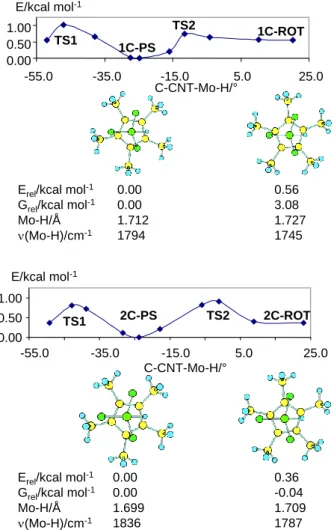
The calculations on the BPR, AC and AP hypotheses show that these cannot rationalize the experimental observation, because either no second minimum could be found or this occurred at a prohibitively high energy (details in the Supporting Information). The hypothesis of a Cp* rotamer, on the other hand, is supported by the computational results. These calculations were carried out only on the full molecule (C model). A scan of the energy as a function of the rotation angle is illustrated in Figure 6. The calculations show two minima around the C-CNT-Mo-H dihedral angle, with the second rotamer being located 0.56 kcal mol-1 higher in energy (3.08 kcal mol-1 in free energy) for 1C-ROT and 0.36 kcal mol-1 higher in energy (but 0.04 kcal mol-1 lower in free energy) for 2C-ROT. The highest energy corresponds, for both 1C and 2C, to the eclipsed geometry (C-CNT-Mo-H dihedral angle around zero). Note that the calculations reproduce, at least at a qualitative level, the lower energy difference for the two conformers in the cationic complex.
1C-PS
1C-ROT TS1
TS2
Erel/kcal mol-1
Grel/kcal mol-1
Mo-H/Å (Mo-H)/cm-1
Erel/kcal mol-1
Grel/kcal mol-1
Mo-H/Å (Mo-H)/cm-1 0.00 0.00 1.699 1836 0.36 -0.04 1.709 1787 0.00 0.00 1.712 1794 0.56 3.08 1.727 1745 0.00 0.50 1.00 -55.0 -35.0 -15.0 5.0 25.0 C-CNT-Mo-H/° E/kcal mol-1 2C-PS TS2 2C-ROT TS1 0.00 0.50 1.00 -55.0 -35.0 -15.0 5.0 25.0 C-CNT-Mo-H/° E/kcal mol-1
Figure 6. Energy of molecules 1C and 2C as a function of the C-CNT-Mo-H dihedral angle.
Views of the optimized minima emphasizing the Cp* dihedral angle (the PMe3
methyl groups have been removed for clarity) and their calculated Mo-H distances, Mo-H vibrational frequencies, and relative energies and free energies, are also shown.
The calculated Mo-H distances and vibrational frequencies are also reported in Figure 6. Notably, the calculated (Mo-H) of the global minimum of 1C-PS is at higher frequency than that of the higher energy minimum, in agreement with the experiment. The calculated frequencies (1794 and 1745 cm-1) are also in excellent agreement with the experimental values (1794 and 1730 cm-1). For system 2C-PS, the calculated frequencies of the nearly
isoenergetic rotamers (1836 and 1787 cm-1) also compare rather well with the experimentally observed ones (ca. 1800 and 1770 cm-1), being blue shifted relative to the frequencies of the neutral species.
Once likely candidates for the observed species have been found, validation of their independent observation by the IR spectroscopic technique is necessary. First of all, the calculated rotational barriers shown in Figure 6 are much too low for the individual observation of the rotamers by NMR spectrometry (for the diamagnetic compound 1). More typical barriers for processes that can be frozen on the NMR timescale are in the order of 10 kcal mol-1, as found above for the P exchange process in 1. The EPR spectroscopic technique operates on a faster timescale than NMR and fluxional processes with lower activation barriers have been investigated,33, 34 including a substituted cyclopentadienyl ring rotation in compound (C5Ph4H)Mo(CO)2(L2) (L2 = 2,3-bis(diphenylphosphino)maleic anhydride) (H‡ =
2.2 kcal mol-1).35 However, the broadness of the EPR resonance of 2 does not permit to reveal any coalescence phenomenon. Thus, the energy profiles shown in Figure 6 are entirely consistent with the observation of only one species in solution for both 1 and 2 even at low temperatures.
Concerning the IR experiments, in order to observe separate bands for exchanging species with a frequency difference of ca. 60 cm-1 (as observed in 1) or ca. 30 cm-1 (as observed in 2), the exchange rate must be slower than 1.8∙1012 s-1 or 9∙1011 s-1, respectively, corresponding to activation barriers above 0.7 or 1.1 kcal mol-1 according to the Eyring equation. The calculated free energy barriers for the Cp* ring rotation (Figure 6) are indeed higher than these values (3.3 kcal mol-1 for 1C and 1.51 kcal mol-1 for 2C). The smaller frequency difference and the smaller calculated activation barrier for the two rotamers of 2 suggests that the spectrum of this compound is closer to the coalescence region. However, warming up this sample to 350 K in dichloroethane shows no clear evidence for the beginning of a coalescence phenomenon (Figure S5). It should be recalled that the coalescence of IR bands caused by molecular dynamics, though not as familiar as the same phenomenon in NMR spectroscopy, is well established and appreciated.36-41
The separate observation of different rotamers for these two redox-related compounds is worth a brief discussion because it is unusual. This is apparently the first reported case of the simultaneous IR observation of metal-hydrogen stretching vibrations in different rotamers of a cyclopentadienyl hydride complex. We attribute this observation to the concurrence of a few favourable circumstances. The first one is the highly crowded nature of the molecule, with the PMe3 and Cp* methyl groups imposing relatively large barriers to go past each other
through the Cp* ring rotation. The second one is the relatively high frequency difference for the two rotamers, created by a sufficiently different transmission of electronic and steric effects from the Cp* conformation to the Mo-H bond in the two separate minima. In addition to the above factors, we point out that metal-hydrogen stretching vibrations typically have relatively low extinction coefficients, requiring high concentrations (thus high solubility) and long optical paths for measurement of the absorption bands at reasonably high signal/noise ratio. Thus, this phenomenon may have been missed in previously investigated complexes of this type. Other hydride complexes containing the Cp* ring that have been investigated in our laboratories, for instance Cp*M(dppe)H (M = Fe,42 Ru,43 Os44) for which high quality (high S/N) spectra were obtained, only show a single band, either because there is only a single minimum around the M-Cp* rotational coordinate, or because the different minima are related by a lower rotational barrier, or because the two structures have much closer vibrational frequencies.
Even though the experimental evidence is against the occurrence of PMe3 dissociation,
the bis-phosphine complexes resulting from the dissociation of one phosphine ligand were also optimized. These are open-shell systems and may adopt two different spin states45
(singlet and triplet for the 16-electron neutral system, doublet and quartet for the 15-electron cationic one). They will be described by codes 16-1, 16-3, 15-2 and 15-4, respectively. The relative energies for each system are also shown in Figure 5. Strictly on the basis of energy, the phosphine dissociation is less favourable than isomerization to the TBP structure for both
oxidation state levels. If we keep into account the entropic contribution, the phosphine dissociation process becomes less disfavoured and indeed becomes the preferred process in the gas phase. However, the free energy lowering caused by the entropic contribution in a condensed phase cannot be precisely estimated, because of the partial quenching of the translational and rotational degrees of freedom.
Note that, independent on the model used for the calculation and contrary to the isomerization to TBP, ligand dissociation is more favourable for the neutral system. It should be noted that the phosphine bond dissociation energy is more strongly dependent on the steric bulk than on the oxidation state: it is lowered by 10 kcal mol-1 on going from system A to system C for 1 and by 15 kcal mol-1 for 2, but only by ca. 5 kcal mol-1 on going from 1C to
2C. Therefore, phosphine dissociation becomes possible for bulkier systems. In this respect,
we point out our recent isolation and crystallographic characterization of a salt of [(1,2,4-C5H2tBu3)MoH(PMe3)2]+, though this was obtained by another route (H2 reductive
elimination from [(1,2,4-C5H2tBu3)MoH3(PMe3)2]+).11, 12 Another point of interest is the
relatively similar energy for the two possible spin configurations for the dissociation product, both for the neutral and for the cationic systems, independent on the chosen model, the difference being less than 3 kcal mol-1. Given the empirical and imprecise handling of
electron pairing by density functional methods,46-50 it is not possible to make reliable predictions as to the spin state adopted by these unsaturated species. The above mentioned cationic 15-electron system containing the bulkier (1,2,4-C5H2tBu3) ligand was found to adopt
a spin quartet ground state.11, 12
(e) Protonation of [Cp*Mo(PMe3)3H]
Protonation of compound 1 with HBF4 in THF affords the cationic dihydride complex
[Cp*Mo(PMe3)3H2]+, 3 (equation 2). An IR study shows that both MoH bands of 1 disappear,
[Cp*Mo(PMe3)3H] + HBF4 [Cp*Mo(PMe3)3H2]+BF4- (2) 0 0.1 0.2 0.3 1700 1750 1800 1850 1900 A /cm-1 (a) (b)
Figure 7. Change of the IR spectrum upon protonation in THF at 200 K. (a)
Cp*Mo(PMe3)3H. (b) Cp*Mo(PMe3)3H in the presence of 1 equiv HBF4.
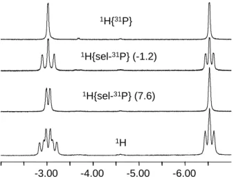
The classical nature of 3 was confirmed by the VT NMR measurements (Figure 8). At 200 K in CD2Cl2, the complex exhibits two separate 1H resonances in the hydride region with
a 1:1 integral ratio (doublet of triplets of small doublets at –3.03 and triplet of small doublets at –6.53) with T1 (200 K, 500 MHz) of 280 and 330 ms, respectively. The
phosphorus atoms in this complex are also non-equivalent at 200 K and appear in the 31P{1H} spectrum as a doublet and a triplet at 7.6 and -1.2 (JPP = 23 Hz) with 2:1 integral ratio. The
coupling pattern for the hydride signals was established by a series of 1H{sel. 31P} experiments, as shown in Figure 8. Thus, the two hydrides are coupled to each other with JHH
= 7 Hz. The hydride at -3.30 shows a stronger coupling (JHP = 65 Hz) to the two equivalent
P nuclei (31P resonance at 7.6) and a weaker one (JHP = 41 Hz) to the unique P nucleus (31P
resonance at -1.2). On the other hand, the hydride at -6.53 is significantly coupled only to the two equivalent P nuclei (JHP = 45 Hz), whereas the coupling to the unique P nucleus is not
clearly discernible in the 1H{sel. 31P: 7.6} spectrum and is estimated as ca. 10 Hz. These data
which hydride ligand. However, a comparison of the observed coupling constants of 3 with those found for 1 (where the assignment is unambiguous) and considering the similarity of the bond angles in the optimized structures of the two compounds (vide infra for compound 3), strongly suggests that the signal at -3.30 should correspond to the axial hydride ligand and that at -6.53 to the equatorial one. A similar structure was reported for the isoelectronic [CpW(CO)2(PMe3)(H)2]+[OTf]- compound51 and later proposed for the related
[CpMoH2(CO)(dppe)]+[OTf]- compound.52 It has also been observed for the isoelectronic
molybdenum complex [CpMo(PMe3)3(MeCN)H]2+ in its BF4- salt, where the
pseudo-equatorial hydride position is replaced by the neutral MeCN molecule.13
-6.00 -5.00 -4.00 -3.00 1H{sel-31P} (7.6) 1H{31P} 1H{sel-31P} (-1.2) 1H
Figure 8. Low temperature (200 K) 1H NMR spectra of compound 3 in CD2Cl2 with various
P decoupling modes.
The 1H hydride resonance and the 31P resonances broaden with the loss of observable coupling upon temperature increase, showing coalescence at ca. 250 K. Knowledge of the full coupling pattern allowed simulation (Figure 9) and an Eyring analysis of the rate constant for the exchange process (supplemental figure S8). The rate constants for the H and P exchange processes that best fit the experimental data are essentially identical at each temperature, suggesting that the same pathway simultaneously exchanges the two types of nuclei. A least-squares Eyring fit to the data yields H‡ = 9.3 ± 0.1 kcal/mol and S‡ = -4.1 ±
0.4 e.u. for the hydride exchange process and H‡ = 9.2 ± 0.1 kcal/mol and S‡ = -4.9 ± 0.5 e.u for the P exchange process.
-13 -11 -9 -7 -5 -3 -1 /ppm 200K 215K 230K 245K 260K 275K 290K 300K -13 -11 -9 -7 -5 -3 -1 /ppm 200K 215K 230K 245K 260K 275K 290K 300K (a) Experimental Simulated -7 -5 -3 -1 1 3 5 7 9 /ppm 200K 215K 230K 245K 260K 275K 290K 300K -7 -5 -3 -1 1 3 5 7 9 /ppm 200K 215K 230K 245K 260K 275K 290K 300K (b) Experimental Simulated
Figure 9. Experimental and simulated NMR spectra for compound 3 at various
temperatures. (a) 1H NMR. (b) 31P{1H} NMR.
Compound 3 has also been subjected to a computational study, with the specific aim to support the proposed ground state geometry and to identify the likely hydride and phosphine exchange pathway. These calculations were carried out for the full system, [Cp*Mo(PMe3)3H2]+. The results are shown in Figure 10. Three different local minima were
found. The global minimum (3a) corresponds indeed to the asymmetric pseudo-octahedral structure, with one pseudo axial (trans to Cp) and one pseudo equatorial (trans to P) hydride ligands, as suggested by the low-temperature NMR study. A second minimum (3b), located 3.3 kcal mol-1 higher, also features a classical dihydride structure. This geometry has Cs
symmetry, with the two hydride ligands and two PMe3 ligands being symmetry related, while
the third PMe3 ligands sits on the mirror plane. The third minimum is a dihydrogen complex
with a four-legged piano stool geometry (3c), at much higher energy (13 kcal mol-1). Four transition states (TS) interconnecting these minima could be located. As shown in Figure 10, the lowest energy TS is that leading from the global minimum to the symmetric classical isomer, TS-3a/b. This rearrangements makes the two H ligands equivalent and at the same time two previously inequivalent P atoms (one trans to the equatorial H atoms and the other one trans to the third P atom) become equivalent. Continuation of this process along the opposite and symmetry related direction exchanges the two H ligands and another pair of P ligands. Thus, this process is in full agreement with the experimental observation of the same activation parameters for the H and P exchange processes. A second exchange mechanism involving the migration of the equatorial hydride to the axial position, thus pushing the axial hydride to the opposite side of the equatorial plane (TS-3a/a), is energetically disfavored compared to the previous one. An additional exchange mechanism, whereby the two H ligands exchange their position via rotation of the H2 ligand in a nonclassical isomer, is also
not operative in this case whereas it is rather common for dihydride compounds.53, 54 Not only is the corresponding transition state (TS-3a/c) located higher in energy, this mechanism is also excluded because it would not lead to the simultaneous exchange of the P nuclei. This
observation emphasizes the strong preference of the classical dihydride structure by this complex. (0.0) (0.0) (3.3) (13.0) (16.3) (12.3) (16.2) (13.2) TS-3a/c (0.0) 3a 3a 3a 3b 3c TS-3b/c TS-3a/b TS-3a/a
Figure 10. Energies (in kcal mol-1) and geometries of optimized minima and transition states for system [Cp*Mo(PMe3)3H2]+.
Conclusions
By playing on the ligand electron donating properties and steric bulk, we have managed to produce a thermally stable paramagnetic hydride complex of MoIII, [Cp*Mo(PMe3)3H]+.
The steric encumbrance of the coordination sphere, in combination with a relatively important effect of the Cp* ring conformation on the Mo-H bond, has allowed the first experimental observation of Cp* distinct rotamers by the IR investigation of a metal-hydrogen stretching vibration. The same phenomenon occurs for the Mo-H stretching vibration of compound 2 and its neutral precursor 1. Thus, these compounds exhibit dynamic behavior on very different timescale: restricted rotation around the Mo-Cp* axis with barriers in the range of 1
mol-1 (millisecond timescale) for compound 1. The ligand mobility in compound 2 cannot be probed experimentally, because of the broadness of the EPR resonance.
Experimental section
General. All preparations and manipulations were carried out with rigorous exclusion
of oxygen and moisture using Schlenk-tube techniques. Solvents were dried by conventional methods and distilled under argon prior to use. The starting material Cp*Mo(PMe3)3H (1) was
prepared by the published method.21
Room temperature (293K) NMR investigations were carried out on a Bruker DPX300 and a Bruker AV300LiQ spectrometers operating at 300.1 MHz (1H), 121.49 MHz (31P {1H}) and 75.47 MHz (13C{1H}). Low-temperature 1H and 31P data were collected with a Bruker
AV500 spectrometer, operating at 500.3 MHz and 202.5 MHz, respectively. The temperature was calibrated using a methanol chemical shift thermometer; the accuracy and stability was ±1 K. All samples were allowed to equilibrate at every temperature for at least 3 min. The spectra were calibrated with the residual solvent resonance relative to TMS (1H, 13C), and with external 85% H3PO4 (31P). The conventional inversion-recovery-pulse method
(180-τ-90) was used to determine the variable-temperature longitudinal relaxation time T1, the
calculations being done with standard Bruker software. Coupling constants, J, are given in hertz. All NMR simulations were performed with SpinWorks software. VT-EPR spectra were measured on a Elexsys E500 BRUKER spectrometer (X-band) equipped with both a frequencemeter and gaussmeter. Cyclic voltammograms were recorded with an EG&G362 potentiostat connected to a Macintosh computer through MacLab hardware/sofware. The electrochemical cell was fitted with an Ag/AgCl reference electrode, a 1 mm-diameter Pt-disk working electrode, and a platinum-wire counter electrode. [Bu4N]PF6 (ca. 0.15 M) was used
couple. Ferrocene was added and measured as an internal standard at the end of each experiment. The solution IR measurements were performed on ‘‘Infralum 801’’ and “Specord M-82” FT-IR spectrometers. Solid state spectra were measured with a Perkin-Elmer Spectrum 100 FTIR spectrometer (2 cm-1 resolution) on the neat solid sample which was pressed inside a quarz crystal. Solution spectra were measured using CaF2 cells of 0.04–0.22
cm path length. All variable temperature IR measurements in the 190–350 K temperature range were carried out by use of a home-modified cryostat (Carl Zeiss Jena). The cryostat modification allows operating under an inert atmosphere and transferring the reagents (premixed either at low or room temperature) directly into the cell which is pre-cooled or warmed to the required temperature. The accuracy of the temperature adjustment was ±1 K.
Preparation of [Cp*Mo(PMe3)3H]PF6 (2): A suspension of [Cp2Fe]PF6 (60 mg, 0.18
mmol) in tetrahydrofuran (4 mL) was added drop by drop over a cold solution (193 K) of Cp*Mo(PMe3)3H (88 mg, 0.19 mmol) in tetrahydrofuran (4 mL). The solution color gradually
changed from green to dark blue and finally to orange in a few minutes. The reaction mixture was warmed up to 253 K and then it was concentrated to ca. 1 mL. Addition of cold diethyl ether (253 K, 8 mL) caused an orange precipitate, which was decanted, washed with cold diethyl ether (253 K, 10 and 8 mL) and vacuum-dried. An orange solid was obtained. Yield: 94 mg (81 %). Anal. Calcd. for C19H37F6MoP4: C, 37.70; H, 7.16. Found: C, 38.01; H, 6.85.
EPR (X-band, THF): g = 2.014 (Hpeak = 22 G) at 110 K; g = 2.011 (Hpeak = 180 G) at 253
K. Cyclic voltammetry (THF): E1/2 = -0.76 V (Ep = 123 mV) [ferrocene: E1/2 = +0.59 V
(Ep = 318 mV)]. Cyclic voltammetry (MeCN): E1/2 = -0.99 V (Ep = 64 mV) [ferrocene: E1/2
= +0.41 V (Ep = 67 mV)].
Preparation of [Cp*Mo(PMe3)3H2]BF4 (3): HBF4 (50% w/w in diethyl ether, 30 L,
0.20 mmol) was added to a cold solution (273 K) of Cp*Mo(PMe3)3H (89 mg, 0.19 mmol) in
diethyl ether (8 mL). Immediate formation of a whitish solid was monitored. The suspension was left to stir for 10 min and then it was decanted. The resulting solid was washed with
diethyl ether (3 x 10 mL) and dried in vacuo. A white solid was obtained. Yield: 96 mg (82 %). Anal. Calcd. for C19H44BF4MoP3: C, 41.63; H, 8.09. Found: C, 41.41; H, 7.94. 31P{1H}
NMR (CDCl3): 4.7 (s). 1H NMR (CDCl3): -4.52 (q, J = 49.0 Hz, 2 H, Mo-H), 1.55
(pseudo q, J = 2.6 Hz, 27 H, P(CH3)3), 1.96 (s, 15 H, C5(CH3)5). 1H NMR (500.3 MHz,
CD2Cl2, 200K): -6.53 (vt, 1H, 50Hz), -3.03 (vdq, 1H), 1.42 (s, 9H, PMe3), 1.47 (s, 18H,
PMe3), 1.88 (s, 15H, Cp*). 31P{1H} (203.5 MHz, CD2Cl2, 200K): -1.2 (t, 1P, J = 23 Hz), 7.6
(d, 1P, J = 23Hz). The compound slowly decomposes in CDCl3 and CD2Cl2 at 290K.
NMR and EPR tube monitoring experiments. For all NMR and EPR tube
experiments, the sample (ca. 10 mg) was charged as a solid in the glovebox under argon and the tube was sealed with a rubber septum and further wrapped with parafilm. The solvents and/or other solutions were added to the NMR tube by cannula transfer on a Schlenk line. All solvents were degassed by three freeze-pump-thaw cycles prior to use.
Computational details.
Quantum mechanical calculations were performed with the Gaussian03 package55 at the DFT/B3LYP level.56-58 Core electrons of the Mo and P atoms were described using the effective core pseudopotentials of Hay-Wadt59, 60 and valence electrons were described with the standard LANL2DZ basis set.55 In the case of the P atoms, a set of d type functions was
added.61 Carbon and hydrogen atoms non-bonded to the metal were described with a 6-31G basis set, carbon atoms linked to the metal were described with a 6-31g* basis set, and the hydride hydrogen atom was described with a 6-31g** basis set.62-71 All minima and transition states were characterized by analytically computing the Hessian matrix and no scaling factors were used for frequencies. Information on atom coordinates (xyz files) for all optimized structures is collected in the supplementary material.
X-ray Structure Determination. A single crystal of [Cp*Mo(PMe3)3H]PF6 was
mounted under inert perfluoropolyether at the tip of glass fibre and cooled in the cryostream of an Oxford-Diffraction XCALIBUR CCD diffractometer and data were collected using the
monochromatic MoK radiation (= 0.71073). The structure was solved by direct methods (SIR97)72 and refined by least-squares procedures on F2 using SHELXL-97.73 All H atoms attached to carbon were introduced in calculation in idealised positions and treated as riding models. The hydride H atom was located in difference Fourier syntheses and was freely refined with isotropic thermal parameter. The drawing of the molecule was realised with the help of ORTEP32.74 Crystal data and refinement parameters are shown in Table 2. Crystallographic data (excluding structure factors) have been deposited with the Cambridge Crystallographic Data Centre as supplementary publication no. CCDC 603848. Copies of the data can be obtained free of charge on application to the Director, CCDC, 12 Union Road, Cambridge CB2 1EZ, UK (fax: (+44) 1223-336-033; e-mail: deposit@ccdc.cam.ac.uk).
Table 2. Crystal data and structure refinement for [Cp*Mo(PMe3)3]PF6.
Empirical formula C19 H43 F6 Mo P4
Formula weight 605.35
Temperature 180(2) K
Wavelength 0.70930 Å
Crystal system Monoclinic
Space group P 21/n
Unit cell dimensions a = 9.9347(11) Å = 90.0°
b = 15.5051(15) Å = 90.827(9)° c = 17.4655(19) Å = 90.0° Volume 2690.1(5) Å3 Z 4 Density (calculated) 1.495 Mg/m3 Absorption coefficient 0.772 mm-1 F(000) 1252 Crystal size 0.25 x 0.102 x 0.071 mm3
Theta range for data collection 3.08 to 24.97°.
Index ranges -9 ≤h ≤11, -18 ≤k ≤18, -20 ≤l ≤20
Reflections collected 18043
Independent reflections 4734 [R(int) = 0.0944] Completeness to theta = 24.97° 99.8 %
Max. and min. transmission 0.9769 and 0.8497
Refinement method Full-matrix least-squares on F2
Data / restraints / parameters 4734 / 0 / 290
Goodness-of-fit on F2 0.932
Final R indices [I>2sigma(I)] R1 = 0.0508, wR2 = 0.1097
R indices (all data) R1 = 0.0779, wR2 = 0.1243
Largest diff. peak and hole 0.796 and -0.887 e.Å-3
Acknowledgement
We thank bilateral grants (PICS and GDRE “CH2D”, Russia; LEA, France-Spain) and National support from the Institut Universitaire de France and CNRS (France), the RFBR (08-03-00464) and the Division of Chemistry and Material Sciences of RAS (Russia) and the Spanish MEC (Project CTQ2005-09000-C02-01 and Consolider Ingenio 2010 CDS2007-00006). MB thanks the Spanish Ministerio de Educación y Ciencia for a post-doctoral fellowship and the Spanish MEC/Universidad de Zaragoza for funding through the “Ramón y Cajal” program.
References
1 Ryan, O. B.; Tilset, M.; Parker, V. D., J. Am. Chem. Soc. 1990, 112, 2618-2626. 2 Smith, K.-T.; Rømming, C.; Tilset, M., J. Am. Chem. Soc. 1993, 115, 8681-8689. 3 Pleune, B.; Morales, D.; Meunier-Prest, R.; Richard, P.; Collange, E.; Fettinger, J. C.;
Poli, R., J. Am. Chem. Soc. 1999, 121, 2209-2225.
4 Poli, R., Paramagnetic mono- and polyhydrides of the transition metals. In Recent Advances in Hydride Chemistry, Poli, R.; Peruzzini, M., Eds., Elsevier Science:
Amsterdam, 2001; 'Vol.' pp 139-188.
5 Foerster, S.; Stein, M.; Brecht, M.; Ogata, H.; Higuchi, Y.; Lubitz, W., J. Am. Chem. Soc. 2003, 125, 83-93.
6 Igarashi, R. Y.; Laryukhin, M.; Dos Santos, P. C.; Lee, H.-I.; Dean, D. R.; Seefeldt, L.
C.; Hoffman, B. M., J. Am. Chem. Soc. 2005, 127, 6231-6241.
7 Gloaguen, F.; Lawrence, J. D.; Rauchfuss, T. B., J. Am. Chem. Soc. 2001, 123,
9476-9477.
8 Mejia-Rodriguez, R.; Chong, D. S.; Reibenspies, J. H.; Soriaga, M. P.; Darensbourg,
M. Y., J. Am. Chem. Soc. 2004, 126, 12004-12014.
9 Kristjánsdóttir, S. S.; Norton, J. R., Acidity of Hydrido Transition Metal Complexes in Solution. In Transition Metal Hydrides, Dedieu, A., Eds., VCH: New York, 1992;
'Vol.' pp 309-359.
10 Jacobsen, H.; Berke, H., Hydridicity of Transition Metal Hydrides and its Implications for Reactivity. In Recent Advances in Hydride Chemistry, Poli, R.; Peruzzini, M., Eds.,
11 Baya, M.; Houghton, J.; Daran, J.-C.; Poli, R., Angew. Chem., Int. Ed. Engl. 2007, 46,
429-432.
12 Baya, M.; Houghton, J.; Daran, J.-C.; Poli, R.; Male, L.; Albinati, A.; Guttman, M., Chem. Eur. J. 2007, 13, 5347-5359.
13 Fettinger, J. C.; Kraatz, H.-B.; Poli, R.; Quadrelli, E. A.; Torralba, R. C., Organometallics 1998, 17, 5767-5775.
14 Abugideiri, F.; Kelland, M. A.; Poli, R.; Rheingold, A. L., Organometallics 1992, 11,
1303-1311.
15 Connelly, N. G.; Geiger, W. E., Chem. Rev. 1996, 96, 877-910.
16 Schultz, A. J.; Stearley, K. L.; Williams, J. M.; Mink, R.; Stucky, G. D., Inorg. Chem.
1977, 16, 3303-6.
17 Tsai, Y.-C.; Johnson, M. J. A.; Mindiola, D. J.; Cummins, C. C., J. Am. Chem. Soc.
1999, 121, 10426-10427.
18 Brammer, L.; Zhao, D.; Bullock, R. M.; Mcmullan, R. K., Inorg. Chem. 1993, 32,
4819-4824.
19 Abugideiri, F.; Fettinger, J. C.; Keogh, D. W.; Poli, R., Organometallics 1996, 15,
4407-4416.
20 Jiménez-Tenorio, M.; Puerta, M. C.; Valerga, P., Organometallics 1994, 13,
3330-3337.
21 Shin, J. H.; Parkin, G., Polyhedron 1994, 13, 1489-1493. 22 Marat, K., SpinWorks Version 2.5.4. ed.; 2006.
23 Kalck, P.; Poilblanc, R., J. Organometal. Chem. 1969, 19, 115-121. 24 Faller, J. W.; Anderson, A. S., J. Am. Chem. Soc. 1970, 92, 5852-5860.
25 Cheng, T. Y.; Brunschwig, B. S.; Bullock, R. M., J. Am. Chem. Soc. 1998, 120,
13121-13137.
26 Baya, M.; Crochet, P.; Esteruelas, M. A.; Gutiérrez-Puebla, E.; Ruiz, N., Organometallics 1999, 18, 5034-5043.
27 Du Plooy, K. E.; Moll, U.; Wocadlo, S.; Massa, W.; Okuda, J., Organometallics 1995,
14, 3129-3131.
28 Liu, A. H.; Murray, R. C.; Dewan, J. C.; Santarsiero, B. D.; Schrock, R. R., J. Am. Chem. Soc. 1987, 109, 4282-4291.
29 Smith, J. M.; Coville, N. J., Organometallics 1996, 15, 3388-3392. 30 Poli, R., Organometallics 1990, 9, 1892-1900.
31 Lin, Z.; Hall, M. B., Organometallics 1993, 12, 19-23.
32 Baya, M.; Maresca, O.; Poli, R.; Coppel, Y.; Maseras, F.; Lledós, A.; Belkova, N. V.;
Dub, P. A.; Epstein, L. M.; Shubina, E. S., Inorg. Chem. 2006, 45, 10248-10262.
33 Ittel, S. D.; Krusic, P. J.; Meakin, P., J. Am. Chem. Soc. 1978, 100, 3264-3266. 34 Krusic, P. J., J. Am. Chem. Soc. 1981, 103, 2131-2133.
35 Mao, F.; Sur, S. K.; Tyler, D. R., J. Am. Chem. Soc. 1989, 111, 7627-7628. 36 Macphail, R. A.; Strauss, H. L., J. Chem. Phys. 1985, 82, 1156-1166. 37 Wood, K. A.; Strauss, H. L., J. Phys. Chem. 1990, 94, 5677-5684.
38 Turner, J. J.; Gordon, C. M.; Howdle, S. M., J. Phys. Chem. 1995, 99, 17532-17538. 39 Grevels, F. W.; Kerpen, K.; Klotzbucher, W. E.; Mcclung, R. E. D.; Russell, G.;
Viotte, M.; Schaffner, K., J. Am. Chem. Soc. 1998, 120, 10423-10433.
40 Londergan, C. H.; Kubiak, C. P., Chem.--Eur. J. 2003, 9, 5962-5969.
41 Cahoon, J. F.; Sawyer, K. R.; Schlegel, J. P.; Harris, C. B., Science 2008, 319,
1820-1823.
42 Belkova, N. V.; Revin, P. O.; Epstein, L. M.; Vorontsov, E. V.; Bakhmutov, V. I.;
Shubina, E. S.; Collange, E.; Poli, R., J. Am. Chem. Soc. 2003, 125, 11106-11115.
43 Belkova, N. V.; Dub, P. A.; Baya, M.; Houghton, J., Inorg. Chim. Acta 2007, 360,
44 Dub, P. A.; Belkova, N. V.; Lyssenko, K. A.; Silantyev, G. A.; Epstein, L. M.;
Shubina, E. S.; Daran, J.-C.; Poli, R., Organometallics in press.
45 Poli, R., J. Organometal. Chem 2004, 689, 4291–4304.
46 Salomon, O.; Reiher, M.; Hess, B. A., J. Chem. Phys. 2002, 117, 4729-4737. 47 Harvey, J.; Aschi, M., Faraday Disc. 2003, 124, 129-143.
48 Poli, R.; Harvey, J. N., Chem. Soc. Rev. 2003, 32, 1-8.
49 Wasbotten, I. H.; Ghosh, A., Inorg. Chem. 2007, 46, 7890-7898. 50 Conradie, J.; Ghosh, A., J. Chem. Theory Comput. 2007, 3, 689-702.
51 Bullock, R. M.; Song, J.-S.; Szalda, D. J., Organometallics 1996, 15, 2504-2516. 52 Cheng, T.-Y.; Szalda, D. J.; Zhang, J.; Bullock, R. M., Inorg. Chem. 2006, 45,
4712-4720.
53 Gusev, D. G.; Berke, H., Chem. Ber. 1996, 129, 1143-1155. 54 Kubas, G. J., J. Organomet. Chem. 2001, 635, 37-68.
55 Frisch, M. J., et al., Gaussian 03, Revision C.02. ed.; Gaussian, Inc.: Wallingford CT,
2004.
56 Becke, A. D., J. Chem. Phys. 1993, 98, 5648-5652.
57 Perdew, J. P., Electronic Structure of Solids. In Ziesche, P.; Eschrig, H., Eds.,
Akademie Verlag: Berlin, 1991; 'Vol.' pp 11.
58 Perdew, J. P.; Wang, Y., Phys. Rev. B 1992, 45, 13244-13249. 59 Wadt, W. R.; Hay, P. J., J. Chem. Phys. 1985, 82, 284-298. 60 Hay, P. J.; Wadt, W. R., J. Chem. Phys. 1985, 82, 299-310.
61 Höllwarth, A.; Bohme, M.; Dapprich, S.; Ehlers, A.; Gobbi, A.; Jonas, V.; Kohler, K.;
Stegmann, R.; Veldkamp, A.; Frenking, G., Chem. Phys. Lett. 1993, 208, 237-40.
62 Ditchfield, R.; Hehre, W. J.; Pople, J. A., J. Chem. Phys. 1971, 54, 724-8. 63 Hehre, W.; Ditchfie, R.; Pople, J., J. Chem. Phys. 1972, 56, 2257-2261. 64 Hariharan, P. C.; Pople, J. A., Mol. Phys. 1974, 27, 209-14.
65 Gordon, M. S., Chem. Phys. Lett. 1980, 76, 163-8.
66 Hariharanan, P.; Pople, J. A., Theor. Chim. Acta 1973, 28, 213-222.
67 Blaudeau, J. P.; Mcgrath, M. P.; Curtiss, L. A.; Radom, L., J. Chem. Phys. 1997, 107,
5016-5021.
68 Francl, M. M.; Pietro, W. J.; Hehre, W. J.; Binkley, J. S.; Gordon, M. S.; Defrees, D.
J.; Pople, J. A., J. Chem. Phys. 1982, 77, 3654-3665.
69 Binning, R. C.; Curtiss, L. A., J. Comput. Chem. 1990, 11, 1206-1216.
70 Rassolov, V. A.; Pople, J. A.; Ratner, M. A.; Windus, T. L., J. Chem. Phys. 1998, 109,
1223-1229.
71 Rassolov, V. A.; Ratner, M. A.; Pople, J. A.; Redfern, P. C.; Curtiss, L. A., J. Comput. Chem. 2001, 22, 976-984.
72 Altomare, A.; Burla, M.; Camalli, M.; Cascarano, G.; Giacovazzo, C.; Guagliardi, A.;
Moliterni, A.; Polidori, G.; Spagna, R., J. Appl. Cryst. 1999, 32, 115-119.
73 Sheldrick, G. M., SHELXL97. Program for Crystal Structure refinement. ed.;
University of Göttingen: Göttingen, Germany, 1997.
TOC Graphic
TOC text
The pair of redox-related complexes [Cp*Mo(PMe3)3H]n+ (n = 0, 1) show exchange of the
phosphine positions (in the ms timescale for n = 0, frozen at low temperature by NMR; not resolved for n = 1) and Cp* ligand rotation (in the ps timescale, frozen in the solution IR spectrum for both complexes).
![Figure 2. Infrared spectra of: (a) complex Cp*Mo(PMe 3 ) 3 H (0.08 M, 0.4 mm pathlength) at room temperature (thinner line: in hexane; thicker line: in THF); (b) complex [Cp*Mo(PMe 3 ) 3 H]PF 6 (0.042 M, 1.2 mm pathlength) in CH 2 Cl 2 at 2](https://thumb-eu.123doks.com/thumbv2/123doknet/13667613.430235/9.892.105.443.581.1035/figure-infrared-spectra-complex-pathlength-temperature-thinner-pathlength.webp)

![Figure 10. Energies (in kcal mol -1 ) and geometries of optimized minima and transition states for system [Cp*Mo(PMe 3 ) 3 H 2 ] +](https://thumb-eu.123doks.com/thumbv2/123doknet/13667613.430235/26.892.104.442.201.603/figure-energies-kcal-geometries-optimized-minima-transition-states.webp)
