HAL Id: hal-00724253
https://hal.archives-ouvertes.fr/hal-00724253
Submitted on 28 May 2021
HAL is a multi-disciplinary open access
archive for the deposit and dissemination of
sci-entific research documents, whether they are
pub-lished or not. The documents may come from
teaching and research institutions in France or
abroad, or from public or private research centers.
L’archive ouverte pluridisciplinaire HAL, est
destinée au dépôt et à la diffusion de documents
scientifiques de niveau recherche, publiés ou non,
émanant des établissements d’enseignement et de
recherche français ou étrangers, des laboratoires
publics ou privés.
Distributed under a Creative Commons Attribution - NonCommercial - ShareAlike| 4.0
Osh4p exchanges sterols for phosphatidylinositol
4-phosphate between lipid bilayers.
Maud de Saint-Jean, Vanessa Delfosse, Dominique Douguet, Gaëtan Chicanne,
Bernard Payrastre, William Bourguet, Bruno Antonny, Guillaume Drin
To cite this version:
Maud de Saint-Jean, Vanessa Delfosse, Dominique Douguet, Gaëtan Chicanne, Bernard Payrastre,
et al.. Osh4p exchanges sterols for phosphatidylinositol 4-phosphate between lipid bilayers.. Journal
of Cell Biology, Rockefeller University Press, 2011, 195 (6), pp.965-978. �10.1083/jcb.201104062�.
�hal-00724253�
JCB:
Article
The Rockefeller University Press $30.00
Maud de Saint-Jean and Vanessa Delfosse contributed equally to this paper. Correspondence to Guillaume Drin: [email protected]; or Bruno Antonny: [email protected]
Abbreviations used in this paper: ALPS, amphipathic lipid packing sensor; DHE, dehydroergosterol; DNS, dansyl; DNS-PE, DNS-phosphatidylethanolamine; DOPC, dioleoyl phosphatidylcholine; DOPS, dioleoyl phosphatidylserine; FRET, fluorescence resonance energy transfer; NBD, 7-nitrobenz-2-oxa-1,3-diazol; Orp, oxysterol-binding protein-related protein; Osh, oxysterol-binding homology protein; PC, phosphatidylcholine; PI, phosphatidylinositol; PI(4)P, PI 4-phosphate; PI(4,5)P2, PI 4,5-bisphosphate; PM, plasma membrane; POPC,
1-palmitoyl-2-oleoyl-PC; POPS, 1-palmitoyl-2-oleoyl-phosphatidylserine; Rhod-PE, rhodamine-phosphatidylethanolamine; rmsd, root mean square deviation.
Introduction
In eukaryotic cells, sterols reside in all compartments but are
unevenly distributed. Cholesterol (ergosterol in yeast) is
syn-thesized in the ER, but is rare in this compartment and more
concentrated at the trans Golgi, endosomes, and the plasma
membrane (PM). Sterols are transferred between organelles
either by transport vesicles or by soluble carriers (Maxfield and
Tabas, 2005; Prinz, 2007; Mesmin and Maxfield, 2009). In
yeast, ergosterol moves from the ER to the PM mostly via
protein-mediated nonvesicular routes (Baumann et al., 2005). Little is
known about the identity of the carriers, but oxysterol-binding
homology protein (Osh) are plausible candidates (Prinz, 2007;
Fairn and McMaster, 2008; Lev, 2010). Suppression of the
entire Osh family is lethal, depriving the PM in ergosterol and
stopping endocytosis. Conversely, expressing any of the seven
Osh restores cell viability (Beh and Rine, 2004).
Osh4p, also known as Kes1p, is the simplest and best
characterized Osh. Its structure consists of a -barrel
form-ing a sterol-bindform-ing pocket (Im et al., 2005). When Osh4p is
empty, the N-terminal segment (29 amino-acids) is unfolded
and leaves the pocket open. Upon sterol binding, this segment
forms a lid that blocks the sterol molecule in the pocket. Thus,
Osh4p seems adapted to transport sterols (Levine, 2005) and,
in accord with this, was found to move sterol between artificial
membranes (Raychaudhuri et al., 2006). Because all Osh
pro-teins and the human oxysterol-binding protein-related protein
(Orp; Ridgway, 2010) contain a sterol-binding domain akin
to Osh4p, better understanding the sterol transport activity of
Osh4p should have general implications.
O
sh/Orp proteins transport sterols between
or-ganelles and are involved in phosphoinositide
metabolism. The link between these two aspects
remains elusive. Using novel assays, we address the
influ-ence of membrane composition on the ability of Osh4p/
Kes1p to extract, deliver, or transport dehydroergosterol
(DHE). Surprisingly, phosphatidylinositol 4-phosphate
(PI(4)P) specifically inhibited DHE extraction because PI(4)P
was itself efficiently extracted by Osh4p. We solve the
structure of the Osh4p–PI(4)P complex and reveal how
Osh4p selectively substitutes PI(4)P for sterol. Last, we
show that Osh4p quickly exchanges DHE for PI(4)P and,
thereby, can transport these two lipids between
mem-branes along opposite routes. These results suggest a model
in which Osh4p transports sterol from the ER to late
com-partments pinpointed by PI(4)P and, in turn, transports
PI(4)P backward. Coupled to PI(4)P metabolism, this
trans-port cycle would create sterol gradients. Because the residues
that recognize PI(4)P are conserved in Osh4p homologues,
other Osh/Orp are potential sterol/phosphoinositol
phos-phate exchangers.
Osh4p exchanges sterols for phosphatidylinositol
4-phosphate between lipid bilayers
Maud de Saint-Jean,
1Vanessa Delfosse,
2Dominique Douguet,
1Gaëtan Chicanne,
3Bernard Payrastre,
3William Bourguet,
2Bruno Antonny,
1and Guillaume Drin
11Institut de Pharmacologie Moléculaire et Cellulaire, Université de Nice Sophia-Antipolis and Centre National de la Recherche Scientifique, 06560 Valbonne, France 2Centre de Biochimie Structurale, Centre National de la Recherche Scientifique, UMR5048 and Institut National de la Santé et de la Recherche Médicale U1054,
34090 Montpellier, France
3Institut National de la Santé et de la Recherche Médicale U1048, Université Toulouse 3, I2MC, and Centre Hospitalier Universitaire de Toulouse, 31432 Toulouse
Cedex 04, France
© 2011 de Saint-Jean et al. This article is distributed under the terms of an Attribution– Noncommercial–Share Alike–No Mirror Sites license for the first six months after the pub-lication date (see http://www.rupress.org/terms). After six months it is available under a Creative Commons License (Attribution–Noncommercial–Share Alike 3.0 Unported license, as described at http://creativecommons.org/licenses/by-nc-sa/3.0/).
THE
JOURNAL
OF
CELL
BIOLOGY
How Osh4p binds PI(4)P is not well understood. Osh4p does
not have any known phosphoinositol phosphate–binding domain,
and in vitro binding assays failed to prove that Osh4p could
distinguish a PI(4)P- from a PI(4,5)P
2-containing membrane
(Li et al., 2002; Fairn and McMaster, 2005; Schulz et al., 2009).
Moreover, no link between PI(4)P and sterol transport by Osh4p
has been found. Here, using a combination of assays for lipid
exchange and x-ray crystallography, we solve this issue by
showing that Osh4p acts as a sterol/PI(4)P exchanger.
Results
Osh4p quickly extracts and solubilizes the fluorescent ergosterol dehydroergosterol (DHE) from neutral liposomes
So far, the ability of Osh4p to extract, deliver, or exchange
sterols has been mostly tested using radiolabeled sterols
(Raychaudhuri et al., 2006; Schulz et al., 2009). However, the
separation procedures required for determining the sterol
dis-tribution between the donor liposomes, the acceptor liposomes,
and the protein do not permit precise kinetics measurements.
To circumvent these limitations, we took advantage of the
fluorescence properties of DHE, a natural ergosterol analogue.
We observed that the intrinsic fluorescence of Osh4p quickly
diminished once added to large liposomes made of dioleoyl
phosphatidylcholine (DOPC) doped with 0.5% DHE (Fig. 1 A).
Spectral analysis revealed that tryptophan quenching at 340 nm
was concomitant with the apparition of three emission peaks at
To properly allocate sterol in the cell, Osh/Orp must
en-sure a one-way transport of sterol between defined
compart-ments. This seems the simplest way to enrich one membrane
with sterol at the expense of another. Such transport must
in-volve transient and sequential interactions between the protein,
the sterol molecule, and two lipid bilayers, one acting as a donor
membrane and one as an acceptor (Lev, 2010). Osh4p adopts
two different structures depending on its state (empty or
sterol-bound), and this is likely critical for proper protein targeting to
donor and acceptor membranes during a cycle of transport. In
line with this hypothesis, Osh4p has been proposed to transport
sterol from the PM to the ER (Raychaudhuri et al., 2006). A key
argument is that sterol uptake in vitro is facilitated by
phospha-tidylinositol (PI) 4,5 bisphosphate (PI(4,5)P
2), a PM lipid.
However, control of Osh4p by PI(4,5)P
2is hard to
recon-cile with other observations. First, the level of ergosterol at the
PM increases upon Osh4p expression (Beh et al., 2001).
Sec-ond, genetic and cellular studies suggest that Osh4p has a specific
function at the Golgi, and that this function depends on another
phosphoinositide, PI 4-phosphate (PI(4)P), present at the trans
side. Osh4p is the only Osh whose expression is lethal in
Sec14p-deficient yeast (Fang et al., 1996; Li et al., 2002). Sec14p
is a key protein, which maintains a lipid composition
permis-sive for vesicular transport (Bankaitis et al., 2005). In the
ab-sence of Sec14p, the amount of PI(4)P is limiting. Osh4p, whose
endogenous expression is high, monopolizes all remaining
PI(4)P molecules at the expense of essential proteins of
vesicu-lar transport (Fairn et al., 2007; LeBlanc and McMaster, 2010).
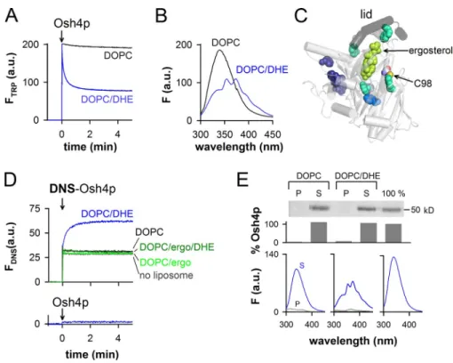
Figure 1. Real-time measurement of DHE load-ing and extraction by Osh4p from large DOPC liposomes. (A) Intrinsic fluorescence at 340 nm of Osh4p (0.5 µM) upon addition to DOPC/ DHE (99.5:0.5 mol/mol) liposomes (blue trace) or to DOPC liposomes (black trace). The final lipid concentration is 0.5 mM. (B) Fluorescence
emission spectra (ex = 285 nm) of the samples
shown in A at the end of the kinetics. (C) Close-up view of the Osh4p structure (PDB accession no. 1ZHZ) showing the lid (segment 1–29, black) covering ergosterol (bright green). Trypto-phans located at ≤15 Å, between 15 and 20 Å, or at ≥20 Å from ergosterol are colored in cyan, blue, and purple, respectively. Cystein C98 is indicated. (D) Fluorescence at 510 nm
(ex = 310 nm) of 0.5 µM DNS-Osh4p added
to buffer alone (gray trace) or to DOPC/DHE (99.5:0.5 mol/mol, blue trace), DOPC (black trace), DOPC/ergosterol (95:5, light green trace), or DOPC/ergosterol/DHE (94.5:5:0.5, dark green trace) liposomes. Bottom trace, unlabeled Osh4p added to DOPC/DHE lipo-somes. (E) 0.5 µM Osh4p was incubated with DOPC or DOPC/DHE (99.5:0.5) liposomes (0.5 mM lipids) filled with sucrose. A reference experiment (100%) was done without lipo-somes. After centrifugation, the percentage of Osh4p in the pellet (P) and supernatant (S) was determined by SDS-PAGE. The fluorescence
spectrum of each sample was recorded (ex =
285 nm). Control spectra corresponding to buffer or liposome alone (0.5 mM lipids) were subtracted from the spectra of the supernatant and pellet, respectively. Data in E are from a single representative experiment out of two in-dependent experiments.
caused much less retention of Osh4p on the liposomes (8–10%).
Surprisingly, the fluorescence spectra of the supernatants
re-vealed a major difference: with PI(4)P, Osh4p failed to fully
extract DHE (I
372/I
340= 1.08). In contrast, DHE loading in the
presence of PI(4,5)P
2was complete and similar to that observed
on pure DOPC liposomes. This suggested that PI(4)P
specifi-cally inhibits DHE extraction.
The fact that PI(4)P had a specific inhibitory effect on
Osh4p was striking in real-time measurements. As mentioned
previously (Fig. 1 A), we recorded the rate of DHE extraction
by Osh4p by measuring the quenching of tryptophan at 340 nm.
In addition, we measured Osh4p binding to the liposomes through
a second and independent fluorescence signal at 525 nm. To
this end, we labeled the ALPS/lid motif with
7-nitrobenz-2-oxa-1,3-diazol (NBD), a probe whose fluorescence increases
upon insertion between lipids. Control experiments indicated
that this probe was well positioned to measure liposome
bind-ing of Osh4p (
Fig. S2
). As shown in Fig. 2 B, PI(4)P at 2 mol%
354, 373, and 393 nm (Fig. 1 B). These peaks are characteristic
of DHE, which suggests fluorescence resonance energy transfer
(FRET) from Osh4p to DHE (Schroeder et al., 1990; Holt et al.,
2008; Liu et al., 2009). Osh4p contains several tryptophans
close to the ergosterol-binding pocket, which should be
respon-sible for the observed FRET signal (Fig. 1 C).
Next, we created a more defined FRET pair by labeling
an endogenous cysteine (C98) with a dansyl (DNS) probe.
C98 is exposed on the external side of Osh4p, at 12.8 Å from
the center of the sterol-binding pocket. Once DNS-Osh4p was
added to DHE-containing liposomes, the fluorescence of DNS
at 510 nm upon DHE excitation increased with kinetics similar
to tryptophan quenching by DHE (1/t
1/2of 0.13 and 0.18 s
1,
respectively; Fig. 1, A and D). No FRET was observed when
Osh4p or DNS-Osh4p was mixed with liposomes lacking DHE
or combining DHE and an excess of nonfluorescent ergosterol
(Fig. 1, A and D). Therefore, the two FRET signals presented in
Fig. 1 monitor the loading of DHE by Osh4p.
The experiments shown in Fig. 1 (A and D) did not allow
us to determine whether Osh4p remained bound to the
lipo-somes or returned in solution after DHE extraction. To
dis-tinguish between these two possibilities, we incubated Osh4p
with sucrose-loaded DOPC liposomes containing DHE. After
15 min, the liposomes were collected by centrifugation. Gel
and spectral analysis indicated that the protein was mostly
re-covered in the supernatant and quenched by DHE (Fig. 1 E).
No spectral change was observed with DHE-free liposomes
(Fig. 1 E) or when a “lidless” form of Osh4p lacking the
am-phipathic lipid packing sensor (ALPS)/lid motif and deficient
in sterol binding was used (
Fig. S1
; Im et al., 2005; Raychaudhuri
et al., 2006). We concluded that Osh4p interacts transiently
with neutral liposomes, traps sterol, and goes back in solution
in a sterol-loaded form stabilized by the lid. “Extraction” will
be used to indicate DHE loading regardless of the retention of
Osh4p on membranes, whereas “solubilization” will be used
to indicate that Osh4p not only extracts DHE but also
dissoci-ates from membranes.
Effect of anionic lipids on DHE solubilization by Osh4p
It has been reported that PI(4,5)P
2specifically stimulates sterol
extraction by Osh4p (Raychaudhuri et al., 2006). We used our
assay combining sedimentation and fluorescence analysis to
examine the effect of various anionic lipids on the ability of Osh4p
to extract and solubilize DHE (Fig. 2 A). To have an index of
DHE binding by Osh4p, we calculated for both the liposome
pellet and the supernatant an intensity ratio I
372/I
340, which
re-flects the extent of FRET between Osh4p and DHE in these
fractions (from 0.5 for DHE-free Osh4p to 1.9 for Osh4p
satu-rated with DHE).
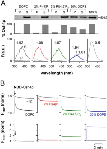
Anionic lipids had contrasting effects on the extraction
and solubilization of DHE by Osh4p. First, dioleoyl
phosphati-dylserine (DOPS), which was used at 30 mol%, caused strong
retention of Osh4p to the liposomes but did not modify the
ex-tent of DHE extraction. Both the soluble and the
membrane-bound forms of Osh4p were loaded with DHE (I
372/I
340= 1.81
and 1.94). PI(4)P or PI(4,5)P
2, which were both used at 2 mol%,
Figure 2. Influence of membrane composition on the solubilization of DHE by Osh4p. (A) Solubilization of DHE by Osh4p from DOPC liposomes doped with the indicated anionic lipid using the same protocol as in
Fig. 1 E. When the fluorescence intensity was high enough, the I372/I340
ratio, which reflects the amount of DHE in complex with Osh4p in the supernatant (S) or in the pellet (P), was determined. (B) 0.5 µM NBD-Osh4p was added to buffer (lip) or to liposomes (0.5 mM lipids) of various
compositions. Trp fluorescence was monitored at 340 nm (ex = 285 nm;
top), whereas NBD fluorescence was followed at 525 nm (ex = 495 nm;
bottom). The contribution of the liposomes to the fluorescence signal was subtracted for each curve, and the fluorescence in buffer at t = 0 was used as a reference for normalization. Data shown in A are from a single representative experiment out of two independent experiments.
PI(4)P caused Osh4p liposome binding (1–5 mol%) indicated
that PI(4)P blocked DHE uptake by a mechanism different
from membrane retention.
Consequently, we hypothesized that PI(4)P could be itself
extracted by Osh4p. As such, PI(4)P would act as a competing
inhibitor of DHE. This mechanism would explain why Osh4p
was recovered in the supernatant in the presence of PI(4)P, but
did not show strong DHE loading (Fig. 2 A). To test this
hypoth-esis, we incubated sucrose-loaded DOPC liposomes containing
radiolabeled PI(4)P with an increasing amount of Osh4p. At
5 µM, Osh4p extracted 40%, i.e., 4 µM of PI(4)P (Fig. 3 B).
Considering the amount of lipids (10 µM PI(4)P) and the fact
that only half was accessible (in the liposome outer leaflet),
this suggested that Osh4p extracted a stoichiometric amount of
PI(4)P. In line with this, the dose response curves of cholesterol
and PI(4)P extraction were very similar. In contrast, less PI or
PI(4,5)P
2was extracted (Fig. 3 B).
In yeast, Osh4p inhibits the recruitment at the Golgi of
a reporter protein for PI(4)P under conditions where this lipid
is limiting (Fairn et al., 2007). Extraction of PI(4)P by Osh4p
could explain this observation. We tested this model using a
two-stage assay. In the first stage, liposomes containing 2 mol%
PI(4)P were incubated with Osh4p and recovered at the top
of a sucrose gradient by centrifugation. In the second stage, the
liposomes were incubated with a GST-tagged Pleckstrin homology
domain that targets PI(4)P (PH
FAPP, 0.75 µM). In the absence of
Osh4p during the first stage, almost all PH
FAPPwas recovered
on the liposomes. When Osh4p was included at a concentration
(10 µM) exceeding the amount of accessible PI(4)P (7.5 µM),
binding of PH
FAPPwas low and undistinguishable from that
ob-served on pure DOPC liposomes (Fig. 3 C). When the
concentra-tion of Osh4p (5 µM) was below the concentraconcentra-tion of accessible
almost completely blocked DHE extraction by NBD-Osh4p (red
trace). In contrast, DOPS and PI(4,5)P
2had no effect on the
extent of DHE loading and, instead, accelerated the rate of sterol
extraction. Intriguingly, when the binding of Osh4p to the
lipo-somes was measured by NBD fluorescence, PI(4)P did not contrast
with other anionic lipids. In agreement with the sedimentation
assay (Fig. 2 A), the NBD intensity was minimal with neutral
liposomes, slightly increased with 2 mol% PI(4)P or 2 mol%
PI(4,5)P
2, and became maximal with liposomes containing a
large amount of DOPS (30 mol%).
In conclusion, anionic lipids affect DHE extraction/
solubilization by Osh4p in very different ways: by stimulating
the rate of sterol extraction (2% PI(4,5)P
2; 30% DOPS), by
re-taining the protein on the liposome surface (30% DOPS), or by
inhibiting DHE extraction (2% PI(4)P). The most surprising
observation was the inhibitory effect of PI(4)P.
Osh4p specifically extracts PI(4)P from liposomes
To further study the inhibitory effect of PI(4)P on DHE
extrac-tion by Osh4p, we performed experiments at increasing surface
density of PI(4)P. The amount of DHE was kept at 0.5 mol%.
As shown in Fig. 3 A, PI(4)P inhibited DHE extraction by
NBD-Osh4p with a half-maximal effect of 0.5–1 mol%. No
inhibition was seen with PI(4,5)P
2, even at 5 mol%.
Interest-ingly, NBD fluorescence indicated negligible protein retention
on the liposomes when the density of PI(4)P did not exceed
1 mol%. At higher density (1–5 mol%), PI(4)P recruited Osh4p
on membranes with similar efficiency as PI(4,5)P
2, which
sug-gests nonspecific electrostatic interactions. The lack of
correla-tion between the concentracorrela-tion range at which PI(4)P inhibited
DHE loading (0–2 mol%) and the concentration range at which
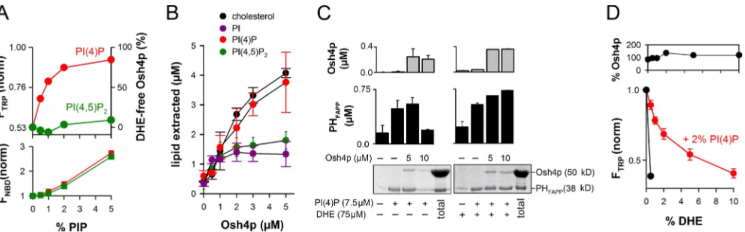
Figure 3. DHE and PI(4)P compete for extraction by Osh4p. (A) 0.5 µM NBD-Osh4p was added to buffer alone or to DOPC liposome (0.5 mM lipids)
containing 0.5 mol% DHE and increasing amounts of PI(4)P or PI(4,5)P2. The changes in Trp and NBD fluorescence, which reflect DHE extraction and
liposome binding, respectively, were monitored as in Fig. 2 B. The plots show the fluorescence levels 9 min after the addition of Osh4p. The amount of DHE-free Osh4p reported on the right axis was determined from the Trp intensity. (B) Lipid extraction assay. Sucrose-loaded DOPC liposomes (0.5 mM
lipids) containing 2% PI(4)P (red curve), 2% PI(4,5)P2 (green curve), 2% PI (purple curve), or 2% cholesterol (black curve) and doped, respectively, with
[32P]PI(4)P, [3H]PI(4,5)P
2, [3H]PI, or [3H]cholesterol were incubated with Osh4p (0–5 µM) for 20 min (room temperature). After centrifugation, radioactivity
in the supernatant and in the pellet was counted to calculate the fraction of extracted lipid. Data are represented as mean ± SEM (error bars; n = 2–3). (C) Two-stage flotation assay. DOPC liposomes containing the indicated amounts of PI(4)P and DHE were mixed with 0, 5, or 10 µM Osh4p, collected
by flotation, and then incubated with 0.75 µM PHFAPP (GST-tagged). The final amount of liposome-bound Osh4p and PHFAPP after the second stage was
determined by SDS-PAGE. (D) Solubilization of DHE by Osh4p from DOPC liposomes containing 2% PI(4)P and increasing amounts of DHE (0–10%).
After centrifugation, the amount of protein in the supernatant (top) was assessed by SDS-PAGE. The bottom shows Osh4p fluorescence at 340 nm (ex =
285 nm). Data are represented as mean ± SEM (error bars; n = 2). For comparison, an experiment was performed with liposomes containing 0.5% DHE but no PI(4)P (black curve).
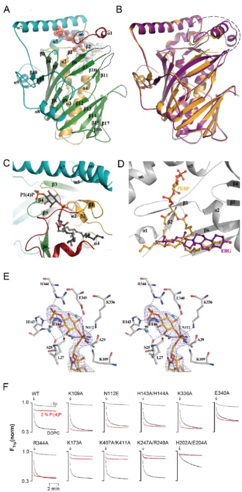
hydrogen-bonds with the side-chains of H143, H144 (4-5),
and R344 (7), and a water-mediated interaction with the main
chain oxygen atom of S25 from the lid. The 1-phosphate group
bridging the inositol ring and the glycerol moiety is hydrogen
bonded to K109 (2), K336 (7), and the backbone amide of
A29 in the lid (Fig. 4 E). Finally, the hydroxyl groups of the
inositol ring are engaged in direct or water-mediated hydrogen
bonds with the main chain oxygen atoms of S25, L27 (lid),
and N112 (2-3 loop), and with the side chain of E340 (7).
Molecular modeling suggested that Osh4p could not bind
PI(4,5)P
2as the 5-phosphate group should sterically clash with
the side-chain of H143 (Fig. S3 B). A phosphate at position 3
seems also unfavorable.
We noted that several residues making contact with the
PI(4)P headgroup are strictly conserved in Osh/Orp proteins.
We tested their functional role by examining the inhibitory
effect of PI(4)P on DHE loading by Osh4p mutants (Fig. 4 F).
The mutants K109A, N112E, H143A/H144A, E340A, R344A,
and K336A loaded DHE from liposomes as efficiently as the
wild-type form but were not inhibited by PI(4)P. As a
con-trol, mutating basic residues (K174A, K407A/K411A, and
K247A/R249A) on the other side of the sterol pocket preserved
both the ability of Osh4p to extract DHE and its inhibition by
PI(4)P. Finally, we noted that loop 204–212, which is defined
in the sterol-bound form of Osh4p but not in the Osh4p–PI(4)P
complex, is likely key for sterol binding but not for PI(4)P: the
H202A/E204A mutant loaded DHE very slowly but remained
sensitive to PI(4)P.
Osh4p delivers DHE to PI(4)P-containing acceptor liposomes
The crystal structures of Osh4p demonstrate that sterols and
PI(4)P are mutually exclusive ligands, which explains why they
compete for Osh4p extraction when present in the same
mem-brane. However, this observation does not necessarily imply
that sterol and PI(4)P act in an antagonist manner. In the context
of lipid transport, Osh4p might exchange sterol for PI(4)P such
that forward transport of sterol between two membranes would
be coupled to backward transport of PI(4)P.
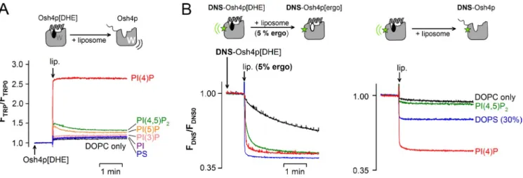
As a first step toward testing this hypothesis, we
per-formed DHE delivery assays. We prepared Osh4p loaded with
DHE (Osh4p[DHE]), then followed the kinetics of DHE release
upon the addition of acceptor liposomes by measuring the
in-crease in tryptophan fluorescence of Osh4p that resulted from
the loss of FRET with DHE. With liposomes containing 2%
PI(4)P, a jump in the fluorescence of Osh4p[DHE] was
ob-served. The signal increase (2.6×) mirrored the signal decrease
observed when Osh4p extracted DHE (1/2.5×; Fig. 1 A), which
suggests the complete release of DHE. The effect of PI(4)P was
very specific. Substituting other anionic lipids for PI(4)P such
as DOPS, PI, PI(3)P, PI(5)P, or PI(4,5)P
2resulted in minor DHE
release (Fig. 5 A).
DHE delivery probably involves sequential steps: binding
of Osh4p[DHE] to the liposome surface, opening of the lid, and
the exit of DHE. To better define this sequence of events and
the role of PI(4)P, we compared experiments in which
DNS-Osh4p[DHE] was mixed with acceptor liposomes with or
PI(4)P, PH
FAPPbound efficiently to the liposomes (Fig. 3 C).
Importantly, under all conditions, the final amount of
liposome-bound Osh4p was low and nearly identical. These observations
suggested that Osh4p can extract a stoichiometric amount of
PI(4)P, thereby preventing the subsequent binding of PH
FAPPto
the liposomes. Last, when 10% mol DHE was included in the
liposome formulation, binding of PH
FAPPto the liposomes was
no longer affected by preincubation with Osh4p. By
compet-ing with PI(4)P, DHE in excess likely prevented Osh4p from
extracting PI(4)P.
As previously mentioned, PI(4)P inhibited the loading
of 0.5 mol% DHE by Osh4p with a half-maximal effect of
0.5–1 mol% (Fig. 3 A). Conversely, when the surface density of
PI(4)P was kept at 2 mol%, the dose response curve of DHE
load-ing was shifted to higher density values (half-maximal effect =
2 mol%; Fig. 3 D). Therefore, PI(4)P and DHE act not only as
competing ligands for extraction by Osh4p, but they also
dis-play similar affinities for the protein.
Structural basis of PI(4)P-specific recognition by Osh4p
Because PI(4)P competes with DHE, the PI(4)P-binding site in
Osh4p should overlap with the sterol-binding pocket. Using
in silico approaches, we noticed that the sterol-binding site may
also house a PI(4)P molecule. This prompted us to perform
structural studies.
We crystallized Osh4p with PI(4)P and solved the
struc-ture of the complex by molecular replacement at 2.6-Å
reso-lution (
Table S1
). The overall structure of Osh4p (Fig. 4 A)
displays a central near-complete -barrel (residues 117–307)
forming a mainly hydrophobic tunnel flanked by an N-terminal
domain (residues 30–116), which consists of a two-stranded
-sheet and three -helices that close the incomplete -barrel,
and a large C-terminal region (residues 308–434). With a root
mean square deviation (rmsd) of 1.14 Å for 382 residues, the
structure of the complex with PI(4)P is highly similar to that
ob-tained in the presence of ergosterol (PDB accession no. 1ZHZ;
Fig. 4 B; Im et al., 2005). However, the lid (residues 1–29) adopts
another conformation to accommodate the PI(4)P molecule
(Fig. 4 B). In addition, several regions of the protein display
poor electron density and could not be modeled reliably. These
unresolved segments in the PI(4)P-bound conformation
corre-spond to loops 99–105, 172–175, 204–212, and 237–242, and
to the N-terminal part of the lid (1–12; Fig. 4, A and B).
PI(4)P binds to Osh4p by inserting its two acyl chains
into the central tunnel of the -barrel while the
phosphory-lated inositol ring lies near the protein surface in a shallow
pocket formed by residues from the C-terminal loop of the
lid, strand 2, the tip of the turn 4-5, and helix 7 (Fig. 4,
A and C). The acyl chains of PI(4)P interact loosely with the
sterol-binding site in a rather nonspecific manner, as suggested
by the faint electron density observed for this part of the ligand
(Fig. 4 D and
Fig. S3 A
). At the opposite, the polar head group
is involved in many direct and water-mediated contacts with
the protein, accounting for the specific recognition of PI(4)P
by Osh4p and the well-defined electron density of this part
of the ligand (Fig. 4 E). The 4-phosphate group makes direct
Figure 4. Crystal structure of Osh4p in complex with PI(4)P. (A) Overall structure. The lid region is shown in red (residues 13–29), the N-terminal domain in orange (30–116), the -barrel in green (117–307), and the C-terminal domain in cyan (308–434). PI(4)P is shown as spheres with carbon atoms colored in white, oxy-gen in red, and phosphorus in orange. The missing loops are represented by dashed lines. (B) Structure superposition of Osh4p in complex with PI(4)P (in orange) or ergosterol (in purple; PDB accession no. 1ZHZ). The black circle indicates the region that differs significantly in the two structures. (C) Close-up view of the PI(4)P binding site. PI(4)P is shown in black with oxygen in red and phosphorus in orange. (D) Super-position of PI(4)P (colored in orange) and ergosterol (purple) molecules in Osh4p. The backbone of Osh4p is shown in light gray. (E) Stereo view of the interaction network of the PI(4)P polar head. The electron density colored in blue represents the Fo-Fc–simulated anneal-ing omit map contoured at 2.5 . Nitrogen, oxygen, and phosphorus atoms are shown in blue, red, and orange, respectively. Key water molecules contacting PI(4)P and the protein are displayed as cyan spheres. (F) Effect of mutations on the ability of Osh4p to extract DHE from DOPC liposomes doped or not doped with 2% PI(4)P. The experimental conditions were the same as in Fig. 2 B.
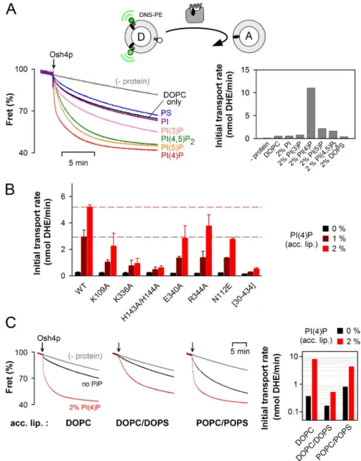
contained only DOPC or were doped with 2 mol%
phospha-tidylserine, PI, or PI(3)P, DHE transport was very slow
(<1 nmol/min). In the presence of 2 mol% PI(4,5)P
2or PI(5)P,
the rate increased approximately fourfold. Finally, with
accep-tor liposomes containing 2 mol% PI(4)P, we observed the
fast-est initial rate of transport (11 nmol /min; Fig. 6 A). Thus, there
was a good correlation between the transport rate under
multi-ple turnover conditions and the ability of Osh4p to deliver DHE
to acceptor liposomes (Fig. 5 A).
Next we tested the DHE transfer activity of several Osh4p
mutants with donor liposomes made of DOPC and acceptor
lipo-somes containing DOPC and 0, 1, or 2 mol% PI(4)P (Fig. 6 B).
In the absence of PI(4)P, the transport activity of all mutants was
as low as that of wild-type Osh4p. Marked differences emerged
when the acceptor membranes included PI(4)P. Some mutants
(K109A, E340A, R344A, and N112E) showed a modest
de-crease in activity (two- to threefold) compared with the wild-type
form, whereas the K336A and H143A/H144A mutant showed a
much stronger defect (five- and ninefold, respectively).
Remark-ably, these two mutants were almost as inactive as lidless Osh4p
([30–434]Osh4p) defective in sterol binding. Because K336A
and H143A/H144A extracted DHE normally but did not respond
to PI(4)P (Fig. 4 F), these results highlighted the importance of
PI(4)P recognition in the sterol transport activity of Osh4p.
In yeast, PI(4)P is synthesized by two PI-4 kinases: Pik1p
at the trans Golgi, and Stt4p at the PM (Strahl and Thorner,
2007). The bulk lipid composition of these compartments
is complex and obviously not properly mimicked by
accep-tor liposomes containing only DOPC and PI(4)P. Compared
with the ER, the PM is highly enriched in phosphatidylserine
(Yeung et al., 2008) and contains more saturated lipids (Zinser
et al., 1991; Schneiter et al., 1999; Pichler et al., 2001; Klemm
et al., 2009). The trans Golgi probably displays intermediate
features between the ER and the PM. To address the influence
of lipid packing and membrane electrostatics, we compared
three acceptor liposome compositions: DOPC (low packing),
without an excess of ergosterol (Fig. 5 B). With DOPC
lipo-somes, the decay in FRET between DHE and DNS-Osh4p
in-dicated a slow (t
1/2= 188 s) replacement of DHE by ergosterol.
In contrast, Osh4p rapidly exchanged DHE for ergosterol
(t
1/2< 5 s) on liposomes containing 2% PI(4)P, PI(4,5)P
2, or
30% DOPS. This implied that Osh4p was able to quickly release
DHE on liposomes containing anionic lipids. However, when
we repeated these experiments with liposomes devoid of
er-gosterol, DHE appeared poorly delivered, except when PI(4)P
was present. These results indicate that DHE release on anionic
liposomes can be followed by recapture of another sterol
mol-ecule, leading to futile exchange. What makes PI(4)P unique
compared with other anionic lipids is its ability to block sterol
re-extraction by being loaded by Osh4p. As such, PI(4)P
en-sures net delivery of sterol.
Fast DHE transport from neutral membranes to PI(4)P-containing membranes
Osh4p optimally solubilized DHE from neutral membranes and
fully delivered DHE to PI(4)P-containing membranes. These
results prompted us to recapitulate sterol transport by Osh4p
from the first to the second type of liposome. For this, donor
liposomes made of DOPC and containing 10% DHE and 2.5%
DNS-phosphatidylethanolamine (DNS-PE) were incubated with
a ninefold excess of acceptor liposomes and with Osh4p.
Al-though the FRET decrease between DHE and DNS-PE
moni-tored DHE extraction from the donor liposomes (John et al.,
2002), a decrease of 60% as seen in Fig. 6 A necessarily
implies DHE delivery to the acceptor liposomes and Osh4p
re-cycling through multiple cycles of transport. Indeed, Osh4p was
used in a substoichiometric amount compared with DHE (ratio
1:20). Without acceptor liposomes, only minor FRET decrease
was observed (see Materials and methods).
Fig. 6 A shows DHE transport experiments where we varied
the composition of the acceptor liposomes. When these liposomes
Figure 5. Osh4p delivers DHE to PI(4)P-containing liposomes. (A) Osh4p loaded with DHE (0.5 µM Osh4p[DHE]) was incubated at 30°C in buffer. At the indicated time, 30 µl of a stock suspension of liposomes (5 mM lipids) was injected (final concentration = 240 µM). The release of DHE was followed
by measuring the increase in Osh4p fluorescence at 340 nm (ex = 285 nm). The liposomes contained DOPC and, when indicated, 2 mol% of DOPS,
PI, PI(3)P, PI(4)P, PI(5)P, or PI(4,5)P2. (B) DNS-Osh4p loaded with DHE (0.5 µM) was mixed with 240 µM liposomes with (left) or without (right) 5 mol%
ergosterol at 30°C. The release of DHE was followed by measuring the diminution in FRET between DHE and DNS (ex = 310 nm; em = 510 nm). In all
assays, the recordings were corrected for the light-scattering signal and the dilution effect was due to liposome addition.
Counter exchange of PI(4)P and DHE by Osh4p
PI(4)P could be very efficiently extracted by Osh4p (Fig. 3),
had a stimulatory effect on DHE delivery (Fig. 5), and strongly
accelerated the rate of DHE transport under multiple turnover
conditions (Fig. 6). Together, these observations suggested that
DHE transport from donor to acceptor liposomes may be
cou-pled to transport of PI(4)P in the opposite direction.
Interest-ingly, this prediction could explain the atypical shape of DHE
transport kinetics: in the presence of PI(4)P in the acceptor
liposomes, the time courses slowed down abruptly once half
of DHE had been transferred (Fig. 6, A and C). PI(4)P, which
promotes DHE delivery on the acceptor liposomes, should, if
transported back to the donor liposomes, become inhibitory by
competing with DHE for Osh4p extraction.
We set up an indirect assay for PI(4)P transport. In a first
step, we mixed Osh4p with two liposome populations: “A1”
containing 6% PI(4)P, and “A2” without PI(4)P. Given the ratio
between these liposomes (1:5), transport and equilibration of
DOPC/DOPS (low packing and high charge density), and
1-palmitoyl-2-oleoyl-phosphatidylcholine (POPC)/1-palmitoyl-
2-oleoyl-phosphatidylserine (POPS; medium packing and high
charge density). In all cases, we observed a strong stimulatory
effect of 2% PI(4)P on DHE transport, which suggests that
PI(4)P retains a specific effect on Osh4p regardless of the bulk
properties of the acceptor membrane (Fig. 6 C). However, the
rate of DHE transport decreased by one order of magnitude
when the acceptor liposomes combined low lipid packing and
high charge density. Because the PI(4)P-dependant sterol
de-livery step was marginally affected by these changes (
Fig. S4
),
the following step of the cycle, recycling of Osh4p from DOPC/
DOPS liposomes, should be rate limiting. This effect is explained
by the fact that Osh4p is strongly retained on these liposomes,
as previously observed (Figs. 2 and S2). Finally, we noticed that
transport occurred in all conditions without liposome aggregation
(Fig. S4, B and C). Thus, Osh4p efficiently transfers sterol from
neutral membranes to membranes containing PI(4)P and a high
density of other anionic lipids without membrane bridging.
Figure 6. Osh4p optimally transports DHE from neutral to PI(4)P-containing membranes. (A) Effect of anionic lipids in the acceptor liposomes. DOPC/DNS-PE/DHE liposomes (87.5:2.5:10 mol/mol, 100 µM lipids) were mixed with DOPC liposomes (900 µM lipids) containing 2 mol% of the indicated lipid. After 3 min, 0.5 µM Osh4p was added. FRET between DHE and DNS-PE in the donor lipo-somes diminishes as DHE is transported to the acceptor liposomes. The initial fluorescence signal was set at 100%. The slow decay ob-served without Osh4p is caused by sponta-neous DHE transfer between the liposomes. (A, right) Initial transport rates. (B) Osh4p WT or mutants (0.5 µM) were added to DOPC/ DNS-PE/DHE liposomes (87.5:2.5:10 mol/mol, 100 µM lipids) mixed with DOPC liposomes (900 µM lipids) containing 0, 1, or 2% PI(4)P. The initial transport rates are indicated on a linear scale for each mutant (black bars, 0% PI(4)P; dark red bars, 1% PI(4)P; red bars, 2% PI(4)P). The broken lines correspond to the value of the initial transport rate measured for the wild-type Osh4p with acceptor liposomes containing 1% or 2% PI(4)P. Data are repre-sented as mean ± SEM (error bars; n = 2). (C) Transport was measured by mixing 0.5 µM Osh4p with DOPC/DNS-PE/DHE liposomes (87.5:2.5:10 mol/mol, 100 µM lipids) and acceptor liposomes (900 µM lipids) of differ-ent compositions (DOPC, DOPC/DOPS or POPC/POPS 70:30 mol/mol) with 2% PI(4)P (red traces) or without PI(4)P (black traces). (C, right) Initial transport rate. Black bars, with-out PI(4)P; red bars, with 2% PI(4)P. Experi-ments in A and C were completed once but include common conditions.
Discussion
Identifying the mechanisms by which lipid transfer proteins
selectively convey lipids between membranes is crucial for
understanding how organelles maintain their composition (Lev,
2010). Osh4p has been suggested to transport sterol from the PM,
marked by PI(4,5)P
2, to the ER (Raychaudhuri et al., 2006). We
propose a different model in which Osh4p physically exchanges
sterols for PI(4)P between membranes. Based on structural and
biochemical data, this model reconciles several cellular
obser-vations on Osh4p and suggests that the distribution of sterol
may be controlled by phosphoinositide metabolism.
Osh4p efficiently solubilizes PI(4)P from membranes and,
for this, uses roughly the same binding site as for sterol
extrac-tion. This unexpected finding is nicely explained by the
struc-ture of Osh4p bound to PI(4)P. First, the sterol-binding pocket
accommodates the PI(4)P acyl chains. Second, a shallow pocket
at the entrance of the tunnel, which contains critical residues
such as K336 and the H143/H144 pair, selects the polar head of
PI(4)P with high specificity. This interaction is probably
essen-tial for compensating loose binding of the PI(4)P acyl chains.
Third, the N-terminal lid secures the bound PI(4)P molecule by
wrapping its glycerol moiety. Overall, the result of these three
interactions is functionally very important: Osh4p displays a
similar affinity for PI(4)P and sterols.
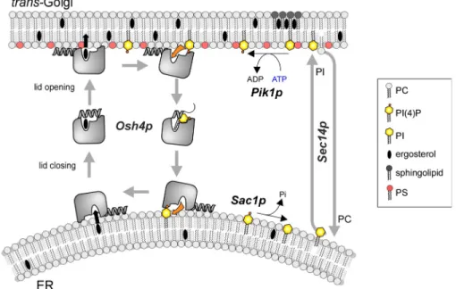
Our kinetic analysis suggests that Osh4p is designed to
promote rapid exchange of sterols for PI(4)P between membranes
according to the cycle shown in Fig. 8. In this scheme, sterol
re-lease in the acceptor membrane is followed by PI(4)P
extrac-tion; conversely, PI(4)P release in the donor membrane precedes
sterol extraction. When integrated in a more general view
in-cluding other lipid-modifying enzymes and lipid transporters at
PI(4)P between A1 and A2 by Osh4p should lead to a final
den-sity of 1 mol% accessible PI(4)P. Thereafter, donor liposomes
(D) containing DHE were added. Because only A2 contained
the fluorescent lipid DNS-PE, the FRET signal monitored DHE
transport from D to A2. As shown in Fig. 7 A, the kinetic of
DHE transport was almost as fast as in a control reaction
per-formed with 1 mol% PI(4)P directly included in liposomes A2.
In contrast, when A1 liposomes were devoid of PI(4)P, the
de-livery of DHE to A2 was very slow. This assay suggested that
Osh4p had efficiently transported PI(4)P from A1 to A2 during
the first step.
Next, we devised a direct assay for PI(4)P transport based
on liposome separation. Donor liposomes containing 2% DHE
were loaded with sucrose and labeled with NBD-PE. Acceptor
liposomes containing 2% radiolabeled PI(4)P were doped with
rhodamine-phosphatidylethanolamine (Rhod-PE). The liposomes
were mixed in a 1:1 ratio, incubated with or without Osh4p, and
then separated by centrifugation. We collected a bottom and top
fraction containing, respectively, mostly donor (80%) or
accep-tor liposomes (90%), as estimated by the relative fluorescence
of NBD-PE and Rhod-PE. We determined that the donor
lipo-somes lost 25% DHE and gained 12–13% PI(4)P when
incu-bated with Osh4p. Conversely, the acceptor liposomes showed
an increase in DHE and a decrease in PI(4)P (Fig. 7 B). If
for-ward transport of sterol was strictly coupled with backfor-ward
trans-port of PI(4)P, the same amount of PI(4)P and DHE should have
been displaced. However, DHE also spontaneously exchanges
be-tween liposomes (see, e.g., the gray trace in Fig. 6 A), making
quantitative comparisons difficult with this assay, which also
lacks time resolution. Notwithstanding these limitations, these
experiments indicate that Osh4p can transport DHE and PI(4)P
in opposite ways.
Figure 7. Osh4p transfers PI(4)P between
membranes. (A) 0.5 µM Osh4p was mixed
at 30°C with DOPC liposomes containing 6% PI(4)P (liposome A1, 15 µM) and DOPC lipo-somes doped with 2.5% DNS-PE (A2, 75 µM). In a second step, DOPC liposomes contain-ing 18% DHE were added (donor liposome, 90 µM). Control experiments were done with A1 and A2 liposomes containing either 0 or 1% PI(4)P (black and dark red trace, re-spectively) or without Osh4p (gray trace). (B) Sucrose-loaded DOPC/DHE liposomes (98:2 mol/mol, 0.5 mM lipids, labeled with 0.1% mol NBD-PE) were incubated with
DOPC/PI(4)P liposomes doped with [32P]PI(4)P
(98:2 mol/mol, 0.5 mM lipids, labeled with 0.1 mol% Rho-PE). After centrifugation on a sucrose gradient, a bottom and top fraction were collected. The fluorescence of NBD-PE, Rho-PE, and DHE was measured and PI(4)P radioactivity was counted for each fraction. (B, bottom) The relative amount of donor and acceptor liposomes in each fraction. (B, top) The gain or loss of DHE and PI(4)P (in percent-ages) for each liposome population. Data are represented as mean ± SEM (n = 2).
explain. The fact that Osh4p also acts as a PI(4)P transporter
suggests that cell survival upon deletion of OSH4 and SEC14 is
caused by reduced backward transport of PI(4)P from the trans
Golgi to the ER. Elimination of this transport pathway
prob-ably compensates for the limiting amount of PI(4)P that can be
synthesized by Pik1p when forward transport of PI by Sec14p
is impaired (Fairn et al., 2007). Again, this model better
plains mutagenesis data: Osh4p mutants that are unable to
ex-tract PI(4)P but behave normally for sterol exex-traction (K336A,
H143A/H144A) are not lethal in Sec14p-deficient yeast, in
contrast to the wild-type form (Im et al., 2005).
In conclusion, we propose a phosphoinositide cycle in
which Sec14p and Osh4p transport PI and PI(4)P along
oppo-site routes, PI(4)P being converted into PI at the ER by Sac1p
and PI being phosphorylated into PI(4)P at the trans Golgi by
Pik1p (Fig. 8). When considered from the sole “point of view”
of phosphoinositides, this cycle seems futile: ATP is used to
continually regenerate PI(4)P. However, from a sterol-centric
point of view, the consumption of ATP by Pik1p ensures net
transfer of sterol in the forward direction and PC in the
back-ward direction. As such, Osh4p could drive sterols up a
concen-tration gradient and promote sterol enrichment of membranes of
the late secretory pathway (TGN, PM) at the expense of the ER,
where sterol is synthesized.
Some aspects of the proposed Osh4p cycle warrant further
examination. Notably, the mechanism by which Osh4p targets
the ER remains to be addressed. Osh4p quickly (t
1/2of 7 s)
and totally solubilizes DHE from neutral (DOPC) liposomes
under conditions where DHE was present at only 0.5 mol%. Thus,
Osh4p should be able to efficiently pick up sterol from the ER,
wherein this lipid is rare. Interestingly, we found previously that
the lid of Osh4p corresponds to an ALPS motif (Drin et al., 2007).
ALPS are unstructured sequences that fold into amphipathic
helices upon binding to neutral membranes whose composition
and high curvature result in loose lipid packing. The ER, which
is characterized by a low content of anionic lipids, a high
content of unsaturated lipids, and a highly tubulated structure,
the ER–Golgi interface, the proposed cycle explains several
genetics and cellular observations as detailed (Fig. 8).
Osh4p rescues yeast lacking the entire Osh family by
re-storing the level of sterol at the PM (Beh and Rine, 2004). By
exchanging sterol for PI(4)P, Osh4p might supply the trans Golgi
and PM, pinpointed by this phosphoinositol phosphate, with
sterol. The coupling between sterol delivery and PI(4)P extraction
also explains why mutants such as K336A and H143A/H144A,
although fully competent for sterol extraction, do not rescue
Osh-depleted yeast (Im et al., 2005). These mutants do not
pro-mote net transfer of sterol to the PM because they are unable to
capture, in turn, PI(4)P.
Osh4p overexpression reduces both the cellular level of
PI(4)P and the Golgi targeting of a PI(4)P reporter (Fairn et al.,
2007). These observations can be explained by retrograde
trans-port of PI(4)P from the Golgi to the ER by Osh4p, where PI(4)P
is metabolized by Sac1p, an ER-resident PI(4)P-phosphatase.
We note that this mechanism offers an alternative explanation
for the interplay between Osh4p and Sac1p. Osh proteins have
been proposed to facilitate the hydrolysis of PI(4)P in “trans”
by Sac1p by forming membrane contact sites between
organ-elles (Stefan et al., 2011). This mechanism is appealing in the
case of Osh proteins with additional domains that can
effec-tively bridge two membranes. However, Osh4p lacks such
do-mains and efficiently exchanges sterol for PI(4)P between
liposomes without tethering them. Therefore, we hypothesize
that Osh4p supplies the ER with PI(4)P, which is then
hydro-lyzed by Sac1p.
Finally, the sterol/PI(4)P transfer activity of Osh4p gives
a straightforward explanation for the long-established genetic
interactions between OSH4, SEC14, SAC1, and PIK1 genes
(Fang et al., 1996; Li et al., 2002; Fairn et al., 2007). SEC14
encodes for a PI/phosphatidylcholine (PC) transfer protein and
is an essential gene. However, yeast cells can survive when
SEC14
is inactivated together with OSH4. As long as Osh4p
was considered as a mere sterol transporter, the functional
antagonism between Osh4p and Sec14p remained difficult to
Figure 8. Model for the coupling between sterol and PI(4)P exchange by Osh4p. Osh4p solubilizes sterols efficiently from neutral and loosely packed membranes. The sterol is locked by the lid. The interaction of Osh4p with an-ionic membranes promotes the opening of the lid and sterol release. Only PI(4)P promotes net sterol delivery because PI(4)P is extracted by Osh4p at the expense of sterol re-extraction. Tight lipid packing in the acceptor membrane limits the retention of Osh4p through its ALPS motif. These observation suggest that Osh4p is suited to transport sterols preferentially from ER to the trans Golgi marked by PI(4)P. Con-versely, Osh4p transports PI(4)P from the trans Golgi to the ER, and another round of trans-port can resume. Although each step of the cycle is reversible by itself, the presence of a PI-4 kinase at the TGN (Pik1p) and of a PI(4)P phosphatase at the ER (Sac1p) favors direc-tionality of lipid transport. The sterol/PI(4)P exchange activity of Osh4p is also coupled to that of Sec14p, a PC/PI exchanger.
PI(4)P (l--PI-4-phosphate), brain PI(4,5)P2 (l--PI-4,5-bisphosphate), liver
PI (l--PI), DNS-PE
(1,2-dioleoyl-sn-glycero-3-phosphoethanolamine-N-(5-dimethylamino-1-naphthalenesulfonyl)), NBD-PE (1,2-dioleoyl-sn-glycero- 3-phosphoethanolamine-N-(7-nitro-2-1,3-benzoxadiazol-4-yl)), and Rhod-PE (1,2-dipalmitoyl-sn-glycero-3-phosphoethanolamine-N-(lissamine rhodamine B sulfonyl)) were obtained from Avanti Polar Lipids, Inc. PI(3)P
inositol-3-phosphate) and PI(5)P
(1,2-dioleoyl-sn-glycero-3-inositol-5-phosphate) were obtained from Echelon. Ergosterol and DHE were obtained from Sigma-Aldrich. The concentration of DHE in stock solu-tion in methanol was carefully determined by UV spectroscopy using an
extinction coefficient of 13,000 M1cm1.
The radioactive lipids [3H]cholesterol, [3H]PI, and [3H]PI(4,5)P
2 were
purchased from PerkinElmer. To prepare radioactive [32P]PI(4)P, BHK cells
were labeled with 1 mCi/ml ortho[32P]phosphate during 36 h in
phos-phate-free DME containing 2% FCS. After one washing step in cold PBS, cells were scrapped off and recovered in 3 ml of HCl (1.6 N). A mixture of methanol and chloroform (6 ml, vol/vol) was immediately added and the sample was vortexed vigorously during 2 min and centrifuged at 300 g for 5 min. The organic phase was collected and dried under a nitrogen stream at 37°C. Lipids were resolved by thin layer chromatography (TLC silica gel
60; Merck) using CHCl3/CH3OH/NH4OH 4N (90/70/20, vol/vol) as a
solvent and identified using appropriate standards. The spot of [32P]PI(4)P
was scraped off and the lipid was extracted from the silica by a modified Blight and Dyer procedure (Bligh and Dyer, 1959). The purity of the PI(4)P preparation was analyzed by HPLC. In brief, an aliquot of the preparation was deacylated by adding 1 ml of methylamine reagent composed of 26.8% (vol/vol) of 40% methylamine, 45.7% (vol/vol) methanol, 11.4% (vol/vol)
n-butanol, and 16% (vol/vol) H2O. After incubation at 53°C for 50 min, the
methylamine reagent was completely evaporated under a nitrogen stream
at 37°C. The samples were resuspended in 1.2 ml H2O and separated
by HPLC using a Whatman Partisphere 5 SAX column 4.6 mm × 125 mm with guard-cartridge anion exchanger units (Ref. 4641 0005; GE
Health-care) and a gradient of 1 M (NH4)2HPO4, pH 3.8, and bi-distilled water
(Payrastre, 2004). The amount of purified PI(4)P was quantified by phos-phorus measurement (Fiske and Subbarow, 1925).
Liposomes
Lipids in stock solutions in chloroform were mixed at the desired molar ratio, and the solvent was removed in a rotary evaporator. For formulations containing phosphoinositides, the mixture was warmed at 30–33°C for 5 min prior to drying under vacuum. For lipid films containing radioactive lipids, the mixture was dried in a hemolysis tube under argon. The lipid film was hydrated in 50 mM Hepes, pH 7.2, and 120 mM potassium ace-tate (HK buffer) to give a suspension of large multilamellar liposomes (lipid concentration: 1–5 mM). The suspension was then frozen and thawed five times (using liquid nitrogen and a water bath) and extruded through polycarbonate filters of 0.2-µm pore size using a mini-extruder (Avanti Polar Lipids, Inc.). For experiments with liposomes of varying curvature, the liposomes were extruded sequentially through 0.4-, 0.2-, 0.1-, 0.05-, and 0.03-µm (pore size) polycarbonate filters. The liposome hydrodynamic
radius (RH) was estimated by dynamic light scattering in a DynaPro
instru-ment (Wyatt Technology). Liposomes were stored at 4°C and in the dark when containing light-sensitive lipids (NBD-PE, DNS-PE, DHE, or ergos-terol) and used within 2 d.
DHE loading assay
For kinetics measurements, Trp fluorescence was measured at 340 nm (bandwidth 5 nm) upon excitation at 286 nm (bandwidth 1.5 nm) in a fluor-ometer (RF 5301-PC; Shimadzu). The sample initially contained liposomes (500 µM total lipids) in HKMD buffer (50 mM Hepes, pH 7.2, 120 mM
potassium-acetate, 1 mM MgCl2, and 1 mM DTT). The liposomes used in this
assay were extruded on a 0.2-µm filter. The sample (volume 600 µl) was placed in a cylindrical quartz cell, continuously stirred with a small mag-netic bar, and thermostated at 30°C. At the indicated time, Osh4p or NBD-Osh4p was injected from stock solutions through a guide in the cover of the fluorometer adapted to Hamilton syringes, such as to not interrupt the fluor-escence recording. Emission spectra were recorded at the end of the kinet-ics upon excitation at 285 nm. Loading experiments with DNS-Osh4p were performed under the same conditions except that DHE was directly excited at 310 nm (bandwidth 1.5 nm) and the fluorescence of DNS was followed at 510 nm (bandwidth 10 nm). We assessed, under conditions where Osh4p was strongly bound to the liposomes (i.e., with DOPC/DOPS lipo-some), that the FRET signal reflected only DHE loading by Osh4p and not the proximity between the protein and DHE embedded in the membrane. In-deed, no FRET occurred when we incubated DNS-Osh4p with DOPS/DOPS liposomes containing DHE (0.5%) and an excess of ergosterol (5%).
seems well adapted to the transient adsorption of the Osh4p
ALPS/lid region. In the future, it will be important to study in a
systematic manner the influence of membrane curvature and
composition on the ability of Osh4p to exchange sterol and
PI(4)P between membranes. In fact, one reason for the
conflict-ing results between this study and previous studies may be related
to membrane curvature. In several in vitro assays for Osh4p
trans-port, very small liposomes obtained by sonication and
contain-ing high amounts of anionic lipids have been used (Raychaudhuri
et al., 2006; Schulz et al., 2009; Stefan et al., 2011). It is likely
that the combination of extreme curvature and high electrostatic
favors Osh4p retention on the liposomes, impairs lipid transport,
and causes membrane aggregation through the multiple potential
membrane-binding sites of Osh4p (ALPS motif, basic patches).
Most of residues that contact the PI(4)P headgroup are
conserved in Osh/Orp proteins, which suggests that extraction
of PI(4)P is a general hallmark of this family. Determining how
this biochemical feature translates to a function will require
further investigation for each Osh/Orp.
Materials and methods
Proteins
Osh4p and Osh4p[30–434] were cloned in a pGEX-4T-3 vector. Site-specific mutations were introduced using the QuikChange mutagenesis protocol and checked by DNA sequencing. Osh4p was expressed in E. coli at 30°C
overnight upon induction with 1 mM IPTG (OD600 = 0.6). All purification
steps were conducted in 50 mM Tris, pH 7.4, 120 mM NaCl, 1 mM MgCl2,
and 1 mM DTT supplemented, during the first purification steps, with 1 mM PMSF, 1 µM bestatine, 10 µM pepstatine, 10 µM phosphoramidon, and protease inhibitor tablets (Roche). Cells were lysed with a French press and the lysate was centrifuged at 200,000 g for 1 h. The supernatant was ap-plied to Glutathione Sepharose 4B beads. After three washing steps, the beads were incubated with thrombin to cleave the GST fusion and allow the release of Osh4p. The purity of the proteins was >95% as assessed by SDS-PAGE. The protein concentration was determined by a Bradford assay and by SDS-PAGE analysis using a BSA standard curve. All constructs contain an N-terminal GS sequence from the thrombin cleavage site.
5-((((2-iodoacetyl)amino)ethyl)amino)naphthalene-1-sulfonic acid (IAEDANS) labeling of Osh4p was performed by mixing the protein (after DTT removal by dialysis) with a 10-fold molar excess of IAEDANS (Invitro-gen). After 90 min on ice, the reaction was stopped by adding a 10-fold
excess of l-cysteine over the probe. The free probe was removed by gel
filtration on a Sephacryl S200 HR XK16-50 column (GE Healthcare). The labeled protein was analyzed by SDS-PAGE and UV-visible spectros-copy. The gel was directly visualized in a fluorescence imaging system (U-GENIUS; Syngene) to detect DNS-labeled Osh4p excited in near-UV and then stained with Sypro Orange to determine the purity of DNS-Osh4p. The percentage of labeling was estimated from the optical density (OD)
of tryptophan at 280 nm ( = 73,900 M1cm1) and DNS at 340 nm
( = 5,700 M1cm1). In a control experiment, we observed no labeling of
the Osh4p(C98S) mutant, which indicates that CYS-98 was the sole endog-enous cysteine that could be labeled by a thiol-reactive probe.
NBD labeling of Osh4p was performed on a double mutant lacking the solvent-exposed endogenous cysteine (C98S) and including a cysteine in the N-terminal lid (A5C). After DTT removal, this mutant was labeled with a 10-fold excess of N,N’-dimethyl-N-(iodoacetyl)-N’-(7-nitrobenz-2-oxa-1,3-diazol-4-yl)ethylenediamine (IANBD-amide; Invitrogen) and pu-rified according to the instructions used for DNS-Osh4p. The percentage of labeling was estimated from the optical density at 280 and 495 nm ( =
25,000 M1cm1 for NBD according to the manufacturer). The PH domain
of FAPP in fusion with GST was provided by J. Bigay (Centre National de la Recherche Scientifique, Institut de Pharmacologie Moléculaire et Cellu-laire, Valbonne, France).
Lipids
DOPC (1,2-dioleoyl-sn-glycero-3-phosphocholine), POPC
(1-palmitoyl-2-oleoyl-sn-glycero-3-phosphocholine), DOPS (1,2-dioleoyl-sn-glycero-3-phospho-
l-serine), POPS (1-palmitoyl-2-oleoyl-sn-glycero-3-phospho-l-serine), brain