HAL Id: hal-03172062
https://hal.sorbonne-universite.fr/hal-03172062
Submitted on 17 Mar 2021
HAL is a multi-disciplinary open access
archive for the deposit and dissemination of
sci-entific research documents, whether they are
pub-lished or not. The documents may come from
teaching and research institutions in France or
abroad, or from public or private research centers.
L’archive ouverte pluridisciplinaire HAL, est
destinée au dépôt et à la diffusion de documents
scientifiques de niveau recherche, publiés ou non,
émanant des établissements d’enseignement et de
recherche français ou étrangers, des laboratoires
publics ou privés.
Site-specific contacts enable distinct modes of TRPV1
regulation by the potassium channel Kvβ1 subunit
Yuanyuan Wang, Xiaoyi Mo, Conghui Ping, Qian Huang, Hao Zhang, Chang
Xie, Bo Zhong, Dongdong Li, Jing Yao
To cite this version:
Yuanyuan Wang, Xiaoyi Mo, Conghui Ping, Qian Huang, Hao Zhang, et al.. Site-specific contacts
enable distinct modes of TRPV1 regulation by the potassium channel Kvβ1 subunit. Journal of
Bio-logical Chemistry, American Society for Biochemistry and Molecular Biology, 2020, 295 (50), pp.17337
- 17348. �10.1074/jbc.ra120.015605�. �hal-03172062�
Site-specific contacts enable distinct modes of TRPV1
regulation by the potassium channel Kv
b1 subunit
Received for publication, August 11, 2020, and in revised form, October 13, 2020Published, Papers in Press, October 15, 2020, DOI 10.1074/jbc.RA120.015605 Yuanyuan Wang1,‡ ,Xiaoyi Mo1,‡,Conghui Ping1,Qian Huang1,Hao Zhang1,Chang Xie1,Bo Zhong1,
Dongdong Li2 , andJing Yao1,*
From the1State Key Laboratory of Virology, Hubei Key Laboratory of Cell Homeostasis, College of Life Sciences, Frontier Science Center for Immunology and Metabolism, Wuhan University, Wuhan, Hubei, China and the2Sorbonne Université, Institute of Biology Paris Seine, Neuroscience Paris Seine, Paris, France
Edited by Michael J. Shipston
Transient receptor potential vanilloid 1 (TRPV1) channel is a multimodal receptor that is responsible for nociceptive, ther-mal, and mechanical sensations. However, which biomolecular partners specifically interact with TRPV1 remains to be eluci-dated. Here, we used cDNA library screening of genes from mouse dorsal root ganglia combined with patch-clamp electro-physiology to identify the voltage-gated potassium channel aux-iliary subunit Kvb1 physically interacting with TRPV1 channel and regulating its function. The interaction was validatedin situ using endogenous dorsal root ganglia neurons, as well as a recombinant expression model in HEK 293T cells. The presence of Kvb1 enhanced the expression stability of TRPV1 channels on the plasma membrane and the nociceptive current density. Surprisingly, Kvb1 interaction also shifted the temperature threshold for TRPV1 thermal activation. Using site-specific mapping, we further revealed that Kvb1 interacted with the membrane-distal domain and membrane-proximal domain of TRPV1 to regulate its membrane expression and temperature-activation threshold, respectively. Our data therefore suggest that Kvb1 is a key element in the TRPV1 signaling complex and exerts dual regulatory effects in a site-specific manner.
TRPV1 is a Ca21-permeable cation channel and plays impor-tant roles in pain sensation and transduction. It is abundantly expressed in sensory neurons in the dorsal root ganglia (DRG) and trigeminal ganglia (1). As a multimodal sensor, TRPV1 responds to a variety of physical or chemical stimuli such as heat (.42 °C), voltage, low pH, capsaicin, and analogs like noni-vamide and resiniferatoxin (1–3). During injury and inflamma-tion, inflammatory mediators (e.g. bradykinin, nerve growth factor) enhance the responses of TRPV1 to noxious stimuli, thereby heightening pain experience, a process called hyperal-gesia. TRPV1 knockout mice show impaired thermal nocicep-tion and vanilloid-induced pain (4,5).
TRPV1 appears usually as a homotetramer channel and also forms heterotetramers with other TRP family members (6,7). At the channel level, TRPV1 function is delicately controlled by multiple mechanisms including protein kinase–mediated
intra-channel phosphorylation or SUMOylation (8–10) and subcel-lular trafficking (11–14). In addition, because ion channels are organized in multiprotein assemblies, termed signaling com-plexes, channel activity is also regulated by local protein –pro-tein interactions (15). TRPV1 function has been suggested to be influenced by a variety of proteins. For example, GABAA
re-ceptor–associated protein is implicated in TRPV1 clustering on the plasma membrane, a process relying on the intermediate interaction with tubulin (16). Cyclin-dependent kinase 5 was reported to alter TRPV1 membrane transport via the involve-ment of kinesin-3 family member 13B protein (17). However, the molecular components directly interacting with TRPV1 channels remain to be determined.
Here, we screened a cDNA library of mouse DRG by yeast two-hybrid assay and identified the voltage-gated K1channel b1 subunit (Kvb1) directly interacting with TRPV1 protein. By patch-clamp recording, Kvb1 was observed to regulate the temperature threshold for TRPV1 thermal activation and the whole-cell current density. Mechanistically, Kvb1 played a role in sustaining the availability of TRPV1 on the plasma mem-brane. Site-specific mapping identified that Kvb1 interacted with the proximal domain (MPD) and membrane-distal domain (MDD) of TRPV1 to orchestrate its temperature-activation threshold and surface expression, respectively. These data suggest that Kvb1 subunit exerts dual regulatory effects on TRPV1 function and represents a functional component in the signaling complex. Targeting Kvb1–TRPV1 interaction would help to regulate nociceptive transduction.
Results
Kvb1 physically interacts with TRPV1 channel
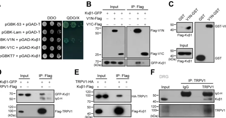
In an attempt to identify the protein that directly interact with TRPV1 channel in DRG neurons, a yeast two-hybrid assay was performed to screen a mouse DRG cDNA library while using the cytosolic N-terminal domain of mouse TRPV1 as the bait. Sequence analysis of the positive clones revealed that 18 proteins interacted with the N terminus of TRPV1 (V1N). Among them, Kvb1, the auxiliary subunit of voltage-gated potassium channels showed strong interaction with TRPV1. To directly verify this interaction, we co-transformed Kv b1-pGADT7 with V1-Nt-pGBKT7 or V1-Ct-pGBKT7 into the yeast reporter strain AH109. Indeed, Kvb1 interacted with the V1N, but not the C terminus (V1C) of TRPV1 (Fig. 1A).
This article containssupporting information.
Author's Choice—Final version open access under the terms of the Creative CommonsCC-BYlicense.
‡These authors contributed equally to this work. * For correspondence: Jing Yao,jyao@whu.edu.cn.
Then the co-immunoprecipitation (co-IP) experiment was conducted to examine Kvb1–TRPV1 interaction in HEK 293T cells. As shown inFig. 1B, Kvb1–GFP was
co-immunoprecipi-tated by V1-Nt-FLAG, but not V1-Ct-FLAG or the FLAG vec-tor alone, confirming that Kvb1 interacts with the N terminus of TRPV1 rather than the C terminus.
Because co-IP experiment cannot exclude the possibility of indirect protein–protein interaction with accessory proteins, we carried out the gluatathione S-transferase (GST) pulldown experiment to examine the direct interaction between Kvb1 and the N terminus of TRPV1. We generated the GST-fusion protein containing the N terminus of TRPV1 (named V1N-GST). Then V1N-GST and GST were immobilized on GSH-Sepharose beads and incubated with the HEK 293T extracts that expressed FLAG-Kvb1. As illustrated inFig. 1C, V1N-GST but not GST specifically retained the recombinant Kvb1, thus corroborating the yeast two-hybrid screen and co-IP results. Next, we evaluated the capacity of Kvb1 to bind the full length of TRPV1 channel in HEK 293T cells using a co-IP strategy. As shown inFig. 1 (D and E), Kvb1-GFP could be
co-immunoprecipitated by TRPV1–FLAG with FLAG-aga-rose beads. Reciprocally, TRPV1-HA could be precipitated by Kvb1-FLAG. These results showed that Kvb1 and TRPV1 interact with each other in situ in HEK 293T cells. Further, the interaction between Kvb1 and TRPV1 was validated with co-IP assay in DRG neurons. We homogenized mouse DRGs
and observed the co-precipitation of the endogenous TRPV1 and Kvb1 proteins by using the specific antibodies (Fig. 1F). Together, Kvb1 and TRPV1 are physically associated in native DRG neurons.
Kvb1 up-regulates TRPV1 current responses
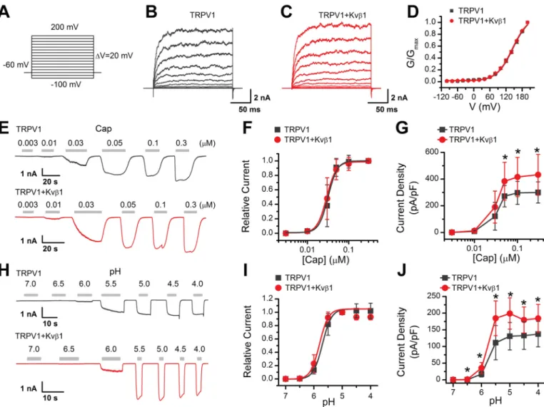
Because HEK 293T cells are commonly used as an expression system for exogenous ion channel and they lack endogenous Kvb1 expression (Fig. S1), we next investigated the effects of Kvb1 on TRPV1 activity by co-expressing both components in this cell line. Kvb1, as an auxiliary subunit of the voltage-gated potassium channel, regulates the rapid inactivation of Kv1 fam-ily channels (18). Because TRPV1 can be activated by mem-brane depolarization, we examined the effect of Kvb1 on the voltage-dependent TRPV1 currents using a voltage step proto-col (Fig. 2A). Whole-cell currents were elicited with 200-ms depolarizing pulses ranging from2100 mV to 200 mV with 20-mV increments in HEK 293T cells that expressed either TRPV1 alone or TRPV1 and Kvb1 (Fig. 2, B and C). The current at the end of the 200-ms pulse was converted to conductance and normalized to that obtained at 200-mV stimulation. Subse-quent fitting with the Boltzmann function yielded similar con-ductance–voltage (G–V) relationships for TRPV1 response regardless of whether Kvb1 was present or not (Fig. 2D). We observed no currents in HEK 293T cells solely expressing Kvb1
Figure 1. Kvb1 physically interacts with TRPV1 channel. A, yeast two-hybrid results showing the interaction between Kvb1 and the TRPV1 N terminus (V1N) rather than the C terminus (V1C). The 53-T and Lam-T were served as positive and negative controls, respectively. DDO, double dropout medium (SD/ 2Leu/2Trp); QDO, quadruple dropout medium (SD/2Leu/2Trp/2His/2Ade); X, X-a-gal. B, IP and IB analysis of HEK 293T cells transfected with plasmids encoding FLAG-tagged TRPV1-Nt, TRPV1-Ct, and GFP-tagged Kvb1. Cell lysates were IP with FLAG antibody and analyzed by IB using anti-GFP and anti-FLAG, respectively. Whole-cell lysates were also used for IB with anti-GFP and anti-FLAG as input. C, GST pulldown analysis of Kvb1 with GST fusion N-terminal of TRPV1 (V1N-GST). GST-fusion proteins encompassing TRPV1 N terminus were constructed and expressed in E. coli strain BL21. The TRPV1 N terminus fusion protein bound to GSH-Sepharose beads was then incubated with HEK 293T cell lysate. GST only was used as a negative control. D and E, HEK 293T cells were transfected with the indicated plasmids. Immunoprecipitation (with anti-FLAG) and immunoblot analysis (with anti-GFP and anti-FLAG in D or anti-HA and anti-FLAG in E) of the interaction between TRPV1 and Kvb1. F, immunoprecipitation (with TRPV1 or IgG as a control) and immunoblot analysis (with anti-TRPV1 or anti-Kvb1) of DRG neurons. The data are representative of three independent experiments. Full images of the Western blots are shown inFig. S4.
Dual regulation of TRPV1 by Kv
b
1
(Fig. S2). Thus, co-expression of Kvb1 did not change the volt-age dependence of TRPV1 under basal conditions.
Then we examined the effect of Kvb1 on TRPV1 channel gating as evoked by capsaicin or acidic extracellular solutions. We compared TRPV1 function by activating the channel with increasing concentrations of capsaicin at the holding potential (Vh) of 260 mV. The amplitude of the evoked currents was
normalized to that evoked by 0.3 mM capsaicin. Fitting the dose-response curves with the Hill equation yielded similar EC50values and Hill coefficients (nH) for TRPV1 co-expressed
with Kvb1 or not (Fig. 2, E and F), EC50= 0.036 0.01mM, nH=
3.96 0.2 for TRPV1 and EC50= 0.036 0.01mM, nH= 3.86 0.4
for TRPV11 Kvb1). The maximum current density, however, showed a significant difference between the absence (300.96 79.4 pA/pF, n = 12) and the presence of Kvb1 (432.5 6 153.5 pA/pF, n = 11;Fig. 2G). We also investigated TRPV1 responses
to protons by applying variable acidic solutions covering a broad pH range from 7.0 to 4.0 and determined pH50and nH.
Akin to capsaicin induced gating, co-expression of Kvb1 with TRPV1 did not alter the concentration dependence to proton but increased the current density, e.g. 130.86 41.2 pA/pF (n = 7) and 199.06 46.8 pA/pF (n = 7) evoked by pH 5.0 for TRPV1 and TRPV11 Kvb1, respectively (Fig. 2, H–J). Taken together, these results show that Kvb1 modulates whole-cell current density of TRPV1.
Kvb1 sustains the surface expression of TRPV1
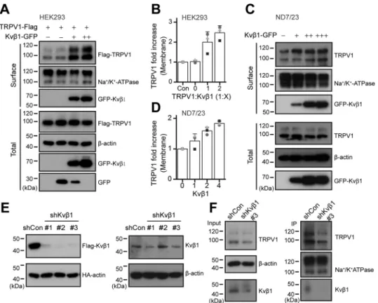
Because the presence of Kvb1 increased TRPV1 current den-sity without altering its agonist sensitivity, we reasoned that Kvb1 might have increased TRPV1 surface expression level. To test this hypothesis, we performed a surface biotinylation
Figure 2. Kvb1 enhances TRPV1 activity. A, voltage protocol with 200-ms test pulses ranging from 2100 to 1200 mV in 20-mV increments from holding potential of260 mV. B and C, representative whole-cell recordings in HEK 293T cells expressing TRPV1 alone (B) and TRPV1 1 Kvb1 (C). TRPV1 currents were eli-cited by the voltage protocol as A. D, normalized conductance–voltage (G–V) relations. Boltzmann fits correspond to V1/2= 155.86 1.9 mV, andk= 34.66 0.9 for TRPV1 (n = 6), and V1/2= 153.56 1.9 mV, andk= 34.16 1.0 for TRPV1 1 Kvb1 (n = 9), respectively. E, representative whole-cell recordings from HEK 293T cells that expressed TRPV1 (top panel) and TRPV11 Kvb1 (bottom panel). The cells were exposed to varied concentrations of capsaicin (Cap) as indicated. The holding potential was260 mV. F, concentration-response curves for capsaicin-evoked currents. Solid lines indicate fits with the Hill equation, which yielded EC50= 0.036 0.01 mMand nH= 3.96 0.2 for TRPV1 (n = 12) and EC50= 0.036 0.01mMand nH= 3.86 0.4 for TRPV1 1 Kvb1 (n = 11). G, summary plot of current density for TRPV1 (n = 12) and TRPV11 Kvb1 (n = 11). The peak current densities evoked by capsaicin are normalized by membrane capacitance. *, P, 0.05. H, parallel recordings of H1-evoked responses in transiently transfected HEK 293T cells held at260 mV. I, dose-response curves of low pH. The solid lines are fits to Hill’s equations with pH50= 5.706 0.02 and nH= 2.76 0.3 for TRPV1 (n = 7) and pH50= 5.806 0.07 and nH= 2.86 0.8 for TRPV1 1 Kvb1 (n = 7). J, comparison of current density evoked by pH 5.0 between TRPV1 (n = 7) and TRPV11 Kvb1 (n = 7). *, P, 0.05. Holding potential was 260 mV. Error bars represent S.D.
experiment. As shown inFig. 3 (A and B), in HEK 293T cells, Kvb1 proportionally increased the surface expression but not the total amount of TRPV1 channel.
We further used the ND7/23 neuroblastoma cell line that is a hybridization of mouse neuroblastoma and rat DRG neuron (19). This immortal cell line inherits the endogenous TRPV1 expression of DRG neurons. Then we explored the effect of overexpressed Kvb1 subunit on TRPV1 protein expression in the ND7/23 cell line. As shown in Fig. 3C, along with the amount of transfected Kvb1 increased, the expression level of TRPV1 on the plasma membrane was proportionally up-regu-lated. Transfection of 4 mg of Kvb1 caused a nearly 2-fold increase TRPV1 plasma membrane expression (Fig. 3D). In this condition, however, the total amount of TRPV1 was unaffected. Next, we evaluated the regulatory effect of Kvb1 on TRPV1 expression using shRNA-mediated knockdown. To test the knockdown efficiency of three groups of shRNAs designed against Kvb1, we co-transfected them or empty vector shCon with FLAG-Kvb1 and HA-actin into HEK 293T cells, respec-tively. HA-actin was used to calibrate the transfection efficiency between groups. The expression of the two proteins were eval-uated by immunoblotting. All the three shRNAs efficiently reduced Kvb1 protein expression levels (Fig. 3E). We also vali-dated the inhibitory efficiency of the shRNAs in ND7/23 cells
that express Kvb1 subunit, although at a relatively low level (Fig. S1). As shown in the right panels ofFig. 3E, shKvb1#1 and shKvb1#3 both inhibited the endogenous expression of Kvb1. We then performed surface biotinylation experiment in ND7/ 23 cells to detect the impact of shKvb1#3 on the total and sur-face expression of TRPV1. We observed that inhibition of Kvb1 expression concomitantly reduced the expression of TRPV1 on the cell surface (Fig. 3F, right panels). These results together suggest that Kvb1 sustains the functional expression of TRPV1 on the plasma membrane.
Molecular domains underlying the association of Kvb1 and TRPV1
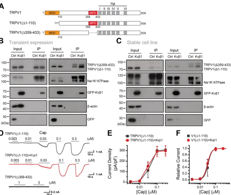
To define the critical domains within the N terminus of TRPV1 that mediate Kvb1-TRPV1 association, a series of Nt-TRPV1 truncations were constructed, including FLAG-1–359, FLAG-1–110, FLAG-111–359, and FLAG-111–433 (Fig. 4A). They were individually co-expressed with Kvb1-GFP in HEK 293T cells for co-IP evaluation. Kvb1-GFP was strongly pre-cipitated by 1–110, as was 1–359 and FLAG-111–433 but not FLAG-111–359 (Fig. 4A). These data indicate that both the MDD (aa 1–110) and MPD (aa 359–433) of TRPV1 channel are required for associating with Kvb1.
Figure 3. Kvb1 sustains the surface expression of TRPV1. A, increased surface expression of TRPV1 by Kvb1. HEK 293T cells were transiently co-transfected with different molar ratios of TRPV1-FLAG and Kvb1-GFP as indicated. Note that the amount of TRPV1-FLAG was fixed. Surface levels of TRPV1 were measured by IB after plasma membrane proteins were biotinylated and purified with NeutrAvidin-agarose beads.b-Actin and Na1/K1ATPase were used as controls for cytoplasmic and membrane proteins, respectively. B, quantitative analysis of the fold increase of TRPV1 on the plasma membrane following co-expression of Kvb1 in HEK 293T cells (n = 3; means6 S.D.). C and D, ND7/23 cells were transiently transfected with increasing amounts of Kvb1-GFP cDNA. Similarly, surface levels of TRPV1 were measured by IB after plasma membrane proteins were biotinylated and purified with NeutrAvidin-agarose beads (n = 3; means6 S.D.). E, immunoblot analysis (with anti-FLAG or anti-HA) of HEK 293T cells transfected for 36 h with plasmids encoding FLAG-tagged Kvb1 and HA-b-actin and either Kvb1-targeting shRNA (shKvb1#1, shKvb1#2, and shKvb1#3) or shCon to test knockdown efficiency of shRNA (left panel). Immunoblot analysis (with anti-Kvb1 or anti-b-actin) of ND7/23 cells stably transfected with shCon, shKvb1#1, shKvb1#2, or shKvb1#3 (right panel). F, the cell-surface biotinylation assay was used to measure the surface levels of TRPV1 in ND7/23 cells that were stably transfected with shCon or shKvb1#3. Full images of the Western blots are shown inFig. S5.
Dual regulation of TRPV1 by Kv
b
1
We further mapped the molecular domains of Kvb1 that mediated its interaction with TRPV1. Three truncations were constructed, including FLAG-1–72, 73–217, and FLAG-218–401. In a yeast two-hybrid experiment, the aa 218–401 segment had been noted to mediate the interaction between Kvb1 and TRPV1 N terminus and therefore was used as a posi-tive control. As shown inFig. 4B, TRPV1-HA could be precipi-tated by both the middle (aa 73–217) and ending part (aa 218– 401) of Kvb1, suggesting that the segment between aa 73 and 401 of Kvb1 interacts with TRPV1.
Kvb1 interacts with MDD to regulate TRPV1 surface expression
To determine the relative role of N-terminal domains in the regulation of TRPV1 surface expression level, we constructed TRPV1 truncations that lack the Kvb1 interaction segments: TRPV1(D1–110) and TRPV1(D359–433) (Fig. 5A). We first transfected the FLAG-TRPV1(D1–110) into HEK 293T cells and 12 h later equally divided the cells into two groups that were then transfected with Kvb1-GFP or GFP, respectively.
Similar experimental procedures were performed for the FLAG-TRPV1(D359–433) construct. When the introduced proteins were fully expressed (i.e. 24 h after the last transfec-tion), we performed surface biotinylation analysis. As shown in
Fig. 5B, although the total amount of both truncated TRPV1 proteins was unaffected by Kvb1, the amount of surface-expressed FLAG-TRPV1(D359–433) (i.e. comprising the MDD aa 1–110) was steadily increased. In contrast, the absence of the MDD within the FLAG-TRPV1(D1–110) construct rendered it irresponsive to Kvb1 regulation.
To further explore the effect of the MDD on TRPV1 mem-brane targeting, we developed HEK 293T cell lines stably expressing phage-TRPV1(D1–110) and phage-TRPV1(D359– 433), respectively. We then transfected Kvb1-GFP or GFP into the stable-expression cell lines separately and conducted a sur-face biotinylation experiment after 24 h. As shown inFig. 5C, the presence of Kvb1 displayed no effect on both the cytoplasm and plasma membrane expression of the MDD-devoid TRPV1 (D1–110) construct but increased the surface expression of the TRPV1(D359–433) construct. Thus, Kvb1 interacts with the MDD region to regulate TRPV1 surface expression.
Figure 4. Two distinct domains mediate the interaction between the N terminus of TRPV1 and Kvb1. A, schematic representation of the truncation mutants of N terminus of TRPV1 including 1–359, 1–110, 111–359, and 1112433 segments, respectively. HEK 293T cells were transiently co-transfected with different truncation mutants tagged with FLAG and Kvb1-GFP as indicated. Cell lysates were immunoprecipitated with anti-FLAG and analyzed by IB using anti-GFP and anti-FLAG, respectively. Whole-cell lysates were also used for IB with anti-GFP and anti-FLAG as input. B, schematic diagram of composition of mutant Kvb1 including the 1–72, 73–217, and 218–401 segments. HEK 293T cells were transfected with TRPV1-HA and different mutant Kvb1 fused with FLAG as indicated. The cell lysates were subjected to IP by anti-FLAG, followed by IB for anti-HA and anti-FLAG, respectively. All blot images are representatives of three independent experiments. Full images of the Western blots are shown inFig. S6.
We also examined the effect of Kvb1 on the electrophysio-logical activity of the TRPV1 mutants. Whole-cell recordings revealed that TRPV1(D359–433) showed no channel activity, whereas TRPV1(D1–110) showed current responses similar to TRPV1(Fig. 5D). We then explored the effect of Kvb1 on the channel gating of TRPV1(D1–110). We recorded TRPV1(D1– 110) responses to increasing concentrations of capsaicin at Vh
=260 mV. The current density showed no significant change in the presence of Kvb1 (Fig. 5E), distinct from its facilitatory effect observed for WT TRPV1 (Fig. 2G). Fitting the normal-ized dose-response curves with the Hill equation yielded
simi-lar EC50values and Hill coefficients (nH) for TRPV1(D1–110)
co-expressed with Kvb1 or not (Fig. 5F). This result suggests that Kvb1 interacts with MDD to regulate TRPV1 surface expression and current response.
Kvb1 modulates TRPV1 temperature threshold by interacting with MPD domain
We have previously demonstrated that the N-terminal region connecting ankyrin repeats to the first transmembrane segment is critical for TRPV1 temperature sensing (20). Because the
Figure 5. Kvb1 regulates the surface expression of TRPV1 by interacting with MDD of TRPV1. A, schematic representation of composition of mutant channels. Residue numbers are shown. TM, transmembrane. B, HEK 293T cells were transiently transfected with the following plasmids as indicated: TRPV1 (D1–110)-FLAG and TRPV1(D359–433)-FLAG together with Kvb1-GFP or GFP. Surface level of TRPV1 were measured by IB after plasma membrane proteins were biotinylated and purified with NeutrAvidin-agarose beads.b-Actin and Na1/K1ATPase were used as controls for cytoplasmic and membrane proteins, respectively. C, stable HEK 293T cell lines expressing phage-TRPV1(D1–110) or phage-TRPV1(D359–433) were transiently transfected with Kvb1-GFP and GFP, respectively. Similar to A, surface levels of TRPV1 were measured by IB after plasma membrane proteins were biotinylated and purified with NeutrAvidin-aga-rose beads. All blot images are representative of at least three independent experiments. D, representative whole-cell currents evoked by increasing concen-trations of capsaicin (Cap) for HEK 293T cells that expressed TRPV1(D1–110) (top panel), TRPV1(D1–110) 1 Kvb1 (middle panel), and TRPV1(D359–433) (bottom panel). Holding potential was260 mV. E, comparison of current density evoked by capsaicin between TRPV1(D1–110) (n = 6) and TRPV1(D1–110) 1 Kvb1 (n = 6). Note that there were no detectable responses for TRPV1(D359–433) (n = 4). F, dose-response curves of capsaicin. Fitting by Hill’s equation resulted in the following: EC50= 0.036 0.01mMand nH= 3.66 0.2 (n = 6) for TRPV1(D1–110) and EC50= 0.036 0.01mMand nH= 3.46 0.2 (n = 6) for TRPV1(D1–110) 1 Kvb1. Error bars indicate S.D. Full images of the Western blots are shown inFig. S7.
Kvb1 analog Kvb2 has been suggested to modulate TRPV1 ac-tivity (21), we sought to compared their effect on TRPV1 ther-mal activation. Kvb2 protein sequence is 85% identical to Kvb1 except the first 72 amino acids at the N terminus. By similar ex-perimental procedure to explore Kvb1-TRPV1 interactions, we observed that Kvb2 could only be precipitated by FLAG-1–359, i.e.the region from the initiating N terminus of TRPV1 to the end of the ankyrin repeats, whereas the TRPV1 MPD domain (aa 359–433) domain was only precipitated by Kvb1-FLAG (Fig. S3). Hence, distinct from Kvb1 that interacts with MDD and
MPD domains, Kvb2 shows interaction with TRPV1 N-termi-nal 1–359 segment.
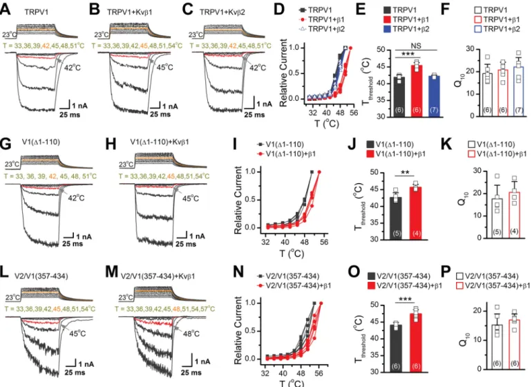
We then comparatively studied the effect of Kvb1 and Kvb2 binding on TRPV1 temperature sensing. Using laser irradia-tion-based temperature controlling and whole-cell patch-clamp recording, we recorded current responses in three groups of HEK 293T cells expressing TRPV1 alone, TRPV1 with Kvb1 or Kvb2, respectively. The presence of Kvb2 showed no effect on the temperature threshold (Tthreshold); currents started to be
induced at 42 °C as observed for TRPV1 alone. In contrast, cells expressing TRPV1 and Kvb1 did not exhibit significant re-sponses until 45 °C (Fig. 6, A–C). An appreciable right shift of the temperature dependence curve of TRPV1 was observed with Kvb1, but not Kvb2 (Fig. 6D). The expression of Kvb1 resulted in a rise of;3°C in TRPV1 Tthreshold, without affecting
the temperature coefficient (Q10), whereas Kvb2 showed no
effect (Fig. 6, E and F).
Our domain mapping analysis showed that Kvb1 interacted with both MDD and MPD of TRPV1 channel (Fig. 4). To exam-ine their involvement in Kvb1-induced Tthreshold change, we
used the truncated TRPV1(D1–110) construct that is specifi-cally devoid of the MDD. Co-expression of Kvb1 still elevated the Tthresholdby;3°C (Fig. 6, G–K), indicating the dispensable
role of MDD. Further we generated the chimera construct TRPV2/TRPV1(357–434) that introduced specifically the TRPV1 MPD domain into the TRPV2 channel. This engi-neering rendered the chimera channel with an activation Tthreshold of ;45 °C, which is consistent with the previous
report (20). With the insertion of the TRPV1 MPD, we also observed that the Tthresholdof TRPV2/TRPV1(357–434) was
raised by ;3°C when co-expressed with Kvb1 (Fig. 6, L–P). Taken together, these results suggest that Kvb1 interacts with MPD domain to regulate TRPV1 temperature sensitivity.
Discussion
As a polymodal sensor, TRPV1 ion channel responds to a va-riety of exogenous stimuli and is implicated in pain sensing and transduction (2,22–24). In addition to being regulated at the channel level, TRPV1 function is also controlled by multipro-tein signaling assemblies (25, 26). Illustrating the molecular components of the TRPV1 signal complexes helps to under-stand its function in physiopathological conditions.
We here demonstrate that two discrete domains at N termi-nus of TRPV1 are responsible for the interaction between TRPV1 and Kvb1. By site-specific mapping, we revealed that Kvb1 interacted with the membrane-distal domain (MDD, aa 1–110) and membrane-proximal domain (MPD, aa 359–433)
of TRPV1 to, respectively, regulate its surface expression levels and temperature-activation threshold by 3 °C. These data sug-gest that Kvb1 is a molecular element in the TRPV1 signaling complex and exerts site-specific dual regulatory effects (Fig. 7).
Previous studies have shown that Kvb1 regulates Shaker-like potassium channels (e.g. Kv1) by the interaction with the N-ter-minal A and B box domain of the Shaker family channels (27). Kvb subunits had also been found to increase the expression of Kv4.3 channels by interacting with the C termini (28). Here we show that the surface expression of TRPV1 is enhanced by the interaction of Kvb1 with the N-terminal MDD. We, however, found no homologous sequences among the N terminus of TRPV1, Kv1 channel, and C terminus of Kv4.3 channels, sug-gesting a specific regulatory effect of Kvb1 on TRPV1. Here we observed that Kvb1 facilitates the surface expression of TRPV1, thereby up-regulating whole-cell current density. Kvb1 may have a similar regulatory mechanism for potassium channels (28–30), an issue requiring additional investigations.
As a Kvb1 analog, Kvb2 has been suggested to influence TRPV1 expression (21). Different regulatory effects have been noted for these two subunits. For instance, Kvb1, but not Kvb2, confers rapid A-type inactivation to noninactivating Kv1 chan-nels (18). Here, we observed that Kvb1 regulates TRPV1
func-tions in a manner distinct from Kvb2. Kvb1 interacts with both MDD and MPD regions of TRPV1, whereas Kvb2 interacts with the aa 1–359 segment. Notably, the thermal activation threshold of TRPV1 was up-regulated by;3°C by the presence of Kvb1, an effect attributed to the interaction with TRPV1 MPD domain and not seen with Kvb2. This result confirms the importance of the MPD domain in setting the thermal activa-tion property of TRPV1 (20). It should be noted that under the condition of exogenous expression, Kvb1 elevates the TRPV1 Tthresholdto;45°C, whereas it remains at 42 °C in endogenous
DRG neurons. It seems that a relatively high amount of Kvb1 is required to change TRPV1 temperature threshold, which might not be the case for DRG neurons. Another possibility is that although Kvb1 tends to increase TRPV1 thermal activa-tion threshold, there are other mechanisms to counterbalance the effect of Kvb1 in sensory neurons. Notwithstanding, our results unveil an important role for Kvb1 in sculpturing TRPV1 thermal responses.
In 13-lined ground squirrels and Bactrian camels, changing a single amino acid raised the TRPV1 activation threshold to ;46°C or even higher (31). The increased heat tolerance may prove beneficial by conferring animals the ability to inhabit oth-erwise prohibitive ecological niches. Thus, modulation of the TRPV1 temperature-activation threshold by Kvb1 might have physiological implications. Targeting the Kvb1-TRPV1 interac-tion would help to tune the thermal reacinterac-tions along the body sensory input pathways.
Experimental procedures
Constructs, cell culture, and transfection
The WT rat TRPV1 cDNA was a gift from Dr. Feng Qin (State University of New York at Buffalo, Buffalo, NY, USA). The full lengths of mouse Kvb1 and Kvb2 were obtained and subcloned into pEGFP-N1 vector, respectively. To express in
mammalian cells, the cDNAs of TRPV1, Kvb1, and Kvb2 were introduced into p33FLAG-cmv-7.1, pEGFP-N1, or pcDNA5-HA, as indicated. To express GST-tagged protein of TRPV1 N terminus in prokaryotic cells, the open-reading frame of the TRPV1 N-terminal cDNA was placed into the pGEX-6P-1 vec-tor. All recombinant constructs and mutations were carried out using the overlap-extension PCR method. The resulting constructs and mutations were then verified by DNA sequenc-ing. Oligonucleotide DNAs targeting Kvb1 were synthesized, annealed, and inserted into pLenti-GFP vector. The sequences
of Kvb1 shRNA are as follows: #1, 5´-GCTTGGTCATCA-CAACCAA-3´; #2, 5´-GATGTGGTCTTTGCAAATC-3´; and #3, 5´-GGAGTTGGTGCAATGACAT-3´. HEK 293T and ND7/23 cells were grown in Dulbecco’s modified Eagle’s me-dium (Thermo Fisher Scientific) containing 4.5 mg/ml glucose, 10% heat-inactivated fetal bovine serum (Gibco, Thermo Fisher Scientific), 50 units/ml penicillin, and 50mg/ml streptomycin and were incubated at 37 °C in a humidified incubator gassed with 5% CO2. HEK 293T cells stably expressing phage-TRPV1,
phage-TRPV1(D1–110), and phage-TRPV1(D359–433) were
Figure 6. Kvb1 modulates temperature threshold for TRPV1 activation by interacting with MPD of TRPV1. A–C, representative responses to a family of temperature jumps for HEK 293T cells that expressed TRPV1 (A), TRPV11 Kvb1 (B), and TRPV11 Kvb2 (C). Temperature pulses stepped from room tempera-ture were generated by local laser irradiation were 100 ms long and had a rise time of 1.5 ms. Temperatempera-tures were calibrated offline from the pipette current using the temperature dependence of electrolyte conductivity (see“Experimental Procedures”). The red traces indicate the temperature threshold for heat activation of TRPV1. D, temperature dependence of steady-state response. Each curve represents measurements from an individual cell, and the responses were normalized to its maximum responses. E, comparison of temperature threshold for activation of TRPV1. Different symbols represent individual data points. The mean temperature thresholds (Tthreshold) of activation were 41.96 0.7 °C for TRPV1 (n = 6), 45.4 6 0.8 °C for TRPV1 1 Kvb1 (n = 6), and 42.36 0.3 °C for TRPV11 Kvb2 (n = 7), respectively. P, 0.001 for Tthresholdof TRPV1 versus TRPV11 Kvb1 and P = 0.37 for Tthresholdof TRPV1 versus TRPV11 Kvb2 by one-way ANOVA test. F, comparison of temperature coefficient. The values of Q10derived from the linear fits in Arrhenius plot of steady-state currents were as fol-lowing, Q10= 19.26 4.2 for TRPV1 (n = 6), Q10= 20.86 3.5 for TRPV1 1 Kvb1 (n = 6), and Q10= 22.26 4.2 for TRPV1 1 Kvb2 (n = 7). Colored symbols indicate individual data points. P = 0.49 for Q10of TRPV1 versus TRPV11 Kvb1 and P = 0.20 for Q10of TRPV1 versus TRPV11 Kvb2 by one-way ANOVA test. G and H, sim-ilar recordings in HEK 293T cells that expressed TRPV1(D1–110) (G) and TRPV1(D1–110) 1 Kvb1 (H). I, temperature-response curves for TRPV1(D1–110) and TRPV1(D1–110) 1 Kvb1. J, summary plot of threshold changes. Tthreshold= 42.66 1.4 °C for TRPV1(D1–110) alone (n = 5) and Tthreshold= 45.76 0.9 °C for TRPV1 (D1–110) 1 Kvb1 (n = 4). P = 0.008 by Student’s t test. K, comparison of Q10. Q10= 17.76 6.1 for TRPV1(D1–110) alone (n = 5), Q10= 20.66 4.8 for TRPV1(D1– 110)1 Kvb1 (n = 4). P = 0.47. L and M, parallel recordings of heat responses in HEK 293T cells transfected with TRPV2/TRPV1(357–434) and TRPV2/TRPV1(357– 434)1 Kvb1. N, temperature-response curves for TRPV2/TRPV1(357–434) and TRPV2/TRPV1(357–434) 1 Kvb1. O and P, comparison of temperature threshold and Q10, respectively. Tthreshold= 44.16 0.8 °C for TRPV2/TRPV1(357–434) alone (n = 6), Tthreshold= 47.46 1.3 °C for TRPV2/TRPV1(357–434) 1 Kvb1 (n = 6). P = 0.0002. Q10= 15.36 3.8 for TRPV2/TRPV1(357–434) alone (n = 6), and Q10= 17.06 2.0 for TRPV2/TRPV1(357–434) 1 Kvb1 (n = 6). P = 0.35. Different symbols represent individual recordings. Error bars indicate S.D.
used in this study. To generate 293T/phage-TRPV1, 293T/ phage-TRPV1(D1–110), and 293T/phage-TRPV1(D359–433) cells, HEK 293T cells were transiently transfected with the indi-cated construct using Lipofectamine 2000 (Invitrogen). Trans-fected cells were grown for 2 weeks in puromycin to select for stable expressing cells. Nontransfected cells were killed by the addition of puromycin (2 mg/ml) 2 days after treatment. The medium was replaced every 2–3 days until single colonies were formed, and then the stable cell lines were placed in a complete medium containing 1mg/ml puromycin. In another set of experiments, cells grown into ;80% confluence were transiently transfected with the desired DNA constructs using either the standard calcium phosphate precipitation method or Lipofectamine 2000 (Invitrogen) following the protocol pro-vided by the manufacturer. In the membrane separation experi-ment, we transfected HEK 293T cells with the same amount (3 mg) of TRPV1 cDNA in all four groups, whereas the amount of Kvb1 cDNA was adjusted to achieve a molar ratio of TRPV1 and Kvb1 at 1:1 and 1:2, respectively. For the membrane sepa-ration experiment in ND7/23 cells, we transfected Kvb1-GFP in amounts of 0, 1, 2, and 4mg, respectively. Transfected cells were reseeded on 12-mm round glass coverslips coated by poly-L-lysine. Experiments took place usually 12–24 h after transfection.
Isolation of DRG neurons
All mice were housed in the specific pathogen-free animal fa-cility at Wuhan University, and all animal experiments were in accordance with protocols approved by the Institutional Ani-mal Care and Use Committee of Wuhan University. DRG
neu-rons were prepared as previously described with minor modifi-cations (13). Briefly, 6–8-week-old adult C57BL/6 male mice were deeply anesthetized and decapitated. L3–L4 DRGs were isolated from the spinal column. After removing the attached nerves and surrounding connective tissues, DRG neurons were rinsed with ice-cold PBS. Then total RNA was extracted from the intact DRG for yeast cDNA library construction using TRIzol (Life Technologies) per the manufacturer’s instruc-tions or directly homogenized DRG neurons for immunopre-cipitation experiments.
Yeast two-hybrid assay
Yeast two-hybrid screen was conducted using the Match-maker GAL4-based two-hybrid system (Clontech). The mouse DRG cDNA library was fused to the GAL4 activation domain of plasmid pGADT7 (Clontech). The titer of the primary cDNA library was calculated using the number of clones on plates. Colony PCR was used to confirm the size of the inserted fragments in the library. Bait plasmids were constructed by introducing the N terminus (aa 1–433) or C terminus (aa 688– 839) of TRPV1 into the GAL4 DNA-binding domain of the pGBKT7 vector (Clontech). The pGBK bait was transformed into yeast strain Y187, and pGAD prey was expressed in AH109. Cytotoxicity and self-activation activity were deter-mined via observing yeast clone growth and size. Diploid yeast cells from yeast mating were selected on the triple dropout medium (SD/2His/2Leu/2Trp). Then replica plate colonies were transferred onto quadruple dropout medium (SD/2Ade/ 2His/2Leu/2Trp) containing X-a-gal (4 mg/ml). X-a-gal was used as substrate for colorimetric detection ofa-Galactosidase activity. The plates were incubated at 30 °C for 7 days. Plasmids extracted from the positive clones were transformed into Esch-erichia coli cloning host DH5a to be amplified, and samples were then sequenced individually. BLAST comparisons and other bioinformatics methods were applied for sequence analy-sis. Thereafter, the bait and prey plasmids in different combina-tions were sequentially co-transformed into yeast stain AH109 and selected on double dropout medium (SD/2Leu/2Trp) and incubated at 30 °C for 3–4 days. Then the positive clones were diluted and equally coated onto SD/2Leu/2Trp/2His/ 2Ade medium with X-a-gal and SD/2Leu/2Trp medium and cultured at 30 °C for 3–4 days. In parallel, the combination of pGBKT7–53/pGADT7–T and pGBKT7–Lam/pGADT7–T were used as positive and negative controls, respectively. Immunoprecipitation and immunoblotting assays
Denaturing immunoprecipitation was performed as previ-ously described (9). Briefly, HEK 293T or ND7/23 cells were harvested 24 h post-transfection and solubilized in Nonidet P-40 lysis buffer (20 mMTris-HCl, pH 7.4, 150 mMNaCl, 1 mM EDTA, 1% Nonidet P-40) containing 1% protease inhibitor mixture. Cell lysates were subjected to SDS-PAGE, and immu-noblot analysis was performed with the appropriate antibodies. For immunoprecipitation assays, the solubilized supernatants were incubated with the control IgG or specific antibodies and protein G–agarose beads for 2 h. Immunocomplexes were washed three times with prelysis buffer (20 mMTris-HCl, pH
Figure 7. Schematic model of the dual regulatory mechanism of Kvb1. Two discrete domains at N terminus of TRPV1 are responsible for the interac-tion between TRPV1 and Kvb1. Mechanistically, Kvb1 interacts with the MDD (aa 1–110) and MPD (aa 359–433) of TRPV1 to respectively regulate its surface expression and temperature-activation threshold, respectively.
7.4, 150 mMNaCl, 1 mMEDTA, 1% Triton X-100) containing 0.5MNaCl, followed by immunoblot analysis.
GST pulldown assay
GST-fusion protein GST-Nt-TRPV1 or GST alone were expressed in BL21 competent cells that were induced with isopropylb-D-thiogalactopyranoside (1 mM) at 16 °C for 18 h, and the proteins were purified using GSH-Sepharose 4B (Transgen Biotech, Beijing, China). HEK 293T cells were transiently transfected with FLAG-tagged Kvb1 (Kvb1-FLAG) cDNA and cultured for 24 h. GST-Nt-TRPV1 or GST were incubated with HEK 293T extracts that expressing Kvb1-FLAG at 4 °C overnight in Nonidet P-40 lysis buffer containing protease inhibitors. The precipitates were washed three times with the prelysis buffer containing 0.5 M NaCl, followed by immunoblot analysis.
Surface biotinylation assay
Surface biotinylation was performed following the estab-lished protocols (9). Cells that expressed TRPV1-FLAG and Kvb1-GFP (or GFP as control) were washed three times with ice-cold PBS (pH 8.0) supplemented with 1 mMMgCl2and 2.5
mMCaCl2. Then the sulfo-NHS-LC-Biotin (Thermo Fisher
Sci-entific) was added to the same solution at 0.25 mg/ml and incu-bated with cells for 25 min at 4 °C with gentle rocking. The cells were rinsed with PBS containing 0.1Mglycine for 20 min at 4 °C to quench unbounded biotin. Biotin-labeled proteins were isolated by incubating whole-cell lysates with NeutrAvidin-aga-rose beads (Thermo Fisher Scientific) overnight at 4 °C with rocking. The beads were washed three times with PBS (pH 8.0), and bound proteins were eluted with the boiling SDS sample buffer and used for immunoblotting.
Antibodies and chemicals
Rabbit anti-TRPV1 (Alomone laboratory, catalog no. ACC-030), mouse anti-Kvb1 (Santa Cruz, catalog no. sc-373986), rabbit anti-Na1/K1 ATPase (Abcam, catalog no. ab76020), mouse GST (Abclonal, catalog no. AE001), mouse b-actin (Transgen Biotech, catalog no. 6609-1), mouse anti-FLAG (Sigma, catalog no. F3165), mouse anti-anti-FLAG (MBL, cat-alog no. M185-3L), rabbit anti-FLAG (Proteintech, catcat-alog no. 20543-1-AP), mouse anti-HA (MBL, catalog no. M180-3), rab-bit anti-HA (Earthox, catalog no. E02218002), mouse anti-GFP (Tianjin Sungene Biotech, catalog no. KM8009), rabbit anti-GFP (WB: Solarbio, catalog no. B1025F), anti-FLAG affinity gel (Bimake, catalog no. B23100), anti-HA affinity gel (Bimake, alog no. B23301), ProteinIso GST resin (Transgen Biotech, cat-alog no. DP201-01), goat anti-mouse IgG (H 1 L) (Jackson Immunoresearch, catalog no. 115-035-003), and goat anti-rab-bit IgG (H 1 L) (Jackson Immunoresearch, catalog no. 111-005-003) were purchased from the indicated manufactures. The EZ-Link Sulfo-NHS-LC-Biotin (catalog no. 21335) and high-capacity NeutrAvidin-agarose (catalog no. 29202) were ordered from Thermo Fisher Scientific.
Electrophysiology
Conventional whole-cell patch-clamp recording methods were used. For the recombinant expressing system, green fluo-rescence from GFP was used as a marker for gene expression. Patch-clamp recordings were voltage clamped using an EPC10 amplifier (HEKA, Lambrecht, Germany). Voltage commands were made from the Patchmaster program. For a subset of recordings, currents were amplified using an Axopatch 200B amplifier (Molecular Devices, Sunnyvale, CA, USA) and recorded through a BNC-2090/MIO acquisition system (National Instruments, Austin, TX, USA) using QStudio devel-oped by Dr. Feng Qin at State University of New York at Buf-falo. Recording pipettes were pulled from borosilicate glass capillaries (World Precision Instruments) and fire-polished to a resistance of 2–4 MV when filled with electrode saline contain-ing 140 mMCsCl, 5 mM EGTA, and 10 mM HEPES, pH 7.4, adjusted with CsOH, and the bath solution for whole-cell re-cording consisted of 140 mMNaCl, 5 mMKCl, 3 mMEGTA, and 10 mMHEPES, pH 7.4, adjusted with NaOH. HEPES was used for pH 7.0–7.4, and instead of HEPES, MES was used as the pH buffer when pH , 6.5. For recordings under low pH conditions, 50mMamiloride was also included to inhibit native acid–sensing ion channels. Exchange of external solutions was performed using a gravity-driven local perfusion system. As determined by the conductance tests, the solution around the patched cell was fully controlled by the application of a solution with a flow rate of 100ml/min or greater. For voltage-depend-ence experiments, a voltage step protocol consisting of 200-ms depolarizing pulses ranging from2100 to 200 mV with 20-mV increments was triggered from the holding potential of260 mV. The compensation of pipette series resistance and capaci-tance were taken by using the circuitry of the amplifier (. 80%) to reduce voltage errors. Capsaicin was dissolved in pure etha-nol to make a stock solution and diluted in the bath solution to the desired concentrations right before the experiments. The final concentrations of ethanol did not exceed 0.3%, which had no effect on the currents. Whole-cell recordings were sampled at 5 kHz and filtered at 1 kHz. Unless otherwise noted, all chemicals were purchased from Sigma (Millipore–Sigma). All patch-clamp recordings except heat activation were made at room temperature (22–24°C).
Ultrafast temperature jump achievement
A rapid temperature jump system is produced by using a sin-gle emitter IR laser diode (1470 nm) as previously described (32). Briefly, a multimode fiber with a core diameter of 100mm was used to transmit the launched laser beam. The other end of the exposed fiber core was placed close to the cell of interest where the perfusion pipette is typically positioned. The laser diode was driven by a pulsed quasi-CW current power supply (Stone Laser, Beijing, China), and the pulsing of the controller was controlled from a computer through the data acquisition card (National Instruments), which was also responsible for patch-clamp recordings. A blue laser line (460 nm) was coupled into the same fiber to aid alignment. Constant temperature steps were generated by irradiating the tip of an open pipette filled with the pipette solution and using the current through
the electrode as a readout for feedback control. The laser diode was first powered on for a brief duration to get the target tem-perature and was then modulated to achieve a constant pipette current. The profile of the modulation pulses was stored and subsequently played back to apply the temperature jump to the cell of interest. Temperature was calibrated offline from the electrode current based on the temperature dependence of electrolyte conductivity. The threshold temperature for heat activation of TRPV1 was determined as the temperature at which the slow inward current (;10% of its maximum re-sponse) was evoked.
Statistical analysis
The data were analyzed offline with Clampfit (Molecular Devices), IGOR (Wavemetrics, Lake Oswego, OR, USA), Sig-maPlot (SPSS Science, Chicago, IL, USA), OriginPro (Origin-Lab Corporation, MA, USA), and ImageJ (National Institutes of Health). Conductance–voltage (G–V) curves for activation were derived from steady-state currents, converted to conduct-ance, and then fitted by Boltzmann function: G/Gmax=1/(11
exp((V2 V1/2)/k)), in which Gmaxis the maximal conductance,
V1/2 is the voltage at which the conductance (G) is half the
maximum conductance (Gmax), andk is the slope factor. For
dose-response analysis, the modified Hill equation was used: Y = A11 (A22A1)/(1 1 (EC50/[toxin])nH), in which nHis the
Hill coefficient. All data are expressed as means6 S.D., with statistical significance assessed using unpaired Student’s t test for two-group comparison.Fig. 6 (E and F)were analyzed with one-way analysis of variance (ANOVA) test for multiple group comparison. Significant difference is indicated by p values less than 0.05: *, p, 0.05; **, p , 0.01; and ***, p , 0.001.
Data availability
The data that support the findings of this study are available from the corresponding author upon reasonable request.
Acknowledgments—We are grateful to our colleagues and members of Yao lab for comments and discussions.
Author contributions—Y. W., X. M., C. P., Q. H., H. Z., C. X., B. Z., D. L., and J. Y. data curation; Y. W., X. M., C. P., Q. H., H. Z., C. X., B. Z., D. L., and J. Y. formal analysis; Y. W., X. M., C. P., Q. H., H. Z., C. X., B. Z., D. L., and J. Y. validation; Y. W., X. M., C. X., D. L., and J. Y. investigation; Y. W., X. M., C. P., Q. H., H. Z., C. X., B. Z., D. L., and J. Y. methodology; Y. W., X. M., and J. Y. writing-original draft; Y. W., X. M., and J. Y. writing-review and editing; C. X. and J. Y. resources; B. Z. and J. Y. software; J. Y. conceptualization; J. Y. supervision; J. Y. funding acquisition; J. Y. visualization; J. Y. project administration.
Funding and additional information—This work was supported by Grants 31830031, 31929003, 31671209, 31871174, 31601864, and 31271209 from the National Natural Science Foundation of China, Grants 2017CFA063 and 2018CFA016 from the Natural Science Foundation of Hubei Province, and funds from the Fundamental Research Funds for the Central Universities.
Conflict of interest—The authors declare that they have no conflicts of interest with the contents of this article.
Abbreviations—The abbreviations used are: TRPV1, transient re-ceptor potential vanilloid 1; DRG, dorsal root ganglia; MDD, mem-brane-distal domain; MPD, membrane-proximal domain; X-a-gal, 5-Bromo-4-Chloro-3-indolyla-D-galactopyranoside; IP, immuno-precipitation; IB, immunoblot; GST, glutathione S-transferase; HA, hemagglutinin; aa, amino acid(s); ANOVA, analysis of variance; shCon, control shRNA.
References
1. Caterina, M. J., Schumacher, M. A., Tominaga, M., Rosen, T. A., Levine, J. D., and Julius, D. (1997) The capsaicin receptor: a heat-activated ion channel in the pain pathway. Nature 389, 816–824CrossRef Medline 2. Tominaga, M., Caterina, M. J., Malmberg, A. B., Rosen, T. A., Gilbert, H.,
Skinner, K., Raumann, B. E., Basbaum, A. I., and Julius, D. (1998) The cloned capsaicin receptor integrates multiple pain-producing stimuli. Neuron 21, 531–543CrossRef Medline
3. Thomas, K. C., Ethirajan, M., Shahrokh, K., Sun, H., Lee, J., Cheatham, T. E., 3rd, Yost, G. S., and Reilly, C. A. (2011) Structure–activity relation-ship of capsaicin analogs and transient receptor potential vanilloid 1– mediated human lung epithelial cell toxicity. J. Pharmacol. Exp. Ther. 337, 400–410CrossRef Medline
4. Caterina, M. J., Leffler, A., Malmberg, A. B., Martin, W. J., Trafton, J., Petersen-Zeitz, K. R., Koltzenburg, M., Basbaum, A. I., and Julius, D. (2000) Impaired nociception and pain sensation in mice lacking the capsa-icin receptor. Science 288, 306–313CrossRef Medline
5. Davis, J. B., Gray, J., Gunthorpe, M. J., Hatcher, J. P., Davey, P. T., Overend, P., Harries, M. H., Latcham, J., Clapham, C., Atkinson, K., Hughes, S. A., Rance, K., Grau, E., Harper, A. J., Pugh, P. L., et al. (2000) Vanilloid recep-tor-1 is essential for inflammatory thermal hyperalgesia. Nature 405, 183– 187CrossRef Medline
6. Hellwig, N., Albrecht, N., Harteneck, C., Schultz, G., and Schaefer, M. (2005) Homo- and heteromeric assembly of TRPV channel subunits. J. Cell Sci. 118, 917–928CrossRef Medline
7. Staruschenko, A., Jeske, N. A., and Akopian, A. N. (2010) Contribution of TRPV1–TRPA1 interaction to the single channel properties of the TRPA1 channel. J. Biol. Chem. 285, 15167–15177CrossRef Medline
8. Amadesi, S., Cottrell, G. S., Divino, L., Chapman, K., Grady, E. F., Bautista, F., Karanjia, R., Barajas-Lopez, C., Vanner, S., Vergnolle, N., and Bunnett, N. W. (2006) Protease-activated receptor 2 sensitizes TRPV1 by protein kinase Ce– and A–dependent mechanisms in rats and mice. J. Physiol. 575,555–571CrossRef Medline
9. Wang, Y., Gao, Y., Tian, Q., Deng, Q., Wang, Y., Zhou, T., Liu, Q., Mei, K., Wang, Y., Liu, H., Ma, R., Ding, Y., Rong, W., Cheng, J., Yao, J., et al. (2018) TRPV1 SUMOylation regulates nociceptive signaling in models of inflam-matory pain. Nat. Commun. 9, 2593CrossRef Medline
10. Zhang, X., Li, L., and McNaughton, P. A. (2008) Proinflammatory media-tors modulate the heat-activated ion channel TRPV1 via the scaffolding protein AKAP79/150. Neuron 59, 450–461CrossRef Medline
11. Morenilla-Palao, C., Planells-Cases, R., García-Sanz, N., and Ferrer-Mon-tiel, A. (2004) Regulated exocytosis contributes to protein kinase C poten-tiation of vanilloid receptor activity. J. Biol. Chem. 279, 25665–25672 CrossRef Medline
12. Van Buren, J. J., Bhat, S., Rotello, R., Pauza, M. E., and Premkumar, L. S. (2005) Sensitization and translocation of TRPV1 by insulin and IGF-I. Mol. Pain 1, 17CrossRef Medline
13. Tian, Q., Hu, J., Xie, C., Mei, K., Pham, C., Mo, X., Hepp, R., Soares, S., Nothias, F., Wang, Y., Liu, Q., Cai, F., Zhong, B., Li, D., and Yao, J. (2019) Re-covery from tachyphylaxis of TRPV1 coincides with recycling to the surface membrane. Proc. Natl. Acad. Sci. U.S.A. 116, 5170–5175CrossRef Medline 14. Zhang, X., Huang, J., and McNaughton, P. A. (2005) NGF rapidly increases
membrane expression of TRPV1 heat-gated ion channels. EMBO J. 24, 4211–4223CrossRef Medline
15. Levitan, I. B. (2006) Signaling protein complexes associated with neuronal ion channels. Nat. Neurosci. 9, 305–310CrossRef Medline
16. Laínez, S., Valente, P., Ontoria-Oviedo, I., Estévez-Herrera, J., Camprubí-Robles, M., Ferrer-Montiel, A., and Planells-Cases, R. (2010) GABAA re-ceptor associated protein (GABARAP) modulates TRPV1 expression and channel function and desensitization. FASEB J. 24, 1958–1970CrossRef Medline
17. Xing, B. M., Yang, Y. R., Du, J. X., Chen, H. J., Qi, C., Huang, Z. H., Zhang, Y., and Wang, Y. (2012) Cyclin-dependent kinase 5 controls TRPV1 mem-brane trafficking and the heat sensitivity of nociceptors through KIF13B. J. Neurosci. 32, 14709–14721CrossRef Medline
18. Rettig, J., Heinemann, S. H., Wunder, F., Lorra, C., Parcej, D. N., Dolly, J. O., and Pongs, O. (1994) Inactivation properties of voltage-gated K1 channels altered by presence ofb-subunit. Nature 369, 289–294CrossRef Medline
19. Wood, J. N., Bevan, S. J., Coote, P. R., Dunn, P. M., Harmar, A., Hogan, P., Latchman, D. S., Morrison, C., Rougon, G., Theveniau, M., and Wheatley, S. (1990) Novel cell-lines display properties of nociceptive sensory neu-rons. Proc. R. Soc. Lond. B Biol. Sci. 241, 187–194CrossRef Medline 20. Yao, J., Liu, B., and Qin, F. (2011) Modular thermal sensors in
tempera-ture-gated transient receptor potential (TRP) channels. Proc. Natl. Acad. Sci. U.S.A. 108, 11109–11114CrossRef Medline
21. Bavassano, C., Marvaldi, L., Langeslag, M., Sarg, B., Lindner, H., Klima-schewski, L., Kress, M., Ferrer-Montiel, A., and Knaus, H.-G. (2013) Iden-tification of voltage-gated K1 channelb2 (Kvb2) subunit as a novel interaction partner of the pain transducer transient receptor potential vanilloid 1 channel (TRPV1). Biochim. Biophys. Acta Mol. Cell Res. 1833, 3166–3175CrossRef Medline
22. Caterina, M. J., and Julius, D. (2001) The vanilloid receptor: a molecular gateway to the pain pathway. Annu. Rev. Neurosci. 24, 487–517CrossRef Medline
23. Huang, J., Zhang, X., and McNaughton, P. A. (2006) Inflammatory pain: the cellular basis of heat hyperalgesia. Curr. Neuropharmacol. 4, 197–206 CrossRef Medline
24. Julius, D. (2013) TRP channels and pain. Annu. Rev. Cell Dev. Biol. 29, 355–384CrossRef Medline
25. Zhao, R., and Tsang, S. Y. (2017) Versatile roles of intracellularly located TRPV1 channel. J. Cell. Physiol. 232, 1957–1965CrossRef Medline 26. Ferrandiz-Huertas, C., Mathivanan, S., Wolf, C. J., Devesa, I., and
Ferrer-Montiel, A. (2014) Trafficking of thermoTRP channels. Membranes 4, 525–564CrossRef Medline
27. Yu, W., Xu, J., and Li, M. (1996) NAB domain is essential for the subunit assembly of botha–aanda–bcomplexes of Shaker-like potassium chan-nels. Neuron 16, 441–453CrossRef Medline
28. Yang, E. K., Alvira, M. R., Levitan, E. S., and Takimoto, K. (2001) Kvb subu-nits increase expression of Kv4.3 channels by interacting with their C ter-mini. J. Biol. Chem. 276, 4839–4844CrossRef Medline
29. Accili, E. A., Kiehn, J., Yang, Q., Wang, Z., Brown, A. M., and Wible, B. A. (1997) Separable Kvbsubunit domains alter expression and gating of po-tassium channels. J. Biol. Chem. 272, 25824–25831CrossRef Medline 30. Aimond, F., Kwak, S. P., Rhodes, K. J., and Nerbonne, J. M. (2005)
Acces-sory Kvb1 subunits differentially modulate the functional expression of voltage-gated K1channels in mouse ventricular myocytes. Circ. Res. 96, 451–458CrossRef Medline
31. Laursen, W. J., Schneider, E. R., Merriman, D. K., Bagriantsev, S. N., and Gracheva, E. O. (2016) Low-cost functional plasticity of TRPV1 supports heat tolerance in squirrels and camels. Proc. Natl. Acad. Sci. U.S.A. 113, 11342–11347CrossRef Medline
32. Yao, J., Liu, B., and Qin, F. (2009) Rapid temperature jump by infrared diode laser irradiation for patch-clamp studies. Biophys. J. 96, 3611–3619 CrossRef Medline