HAL Id: hal-01009683
https://hal.archives-ouvertes.fr/hal-01009683
Submitted on 25 Jun 2014HAL is a multi-disciplinary open access archive for the deposit and dissemination of sci-entific research documents, whether they are pub-lished or not. The documents may come from teaching and research institutions in France or abroad, or from public or private research centers.
L’archive ouverte pluridisciplinaire HAL, est destinée au dépôt et à la diffusion de documents scientifiques de niveau recherche, publiés ou non, émanant des établissements d’enseignement et de recherche français ou étrangers, des laboratoires publics ou privés.
The Effect of Ag+ on the Excited State Properties of
Gas Phase (Cytosine)2Ag+ Complex: Electronic
Transition and Estimated Lifetime
Matias Berdakin, Géraldine Féraud, Claude Dedonder-Lardeux, Christophe
Jouvet, Gustavo A. Pino
To cite this version:
Matias Berdakin, Géraldine Féraud, Claude Dedonder-Lardeux, Christophe Jouvet, Gustavo A. Pino. The Effect of Ag+ on the Excited State Properties of Gas Phase (Cytosine)2Ag+ Complex: Elec-tronic Transition and Estimated Lifetime. Journal of Physical Chemistry Letters, American Chemical Society, 2014, 5, 2295-2301 Reprinted (adapted) with permission from Journal of Physical Chemistry Letters. �10.1021/jz5009455�. �hal-01009683�
The Effect of Ag
+on the Excited State Properties of Gas Phase
(Cytosine)
2Ag
+Complex: Electronic Transition and Estimated Lifetime
Matias Berdakin,
aGéraldine Féraud,
bClaude Dedonder-Lardeux,
bChristophe Jouvet
band Gustavo A. Pino
a*
a) INFIQC (CONICET – Universidad Nacional de Córdoba). Dpto. de Fisicoquímica – Facultad de Ciencias Químicas – Centro Láser de Ciencias Moleculares – Universidad Nacional de Córdoba, Ciudad Universitaria, X5000HUA Córdoba, Argentina
b)
CNRS, Aix Marseille Université, Physique des Interactions Ioniques et Moléculaires (PIIM): UMR-
7345, 13397 Marseille, France
.Abstract
Recently, DNA molecules have received great attention because of their potential applications in material science. One interesting example is the production of highly fluorescent and tunable DNA-Agn clusters
with cytosine (C) rich DNA strands. Here, we report the UV photofragmentation spectra of gas phase C tosi e…Ag+…C tosi e C
2Ag+) and C tosi e…H+…C tosi e C2H+) complexes together with theoretical
calculations. In both cases the excitation energy does not differ significantly from that of isolated cytosine or protonated-cytosine indicating that the excitation takes place on the DNA base. However, the excited state lifetime of the C2H+ ( = 85 fs), estimated from the bandwidth of the spectrum, is at least two orders of
magnitude shorter than that of the C2Ag+ ( > 5300 fs).
The increased excited state lifetime upon silver complexation is quite unexpected and it clearly opens the question about what factors are controlling the non-radiative decay in pyrimidine DNA bases? This is an important result for the expanding field of metal-mediated base pairing, and may also be important to the photophysical properties of DNA-templated, fluorescent silver clusters.
Keywords: DNA-Agn cluster, excited state, photofragmentation spectroscopy, DNA bases,
In the past years, natural and artificial DNA molecules have been of great interest because of their potential applications in biological and material science.12
-34
5 The central idea is to exchange the natural
canonical interaction between the DNA bases for new non-covalent interactions (i.e hydrogen bonding, hydrophobic interactions, structure complementarity and site-specific functionalization),6 leading to artificial
base pairing. One of the recently established methods for site-specific functionalization is metal-mediated base pairing, that is to replace the hydrogen atom within hydrogen-bonded base pairs by metal ions.6
Metal mediated base pairing constitutes a major advance in the attempt of expanding the genetic code, at least twenty three artificial metal mediated base pairs have been reported to stabilize the double helix structure of DNA (for a complete review the readers are referred to a recent work on metal-mediated DNA base pairs that address all the studies on this topics all over the time).6 Besides, the incorporation of metal mediated base pairing has proven to be a suitable and powerful tool for the potential development of artificial DNA based devices.7,8
It is recognized that the T-T and C-C mismatch pairs can be transformed into very stable T-Hg2+-T9 and C-Ag+-C10,11 metallo-mediated base pairs, by incorporating Hg2+ or Ag+ cations, respectively. The strong
metal-base interactions are highly specific in both cases, and it can interfere in the replication and transcription of DNA and induce conformational changes (i.e. from random coil conformation to duplex conformation). This highly specific conformational changes have been used as analytical probe of Ag+ upon bio-recognition by C-C
mismatching, by using circular dichroism spectroscopy to sense real time conformational changes12 or
electrochemical methods to follow the conformational-dependent activity of Exonuclease III.13
One interesting application of the strong nucleobase-metal binding feature of DNA is the production of highly fluorescent and tunable hybrid DNA-Agn clusters. The optical properties of these hybrid systems (e.g.
high fluorescence yield and absorption/emission wavelength tuning) are strongly dependent on the nucleobases sequence,1415
-16
17 cluster size,18 pH,19 temperature20 etc. These photophysical properties of silver
labels, that exceed the commonly used semiconductors quantum dots and organic dyes in regard to fluorescence quantum yield, photostability and biocompatibility, due to the low toxicity and very small size of the Agn clusters.14,21 For more information about these fascinating systems the readers are recommended to
refer to recent reviews on this topic. 14,22 23 24
-2526
27
The hybrid DNA-Agn clusters have two intense absorption bands, one in the visible which is tunable
with DNA bases sequence and the other in the UV spectral region which is common to all of them, regardless of the position of the visible band.14,17,19,28 The UV excitation band is located in the spectral region where the
DNA bases absorb (260 – 270 nm). Very interestingly, the excitation of the common UV band leads to the same fluorescence spectrum as in the case of excitation of the tunable visible band. It was previously suggested that this peak could be due to excitation to higher lying states,28 however, recent evidence indicate that the UV absorption band corresponds to excitation of the nucleobases.17
While DNA nucleobases29 and protonated nucleobases30 present mostly very short excited state
lifetime on the sub-ps to ps time scale, highly fluorescent DNA-Agn clusters have excited state lifetime in the ns
regime,10 although according to a previous report17 their excitation in the UV spectral region is on the DNA
moiety for which short excited state lifetimes are expected. This is clear evidence of the effect of Ag on the excited state lifetime of these systems. In addition, it has been reported that the formation of highly fluorescent DNA-Agn clusters is favored when using C rich DNA strands as template.15,19
The detailed mechanism responsible for the fluorescence enhancement is still elusive and whether metal-mediated DNA base pairs will behave in a similar way as natural DNA may be a major issue to take into account when analyzing biocompatibility. In this context, gas phase characterization of optical and structural properties of model systems, together with quantum modeling, has shown to be suitable for understanding the photophysics of related hybrid aminoacids or peptides clustered with Agn+ or Aun+ clusters. In previous
seminal works, it has been shown that complexation of Ag+,31 Au+32 and Agn+3334 -35
36 with aminoacids and small
strong absorption band around 300 – 450 nm spectral region attributed to Charge Transfer (CT) excitation. For
more information on this topic, readers are referred to a very recent and complete review that deals with these systems from basics towards sensor development.37
Here we report the spectroscopic characterization in the UV region, where the nucleobase is expected to absorb, of the (Cytosine)2Ag+ complex (C2Ag+) and the related protonated cluster (C2H+) for comparison of
their optical properties, as a reductionist approach to gain information about the molecular mechanism that controls the appealing photophysical properties of hybrid DNA-Agn clusters. It must be noted that although it
was recently established that fluorescent DNA-Agn clusters do contain varying amounts of cationic silver, the
presence of neutral silver clusters is crucial for emission of photons following excitation.38 Thus, a direct connection between the present results and fluorescent DNA-Agn clusters is not feasible at this point and
more work considering hemi-reduced silver cluster is necessary. However, it constitutes a first approach to start understanding this interesting phenomenon.
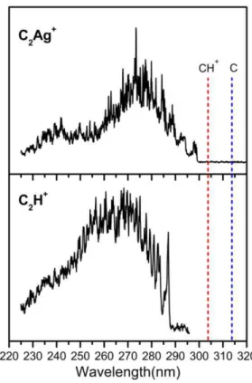
The photofragmentation spectra of C2H+ and C2Ag+ were recorded in the spectral range (225 – 320
nm) under similar experimental conditions for comparison are shown in Figure 1. In both cases the main fragmentation channel was the elimination of one neutral cytosine molecule.
The origins of the electronic transitions 286.9 nm for C2H+ and 299.1 nm for C2Ag+, are close and also
similar to the origin of the electronic transition of CH+ (303.5 nm)30 and C (314.2 nm).39 An enlarged view of
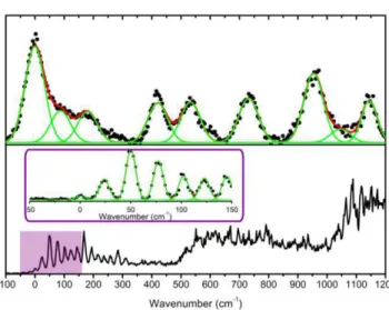
the spectra near the origin is shown in Figure 2, from which a very different vibronic structure and bandwidth can be observed for C2H+ and C2Ag+. The spectrum of the C2Ag+ complex shows a very low vibrational
frequency progression ( = 24 cm-1) and another one at 123 cm-1 which correspond to in-plane intermolecular bending modes (N-Ag+-N angle), 2= 33 cm-1 and 5= 95 cm-1 as calculated in the ground state geometry at the MP2/SV(P) theory level (see Figure S1 in the Supporting Information). It should be noted that this angle changes from 159° in the ground state optimized geometry to 165° in the A´ state optimized geometry which is the main geometrical change between both equilibrium structures.
Figure 1: Photofragmentation spectra of the C2Ag +
and C2H +
complexes in the whole spectral range analyzed in this work (225 - 320) nm. Vertical dashed lines show the wavelengths of the origins of the electronic transition of the individual components of both complexes, C39 and CH+30.The electronic transitions wavelength of Ag (328.2 nm)40 and Ag+ (110.7 nm,111.2 nm and 119.6 nm)41,42 fall out of scale.
The excited state lifetimes have been estimated, as in a previous study,30 from the widths of the bands
fitted to Voigt profiles to account for the rotational contour and the laser Gaussian bandwidth (11 cm-1)
convolution. The intrinsic Gaussian profile of the laser is shown in the Figure S2 (see Supporting Information) along with the experimental profiles recorded for both complexes. The relationship between the Lorentzian full width at half maximum (FWHM) and the excited state lifetime ( ) is given by the uncertainty principle, which can be written as:
12
1 1 5.3 10
( ) (2 c ) x ( )
where c stands for the speed of the light. This procedure is valid if it can be assumed that the broadening is only due to the excited state lifetime and not to spectral congestion (e.g. rotational contour or low frequency active vibrational modes). The spectrum of the C2Ag+ complex clearly shows that the spectral resolution is
good enough to resolve the different vibronic transitions, even in the case of vibrational frequencies as low as 24 cm-1, while the rotational broadening can be neglected since the temperature of the experiment is low (40
K)30. Therefore, the broadening in the C2H+ spectrum is due to non-radiative process leading to a short excited
state lifetime. From the bandwidth analysis, the Lorentzian FWHM for the C2Ag+ complex is less than 1 cm-1
and can be associated to an excited state lifetime > 5000 fs, while the bandwidth for the C2H+ complex is
FWHM = 62 cm-1 with an associated lifetime ≈ 85 fs, which is at least two orders of magnitude shorter than that of the former complex. This remarkable difference shows the effect of the Ag+ cation on the excited state dynamics of the complex. The long excited state lifetime estimated for the C2Ag+ complex is compatible with
high fluorescence quantum yield of DNA-Agn clusters, with C rich oligonucleotide strands.9,10,14,16 The lifetime
of the C2H+ complex is quite similar to the ones observed for the free protonated cytosine (133 ± 20) fs,30
indicating that it is not strongly perturbed by the dimer formation.
The electronic ground state structures of the protonated cytosine dimer43,44 and modified cytosine
dimer,43-44
45 and C
2Ag+46 in the gas phase have been previously determined by Infrared Multiphoton Dissociation
(IR-MPD) spectroscopy together with quantum chemical calculations. Both complexes are planar (Cs symmetry) and have equivalent structures, in which each C molecule is found in the keto-amino form with the NH groups where the glycosidic bonds are expected, in transoid orientation as in the case of the i-motif structure of DNA. The H+ as well as the Ag+ cation acts as a bridge between the hetero-nitrogen atoms in each C (Table 1). In addition, theoretical calculations have shown that these structures are the most stable ones in the electronic ground state.43,44,46,47
Figure 2: Low energy part of the spectra of the C2H +
(a) and C2Ag +
(b) complexes. In panel (b) the inset shows an amplification with better resolution of the first 150 cm-1 of the spectrum of the C
2Ag +
complex, in which a low vibrational frequency progression composed of narrow bands is observed. The vibrational bands were fitted to Voigt profiles.
To help the interpretation of the results and get more insight into the structure and excited state dynamics of these complexes, ground and excited state optimizations were performed at the MP2 and RI-ADC(2) (SV(P) basis set) theory levels, respectively.
The ground state optimization at the MP2 level, lead to the same planar structures as in previous studies at other levels of theory.43,44,46,47 The vertical energies (Evert) of the first excited states of
A´(symmetrical versus the molecular plane) and A” (antisymmetrical versus the molecular plane) symmetry
were also calculated (Table 1).
In many free neutral or protonated DNA bases, the S1 optimization leads to out-of-plane deformations
and to an avoided crossing between S1 and S0 states, which is responsible for a fast non-radiative decay.48 For
the C2H+ and C2Ag+ complexes, the size and complexity of the systems imposed to limit the calculation to the
accessible Franck-Condon region, so the excited states were only optimized in Cs (planar) symmetry since the ground state geometry is planar. Moreover, the A” and A’ states being very close in energy, any optimization
The optimization of the geometry in the A´ and A´´ excited states allows determining the equilibrium (lowest energy) structure in each excited state and then the Cs adiabatic transition energies (Ead), which is the
energy involved to reach the equilibrium geometry in the excited state from the equilibrium geometry in the ground state. Since the ground state is planar, they can be considered as the adiabatic potential accessible in the Franck–Condon window from the ground states and indeed the calculated Ead values are in good
agreement with the experimental transition energies (0-0exp). All the results are summarized in Table 1 and
Figure 3. As usual for this type of calculations the calculated adiabatic values are within 0.2 eV from the experimental ones.30
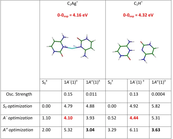
Table 1. Experimental and theoretical electronic excitations energies of C2Ag+ and C2H+ clusters for the first
A´and A” symmetry excited electronic states in planar Cs symmetry.
C2Ag+ 0-0exp = 4.16 eV C2H+ 0-0exp = 4.32 eV S0a 1A´(1)a 1A a S0a 1A´(1) a 1A a Osc. Strength 0.15 0.011 0.13 0.0004 S0 optimization 0.00 4.79 4.88 0.00 4.92 5.82 A´ optimization 1.10 4.10 3.93 0.52 4.44 5.31 A” optimization 2.00 5.32 3.04 3.29 6.11 3.63
Highlighted in bold-black are the Ead from the S0 to the first A” excited state and in bold-red those that
best match with the experimental values of the 0-0 transition that correspond to the transition from the S0 to
the first A´ symmetry excited state in both complexes. The locally excited electronic state (A’ symmetry) is a
* state in the C2H+ case while for C2Ag+, in addition it has a small partial CT character between both cytosine
molecules (Figure 4).
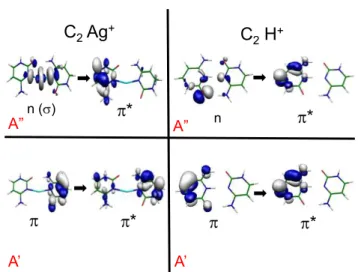
Figure 3: Scheme of the energy levels of C2Ag +
and C2H +
. For each complex, the left side corresponds to the vertical energies at the ground state equilibrium geometry and the right side corresponds to the energies of the ground, * and n * states at the A´( *) excited state optimized structure conserving the planar geometry. In the C2Ag
+
complex the * and n * states are very close in energy which might be related to the observed longer lifetime as compared with the C2H
+ complex. * * n ( ) C2Ag+ * n * C2H+ A’ A” A’ A”
In the case of the C2Ag+ complex, at the ground state equilibrium geometry the * state (A’ is lo er
in energy than the first (A Ag * state. Upon excited state optimization of the A’ states in Cs symmetry, the
first A” state becomes lower in energy than the optically allowed A’ state (Figure 3). We have observed a
similar behavior between two tautomers of protonated uracil: the enol/enol tautomer has a short excited state lifetime while the enol/keto has a longer one.30 In this latter tautomer the n * is lower in energy than
the * at the Cs * optimized geometry as in the case of C2Ag+. Upon slight out-of-plane deformation, the
optimization process of the A” state brings it back in its planar geometry, hile the A’ state te ds to be stabilized upon out-of-plane deformation. This may lead to a crossing between these states and to a barrier along the out-of-plane deformation coordinate.
On the other hand, in the case of the C2H+ complex, the nO * state (A stays higher in energy than the
* (A’ upon optimization of the A’ states in Cs symmetry. In absence of Cs symmetry both states are mixed and optimization of the S1 state leads to a crossing with the ground state. Maybe, there is a correlation
between the energy gap between the * and the n *, the lifetime being longer when the n * is slightly lower in energy at the * optimized geometry than when the n * is higher in energy. This should be tested theoretically but on simpler systems which less spectral congestion.
One may tentatively rationalize the variation of the excited state lifetime in the following way. The optical excitation prepares the * state in its Cs optimized geometry, i.e. planar or nearly planar. If there is no n * state lower in energy, the S1 * state will easily undergo out-of-plane deformations, which leads to
internal conversion. At the opposite if the n * is lower in energy than the * state, out-of-plane deformations will induce a crossing between these states, resulting in a barrier for the * state along the out-of-plane coordinate that will prevent the second crossing with the ground state at low excitation energies. Thus this state should have a longer lifetime.
This idea has to be tested by more elaborate calculations on this system or even on the simplest protonated DNA bases for which there is now some experimental information.30
In summary, the complexation of two cytosine molecules with Ag+ does not change drastically the
character and energy of the electronic transition as compared to C2H+ or CH+, contrary to the silver/gold
aminoacids case, for which a new absorption band appears in the near UV/visible spectral region (300 – 450 nm), depending on the specific system, attributed to CT excitations.31
323334
-3536
37 The present results are in agreement
with previous results17 that suggested that, in bulk systems, the UV excitation of highly fluorescent DNA-Ag n
clusters is due to absorption of the DNA bases. On the other hand, the Ag+ complexation significantly changes the excited state lifetime of the complex as compared with H+ complexation. This change is explained as a
consequence of the removal of a conical intersection between the excited and the ground states of C2Ag+
complex, which takes place in the C2H+ complex and leads to a fast non-radiative decay of the excited state of
the latter. Therefore, a higher fluorescence quantum yield is expected upon Ag+ complexation and this could be the reason for the high fluorescence of DNA-Agn clusters, although in the latter case the fluorescence is
observed upon chemical reduction of Ag+. However, it has been shown that cationic systems can give relevant
and detailed information to understand the optical properties of hybrid biomolecules-noble metal clusters. 31323334 -3536
37
The ultimate goal of noble-metal bioconjugation is to develop biosensing at the molecular level and gas phase studies can help to build model systems in a bottom-up strategy. In this regard, the next step will be to generate larger Agn+ clusters conjugated with DNA bases, in which some reduction processes has taken place.
Methodology
a. Experiment
The electronic spectra of the C2Ag+ and C2H+ complexes were obtained via parent ion photo-fragment
spectroscopy in a cryogenically-cooled quadrupole ion trap (Paul Trap from Jordan TOF Products, Inc.).49 The
setup is similar to the one developed in several groups based on the original design by Wang and Wang. 50-51
a solution of cytosine (500 µM) and silver nitrate (250 µM) in a methanol (50%)/water (50%) solvent . At the exit of the capillary, ions are trapped in an octopole trap for 90 ms. They are extracted by applying a negative pulse of c.a. 50 V and are further accelerated to 190 V by a second pulsed voltage just after the exit electrode. This time sequence of pulsed voltages produces ion packets with duration between 500 ns and 1 µs. The ions are driven by a couple of electrostatic lenses toward the Paul trap biased at 190 V so that the ions enter the trap gently avoiding fragmentation induced by collisions. A mass gate placed at the entrance of the trap allows selecting the parent ion. The Paul trap is mounted on the cold head of a cryostat (Coolpak Oerlikon) connected to a water-cooled He compressor. Helium as buffer gas is injected in the trap using a pulsed valve (General Valve) triggered 1 ms before the ions enter the trap as previously reported by Kamrath et al.51 The ions are trapped and thermalized at a temperature between 20 and 50 K through collisions with the cold buffer gas. The ions are kept in the trap for several tens of ms before the photodissociation laser is triggered. This delay is necessary to ensure thermalization of ions and efficient pumping of the He buffer gas from the trap to avoid collision induced dissociation of the ions during the extraction towards the 1.5 m long time-of-flight mass spectrometer. After laser excitation, the ions are stored in the trap for a delay that can be varied between 20 and 90 ms before extraction to the TOF mass spectrometer. The complete mass spectrum is recorded on a micro channel plate (MCP) detector with a digitizing storage oscilloscope interfaced to a PC. The photofragment yield spectrum of each detected ion is normalized to the parent ion signal and the laser power. The photo-dissociation laser is an OPO laser from EKSPLA, which has a 10 Hz repetition rate, 10 ns pulse width, a resolution of 10 cm-1 and a scanning step of 0.02 nm. The laser is shaped to a 1mm2 spot to fit the entrance hole of the trap and the laser power is around 20 mW in the UV spectral region.
b. Calculations
Ab initio calculations have been performed with the TURBOMOLE program package,54making use of the resolution-of-the-identity (RI) approximation for the evaluation of the electron-repulsion integrals.55 The
equilibrium geometry of the clusters and the vibrational frequencies in their ground electronic state (S0) were
determined at the MP2/ SV(P) level. Excitation energy and equilibrium geometry of the lowest excited singlet state (S1) were determined at the RI-ADC(2)/SV(P) level.
Structure optimizations were done by the quasi-Newton Raphson methods using the exact gradient vector and an approximation to the Hessian matrix as implemented in the TURBOMOLE program package.
Acknowledgements
This works was supported by ECOS-MinCyT cooperation program (A11E02) the ANR Research Grant (ANR2010BLANC040501), FONCyT, CONICET and SeCyT-UNC. We acknowledge the use of the computing facility cluster GMPCS of the LUMAT federation (FR LUMAT 2764).
Supporting Information Available:
Description and assignment of the low frequency vibrationalactive modes of the C2Ag+ cluster, and the intrinsic Gaussian profile of the laser together with the
experimental profiles recorded for both complexes. This material is available free of charge via the Internet at http://pubs.acs.org.
References
(1) Seeman, N. C. DNA in a Material World. Nature 2003, 421, 427–431.
(2) Li, X.; Liu, D. R. DNA-Templated Orga i “ thesis: Nature’s “trateg for Co trolli g Che i al Rea ti it Applied to Synthetic Molecules. Angew. Chem. Int. Ed. 2004, 43, 4848–4870.
(3) Lu, Y.; Liu, J. Functional DNA Nanotechnology: Emerging Applications of DNAzymes and Aptamers. Curr.
Opin. Biotechnol. 2006, 17, 580–588.
(4) Feldkamp, U.; Niemeyer, C. M. Rational Design of DNA Nanoarchitectures. Angew. Chem. Int. Ed. 2006,
45, 1856–1876.
(5) Niemeyer, C. M.; Mirkin, C. A. Nanobiotechnology: Concepts, Applications and Perspectives; Wiley-VCH: Weinheim, 2004.
(6) Takezawa, Y.; Shionoya, M. Metal-Mediated DNA Base Pairing: Alternatives to Hydrogen-Bonded Watson-Crick Base Pairs. Acc. Chem. Res. 2012, 45, 2066–2076.
(7) Liu, S.; Clever, G. H.; Takezawa, Y.; Kaneko, M.; Tanaka, K.; Guo, X.; Shionoya, M. Direct Conductance Measurement of Individual Metallo-DNA Duplexes within Single-Molecule Break Junctions. Angew.
Chem. Int. Ed. 2011, 50, 8886–88890.
(8) Park, K. “.; Ju g, C.; Park, H. G. Illusio ar Pol erase A ti it Triggered Metal Ions: Use for Molecular Logic-Gate Operations. Angew. Chem. Int. Ed. 2010, 49, 9757–9760.
(9) Miyake, Y.; Togashi, H.; Tashiro, M.; Yamaguchi, H.; Oda, S.; Kudo, M.; Tanaka, Y.; Kondo, Y.; Sawa, R.; Fujimoto, T.; et al. Mercury II-Mediated Formation of Thymine - HgII - Thymine Base Pairs in DNA
Duplexes. J. Am. Chem. Soc. 2006, 128, 2172–2173.
(10) Ono, A.; Shiqi, C.; Humika, T.; Tashiro, M.; Fujimoto, T.; Machinami, T.; Oda, S.; Miyake, Y.; Okamoto, I.; Ta aka, Y. “pe ifi I tera tio s et ee sil er I Io s and Cytosine–cytosine Pairs. Chem. Commun. 2008, 44, 4825–4827.
(11) Urata, H.; Yamaguchi, E.; Nakamura, Y.; Wada, S. Pyrimidine-Pyrimidine Base Pairs Stabilized by silver(I) Ions. Chem. Commun. 2011, 47, 941–943.
(12) Zheng, Y.; Yang, C.; Yang, F.; Yang, X. Real-Time Study of Interactions between Cytosine-Cytosine Pairs in DNA Oligonucleotides and Silver Ions Using Dual Polarization Interferometry. Anal. Chem. 2014, 86, 3849–3855.
(13) Xu, G.; Wang, G.; He, X.; Zhu, Y.; Chen, L.; Zhang, X. An Ultrasensitive Electrochemical Method for Detection of Ag+ Based on Cyclic Amplification of Exonuclease III Activity on Cytosine-Ag+-Cytosine.
(14) Petty, J. T.; Story, S. P.; Hsiang, J. C.; Dickson, R. M. DNA-Templated Molecular Silver Fluorophores. J.
Phys. Chem. Lett. 2013, 4, 1148–1155.
(15) Richards, C. I.; Choi, S.; Hsiang, J.; Antoku, Y.; Vosch, T.; Bongiorno, A.; Tzeng, Y.; Dickson, R. M. Oligonucleotide-Stabilized Ag Nanocluster Fluorophores. J. Am. Chem. Soc. 2008, 8, 5038–5039.
(16) Petty, J. T.; Sergev, O. O.; Nicholson, D. A.; Goodwin, P. M.; Giri, B.; McMullan, D. R. A Silver Cluster-DNA Equilibrium. Anal. Chem. 2013, 85, 9868–9876.
(17) O’Neill, P. R.; G i , E. G.; F ge so , D. K. UV E itatio of DNA “ta ilized Ag Cluster Fluorescence via the DNA Bases. J. Phys. Chem. C. 2011, 115, 24061–24066.
(18) Copp, S. M.; Schultz, D.; Swasey, S.; Pavlovich, J.; Debord, M.; Chiu, A.; Olsson, K.; Gwinn, E. Magic Numbers in DNA-Stabilized Fluorescent Silver Clusters Lead to Magic Colors. J. Phys. Chem. Lett. 2014,
5, 959–963.
(19) Ritchie, C. M.; Johnsen, K. R.; Kiser, J. R.; Antoku, Y.; Dickson, R. M.; Petty, J. T. Ag Nanocluster Formation Using a Cytosine Oligonucleotide Template. J. Phys. Chem. C. 2007, 111, 175–181.
(20) Oe ra si gh, “. “. R.; Markeše i , N.; G i , E. G.; Eliel, E. R.; dou eester, D. “pe tral Properties of Individual DNA-Hosted Silver Nanoclusters at Low Temperatures. J. Phys. Chem. C. 2012, 116, 25568– 25575.
(21) Vosch, T.; Antoku, Y.; Hsiang, J.; Richards, C. I.; Gonzalez, J. I.; Dickson, R. M. Strongly Emissive Individual DNA-Encapsulated Ag Nanoclusters as Single-Molecule Fluorophores. Proc. Natl. Acad. Sci.
U.S.A. 2007, 104, 12616–12621.
(22) Choi, S.; Dickson, R. M.; Yu, J. Developing Luminescent Silver Nanodots for Biological Applications.
Chem. Soc. Rev. 2012, 41, 1867–1891.
(23) Díez, I.; Ras, R. H. a. Fluorescent Silver Nanoclusters. Nanoscale 2011, 3, 1963–1970.
(24) Xu, H.; Suslick, K. S. Water-Soluble Fluorescent Silver Nanoclusters. Adv. Mater. 2010, 22, 1078–1082.
(25) Guo, S.; Wang, E. Noble Metal Nanomaterials: Controllable Synthesis and Application in Fuel Cells and Analytical Sensors. Nano Today 2011, 6, 240–264.
(26) Shang, L.; Dong, S.; Nienhaus, G. U. Ultra-Small Fluorescent Metal Nanoclusters: Synthesis and Biological Applications. Nano Today 2011, 6, 401–418.
(27) Latorre, A.; Somoza, Á. DNA-Mediated Silver Nanoclusters: Synthesis, Properties and Applications.
ChemBioChem 2012, 13, 951–958.
(28) Petty, J. T.; Zheng, J.; Hud, N. V; Dickson, R. M. DNA-Templated Ag Nanocluster Formation. J. Am. Chem.
(29) Kleinermanns, K.; Nachtigallová, D.; de Vries, M. S. Excited State Dynamics of DNA Bases. Int. Rev. Phys.
Chem. 2013, 32, 308–342.
(30) Berdakin, M.; Dedonder-lardeux, C.; Jouvet, C.; Pino, G. A. Excited States of Protonated DNA/RNA Bases. Phys. Chem. Chem. Phys. 2014, 16, 10643–10650.
(31) A toi e, R.; Ta ari , T.; dro er, M.; Dugourd, P.; Mitri , R.; do ači -Koutecký, V. Optical Properties of Gas-Phase Tryptophan – Silver Cations: Charge Transfer from the Indole Ring to the Silver Atom.
ChemPhysChem 2006, 7, 524 – 528.
(32) A toi e, R.; dertorelle, F.; dro er, M.; Co pag o , I.; Dugourd, P.; Kulesza, A.; Mitri , R.; do ači -Koutecký, V. Gas-Phase Synthesis and Intense Visible Absorption of Tryptophan –Gold Cations. Angew.
Chem. Int. Ed. 2009, 48, 7829–7832.
(33) Compagnon, I.; Tabarin, T.; Antoine, R.; Broyer, M.; Dugourd, P. Spectroscopy of Isolated, Mass-Selected Tryptophan-Ag3 Complex: A Model for Photoabsorption Enhancement in
Nanoparticle-Biomolecule Hybrid Systems. J. Chem. Phys. 2006, 125, 164326–164331.
(34) Mitri , R.; Peterse , J.; Kulesza, A.; do ači -koutecký, V.; Tabarin, T.; Compagnon, I.; Antoine, R.; Broyer, M.; Dugourd, P. Photoabsorption and Photofragmentation of Isolated Cationic Silver Cluster – Tryptophan Hybrid Systems. J. Chem. Phys. 2007, 127, 134301–13410.
(35) Mitri , R.; Peterse , J.; Kulesza, A.; do ači -Koutecký, V.; Tabarin, T.; Compagnon, I.; Antoine, R.; Broyer, M.; Dugourd, P. Absorption Properties of Cationic Silver Cluster – Tryptophan Complexes: A Model for Photoabsorption and Photoemission Enhancement in nanoparticle–Biomolecule Systems.
Chem. Phys. 2008, 343, 372–380.
(36) “a ader, ).; Mitri , R.; do ači -Koutecký, V.; Bellina, B.; Antoine, R.; Dugourd, P. The Nature of Electronic Excitations at the Metal–bioorganic Interface Illustrated on Histidine–silver Hybrids. Phys.
Chem. Chem. Phys 2014, 16, 1257–1261.
(37) do ači -Koute ký, V.; Kulesza, A.; Gell, L.; Mitri , R.; A toi e, R.; dertorelle, F.; Ha ouda, R.; Ra a e, D.; Broyer, M.; Tabarin, T.; et al. Silver Cluster-Biomolecule Hybrids: From Basics towards Sensors. Phys.
Chem. Chem. Phys 2012, 14, 9282–9290.
(38) “ hultz, D.; Gard er, K.; Oe ra si gh, “. “. R.; Markeše i , N.; Olsso , K.; De ord, M.; dou eester, D.; Gwinn, E. Evidence for Rod-Shaped DNA-Stabilized Silver Nanocluster Emitters. Adv. Mater. 2013,
25, 2797–2803.
(39) De Vries, M. S.; Hobza, P. Gas-Phase Spectroscopy of Biomolecular Building Blocks. Annu. Rev. Phy.
Chem. 2007, 58, 585–612.
(40) Fuhr, J. R.; Wiese, W. L. CRC Handbook of Chemistry and Physics; CRC Press: Boca Raton, USA, 1998. (41) Biémont, E.; Pinnington, E. H.; Kernahan, J. A.; Rieger, G. Beam-Laser Measurements and Relativistic
(42) Kramida, A. A Critical Compilation of Energy Levels, Spectral Lines, and Transition Probabilities of Singly Ionized Silver, Ag II. J. Res. Natl. Inst. Stand. Technol. 2013, 118, 168–198.
(43) Yang, B.; Rodgers, M. T. Base-Pairing Energies of Proton-Bound Heterodimers of Cytosine and Modified Cytosines: Implications for the Stability of DNA I-Motif Conformations. J. Am. Chem. Soc. 2013, 136, 282–290.
(44) Yang, B.; Wu, R. R.; Berden, G.; Oomens, J.; Rodgers, M. T. Infrared Multiple Photon Dissociation Action Spectroscopy of Proton-Bound Dimers of Cytosine and Modified Cytosines: Effects of Modifications on Gas-Phase Conformations. J. Phys. Chem. B 2013, 117, 14191–14201.
(45) Oomens, J.; Moehlig, A. R.; Morton, T. H. Infrared Multiple Photon Dissociation (IRMPD) Spectroscopy of the Proton-Bound Dimer of 1-Methylcytosine in the Gas Phase. J. Phys. Chem. Lett. 2010, 1, 2891– 2897.
(46) Berdakin, M.; Steinmetz, V.; Maitre, P.; Pino, G. A. Gas Phase Structure of Metal Mediated
(Cytosine)2Ag+ Mimics the Hemiprotonated (Cytosine)2H+ Dimer in I-Motif Folding. J. Phys. Chem. A
2014, 118, − .
(47) Megger, D. A.; Fonseca Guerra, C.; Bickelhaupt, F. M.; Müller, J. Silver(I)-Mediated Hoogsteen-Type Base Pairs. J. inorg. Biochem. 2011, 105, 1398–1404.
(48) Delchev, B. V.; Sobolewskib, A. L.; Domckea, W. Comparison of the Non-Radiative Decay Mechanisms of 4-Pyrimidinone and Uracil: An Ab Initio Study. Phys. Chem. Chem. Phys 2010, 12, 5007–5015.
(49) Alata, I.; Bert, J.; Broquier, M.; Dedonder, D.; Feraud, G.; Grégoire, G.; Soorkia, S.; Marceca, E.; Jouvet, C. J. Electronic Spectra of the Protonated Indole Chromophore in the Gas Phase. J. Phys. Chem. A 2013,
117, 4420–4427.
(50) Wang, X. B.; Wang, L. S. Development of a Low-Temperature Photoelectron Spectroscopy Instrument Using an Electrospray Ion Source and a Cryogenically Controlled Ion Trap. Rev. Sci. Instrum. 2008, 79, 073108–073109.
(51) Kamrath, M. Z.; Relph, R. A.; Guasco, T. L.; Leavitt, C. M.; Johnson, M. A. Vibrational Predissociation Spectroscopy of the H2-Tagged Mono- and Dicarboxylate Anions of Dodecanedioic Acid. Int. J. Mass
Spectrom. 2011, 300, 91–98.
(52) Choi, C. M.; Kim, H. J.; Lee, J. H.; Shin, W. J.; Yoon, T. O.; Kim, N. J.; Heo, J. Ultraviolet Photodepletion Spectroscopy of Dibenzo-18-Crown-6-Ether Complexes with Alkali Metal Cations. J. Phys. Chem. A 2009, 113, 8343–8350.
(53) Andersen, J. U.; Cederquist, H.; Forster, J. S.; Huber, B. A.; Hvelplund, P.; Jensen, J.; Liu, B.; Manil, B.; Maunoury, L.; Brøndsted Nielsen, S.; et al. Photodissociation of Protonated Amino Acids and Peptides in an Ion Storage Ring. Determination of Arrhenius Parameters in the High-Temperature Limit. Phys.
(54) Ahlrichs, R.; Bär, M.; Häser, M.; Horn, H.; Kölmel, C. Electronic Structure Calculations on Workstation Computers: The Program System Turbomole. Chem. Phys. Lett. 1989, 162, 165–169.
(55) , C. Geometry Optimizations with the Coupled-Cluster Model CC2 Using the Resolution-of-the-Identity Approximation. J. Chem. Phys. 2003, 118, 7751–12.