HAL Id: tel-01556586
https://tel.archives-ouvertes.fr/tel-01556586
Submitted on 5 Jul 2017
HAL is a multi-disciplinary open access archive for the deposit and dissemination of sci-entific research documents, whether they are pub-lished or not. The documents may come from teaching and research institutions in France or abroad, or from public or private research centers.
L’archive ouverte pluridisciplinaire HAL, est destinée au dépôt et à la diffusion de documents scientifiques de niveau recherche, publiés ou non, émanant des établissements d’enseignement et de recherche français ou étrangers, des laboratoires publics ou privés.
Biomarkers of Exposure in Environmental
Epidemiology : the case of the effects of prenatal
exposure to phenols and phthalates on pre- and
post-natal growth.
Claire Philippat
To cite this version:
Claire Philippat. Biomarkers of Exposure in Environmental Epidemiology : the case of the effects of prenatal exposure to phenols and phthalates on pre- and post-natal growth.. Human health and pathology. Université de Grenoble, 2013. English. �NNT : 2013GRENS014�. �tel-01556586�
Université Joseph Fourier / Université Pierre Mendès France / Université Stendhal / Université de Savoie / Grenoble INP
THÈSE
Pour obtenir le grade de
DOCTEUR DE L’UNIVERSITÉ DE GRENOBLE
Spécialité : Modèle, méthode et algorithmes en biologieArrêté ministériel : 7 août 2006
Présentée par:
Claire PHILIPPAT
Thèse dirigée par Rémy Slama
Préparée au sein de l'Institut Albert Bonniot (CRI INSERM/UJF U823). Equipe d'Epidémiologie Environnementale appliquée à la Reproduction et la Santé Respiratoire
dans l'école doctorale EDISCE
Utilisation des Biomarqueurs d'Exposition en
Epidémiologie Environnementale ; application à
l'étude des effets des expositions intra-utérines aux
phénols et aux phtalates sur la croissance pré- et
post-natale.
Biomarkers of Exposure in Environmental
Epidemiology; the case of the effects of prenatal
exposures to phenols and phthalates on pre- and
post-natal growth.
Thèse soutenue publiquement le 26 août 2013 devant le jury composé de :
Sylvaine Cordier Président et rapporteur Enrique Schisterman Rapporteur Joe Braun Membre du jury Jean-Luc Volatier Membre du jury Christophe Ribuot Membre du jury Rémy Slama
2
Acknowledgement
The general design of the EDEN cohort was made possible by financial support from la Fondation pour la Recherche Médicale, l'INSERM, l’Institut de Recherche en Santé Publique, Nestlé, The French Ministry of Health, l’Agence Nationale de la Recherche (ANR), l’Université Paris-Sud, l’Institut de veille sanitaire (InVS), the French Agency for Food, Environmental and Occupational Health and Safety (ANSES) and la Mutuelle Générale de l’Education Nationale. The PELAGIE cohort is funded by ANR, InVS, INSERM, and the Regional Council of Brittany. The SARAEH cohort was supported by funding from NIEHS. During my PhD, I was supported by grants from the French Ministry of Research and Region Rhône-Alpes.
I would like to thank first Rémy Slama, who supervised this PhD and introduced me to many concepts in Environmental Epidemiology. Many of the results presented here could not have been developed without his support. I also would like to thank the members of the team of Environmental Epidemiology (Inserm - University Joseph Fourier, U823), with whom I shared great scientific and human experiences and Stephanie Engel from University of North Carolina who welcomed me in her group during a few months of my PhD.
Finally, I would like to thank Sylvaine Cordier, Enrique Schisterman, Joseph Braun, Christophe Ribuot and Jean-Luc Volatier for having accepted to be members of the jury of my PhD.
4
Contents
1
CHAPTER I: INTRODUCTION ... 10
I. Pre- and post-natal growth in humans ... 14
1.Pre and post-natal growth as markers of well being ... 14
2.Determinants of pre- and early post-natal growth ... 17
II. Phenols and phthalates ... 21
1.Exposure in the general population ... 21
2.Fetal exposure ... 23
3.Metabolism ... 23
4.Suspected effects of prenatal exposure to phthalates and phenols on health ... 24
III. Effects of prenatal exposures to phthalates and phenols on pre- and post-natal growth...25
1.In vitro studies suggesting that phthalates and phenols interact with mechanisms implied in growth .. 25
2.Effects of prenatal exposures to phenols and phthalates on growth; evidence from animal studies ... 26
3.Effects of prenatal exposures to phenols and phthalates on growth, epidemiological studies ... 28
IV. Assessment of prenatal exposure to phenols and phthalates in Epidemiological studies ... 33
1.Exposure assessment to chemicals in epidemiological studies ... 33
2.Specific issues related to the use of biomarkers to assess prenatal exposure to phenols and phthalates 34 V. Objectives of the thesis ... 38
CHAPTER II: EFFECTS OF PRENATAL EXPOSURES TO PHTHALATES AND PHENOLS ON HUMAN GROWTH ... 40
I. Effects of prenatal exposures to phthalates and phenols on birth outcomes ... 44
1.Aims of the chapter ... 44
2.Population and Methods ... 44
3.Results... 51
4.Discussion... ... 61
5.Conclusion ... 64
II.Effects of prenatal exposures to phenols on fetal growth, estimated using repeated ultrasound measurements and early postnatal growth ... 65
1.Aims of the chapter ... 65
2.Population and methods ... 65
3.Results... ... 73
4.Discussion... . ...85
5.Conclusion... .. .89
1 Conformément aux consignes de l'école doctorale Edisce concernant les thèses rédigées en anglais, chaque chapitre est précédé d'un résumé en français.
5
CHAPTER III: ASSESSMENT OF PRENATAL EXPOSURE TO PHENOLS AND
PHTHALATES: METHODOLOGICAL ISSUES ... 90
I. Prenatal exposure to short half-life chemicals: concentrations in amniotic fluid and variability in phenol urinary concentrations during pregnancy. ... 93
1.Aims of the chapter ... 93
2.Population and methods ... 93
3.Results... .... 97
4.Discussion... ... 109
5.Conclusion...113
II. Impact of exposure misclassification and of number of urine samples collected on bias and power in biomarkers based epidemiological studies; a simulation study applied to short half-life endocrine disruptors ... 114
1.Background ... 114
2.Methods... 115
3.Results... 117
4.Discussion... 118
CHAPTER IV: OVERALL DISCUSSION ... 120
I. Prenatal exposure to phthalates and phenols and pre- and post-natal growth ... 122
1.Associations with birth outcomes ... 122
2.Associations with fetal biometry and early postnatal growth ... 123
3.Methodological issues ... 124
II. Variability in urine concentration and correspondence between urine and amniotic fluid concentration ... 125
III. Conclusion and research perspectives ... 127
1.Improving exposure assessment during fetal life ... 127
2.Link between the toxicological and epidemiological studies... 128
REFERENCES ... 130
ANNEXES ... 142
I. Curriculum vitae ... 144
II. PhD training and teaching ... 146
1.Catégorie : Insertion professionnelle ... 146
2.Formations... .146
III.Publications ... 147
1.Related to the thesis ... 147
6
List of abbreviations
AhR: Aryl hydrocarbon receptorARNT: Aryl hydrocarbon receptor nuclear translocator BP: Butyl paraben
BP3: Benzophenone-3 BPA: Bisphenol A
CAR: Constitutive Androstane Receptor
CDC: Centers for Disease Control and Prevention CI: Confidence interval
DEHP: Di(2-ethylhexyl) phthalate EP: Ethyl paraben
ER: Estrogen receptors GR: Glucocorticoid receptor
ICC: Intraclass correlation coefficient IOTF: International Obesity Task Force LOD: limit of detection
MBP: Mono-n-butyl phthalate MBzP: Monobenzyl phthalate
MCNP: Monocarboxy-isononyl phthalate MCOP: Monocarboxy-isooctyl phthalate MCPP: Mono(3-carboxypropyl) phthalate
MECPP: Mono(2-ethyl-5-carboxypentyl) phthalate MEHHP: Mono(2-ethyl-5-hydroxyhexyl) phthalate MEHP: Mono(2-ethylhexyl) phthalate
MEOHP: Mono(2-ethyl-5-oxohexyl) phthalate MEP: Monoethyl phthalate
MiBP: Mono-isobutyl phthalate MP: Methyl paraben
MW: Molecular weight PP: Propyl paraben
PPAR: Peroxisome Proliferator–Activated Receptors PXR: Xenosensors Pregnane X Receptor
RXR: Retinoid X receptor TCS: Triclosan
TR: Thyroid hormone receptor US: Ultrasound examination WHO: World Health Organization 2,4-DCP: 2,4-dichlorophenol 2,5-DCP: 2,5-dichlorophenol
∑HMW: Molecular sum of high molecular weight phthalates ∑LMW: Molecular sum of low molecular weight phthalates ∑PB: Molecular sum of parabens
7
List of figures
Figure 1: Summary of the complications related to childhood obesity (Lakshman et al. 2012). ... 16 Figure 2: Odds ratios (ORs) and 95% CIs of being overweight (IOTF) in 325 adolescent boys and girls for a
1-SD increase in weight growth velocity at different ages between 3 and 36 months (Botton et al. 2008). ... 17 Figure 3: Nuclear receptors implied in the control of growth and adipogenesis, and endocrine disruptors likely to interact with them (Casals-Casas and Desvergne 2011). ... 21 Figure 4: Bisphenol A concentration in maternal and fetal compartments during three hours of perfusion in an ex-vivo human placental perfusion system (Balakrishnan et al. 2010). ... 23 Figure 5: Metabolism of di(2-ethylhexyl) phthalate (DEHP) in Human (Koch et al. 2005) ... 24 Figure 6: Body weights of female (A, B) and male (C, D) offspring of Sprague-Dawley female rats exposed to 0.1 mg/kg of b.w/day ("low dose"), 1.2 mg/kg of b.w /day ("high dose ") of bisphenol A or to vehicle only (controls) during gestation and lactation (Rubin et al. 2001). ... 27 Figure 7: Schematic view of the different tools allowing to quantify exposure to the environmental chemicals in Environmental Epidemiology. ... 34 Figure 8: Concentrations of BPA and MEP (µg/g creatinine) for all of the spot urine samples collected from one participant over one week (Preau et al. 2010; Ye et al. 2011a) ... 37 Figure 9: Design of the study population for the birth outcome analyses (Gona_PE study, EDEN and PELAGIE cohorts, n = 287) ... 45 Figure 10: Birth weight as a function of MCPP and MECPP concentrations standardized for sampling conditions and coded as restricted cubic splines (Gona_PE study, n = 287 mother-child pairs from EDEN and PELAGIE cohorts, 2002-2006). ... 53 Figure 11: Adjusted associations between 2,5-DCP, 2,4-DCP and BP3 maternal urinary concentrations (tertiles) standardized for sampling conditions and birth weight (Gona_PE study, n = 191 mother-child pairs from EDEN cohort, 2003-2006). ... 56 Figure 12: Birth weight as a function of bisphenol A concentrations standardized for sampling conditions, categroized in tertile and coded as restricted cubic splines (Gona_PE study, n = 191 mother-child pairs from EDEN cohort, 2003-2006). ... 57 Figure 13: Study population for fetal and postnatal growth analyses (PEnDevE study, n = 520 mother-child pairs from the EDEN cohort). ... 66 Figure 14: Fitted weight and height growth curves between birth and 3 years for 5 randomly selected children of the EDEN cohort. ... 68 Figure 15: Biparietal diameter (mm) and estimated fetal weight (kg) as a function of gestational age at measurement among 520 mother-child pairs from EDEN cohort. ... 71 Figure 16: Adjusted associations between phenols and weight and head circumference at birth in different subpopulations of the EDEN cohort (transversal analysis). ... 84 Figure 17: Assumed associations of maternal pregnancy bisphenol A level with child weight at 12 months and potential confounding by maternal and child eating behaviors. ... 88 Figure 18: Box plots of the distribution of specific-gravity corrected urinary phenol concentrations are presented according to whether detectable levels of phenol metabolites were measured in amniotic fluid taken at the same time.. ... 100 Figure 19: Means of urine specific gravity and urine creatinine concentrations by gestational age (SARAEH study, 2005 to 2008). ... 103 Figure 20: Distribution of the β parameter characterizing the association between exposure and birth weight using the average pregnancy exposure Ei or only one urine sample to assess exposure to chemicals with ICC of 0.6 and to chemicals with ICC of 0.15. ... 117
8
List of tables
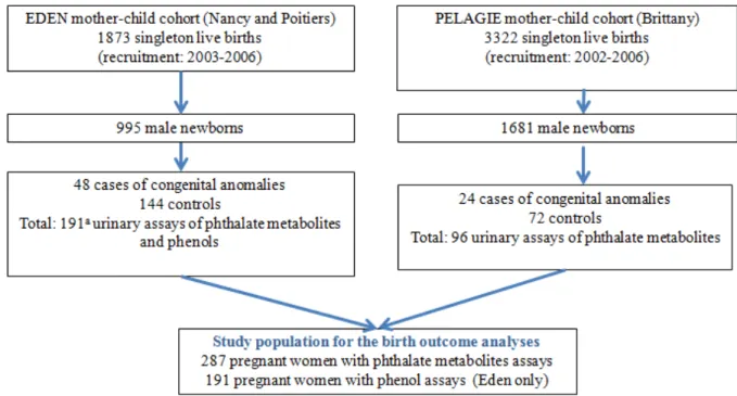
Table 1: Definitions of overweight and obesity commonly used in study of weight and adiposity in children. ... 15 Table 2: Non exhaustive list of sources of exposure to the phthalates and phenols studied in this thesis ... 22 Table 3: Studies of associations between prenatal exposures to phthalates and fetal growth in the general population ... 31 Table 4: Studies regarding the effects of prenatal exposures to phenols on pre- and post-natal growth in the general population ... 32 Table 5: Name of the metabolites (and of their parent compounds) assessed in urine of the pregnant women from the EDEN and PELAGIE cohorts. ... 46 Table 6: Characteristics of French pregnant women and of their offspring (Gona_PE study, n = 287 mother-child pairs from EDEN and PELAGIE cohorts, 2002-2006). ... 51 Table 7: Urinary phenol (n = 191) and phthalate (n = 287) biomarker concentrations after correction for
case-control sampling (Gona_PE study, n = 287 mother-child pairs from EDEN and PELAGIE cohorts, 2002-2006)... 52 Table 8: Adjusted associations between maternal urinary concentrations of phthalate biomarkers standardized for sampling conditions and birth outcomes (Gona_PE study, n = 287 mother-child pairs from EDEN and PELAGIE cohorts, 2002-2006). ... 54 Table 9: Adjusted associations between maternal urinary concentrations of phenol biomarkers standardized for sampling conditions and birth outcomes (Gona_PE study, n = 191 mother-child pairs from EDEN cohort, 2003-2006). ... 58 Table 10: Characteristics of the whole EDEN cohort delivering boys and comparison with PEnDevE and Gona_PE population. ... 74 Table 11: Descriptive statistics of growth measurements in the PEnDevE population (n = 520, EDEN cohort, 2003-2006). ... 75 Table 12: Correlations between pre- and post-natal growth measurements in the PEnDevE population (n = 520 male newborns from the EDEN cohort, 2003-2006). ... 77 Table 13: Concentrations of phenol biomarkers in maternal urine in the PEnDevE population (EDEN cohort, 2003-2006, n = 520). ... 78 Table 14: Adjusted associations between phenol maternal urinary concentrations and pre- and post-natal size in the PEnDevE population (EDEN cohort, 2003-2006, n = 520 male newborns)... 81 Table 15: Adjusted associations between phenol maternal urinary concentrations and growth rate in the PEnDevE population (EDEN cohort, 2003-2006, n = 520 male newborns). ... 82 Table 16: Characteristics of the 71 women with three repeated urine measurements during pregnancy (SARAEH study, 2005 to 2008).. ... 97 Table 17: Distribution of phenol concentrations (µg/L) measured in amniotic fluid and in urine collected at three time periods during pregnancy among women of the SARAEH study, 2005 to 2008. ... 99 Table 18: Adjusted associations between ln-transformed amniotic fluid concentrations of selected phenols and their potential predictors among 68 pregnant women from the SARAEH study, 2005 to 2008. ... 102 Table 19: Intraclass Correlation Coefficients for phenol biomarker concentrations measured in urine samples collected at several times during pregnancy (n = 71 women from the SARAEH study, 2005 to 2008). ... 104 Table 20: Spearman correlation coefficients between urine samples collected at specific time intervals using specific gravity corrected, creatinine corrected and uncorrected phenol concentrations (SARAEH study, 2005 to 2008). ... 105 Table 21: Spearman correlation coefficients between pairs of ln-transformed phenol concentrations from the first, second and third samplings (specific gravity corrected, creatinine corrected and uncorrected for urine dilution), SARAEH study, 2005 to 2008. ... 106 Table 22: Adjusted geometric mean of urinary phenol concentrations according to collection conditions among 213 urine samples from the 71 women of Study of Advanced Reproductive Age and Environmental Health, 2005 to 2008. ... 108 Table 23: Statistical power and effect estimate (β) characterizing the association between birth weight and exposure assessed during pregnancy (simulation study, n = 500 women, 1000 simulations) ... 118
10
12 Introduction
La période prénatale ainsi que les premières années de vie sont considérées comme des périodes de sensibilité importante aux expositions à de nombreux facteurs environnementaux. Des modifications survenues durant cette période pourraient entrainer des effets à court mais aussi à long terme sur la santé, ainsi que formulé dans le contexte de l'hypothèse DOHaD ("Developmental Origins of Health
and Diseases", (Hanson and Gluckman 2008)). Cette hypothèse considère que les effets chez l'adulte
associés à des expositions survenues pendant la vie fœtale pourraient s'expliquer par une adaptation du développement en réponse à un environnement intra-utérin défavorable. Cette adaptation développementale est favorable pendant la période intra-utérine mais, à cause de modification physiologiques et/ou métaboliques, pourrait entrainer des conséquences ultérieures néfastes (Barker 2004, 2007; Gluckman and Hanson 2004). C'est une des raisons pour lesquelles il est important de caractériser les événements et expositions survenant durant la grossesse et leur impact potentiel sur le développement du fœtus et de l'enfant dans les premières années de vie.
L'événement de santé étudié dans le cadre de ce travail de thèse est la croissance durant la vie fœtale et les premières années de vie. Une altération de la croissance fœtale est considérée comme un marqueur non spécifique des agressions subies pendant la vie fœtale. Un faible poids de naissance a été associé à un risque plus élevé de mortalité infantile, ainsi qu'à un risque plus élevé de développer des pathologies de type obésité, diabète, troubles cardiovasculaire à l'âge adulte (Barker 2007). Le poids à la naissance est un paramètre facilement mesurable et est couramment utilisé comme estimateur de la croissance fœtale dans les études épidémiologiques. Néanmoins, le poids est une mesure globale ne permettant pas d'identifier un effet sur une partie spécifique du corps et des mesures complémentaires telles que la taille et le périmètre crânien à la naissance sont souvent utilisées de concert. Une limitation à l'utilisation des mesures à la naissance est qu'elles ne fournissent pas d'information directe concernant des périodes spécifiques du développement du fœtus. L'utilisation de mesures échographique répétées pendant la grossesse permet de mettre en évidence des périodes de sensibilité au cours la vie fœtale. De telles mesures ont déjà été utilisées en épidémiologie environnementale pour évaluer les effets des polluants atmosphériques sur le développement fœtal (Aguilera et al. 2010; Hansen et al. 2008; Iniguez et al. 2012b; Slama et al. 2009; van den Hooven et al. 2012), mais encore très peu en relation avec des perturbateurs endocriniens à courte demi-vie (Snijder et al. 2013). La croissance post-natale précoce a été identifiée comme un facteur de risque d'obésité infantile (Botton et al. 2008; Giles et al. 2013). L'obésité infantile est un problème de santé publique important. Cette pathologie, dont la prévalence augmente dans le monde, est associée à d'autres problèmes de santé graves dans la jeunesse, mais aussi plus tard dans la vie adulte, comme le diabète, l'hypertension artérielle et les maladies coronariennes (Barker 2007; Bibbins-Domingo et al. 2007). En plus des déterminants connus de la croissance pré- et post-natal (génétique, alimentation...), l'exposition à des polluants chimiques est de plus en plus suspectée d'affecter les trajectoires de croissance.
Les contaminants environnementaux étudiés dans ce travail appartiennent aux familles des phénols et des phtalates. Ils font partie des composés chimiques dont le niveau de production est très élevé (de l'ordre de 4 millions de tonnes dans le monde pour le Bisphénol A en 2006) et sont présents dans un vaste éventail de produits de consommation courante. Les phtalates de poids moléculaire inférieur à < 250 g / mol sont utilisés dans les produits de soins personnels (parfums, cosmétiques) et dans les industries pharmaceutiques (enrobage de certain médicaments) tandis que les phtalates de haut poids moléculaire (> 250 g / mol) sont plutôt retrouvés dans les revêtements de sols et de murs, les emballages alimentaires et des dispositifs médicaux (A. M. Calafat et al. 2006; Hauser and Calafat 2005).
13
Pour ce qui est des phénols, le bisphénol A est utilisé dans la fabrication des résines époxy couvrant l'intérieur des boîtes de conserve et dans certains plastiques de type polycarbonate. D'autres phénols sont utilisés pour leurs propriétés antibactériennes et de conservateurs dans les cosmétiques (parabènes) et les savons (triclosan). La benzophénone-3 est utilisée comme filtre anti rayons ultra-violets dans les crèmes solaires (Calafat et al. 2008). Ainsi l'exposition à ces composés est ubiquitaire. Ces substances et / ou leurs métabolites ont été mis en évidence dans la quasi totalité des urines testées en population générale, incluant des femmes enceintes (Casas et al. 2012; Woodruff et al. 2011). Le fœtus est lui aussi exposé et certains métabolites ont été détectés dans le sang du cordon ombilical, le liquide amniotique ou le méconium des nouveau-nés (Latini et al. 2003; Zhang et al. 2009).
Les phénols et les phtalates ont été identifiés comme étant des perturbateurs endocriniens. Ils peuvent interagir avec les récepteurs hormonaux, mais aussi la synthèse, la sécrétion, le transport, la fixation et l'élimination des hormones naturelles qui jouent un rôle important dans de nombreuses fonctions telles que le métabolisme, le comportement, la fertilité ou la croissance.
Une étude réalisée au sein d'une population de femmes enceintes new-yorkaise avait suggéré un effet de ces composés sur la croissance fœtale estimée à l'aide de mesures à la naissance; l'effet de certains phénols sur le poids de naissance était différent suivant le sexe de l'enfant (Wolff et al. 2008). Une seule étude s'était intéressée aux effets des expositions prénatales aux phénols sur la croissance postnatale (Harley et al. 2013), les autres études étant transversales (Teitelbaum et al. 2012; Trasande et al. 2012). Dans l'étude publiée par Harley et al (2013) une association négative entre les concentrations maternelles de bisphénol A et le risque d'être en surpoids à 9 ans a été mis en évidence chez les filles.
Les objectifs de ce doctorat étaient:
1) d'étudier les associations entre les concentrations urinaires maternelles de phenols et de phtalates et la croissance pré et post-natale de l'enfant (chapitre II);
2) de comparer les concentrations de phénols dosées dans l'urine maternelle à celle dosées dans le liquide amniotique, recueillis le même jour (Chapitre III);
3) d'étudier la variabilité des concentrations urinaires de polluants environnementaux non persistants (phénols) durant la grossesse (Chapitre III);
4) de caractériser le biais associé à l'utilisation d'un seul échantillon urinaire pour estimer l'exposition durant la grossesse, qui est l'approche utilisée dans la plupart des études épidémiolgiques publiées à cette date (Chapitre III).
Introduction
14
I.
Pre- and post-natal growth in humans
1. Pre and post-natal growth as markers of well being a) Prenatal growth
Fetal growth is the reflect of healthy development and is considered to be a non-specific marker of exposures and mechanisms taking place during fetal life (Gluckman and Hanson 2004; Hanson and Gluckman 2008). Birth weight is easy to collect and is often used as a surrogate measure for fetal growth in epidemiological studies. Term low birth weight (< 2500 g) has been linked with a higher risk of baby's mortality (Wilcox 2001), but also with occurrence of several chronic conditions including cardiovascular diseases, type 2 diabetes, obesity and hypertension (Barker 1995, 2004; Godfrey and Barker 2001). These results are in line with the Developmental Origins of Health and Diseases (DOHaD) hypothesis, which suggests that exposures during fetal life can result in long term effects on health. These long term effects may result from fetal developmental adaptations, which are beneficial during the intrauterine period, but may lead to permanent changes in body’s structure, physiology and metabolism and thus may promote development of diseases later in life (Barker 2004, 2007; Gluckman and Hanson 2004).
Historically birth weight was analyzed independently of gestational duration, making it difficult to distinguish low birth weight due to restricted fetal growth from low birth weight due to preterm birth (Adams et al. 2010; Savitz et al. 2002). In studies on the effects of environmental factors on fetal growth, birth weight is now usually corrected for gestational age. In addition to low birth weight, metrics including reduction in birth weight (continuous variable), very low birth weight (< 1500 g) and small for gestational age (birth weight lower than the 10th percentile of a suitable sex- and gestational age-specific weight reference distribution) are commonly used in epidemiological studies. Weight at birth does not allow to identify an effect on a specific body part or organ and additional measurements of neonatal anthropometry at birth such as length and head circumference provide additional information. A limitation to the use of birth outcomes in epidemiological studies is that they are measured at the end of intrauterine life. They therefore do not provide direct information on specific periods of fetal development (Hemachandra and Klebanoff 2006) and effects occurring at specific times of pregnancy could be missed. In addition, developmental
Introduction
15
programming may occur independently of birth weight. Animal experiments have indeed shown that fetuses from mothers with nutrition restriction during early pregnancy may have normal weight at birth, but exhibited increased body weight and fat deposition later in life (Ford et al. 2007; Reynolds and Caton 2012). Repeated measurements of fetal anthropometry during pregnancy by ultrasound examinations may allow to highlight windows of heightened sensitivity to environmental contaminants during fetal life. Such measurements have already been used in environmental epidemiology to assess the effects of air pollutants on fetal development (Aguilera et al. 2010; Hansen et al. 2008; Iniguez et al. 2012b; Slama et al. 2009; van den Hooven et al. 2012) and very recently to characterize the effect of short half-life endocrine disruptors (Snijder et al. 2013).
b) Early postnatal growth and link with childhood obesity
The definition of overweight and obesity given by the World Health Organization (WHO) is an abnormal or excessive fat accumulation that presents a risk to health. A crude measure of overweight and obesity is given by the body mass index (BMI): an adult with a BMI above 25 kg/m2 is defined as overweight; a BMI above 30 kg/m2 is defined as obesity. During childhood, body composition strongly varies with age and sex. Child's weight status is therefore usually determined using an age- and sex-specific references rather than the BMI categories used for adults. Several definitions of child's obesity and overweight exist; the most common used are those from the WHO, the Centers for Disease Control and Prevention (CDC) and the International Obesity Task Force (IOTF) (Flegal and Ogden 2011).
Table 1: Definitions of overweight and obesity commonly used in study of weight and
adiposity in children.
Organization Definition of Childhood Obesity and overweight
World Health Organization Between birth to age 5:
Obese: BMI > 3 standard deviations above the WHO growth standard median
Overweight: BMI > 2 standard deviations above the WHO growth standard median
Centers for Disease Control and
Prevention Between 2 to 19 years: Obese: BMI ≥ 95th percentile.
Overweight: BMI between the 85th and the 95th percentiles International Obesity Task
Force Age 2 to 18: Provides international BMI cut points by age and sex for overweight and obesity, the cut points correspond to an adult BMI of 25 (overweight) or 30 (obesity)
Introduction
16
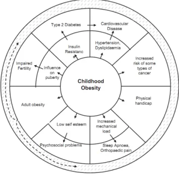
The prevalence of childhood overweight and obesity has strongly increased in developed countries. Surveys during the 1990s show that in Europe an additional 1 % of the entire child population became overweight each year and 15 % of the European school-aged children are estimated to be carrying excess body fat; among them a quarter are obese (IOTF definition (Lobstein et al. 2004)). Obesity is associated with other health troubles during youth and obese children and adolescents are likely to suffer from related comorbidities (Figure 1). These children are also more likely to be obese as adults (Bibbins-Domingo et al. 2007; Haslam and James 2005; Lakshman et al. 2012; Reilly et al. 2003; Singh et al. 2008).
Figure 1: Summary of the complications related to childhood obesity (Lakshman et al. 2012).
Comorbidities of childhood obesity are depicted in the outer ring with their intermediate processes in the inner ring.
Several epidemiological studies have shown an association between rapid growth during the first years of life and the development of obesity later in the childhood (Botton et al. 2008; Giles et al. 2013; Ong et al. 2000; Taveras et al. 2009), suggesting that early post-natal growth is a risk factor for childhood obesity (Figure 2). Two periods in particular (up to 6 months and from 2 years onward) have been associated with a later risk of overweight (Botton et al. 2008). Such results highlight the importance of studying determinants of overweight in early life.
Introduction
17
Figure 2: Odds ratios (ORs) and 95% CIs of being overweight (IOTF) in 325 adolescent boys
and girls for a 1-SD increase in weight growth velocity at different ages between 3 and 36 months (logistic model adjusted for sex, (Botton et al. 2008)).
Early post-natal growth can be studied by collecting repeated measures of weight and height during childhood. Such measurements are easy to collect, especially in countries where they are regularly reported by the family pediatrician in the personal child health booklet (such as France). Skin fold thickness and abdominal circumference are not routinely measured. They can provide complementary information on the body fat composition and are also interesting to study early post-natal growth (Hauspie et al. 2004).
2. Determinants of pre- and early post-natal growth a) Fetal growth
Fetal growth results from a complex interplay among factors including genetic, maternal, placental, hormonal and environmental factors (Kramer 1987; Sacks 2004; Williams 2005). A non-exhaustive overview of these determinants is given in this chapter.
Genetic influences
A study has reported that around 50% of the variation in weight, length and head circumference at birth may be explained by genetic factors (n = 101,748 parent-offspring pairs, data from Medical Birth Registry of Norway from 1967 to 2004). Among this share of
Introduction
18
50%, the fetal genome might explain about 30% of the normal variation in birth weight while the maternal genome would explain the other 20% (Lunde et al. 2007). Infant sex is associated with fetal growth and at birth boys are on average 150 g heavier than girls (Valero De Bernabé et al. 2004). Several fetal chromosomal anomalies including trisomy 21, 18 and Turner’s syndrome usually result in fetal growth retardation. Conversely, other genetic disorders can lead to pre- and/or post-natal over-growth. This is the case of the Beckwith-Wiedemann syndrom, caused by the disruption of imprinting of the short arm of chromosome 11, leading to over activity of the Insuline like Growth Factor-2 (IGF-2) gene (Gicquel and Le Bouc 2006).
Placental influences
The placenta is the main interface between mother and fetus during pregnancy and fetal growth is related to its capacity to transfer oxygen and nutrients to the fetus (Jansson and Powell 2007; Reece and Hobbons 2007). The placenta also produces a wide spectrum of hormones involved in fetal growth regulation (e.g., IGF-I, IGF-II, placental growth hormone, (Murphy et al. 2006; Newbern and Freemark 2011)) and modifications in placental functions are likely to lead to fetal growth alterations.
Endocrine regulation of fetal growth
A wide range of hormones influence growth and maturation, beginning in-utero and continuing throughout childhood. Hormones, such as IGFs, insuline, thyroid hormones and oestrogens control the rate of cell proliferation, apoptosis and differentiation in many tissues, they could either stimulate or inhibit growth in-utero.
Maternal (non genetic) influences
Maternal behavioral factors affecting fetal growth include alcohol consumption and active smoking during pregnancy, which has been associated with decrease in offspring birth weight (Mills et al. 1984; Rahmalia et al. 2012; Suzuki et al. 2008). Good maternal health is essential for normal fetal development. Pathologies limiting nutrients and oxygen transfer to the fetus, including preeclampsia and anemia, negatively affect fetal growth. Conversely infants born to mothers with gestational diabetes are more likely to be large for gestational age (Gillman et al. 2003). Other maternal factors associated with size at birth include maternal pre-pregnancy weight, maternal age and parity; the first child weights on average less than the
Introduction
19
second and third children (Reece and Hobbons 2007; Thame et al. 2004; Valero De Bernabé et al. 2004)
Environmental factors
Environmental factors suspected to influence fetal growth include biological factors, microorganisms can cross the placenta and several agents including toxoplasma, rubella, cytomegalovirus and herpes simplex have been associated with fetal growth disorders, and chemical factors. We will focus here on the chemical environment.
There is growing concern that environmental contaminants might interact with developmental processes and fetal growth (Hanson and Gluckman 2008; Woodruff et al. 2010). Research in Humans is at a somewhat advanced stage for air pollutants, metals and persistent endocrine disrupters (DDT, PCBs)). Among them, lead (Bellinger 2005), PCBs
(Govarts et al. 2012) and air pollutants, notably the fine particulate matter of diameter ≤ 2.5 µm (Dadvand et al. 2013; Shah and Balkhair 2011; Slama and Cordier In Press) have
been identified as possibly inhibiting fetal growth. Exposure to environmental tobacco smoke has also been associated with an increased risk of having a baby with low birth weight (Salmasi et al. 2010). Data in Human are much more limited for the so-called "emerging chemicals", including brominated flame retardants, perfluorinated compounds, phthalates and phenols and do not allow to conclude yet about their potential effects on fetal growth (Slama and Cordier In Press). Specific review of the effects of phthalates and phenols, two families of non persistent endocrine disruptors, on fetal growth is available section III of this chapter (definition of endocrine disruptors is detailed page 18).
b) Post-natal growth, childhood obesity
Influences of prenatal growth on postnatal growth
Early postnatal growth is related to fetal growth. Smaller babies at birth are more likely to show an increased growth rates during early infancy compared to normal size newborns, what is commonly called catch up growth (Eleftheriades et al. 2006; Hindmarsh et al. 2008; Mook-Kanamori et al. 2011).
Factors influencing prenatal growth also suspected to influence postnatal growth
As for prenatal growth, postnatal growth is controlled by hormonal (e.g., growth hormone) and genetic factors (Hauspie et al. 2004; K Ong et al. 2002). Infant sex is associated
Introduction
20
with postnatal growth (Regnault et al. 2010). Regarding childhood obesity it has been suggested that inheritance may account for around 25 to 40% of the BMI inter-individual variability (Maffeis 2000). Several other factors affecting fetal growth are suspected to affect postnatal growth. This is in particular the case for maternal smoking, maternal and paternal height and parity. As an example, a lower weight at 3 months (Regnault et al. 2010), but not later in childhood (Hindmarsh et al. 2008; Ong et al. 2000) has been reported for infants prenatally exposed to tobacco smoke. Regarding parity, infants of primiparous pregnancies, which are generally smaller and thinner at birth, exhibit an important postnatal catch-up in weight and length, which results in larger size during childhood (Ong et al. 2000). It is not well known in which extent these effects are mediated by effects on prenatal growth.
Postnatal factors
Postnatal factors, especially nutrition, play an important role in early post-natal growth. Marked differences in growth rates have been reported as early as at the age of 2-3 months between infants who were breast- or bottle-fed. Breastfeed infants usually have lower weight growth velocity than formula-fed infants do. The difference in weights between these two groups is still observed at 31 months of age (KK Ong et al. 2002; Regnault et al. 2010).
Environmental chemicals
Exposure to industrial chemicals during critical periods of development has been recently mentioned as a potential risk factor for obesity development; this particularly concerns endocrine disruptors (Casals-Casas and Desvergne 2011; DiVall 2013; Newbold 2010; Newbold et al. 2008).
Endocrine disruptors have been defined as exogenous substances that alter function(s) of the endocrine system and consequently cause adverse health effects in an intact organism, or its progeny, or (sub)populations (definition from the International Program on Chemicals (IPCS 2002)). Endocrine disruptors interfere with the normal functioning of the endocrine system by affecting the synthesis, secretion, transport, metabolism and elimination of the natural hormones; mimicking their action and / or interfering with their receptors. Both natural (e.g., phytoestrogens) and man-made substances have been identified to be endocrine disruptors.
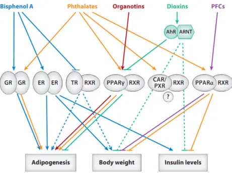
Some endocrine disruptor chemicals are in particular able to interact with receptors implied in adipogenenesis, lipid accumulation and metabolism (e.g., Peroxisome
Proliferator-Introduction
21
Activated Receptors (PPARs), Estrogen Receptors, Figure 3) and may induce epigenetic modifications in obesity-related genes (Casals-Casas and Desvergne 2011; DiVall 2013). For these reasons, they are considered potential obesogens. Endocrine disruptor chemicals can be categorized according to their intended use, their structural properties and / or their persistence in the environment and organism. Persistent endocrine disruptors suspected to act as metabolic disruptors include dioxins, PCBs, organochlorine pesticides (Casals-Casas and Desvergne 2011). We will focus here on two families of short-lived endocrine disruptors: phthalates and phenols.
Figure 3: Nuclear receptors implied in the control of growth and adipogenesis, and endocrine
disruptors likely to interact with them (Casals-Casas and Desvergne 2011).
PPAR: Peroxisome Proliferator–Activated Receptors; ER: estrogen receptors, TR: Thyroid hormone receptor, GR: Glucocorticoid receptor, PXR: xenosensors Pregnane X Receptor, CAR: Constitutive Androstane Receptor, AhR: Aryl hydrocarbon receptor, ARNT: Aryl hydrocarbon receptor nuclear translocator; RXR: Retinoid X receptor.
II.
Phenols and phthalates
1. Exposure in the general population
Esters of phthalic acid, commonly named phthalates, are a group of chemicals with a wide spectrum of industrial applications (Table 2). Low molecular weight (MW) phthalates (MW < 250 g / mol) are primarily used in personal care products (perfumes, cosmetics)and as coating for pharmaceutical products while high MW phthalates (MW > 250 g / mol) are
Introduction
22
mostly used in polyvinylchloride floor and wall covering, food packaging and medical devices (A. M. Calafat et al. 2006; Hauser and Calafat 2005). Bisphenol A, benzophenone-3, triclosan, dichlorophenols and parabens are phenolic compounds used in lot of common products: food packaging and polycarbonate plastics (bisphenol A), cosmetics (parabens), soap (triclosan) and sunscreen (benzophenone-3, (Calafat et al. 2008)). Precursors of dichlorophenols are used as intermediates in the production of several herbicides and insecticides (Agency for Toxic Substances and Disease 2006).
Exposure to these chemicals is prevalent in the general population, including pregnant women (Adibi et al. 2008a; Braun et al. 2011b; Cantonwine et al. 2010; Wolff et al. 2008; Ye et al. 2009; Ye et al. 2008).
Table 2: Non exhaustive list of sources of exposure to the phthalates and phenols studied in
this thesis.
Chemicals Sources of exposure, French and EU regulation
PHTHALATES
Di-ethyl phthalate Personal care products (fragrances, cosmetics), coatings (pharmaceutical products), dyes
Di-butyl phthalate Personal care products, lacquers, vanishes, coating (pharmaceutical products), banned from toys and childcare products (2005/12/14)a
Di-isobutyl phthalate Polish, lacquers
Di-n-octyl phthalate Medical devices, cosmetics, food packaging
Butyl-benzyl phthalate Vinyl flooring, adhesives and sealants, personal care products, food packaging, industrial solvents, banned from toys and childcare products (2005/12/14)a Di(2-ethylhexyl) phthalate PVC plastics used in household products (floor tiles, furniture), food packaging,
medical devices, banned in toys and childcare products (2005/12/14)a Di-isodecyl phthalate Anticorrosive paints
Di-isononyl phthalate Sealants, lacquers, paints
PHENOLS
2,4-dichlorophenol Metabolite of 1,3 dichlorobenzene: herbicides, pesticides, minor contaminant of 1,4-dichlorobenzène
2,5-dichlorophenol Metabolite of 1,4 dichlorobenzene: pesticides, toilet-deodorizer blocks, banned from mothballs (2009/08/21)b
Bisphenol A Polycarbonate plastics and epoxy resins: food packaging, toys, medical tubing, dental fillings, banned from baby bottles (2010/07/01)c and food containers for children below 3 years
Benzophenone-3 Sun cream, food packaging
Triclosan Antibacterial and antifungal agent: soap, deodorant, toothpaste
Parabens
Methyl paraben Ethyl paraben Propyl paraben Butyl paraben
Antibacterial agents, food and cosmetic preservatives
Phthalates were ranged in order of increasing molecular weight. Abbreviations: PVC : polyvinyl chloride a Directive 2005/84/CE
b Directive 2007/565/CE
Introduction
23
2. Fetal exposure
Human studies have shown fetal exposure to phenols and phthalates. Metabolites of these chemicals have been detected in amniotic fluid (Engel et al. 2006; Huang et al. 2009; Jensen et al. 2012; Silva et al. 2004a; Yamada et al. 2002), cord blood (Latini et al. 2003) and meconium (Zhang et al. 2009). This was corroborated by experimental data showing that several phthalates and phenols are able to cross the placenta (Balakrishnan et al. 2010; Mose et al. 2007; Takahashi and Oishi 2000). In an ex-vivo experimental system, where 7 human placentas were in contact with a perfusate enriched in bisphenol A (10 µg/L), authors observed rapid transfer of this phenol across the placenta (Figure 4).
Figure 4: Bisphenol A concentration in
maternal and fetal compartments during three hours of perfusion in an ex-vivo human placental perfusion system
(Balakrishnan et al. 2010).
3. Metabolism
Phenols and phthalates have short half-lives in Human, generally estimated to be less than 24 hours (Janjua et al. 2008; Koch et al. 2004; Sandborgh-Englund et al. 2006; Volkel et al. 2002). To our knowledge none of these studies have specifically explored half-lives in pregnant women, which may differ from those observed in men and non-pregnant women because of the metabolic and physiologic changes occurring during pregnancy. The short half-life reported in Human is a priori a desirable feature in terms of reducing exposure levels, but creates strong challenges when it comes to assessing exposure levels, since these will vary very quickly throughout the day.
After exposure, phthalates and phenols are rapidly metabolized and mostly excreted in urine. Phenols are usually excreted unchanged or conjugated (conjugation increases water solubility and therefore increases urinary excretion). Phthalate biotransformation is related to their molecular weight: low molecular weight phthalates are changed into their hydrolytic monoesters, which can be excreted in urine or undergo conjugation. The metabolism of the
Introduction
24
higher molecular weight phthalates is more complex and results in more metabolites (Figure 5). High molecular weight phthalates are also metabolized to hydrolytic monoesters, but then undergo enzymatic oxidation of the alkyl chain, to produce oxidative metabolites (Hauser and Calafat 2005).
Figure 5: Metabolism of di(2-ethylhexyl) phthalate (DEHP) in Human (major metabolites
are highlighted, (Koch et al. 2005))
4. Suspected effects of prenatal exposure to phthalates and phenols on health
Phthalates and phenols are known to be endocrine disruptors. They interact, among others with sex hormone receptors and therefore research first focused on their potential effects on reproductive development and sex differentiation. Experimental studies have suggested that phthalates may induce developmental abnormalities of the male genital tract (Foster 2006; Swan 2008). One study in 106 boys (Swan 2008) has suggested male reproductive toxicity (decreased anogenital distance) while another did not (cases-control study: n = 21 cases of hypospadias and 50 cases of undescended testis (Chevrier et al. 2012)).
The endocrine system is related to a wide range of organism functions and it has been recently shown that phenols and phthalates may impact other facets of health than reproduction. Experimental studies have reported effects on brain organization and behavior (Wolstenholme et al. 2011; Wolstenholme et al. 2012), respiratory health (Nakajima et al.
Introduction
25
2012) and growth (Rubin and Soto 2009). In Human, epidemiological studies have reported associations between exposures to phthalates and phenols during perinatal periods and child neurodevelopment. For example, low molecular weight phthalates, measured in maternal urine collected during pregnancy, have been associated with a higher risk of developing behavioral, attention and depressive problems in children between 4 and 9 years (n = 137, (Engel et al. 2010; Miodovnik et al. 2011)). Prenatal exposure to bisphenol A has been associated with more anxious and depressed behaviors at 3 years (Braun et al. 2011a). Bisphenol A and phthalates have also been associated with respiratory health (Jaakkola and Knight 2008; Kwak et al. 2009; Spanier et al. 2012) and prenatal growth (Wolff et al. 2008). This thesis focuses on the effects of these chemicals on pre- and early post-natal growth.
III.
Effects of prenatal exposures to phthalates and phenols on pre- and
post-natal growth
1. In vitro studies suggesting that phthalates and phenols interact with mechanisms implied in growth
In vitro, phthalates have shown to be weak agonists of the estrogenic receptors ER∝ and ERβ implied among others in growth and maintenance of a diverse range of tissues (mammary gland, uterus, bone, cardiovascular system...). These chemicals shown antagonist activities on androgen (National Institute of Health and Medical Research 2011) and thyroid receptors (Shen et al. 2009); they also interact with the triiodothyronine (T3) transportation (Boas et al. 2010; Ishihara et al. 2003). Fetal growth is regulated among other by thyroid hormones (Blazer et al. 2003; Shields et al. 2011). Phthalates are partial agonists of peroxisome proliferator-activated receptors (PPAR∝ and PPARγ) involved in lipid metabolism, adipocyte differentiation and energy homoeostasis. PPAR∝ activation has been associated with the induction of fatty acid hydroxylase (CYP4A family) involved in several metabolic pathways (Desvergne et al. 2009; Hurst and Waxman 2003)).
Regarding phenols, affinity with estrogen receptors have been reported for benzophenone-3, triclosan and parabens (Kunz and Fent 2006; Witorsch and Thomas 2010); the estrogenic activity of parabens depends on the length of the alkyl group (ER affinity: butylparaben > propylparaben > ethylparaben > methylparaben (Routledge et al. 1998; Witorsch and Thomas 2010)). A recent in vitro study has reported that parabens promote adipocyte differentiation in mice cells by an activation of glucocorticoid receptors and / or
Introduction
26
PPARγ (Hu et al. 2012; Taxvig et al. 2012). Bisphenol A is one of the most studied phenols. Additionally to its effects on estrogen receptors, it can inhibit androgen (Sohoni and Sumpter 1998) and thyroid receptors (Moriyama et al. 2002; Pearce and Braverman 2009). For this phenol, in vitro studies have also suggested an agonist effect on glucocorticoid receptors, notably implied in adipocyte differentiation (Sargis et al. 2010).
Recent studies have suggested that effects of phthalates and phenols on obesity might be mediated by epigenetic modifications (DiVall 2013; Fleisch et al. 2012; Manikkam et al. 2013; Singh and Li 2012). As an example, prenatal exposure to bisphenol A has induced hypomethylation leading to an increased expression of the Agouti gene, inducing obesity and diabetes in mice (Dolinoy 2008).
2. Effects of prenatal exposures to phenols and phthalates on growth; evidence from animal studies
Originally, animal studies aiming at assessing phthalate effects on health did not specifically focus on growth. However authors usually reported pup's weight as secondary outcome.
a) Phthalates
In rodents, exposure to several phthalates (di-iso-butyl phthalate, di-n-butyl phthalate and benzylbutyl phthalate) during the prenatal period has been inversely associated with fetal weight (Ema and Miyawaki 2002; Saillenfait et al. 2003, 2006, 2008). In another study, exposure to low dose of mono-(2-ethylhexyl) phthalate (MEHP, dose: 0.05 mg/kg of body weight/day), a metabolite of DEHP, has been associated with birth weight and fat mass increases in male offspring; no effect was observed with higher doses of MEHP (0.25 or 0.5 mg/kg of body weight/day, (Hao et al. 2013)). Two other studies, using high dose of DEHP (pregnant rats and mice were exposed to 50 mg/kg/day and 750 mg/kg/day) did not observe effect on weight at birth nor later in life (Gray et al. 2000; Tanaka 2005).
b) Phenols
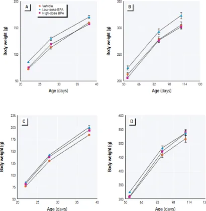
Exposure to bisphenol A (0.1 and 1.2 mg/kg of body weight/day) during perinatal period (day 6 of pregnancy through the period of lactation) has been positively associated with weight at birth (Rubin and Soto 2009; Rubin et al. 2001). This association remained later
Introduction
27
in life and tended to be non-monotonic, especially in female newborns: the effect of bisphenol A was more marked in the group exposed to 0.1 mg compared to the group exposed to 1.2 mg/kg of body weight/day (Figure 6). Birth and postnatal weight increase in association with exposure to bisphenol A during pregnancy has also been reported with lower doses of exposure (around 0.25 µg/kg of body weight/day, (Ryan et al. 2010) and 70 µg/kg/day (Somm et al. 2009)). Reports of body weight reduction at birth existed, however at high doses (300 and 1000 mg/kg) and the authors did not see any effect with the dose of 100 mg/kg (Kim et al. 2001). Data were sparse regarding effects of the other phenols. In rodents, weight at birth and in early life (postnatal day 7) decreased after perinatal (gestational and lactational periods) exposure to 2,4-dichlorophenol (Aoyama et al. 2005). 1,4-dichlorobenzene, a precursor of 2,5-dichlorophenol has been associated with birth weight decrease (Agency for Toxic Substances and Disease 2006).
Figure 6: Body weights of female (A, B) and male (C, D) offspring of Sprague-Dawley
female rats exposed to 0.1 mg/kg of body weight/day ("low dose"), 1.2 mg/kg of body weight/day ("high dose ") of bisphenol A or to vehicle only (controls) during gestation and lactation (Rubin et al. 2001).
Introduction
28
3. Effects of prenatal exposures to phenols and phthalates on growth, epidemiological studies
a) Phthalates
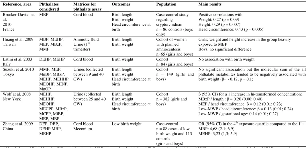
Findings of the studies aiming at studying the associations between prenatal exposure to phthalates and birth outcomes in the general population are summarized in Table 3. Several studies have reported positive associations between phthalates and birth outcomes (Brucker-Davis et al. 2010; Huang et al. 2009; Wolff et al. 2008). In an American cohort study of 404 mother–infant pairs, authors have reported a positive association between monobenzyl phthalate (MBzP), measured in maternal urine and birth length and between monoethyl
phthalate (MEP) and head circumference at birth (Wolff et al. 2008). In another study with a
small sample size (n = 32 female and 33 male newborns), a significant birth weight and birth length increase has been observed in female newborns with the highest concentrations of mono-butyl phthalate (MBP) in amniotic fluid (concentration > median level); similar but not significant associations have been observed in male newborns (Huang et al. 2009). The concentration of MBP, measured in cord blood, has been positively correlated to weight, length and head circumference at birth in a study restricted to male newborns (n = 86, (Brucker-Davis et al. 2010)). Reports of inverse association also exist for the precursor of MBP measured in cord blood, which has been associated with an increase risk of low birth weight (< 2500 g) (Zhang et al. 2009). Suzuki et al. (2010) have studied the association between the molecular sum of phthalate metabolites measured in maternal urine and birth weight, and suggested a negative association (n = 149 boys and girls).
Comparisons across these studies are difficult because different matrices, including maternal urine, cord blood and amniotic fluid, have been used to measure biomarkers of exposure (Table 3). Sometimes the parent phthalate rather than its metabolites has been measured. This could lead to exposure misclassification. In the case of external contamination of the biological samples, levels of parent chemicals will be impacted but not those of their metabolites.
Associations between phthalate exposure and early postnatal growth have only been explored in a cross-sectional study. No data regarding exposure during fetal life was available (Teitelbaum et al. 2012). No clear association has been observed between any of the nine assessed phthalate metabolites (measured once in the child's urine collected between 6 and 8
Introduction
29
years) and body mass index, waist circumference and height between 6 and 8 years (n = 361, (Teitelbaum et al. 2012)).
b) Phenols
The findings of the studies aiming at studying the association between prenatal exposure to phenols and pre- and post-natal growth in human are summarized in Table 4. Most of the studies used birth measurements as proxies of fetal growth and focused on bisphenol A. Only two studies had fetal biometry measured during pregnancy (Lee et al. 2010; Snijder et al. 2013).
The two studies with ultrasound measurements reported a negative association between prenatal exposure to bisphenol A and fetal head circumference (Lee et al. 2010; Snijder et al. 2013). Snijder et al. (2013) further reported a negative association with fetal weight. A negative association with fetal abdominal circumference has been observed in the study by Lee et al (2010). The negative associations with weight and head circumference reported in the study by Snijder et al. (2013) were only observed in the 80 subjects in whom three measurements of bisphenol A concentrations during pregnancy were used to assess exposure and no association was observed when only one (n = 219) or two (n = 120) samples were used. Inversely, in a study among 339 pregnant women from New-York city, maternal urinary concentrations of bisphenol A tended to be positively associated with birth weight (p < 0.2, (Wolff et al. 2008)). Dichlorophenols, benzophenone-3 and triclosan have been measured in this cohort. An inverse association between 2,5-dichlorophenol and birth weight, and a positive association between benzophenone-3 and birth weight in male, but not in female newborns, have been observed.
Only one study explored the potential effects of prenatal exposures to phenols on post-natal growth. Bisphenol A concentrations measured in maternal urine collected during pregnancy have been associated with a decrease risk of being overweight at 9 years. This association was observed in girls but not in boys (Harley et al. 2013). Bisphenol A concentrations were also measured in the urine of the children collected at 9 years. A positive association with overweight at age 9 have been reported in boys and girls. This positive associations as well as those observed in a cross-sectional study (Trasande et al. 2012) have to be interpreted cautiously because of reverse causality. Overweight children may consume more canned drink or processed foods, which are important sources of bisphenol A, than
non-Introduction
30
overweight children. In addition, obesity and associated health disorders might affect bisphenol A metabolism, storage and elimination.
To summarize, only few human studies have explored the effects of prenatal exposures to phenols and phthalates on pre- and early post-natal growth, and most of them had a limited sample size (Tables 3, 4). Some of the reported associations might be due to false positive discovery induced by the high number of comparisons performed (high number of biomarkers assessed and high number of outcomes, especially for studies using repeated measurements of fetal biometry during pregnancy). Another limitation of these studies is the exposure assessment, usually based on a small numbers of maternal urine samples collected during pregnancy.
Introduction
31
Table 3: Studies of associations between prenatal exposures to phthalates and fetal growth in the general population Reference, area Phthalates
considered
Matrices for phthalate assay
Outcomes Population Main results
Brucker-Davis et al.
2010 France
MBP Cord blood Birth length
Birth weight Head circumference at birth Case-control study regarding cryptorchidism n = 86 controls (boys only)
Positive correlations with Weight: 0.27 (p = 0.09) Height: 0.29 (p = 0.007)
Head circumference: 0.43 (p = 0.005) Huang et al. 2009
Taiwan MBP, MEHP, MEP, MBzP,
MMP
Amniotic fluid
Urine (1st
trimester)
Birth length
Birth weight Cohort of women with planned
amniocentesis n=65 (girls and boys)
Girls: weight and height increase in the group heavily exposed to MBP
Boys: no significant difference Latini et al. 2003
Italy DEHP, MEHP Cord blood Birth weight Cohort n=84 (girls and boys) No association with birth weight
Suzuki et al. 2010 Tokyo MMP, MEP, MnBP, MBzP, MEHP, MEHHP MEOHP, MINP, MnOP Urines (collected between 9 and 40 GW) Birth length Birth weight Head circumference at birth Cohort n = 149 (girls and boys)
No significant association but the molecular sum of the all phthalate metabolites tended to be negatively associated with birth weight (β= - 0.12, p = 0.1) Wolf at al. 2008 New York MEHP, MEHHP, MEOHP, MECPP, MBzP, MCPP, MiBP, MEP, MBP Urine (collected between 25 and 40 GW) Birth length Birth weight Head circumference at birth Cohort n = 382 (girls and boys)
β (95% CI) for a 1 increase in ln-transformed concentration: MBzP / length : β = 0.20 (0.00; 0.40)
MEP / head circumference: β = 0.12 (0.01; 0.23) Low-MWP / head circumference: β = 0.13 (0.01; 0.24) Low-MWP / gestational age: 0.14 (0.01; 0.27)
Zhang et al. 2009 China DEP, DBP, DEHP MBP, MEHP Cord blood Meconium
Low birth weight Case-control
n = 88 cases of low birth weight and 113 controls
(girls and boys)
OR (95% CI) in the 4th exposure quartile compared to the 1st:
MBP: 4,68 (2.1; 6.9) MEHP: 3,23 (1,3; 5.9)
Abbreviations : OR : odds ratio, GW: gestational week, MBP: mono-n-butyl phthalate, MBzP: monobenzyl phthalate, MCNP: monocarboxyisononyl phthalate, MCOP: monocarboxy-isooctyl phthalate, MCPP: mono(3-carboxypropyl) phthalate, MECPP: mono(2-ethyl-5-carboxypentyl) phthalate, MEHP: mono(2-ethylhexyl) phthalate, MEHHP: mono(2-ethyl-5-hydroxyhexyl) phthalate, MEOHP: mono(2-ethyl-5-oxohexyl) phthalate, MEP: monoethyl phthalate, MiBP: mono-isobutyl phthalate, ∑LMW: molecular sum of low molecular weight phthalates ∑HMW: molecular sum of high molecular weight phthalates
Introduction
32
Table 4: Studies regarding the effects of prenatal exposures to phenols on pre- and post-natal growth in the general population Reference, area Phenols considered Matrices for phenol assay Outcomes Population Main results
Chou et al. 2011
Taiwan BPA
Maternal and cord blood collected at birth
Birth weight Small for gestational age
Birth cohort n = 97 (girls and boys)
BPA in maternal blood was negatively correlated with weight U-shaped association between BPA (quartile) and the risk to be small for gestational age in males.
Harley et al. 2013
United States BPA Maternal and child urine
BMI, waist
circumference, body fat, obesity
n = 311, 9 year-old children (boys and girls)
Prenatal exposure: negative association with BMI- Z-score, waist circumference and with the risk of being obese in girl but not in boys.
Postnatal exposure (at age 9): positive association with BMI Z-score and waist circumference (not modified by sex). Lee et al. 2010
Korea Abstract only
BPA Maternal blood and urine
Biparietal and abdominal diameter, femur length measurements at 19 and 36 GW n = 125 (girls and boys)
Negative association with:
Head circumference in late pregnancy: β = - 0.06, p = 0.02 Abdominal circumference in late pregnancy: β = - 0.32, p = 0.02) Femur length in middle pregnancy: β = - 0.04, p = 0.04
Padmanabhan et al. 2008
Michigan (USA) BPA
Maternal blood collected at birth
Birth weight General population n = 40 (girls and
boys) No correlation between BPA concentrations and birth weight.
Wolff et al. 2008 New York 2,4-, 2,5-DCP BPA BP3 TCS Maternal urines collected between 25 and 40 GW. Birth length Birth weight Head circumference at birth Cohort n = 339 (girls and boys)
Boys : weight and length decreased with exposure to 2,5-DCP, weight increased with exposure to BP3
Girls: weight decreased with exposure to BP3 Snijder et al. 2013
Netherlands BPA Maternal urines (1 to 3 samples).
Ultrasounds (T2 and T3) and birth measurements of weight and head circumference
n = 80, 120 or 219 depending of the analysis
When analyses were restricted to the 80 fetuses with 3 exposure assessments: Negative association with weight and HC (p <0.1), which disappeared if using only 1 or 2 of the 3 samples were used. Abbreviations: GW: gestational week, BPA: bisphenol A, BP3: benzophenone-3, DCP: dichlorophenol, TCS: Triclosan, T2: second trimester of pregnancy, T3: third trimester of pregnancy,
Introduction
33
IV.
Assessment of prenatal exposure to phenols and phthalates in
Epidemiological studies
1. Exposure assessment to chemicals in epidemiological studies
Exposure assessment is a central issue in epidemiological studies aiming at exploring the effects of environmental contaminants on human health; exposure misclassification may indeed lead to bias in the health risk estimates and impacts statistical power (de Klerk et al. 1989; White et al. 2008). There are several methods to assess exposure to environmental contaminants and the choice of a particular one will depend on the characteristics, sources and routes of exposure to the studied chemical(s) (Figure 7). Studies focusing on atmospheric pollutants usually estimate chemical concentrations to which the participants may have been exposed (external dose) using environmental and/or personal measurements. This approach is not well adapted to chemicals with multiple sources and routes of exposure, such as phenols and phthalates. However, phthalate measurements in indoor air have been used in few previous studies exploring their effects on respiratory health (Bornehag et al. 2004; Kolarik et al. 2008). Other methods, such as questionnaires, could be used to estimate environmental exposure to pollutants. For chemicals with multiple sources of exposure, exposure assessment through questionnaires is not easy and is likely to suffer of important lack of precision. Job-exposure matrices are usually used in studies aiming at assessing professional Job-exposure and allow to infer exposure on the basis of the job title, the industry and / or calendar times of employment (Nieuwenhuijsen 2003; Snijder et al. 2012).
Human biomonitoring consists in the assessment of the levels of chemicals or their metabolites in human biological samples such as blood or urine and provides an estimate of the dose that actually enters the body (internal dose) from all sources and routes of exposures. For these reasons, it is the most relevant option to assess exposure to phenols and phthalates (A.M. Calafat et al. 2006). Biomonitoring was primarily used in occupational medicine. Improvements in analytical techniques have allowed detection of relatively low chemical concentrations in human fluid and biomarkers of exposure are now widely used in epidemiological studies in the general population (Angerer et al. 2007; A. M Calafat et al. 2006; Schisterman and Albert 2012). The use of biomarkers does not guarantee an absence of exposure misclassification. Biomonitoring indeed only provides a snapshot in time of a