HAL Id: hal-00504363
https://hal.archives-ouvertes.fr/hal-00504363
Submitted on 20 Jul 2010
HAL is a multi-disciplinary open access archive for the deposit and dissemination of sci-entific research documents, whether they are pub-lished or not. The documents may come from teaching and research institutions in France or abroad, or from public or private research centers.
L’archive ouverte pluridisciplinaire HAL, est destinée au dépôt et à la diffusion de documents scientifiques de niveau recherche, publiés ou non, émanant des établissements d’enseignement et de recherche français ou étrangers, des laboratoires publics ou privés.
Simulation of dynamic orofacial movements using a
constitutive law varying with muscle activation
Mohammad Ali Nazari, Pascal Perrier, Matthieu Chabanas, Yohan Payan
To cite this version:
Mohammad Ali Nazari, Pascal Perrier, Matthieu Chabanas, Yohan Payan. Simulation of dynamic orofacial movements using a constitutive law varying with muscle activation. Computer Meth-ods in Biomechanics and Biomedical Engineering, Taylor & Francis, 2010, 13 (4), pp.469-482. �10.1080/10255840903505147�. �hal-00504363�
Simulation of dynamic orofacial movements using a constitutive law
varying with muscle activation
NAZARI Mohammad Ali
1,2, PERRIER Pascal
1, CHABANAS Matthieu
1and PAYAN Yohan
31. GIPSA-lab, Speech and Cognition Department, CNRS UMR 5216 / Grenoble Institute of Technology, France
2. Mechanical Engineering Department, Faculty of Engineering, University of Tehran, Iran
3. TIMC-IMAG, CNRS UMR 5525 / Université Joseph Fourier, Grenoble, France1 Correspondance: Mohammad Ali NAZARI, GIPSA-lab Département Parole & Cognition, Domaine universitaire BP 46, 38402 Saint Martin d‟Hères, France
This paper presents a biomechanical model of the face to simulate orofacial movements in speech and non-verbal communication. A 3D finite element model, based on medical images of a subject, is presented. A hyperelastic Mooney-Rivlin constitutive law accounts for the non linear behaviour of facial tissue. Muscles fibres are represented by piece-wise uniaxial tensile elements, which generate force. The stress stiffening effect, an increase of the stiffness of the muscles when activated, is modelled by varying the constitutive law of the tissue with the level of activation of the muscle. A large number of facial movements occurring during speech and facial mimics are simulated. Results show that our modelling approach provides a realistic account of facial mimics. The differences between dynamic versus quasi-static simulations are also discussed, proving that dynamic trajectories better fit experimental data.
Keywords: face biomechanics; orofacial movements; muscle active force; hyperelastic modelling.
1. Introduction
Orofacial gestures, produced by articulators such as the tongue, jaw and lips,
are of primary importance in speech communication. By their position at the
extremity of the vocal tract, lips have a major influence on the acoustic signal
generated by the airflow coming from the lung. In addition, seeing facial gestures
directly influence the perception of speech. Listeners perceive and interpret the
produced speech via a combination of auditory and visual processing, a strategy well
demonstrated by the well-know McGurk effect (McGurk and MacDonald 1976).
Consequently an important field in speech communication research is the
development of synthetic models of the human face. The accuracy of these models is
a major requirement, both for production and perception of speech. In this context,
due to the strong influence of 3D lip horn geometry on the spectral characteristics of
speech signals, special attention has to be devoted the modelling of the lips region.
Moreover, it has been shown that the dynamic of the movements is very important in
the perception of facial expressions (Munhall and Vatikiotis-Bateson 1998; Ambadar
et al. 2005). Hence, synthetic speaking faces have to well account for the temporal
course of face shaping.
Contrary to empirical models based on recorded data or medical images (for
example Lucero et al. (2005) or Badin et al. (2008)), our approach in the last decade
has been to develop biomechanical models of speech articulators, which are as close
as possible to the human anatomy and functional morphology. Special emphasis has
been given to the representation of the muscular structures and the rheological
properties of soft tissues. Another major contribution has dealt with the motor control
system involved in the production of speech and orofacial movements: which muscles
have to be contracted to obtain a given acoustic signal or facial mimic? What must be
the intensities, variations and sequencing of the motor commands? Former works
were focused on a biomechanical modelling of the tongue, first in 2D then in 3D, and
the controlled activation of its muscles to generate complex articulatory paths (Payan
and Perrier 1997; Perrier et al. 2003; Buchaillard et al. 2009). In continuation of these
works, this paper presents a biomechanical model of the face which enables the
generation of facial movements in response to muscles activations. The bases of the
finite element model (mesh, mechanical properties and boundary conditions), which
entirely focused on the active components, namely the muscles: their representation,
mechanism of contraction, and the evolution of their mechanical properties with
activation. Simulations of different orofacial movements are presented in section 4,
before the discussion and conclusion.
2. Main structure of the model
Many physically-based models of the human face were developed in the
framework of computer graphics facial animation (Lee et al. 1995; Sifakis et al.
2005), computer aided surgery (Chabanas et al. 2003; Gladilin et al. 2004) or speech
production study (Lucero and Munhall 1999; Gomi et al. 2006).
The pioneer work of Lee et al. (1995) has made popular a discrete modelling
framework, with sparse mass-spring entities regularly assembled inside facial tissues.
This approach allows fast computations with a simple algorithmic implementation.
However, in addition to the lack of accuracy of such models and to their numerical
instabilities, it seems to be very difficult to set their elastic parameters (the stiffness of
springs) in order to fit the constitutive behaviour that is observed and measured on
living tissues. Recently Kim & Gomi (2007) have improved Gomi et al.‟s (2006)
discrete model by implementing a so-called “continuum compatible” mass-spring
model with stiffness parameters that can be adjusted in order to follow a simple linear
continuum constitutive law. Although this model is interesting in computational
terms, especially for dynamic simulations, it is limited to correctly reproduce the
behaviour of highly non-linear material such as facial tissues (Fung 1993; Gerard et
al. 2005).
In continuity with the works of Chabanas et al. (2003) and Sifakis et al.
(2005), we have chosen to use the Finite Element method to model the continuous
computationally less efficient, it enables in particular the use of non linear mechanical
modelling such as hyperelastic laws to better approximate the tissues behaviour
(Humphrey and Yin 1989; Weiss et al. 1996; Yucesoy et al. 2002; Blemker et al.
2005).
Our implementation is based on the ANSYS release 11.0 software.
2.1 Mesh of the passive tissues
The main mesh is a Finite Element (FE) discretization of the volume defined
by the facial tissues located between the skull and the external skin surface of the
face. It is based on a previous continuous face model developed by Chabanas et al.
(2003) in the context of computer aided maxillo-facial surgery. The outer and inner
surfaces of the mesh were extracted from a CT scan of a female adult subject. The
volume delimitated by these two surfaces was then manually meshed, as regularly as
possible, with hexahedral and wedge elements (figure 1). Anatomically, the face can
be considered as the superposition of three distinct layers of tissues, namely (from the
internal to the external layer) the hypodermis, dermis and epidermis (Stranding 2005).
The mesh is thus also built in three discrete layers of elements. The external one
corresponds to the epidermis (very thin) and dermis parts while the two internal layers
model the hypodermis, which will later include the facial musculature (section 3). The
mesh is composed of 6342 brick elements (6024 hexahedron and 318 wedges) based
on 8720 nodes. In order to reduce the number of DOF during simulation the mesh was
assumed to be symmetrical along the sagittal plane, which seems reasonable in the
context of speech production.
2.2 Mechanical properties
Element material properties are assumed to follow a hyperelastic law (Fung
1993). A simplified 5 parameters Mooney-Rivlin model is used, which is based on a
strain-energy function W defined by:
W=c10(I1-3)+c01(I2-3)+c20(I1-3)2+c11(I1-3)(I2-3)+c02(I2-3)2+((J-1)2/d) (1)
where I1 and I2 are respectively the first and second invariants of the right
Cauchy-Green strain tensor, J is the determinant of the elastic deformation gradient, and d=(1-2ν)/(c10+c01) with ν the Poisson‟s ratio. The derivatives of W with respect to strain
give stress:
Sij=2∂W/∂Cij (2)
Sij are the components of the second Piola-Kirchhoff stress tensor and Cij the
components of the right Cauchy-Green deformation tensor.
In our work, a simplified version of the strain-energy function W is used with
only two constants, c10 and c20, different from zero (Gerard et al. 2005; Buchaillard et
al. 2009). According to Tracqui and Ohayon (2004), c10≈E/6 where E is the Young‟s
modulus. The two coefficients c10 and d have been calculated from the values reported
in Payan and Perrier (1997), with the assumption of mechanical linearity and
incompressibility of tissues, namely E=15 kPa and ν=0.499. The c20 coefficient has
been adapted from the values proposed for tongue tissues by Buchaillard et al. (2009) based on indentation measures from a cadaver‟s tongue (Gerard et al., 2005). The computed constants are shown in table 1.
--- Table 1 around here ---
The modelled passive tissues have been so far considered as homogeneous and
mechanical properties to the different layers of the mesh. The mechanical properties
of the active part of the model, the muscles, will be treated in section 3.4.
2.3 Boundary conditions and contact surfaces
Nodes of the internal layer of the mesh corresponding to the face tissue
attachments to the skull are fixed. Others are free.
During speech and facial mimics, many contacts occur between the upper and
lower lip, and between the lips and the teeth. They are extremely important in lips
shaping. The teeth surfaces on mandible and maxilla, segmented on CT images, have
been approximated with spline surfaces, and then meshed with quadrilateral
undeformable elements (figure 2). Contacts are handled using surface to surface
contact elements (CONTA173 and TARGE170 in Ansys ), which provide collision
detection and sliding reaction, considered here without friction (MU=0). There is no
initial interpenetration between all the contact surfaces.
--- Figure 2 around here ---
3. Muscles
Since orofacial movements are directly generated by facial muscles, a realistic
modelling of their course and mechanical properties is a main challenge. The total
force generated in a muscle is the sum of two components: an active (Fac) one and a
passive (Fpc) one. Due to α-motoneurons depolarization, muscle fibres generate force, which in turn causes change in muscle length. The force generated through the
actin-myosin cross-bridges is the active component of muscle force. Due to their stiffness
the surrounding tissues will resist to the active component thus defining a passive
since the mechanical properties in the direction of muscle collagen fibres are different
from the embedding matrix (McMahon 1984). Hence, the passive material behaviour
should be modelled with a transversely isotropic material (Humphrey and Yin 1989;
Weiss et al. 1996; Yucesoy et al. 2002; Blemker et al. 2005). However, in a first
approach, this behaviour is considered as isotropic.
The very earlier model of the active part of muscle force was proposed by
Hill (1938). According to this basic model a contractile element generates force as a
function of muscle length (F versus L curve) and its velocity (F versus V curve).
These curves are assumed to be scaled up or down as a function of the level of
activation (Zajac 1989). More recently, authors working with finite element
framework have modelled the muscle force by designing new elements which include
both active stress stiffening effect and passive transversal isotropy (Wilhelms-Tricario
1995; Blemker et al. 2005). These elements need to be oriented along the axis of
isotropy (Ng-Thow-Hing and Fiume 2002) to define fibre and cross fibre directions
and also they should be distinguished from the surrounding tissues (Teran et al. 2005).
This method has been implemented by Sifakis et al. (2006) for modelling face
muscles and speech behaviours quasi-statically.
Next subsections present the muscle modelling developed in our work, with
the representation of their active and passive components and the stress stiffening
effect, leading to an evolution of the mechanical properties of muscles during
contraction.
3.1 Muscle contractile fibres or active part
The muscular structure of the face enables huge possibilities of movements, in
speech, eating and facial expressions, with a great dexterity. Its complex structure can
deep, strong muscles that generate the movement of the mandible. Since the mandible
is not handled yet in our modelling, we have only focused on the other group, the
muscles of the lip region, namely the superficial muscles involved in facial mimics
(Hardcastle 1976). Most of them are bilateral, symmetrical, gathered around the lips
with one bony insertion and the other within the facial tissues. A notable exception is
the orbicularis oris, a specific constrictor muscle embedded in the lips without bony
insertions.
In order to ensure anatomical and physical reliability, muscles courses and
insertions were directly defined from medical images and anatomical charts, with the
help of a maxillofacial surgeon. The locations of points describing the muscle fibres
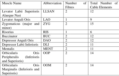
were measured in the different CT scan slices. The number of fibres per muscle
depends on its extent and size. Figure 3 and table 2 show the ten orofacial muscles
that are modelled.
--- Figure 3 around here ---
--- Table 2 around here ---
Muscle fibres are embedded in the facial mesh as continuous sets of uniaxial
cable elements. Since each cable is a line in 3D space, their number per fibre
increases as a function of the muscle fibre curvature, to model this curvature
smoothly. These cable elements (LINK10 in Ansys ) act in tension only and will
become slack under compression. Such properties are consistent with the observations
that in the fibre direction a muscle can resist only tensile forces and not compressive
forces (Loocke et al. 2006).
End points of the cable elements are defined independently of the level of
refinement of the main mesh. They correspond to anatomical landmarks located in
requiring any change in the definition of muscles courses. To couple the fibres with
the main mesh, point to surface contact elements are used. The points (pilot nodes)
are the extremities of the cable elements. They are bilaterally linked to the surfaces of
the mesh elements which centroids are the closest to the cable extremity. Figure 4
displays the cable elements and the corresponding coupling elements for the muscles
in half of the face. The no-displacement boundary condition is also applied to the ends
of cable elements that correspond to the muscles insertions on the skull.
--- Figure 4 around here ---
3.2 Muscle body or passive part
Once the fibres are set, the body of the muscles can be defined in the main
mesh. A neighbourhood is determined around each fibre by an algorithm considering
a sphere, which radius is equal to an estimation of the muscle cross-sectional
dimension, running along the cable elements lines. Each element of the main mesh
intersecting the sphere is then labelled as a part of the muscle body. The resulting
bodies of the muscles in the mesh are displayed in figure 5 for the left half of the face.
Although this definition of the muscle body is a rough approximation, it is enough so
far for our use, which is to account for the stress stiffening effect.
--- Figure 5 around here ---
3.3 Stress stiffening effect
Muscles behave like a transversely isotropic material, with an isotropic
behaviour in the directions orthogonal to the muscle fibres. This means that
mechanical properties in the direction of muscle fibres are different from the ones in
fibres tensile characteristics, the transversal bending stiffness increases with the
tensile force (similarly to the stress stiffening phenomenon in cable members or
membranes).
--- Figure 6 around here ---
This is illustrated in Figure 6 with a simple example of a virtual point P inside
a muscle fibre originally at equilibrium under constant muscle activation (force F1)
and then displaced (by δ) because of the action of a force F applied in the muscle
transversal direction. Once the new equilibrium is reached (Figure 6 lower panel),
assuming a linear relationship between force and displacement, we have:
2 1 1 1 1 1 2 F F (3)
This means that, when δ is negligible as compared to ℓ1, the muscle transversal
stiffness dF/dδ is proportional to muscle force F1.
When a muscle is activated, its fibres generate forces that resist to elongation,
according to a certain tension-length relation (see for example McMahon (1984)), and
in a way that increases when activation increases (see for example Wilhelms-Tricario
(1995)). In real muscle the fibres distribution is so dense, that the resistance to
elongation of the whole muscle body increases with elongation in the fibres direction.
In our model, muscle fibres are not represented in all their details. They are modelled
by a limited number of localized macrofibres (typically from one to three). When the
muscle is activated, each of these macrofibres generates a force and resists to the
elongation, but since the fibres are localized, this resistance does not apply to the
whole body of the muscle. This would not be a realistic behaviour. In order to
(section 3.2) increases with muscle activation in the fibres directions. Hence, muscle
activation is associated both with a resistance to stress in the direction orthogonal to
the fibre direction (the stress stiffening effect) and with a resistance to elongation in
the fibre direction. Consequently, it is modelled by an isotropic increase of the tissues
stiffness, implemented by modifying the parameters of the passive constitutive law
(equation 1), according to the approach explained in the next section.
3.4 Implementation of muscle activation and stress stiffening effect
The cable elements generate the active force Fac of each muscle, following the
relation:
Fac=AEcable (ε-αΔT) (4)
where A is the cable cross sectional area, ε its strain and Ecable its Young‟s modulus. In
standard ANSYS use, parameter T is equivalent to the temperature of the element,
and α to the thermal coefficient of expansion of the cable. In our case, we have used
parameters T and Ecable to specify the level of activation. Parameter Ecable is a scaling
factor specifying the maximal level of activation, which is muscle specific. Parameter
T is used to control the level of activation within the muscle specific maximal range of variation. Thus, parameter T can be considered as a normalized control parameter of
muscle activation. Decreasing T leads to a shortening of the cables lengths, which therefore exert forces on the main mesh through the coupling elements. The activation
level is then a decreasing function of parameter T. The value of α is arbitrarily set to
0.001
To account for the stress stiffening effect, the constitutive law of the elements
of a muscle body varies with the level of muscle activation specified with T. In
hyperelastic law are hence linearly scaled as an increasing function of the activation,
which is a decreasing function of T (Figure 7).
--- Figure 7 around here ---
When different muscles are activated simultaneously, the stiffness of the main mesh elements which are common to these muscles‟ bodies change as a function of the most activated muscle, and not as the result of an accumulation of the stiffness
changes associated with each individual muscle activation. The proposed stress
stiffening modelling is functionally correct, except for the resistance to compression
in the fibres direction. Indeed, it is known that this resistance varies with the strain
rate and is close to zero when this rate is low (Loocke et al. 2006). Further
improvements will be provided along this line in future works.
The muscle activationvaries in time as a ramp function. In further works that
we will develop in the context of speech production, these commands will be handled
by a motor control mechanism integrating voluntary commands and low-level
feedback information sent by the muscles (Feldman 1986; Buchaillard et al. 2006).
3.5 Dynamic parameters
For dynamic transient analysis, viscosity is modelled using proportional
damping:
C=αM+βK (5)
To determine α and β coefficients the first 7000 modes of the main mesh (about a
third of the total number of degrees of freedom) were calculated. Simulations were
run twice, first with the material stiffness used in the absence of muscle activation and
then for a high material stiffness level (10 times more). The corresponding natural
have been tuned such that the damping ratio (ratio of viscous damping factor to
critical damping) is larger than and near to one. The computed values are α=19 sec-1
and β=0.055 sec.
The density of face tissues is set to =1.04E-6 kg/mm3 (Buchaillard et al.
2006). The effect of gravity has not been considered.
Other parameters regarding solver and computational costs are discussed in
the appendix.
4. Simulations and results
Different muscle activation patterns have been used and their influences on facial
gestures and mimics evaluated. Both static and transient analyses have been
performed. In addition to the static analysis that takes into account only the stiffness
matrix, dynamic simulations obtained with full transient analysis also takes into
account the effect of inertia and viscosity.
4.1 Simulation of facial mimics resulting from various orofacial gestures
Activation of muscles taken individually and in coordination has been
investigated. In this section, only the final shapes of the mesh resulting from these
activations are shown. They are the same for the static and the full transient analysis.
These results well comply with the anatomical predictions in the related literature
(Standring 2005).
The result of activating zygomaticus draws the angle of the mouth upwards
and laterally (Figure 8).
Levator labii superioris elevates the upper lip. Acting with other muscles, it modifies
the nasolabial furrow. In some faces, this furrow is a highly characteristic feature
often deepened in expressions of sadness or seriousness. The activation of the levator
labii superioris with zygomaticus and levetor labii superioris alaeque nasi in Figure 9
well satisfies that hypothesis.
--- Figure 9 around here ---
The effect of orbicularis oris peripheralis (OOP) in protruding and rounding
the lips has been shown (Figure 10). The effect of stiffening in producing rounding
with protrusion has been discussed in Nazari et al. (2008): without the stiffening, lips
are protruded but the amount of lip opening is too large.
--- Figure 10 around here ---
Figure 11 shows the consequence of the activation of the risorius and Figure
12 the impact of activation of the buccinator (BUC). In Figure 13 the mimic
associated with the coordinated action of OOP and BUC is illustrated. In all these
figures these actions are qualitatively consistent with predictions made from
anatomical knowledge.
--- Figure 11 around here ---
The risorius is known to stretch the mouth laterally and to retract the corners of the
mouth. This is consistent with the strain depicted in Figure 11.The buccinator has no
or little influence on the lips, and essentially compresses the cheeks against the teeth
12): the lips have the same shape as in our model at rest, while the strain essentially
affects the lower part of the face.
--- Figure 12 around here ---
The OOP has been shown in our model to generate a protrusion and a closing of the
lips which is consistent with usual hypotheses in the literature (Gomi et. al 2006;
Nazari et. al 2008). Meanwhile, the coordinated action of the buccinator and the OOP
generates a closing of the lips only. It can be assumed that the stiffening of the cheeks
due to the buccinator activation limits the amplitude of the lip protrusion, which
would explain that mainly closure is observed.
--- Figure 13 around here ---
4.2 Dynamics versus Quasi-static simulations
We have studied the effect of dynamic versus quasi-static analysis on the lip
protrusion. For this purpose both OOP and mentalis (MENT) muscles are activated.
The same activation level in both dynamic and static analyses is assumed. Figure 14
shows the trajectories, for both conditions, of a node located on the lower lip in the
midsagittal plane.
--- Figure 14 around here ---
While starting and ending points are the same in static and dynamic analysis, the
trajectories are clearly different. The trajectory obtained with the static analysis is
close to a straight line while the dynamic trajectory is noticeably curved. This
difference is large enough to generate significant differences in lip shape variation
acoustic signal. In addition, a large number of human skilled movements have been
shown to follow curved path (Morasso, 1981).
Figure 15 shows the tangential velocity profile for the same point together
with the corresponding activation signal..
--- Figure 15 around here ---
An asymmetrical bell-shaped velocity pattern is generated. This kinematic
pattern is typical for lip movements as shown for example by Shaiman et al. (1997)
for several American English speakers.
Both properties, the curved path and the bell-shaped velocity profiles,
observed in experimental studies and accounted for in dynamic analysis and not in
quasi-static analysis demonstrates the necessity to integrate dynamic factors, such as
inertia and damping, to obtain realistic simulations of lip shape variations in speech
production.
To assess more precisely the realism of the trajectories produced by our
model, they can be compared to lips trajectories measured with video processing
(Abry et al. 1996) from a native speaker of French. As an illustration, let us consider
the sequence /iRy/ embedded in the carrier sentence„Tu dis “ruise” (/tydiRyiz/, you‟re saying “ruise”, /). These data were processed with a low-pass linear phase filter (cut-off frequency 6 Hz). The trajectory of a point located on the lower lip in the
mid-sagittal plane has been extracted in the temporal section corresponding to lip
protrusion from /i/ to /y/ (Figure 16). It can be observed that the path of this point is
qualitatively similar to the path simulated with dynamic analysis (Figure 14). More
specifically, the path is curved, a key feature that could not be predicted from the
--- Figure 16 around here ---
Figure 17 shows the experimental velocity profile: it has, like our simulation,
an asymmetrical bell-shape in agreement with Shaiman et al.‟s (1997) data collected
from speakers of American English.
--- Figure 17 around here ---
This example of a comparison between simulations and real data confirms the general
observation made above: contrary to those obtained in the quasi-static analysis
framework, the simulations obtained in the dynamic analysis framework generate
curved paths and bell-shaped velocity profiles similar to those observed in
experimental lips protrusion movements collected during speech production
The experimental movement and the simulation in dynmica analysis have also
similar ranges of velocity (maximum velocity 3.9cm/s versus 2.4cm/s), durations
(200ms versus 270ms at 20% of the peak velocity), and movement amplitudes
(4.5mm versus 4mm for the horizontal protrusion).
Some discrepancies can be noticed between simulations and experimental
data. In the experimental data, the curved path includes a rising part followed by a
short decline. In the simulation this rising/declining sequence is also observed, but it
is preceded by a horizontal part. It is important to state that these differences are not
intrinsically due to the characteristics of the model but ,more factually, to differences
between the conditions of simulation and the conditions of real speech production. In
the simulations the movement starts from a zero velocity position and ends at a zero
continuum (Figure 16) in which the observed section does not start or end with a zero
velocity position. This phenomenon can be clearly seen in the experimental velocity
profile (Figure 17), in which velocity curve never crosses zero.
5. Discussion and conclusion
The use of a realistic dynamical biomechanical model of the face has allowed
simulating a number of facial movements comparable to those occurring during the
production of speech or of facial mimics in non-verbal communication.
One of the main specificities in our model is the representation of the muscles.
First, their anatomical description, which relies on subject specific medical images
and anatomical data, is independent from the finite element mesh. It enables to easily
modify the structure of the mesh, its number or type of elements, without losing the
anatomical information. The second aspect that makes our model original is the
modelling of elastic muscle properties and more specifically the stress stiffening
effect associated with muscle activation. The elastic characteristics are inherent to the
muscle body definition, and are determined by varying the constitutive law of the muscle‟s tissues with the level of activation of the muscle. Results on the protrusion movement have shown that this approach enhances the generation of accurate facial
movements and shapes (Nazari et al., 2008).
Studies in the literature have shown that articulatory dynamics has a major
impact on the temporal patterning of speech movements. Time characteristics are
important in speech perception. We have shown that lip movement patterns are indeed
different in quasi-static and dynamic simulation frameworks. Interesting results, close
for the quasi-static one, such as the generation of curved paths and bell-shaped
velocity profiles classically observed in unperturbed skilled human movements
(Morasso 1981). The clear differences observed between the trajectories simulated
with dynamic and static analysis demonstrate that the usage of dynamic analysis is a
requirement for speech production studies. The role of dynamics has also been
studied in the literature for non-speech movements. Ambadar et al. (2005) observed
for example that recognition of subtle facial expressions by watching the evolution of
facial gestures in time is much easier than by looking at static shots. Hence, in
modelling studies, if temporal patterning of movements integrates dynamic
constraints like inertia and viscosity, synthetic facial expressions will be deciphered
faster and easier.
Simulations have also highlighted the indirect role of stiffening the face,
mostly in the cheeks area, on the way muscles impact the lip shapes. It was shown
that the stiffening of the cheeks due to the activation of the buccinator induces a
limitation of the amplitude of the upper lip protrusion associated with OOP activation.
The role of muscles, which are not directly involved in lip shaping, was thus
demonstrated. These results, similar to those of Buchaillard et al. (2009) about the
role of mouth floor muscles in tongue elevation, are encouraging for our modelling
approach toward a better understanding of facial mimic mechanisms.
Future improvements of the model will among others concern the mechanical
properties, to account for the non-homogeneity of the tissue layers, and include some
mesh refinement. Also, new muscle elements will be developed to integrate the
displacements and strain dynamically in their constitutive law. This will be used while
implementing a motor control mechanism that integrates voluntary commands and
every muscle to reach specific targets, defined as positions or shapes of the face in
relation with specific spectral patterns of the acoustic signal. Other works will
concern the coupling of the face with a model of the jaw. Finally, experimental data
will be more extensively used to better evaluate, qualitatively and/or quantitatively,
the simulated orofacial movements.
Appendix
The sparse direct solver based on Newton-Raphson method has been used.
Convergence is assumed when ||ΔV||<εV Vref
where V is the variable, which is in our case either force or displacement, εV is the
tolerance and ||ΔV|| the Euclidian norm of the variable difference at each time step
(ANSYS Inc., Theory Reference). The assumed values are given in Table 3.
----Table 3 around here---
In the Newton-Raphson method, line search with adaptive descent is used. The
computation time on a Windows XP (32bit) platform running on a machine with a
Duo CPU E6850 @ 3 GHz for static analysis is around 3000 seconds per simulation
(3183 CPU times) and for dynamic analysis is around 20,000 seconds (10,000 CPU
times) for simulation of one second real time gesture.
Acknowledgement
We are pleased to express our gratitude to Dr. Pierre Badin, Dr. Christophe
Savariaux and Dr. Jean Luc-Schwarz for providing us with experimental movement
FIGURES CAPTIONS
Figure 1: Main mesh of the face soft tissue.
Figure 2: Surfaces of contact elements between lips (a) and lips and teeth (b).
Figure 3: Macrofibers defining the muscles of the face shown in CT data (a), in the main mesh (b), and with their abbreviated names (c).
Figure 4: Coupling elements between the piece-wise fibres of cable elements and the main mesh (only the left half of face is shown).
Figure 5: Body of the muscles: elements of the main mesh in a neighbourhood of the muscles fibres (only the left half of face is shown).
Figure 6: A schematic representation of stress stiffening effect. A point P inside a muscle at equilibrium under constant muscle activation (force F1) (top panel) is
virtually displaced by δ under the action of a transversal force F (bottom panel). Once the new equilibrium is reached with a new force level F1, transversal stiffness dF/dδ is
proportional to that force.
Figure 7: Modelling of the stress stiffening effect: variation of the hyperelastic constitutive law of the tissue with the activation of the muscle.
Figure 8: Face shaping after activation of the zygomaticus muscle
Figure 9: Face shaping from coordinate activation of the zygomaticus, levetor labii superioris alaeque nasi and levetor angulai oris muscles
Figure 10: Face shaping resulting from the orbicularis oris peripheralis activation
Figure 11: Face shaping resulting from the risorius activation
Figure 13: Face shaping resulting from the orbicularis oris peripheralisand buccinator co-activation
Figure 14: Comparison between the trajectories of a point on the lower lip in the mid-sagittal plane in static and dynamic analysis resulting from an orbicularis oris peripheralis and mentalis co-activation (with Ecable=0.3 and T=-500 with spherical neighbourhood radius for OOP 3mm and for MENT 2 mm).
Figure 15: Upper panel: Velocity profile of a point on the lower lip in the mid-sagittal plane resulting from the co-activation of orbicularis oris peripheralis and mentalis in dynamic analysis. Lower panel: Time patterns of the corresponding activations. (with Ecable=0.3 and T=-500 with spherical neighbourhood radius for OOP 3mm and for MENT 2 mm)
Figure 16: Experimental data. Top panel: trajectory of a point on the lower lip in the mid-sagittal plane in /iRy/ sequence; diamond mark is for the starting point and square mark for the ending point. Bottom panel: corresponding acoustic signal with phonetic labelling
Figure 17: Experimental data. Tangential velocity profile corresponding to trajectory and the acoustic signal displayed in Figure 16.
TABLES
Table 1. Constants of the simplified 5-parameter Mooney-Rivlin model for passive tissues
c10 (MPa) c20 (MPa) d (1/MPa)
2.5e-3 1.175e-3 0.8
Table 2. Orofacial Muscles for half of the face
Muscle Name Abbreviation Number of Fibres
Total Number of Cable Elements Levator Labii Superioris
Alaeque Nasi
LLSAN 2 12 Levator Anguli Oris LAO 1 9 Zygomaticus (major and
minor)
ZYG 2 15 Risorius RIS 1 6 Buccinator BUC 2 12 Depressor Anguli Oris DAO 2 12 Depressor Labii Inferioris DLI 2 11 Mentalis MENT 2 11 Orbicularis Oris Peripheralis (Inferioris and Superioris) OOP 2 14 Orbicularis Oris Marginalis (Inferioris and Superioris)
OOM 2 14
Table 3. Tolerance values
ε Vref
Force (N) 0.035 0.01 Displacement (mm) 0.01 0.00
REFERENCES
Abry C, Lallouache MT and Cathiard MA. 1996. How can coarticulation models account for speech sensitivity to audio-visual desynchronization? In Speechreading by Humans and Machines, NATO ASI Series F: Computer and System Sciences, Stork D & Hennecke M eds. 150:pp. 247-255. Springer-Verlag, Berlin, Heidelberg, Tokyo.
Ambadar Z, Schooler J and Cohn JF. 2005. Deciphering the enigmatic face: the importance of facial dynamics to interpreting subtle facial expressions. Psychological Science. 16(5): 403-41.
Ansys, Inc. 2007. Theory Reference Manual. Release 11.
Blanton PL, Biggs NL and Perkins RC. 1970. Electromyographic analysis of the buccinator muscle. J. Dent. Res. 49:389-394.
Badin P, Elisei F, Bailly G and Tarabalka Y. 2008. An audiovisual talking head for augmented speech generation: models and animations based on a real speaker's articulatory data. Proceedings of the 5th Conference on Articulated Motion and Deformable Objects (AMDO 2008). Springer Verlag LNCS 5098:132-143.
Blemker S, Pinsky PM and Delp SL. 2005. A 3D model of muscle reveals the causes of nonuniform strains in the biceps brachii. J. of Biomechanics. 38:657–665. Buchaillard S, Perrier P and Payan Y. 2006. A 3D biomechanical vocal tract model to
study speech production control: How to take into account the gravity? Proc. of the 7th International Seminar on Speech Production, pp. 403-410.
Buchaillard S, Perrier P and Payan Y. 2009. A biomechanical model of cardinal vowel production: Muscle activations and the impact of gravity on tongue positioning. J. Acoustical Society of America, 126(4):2033–2051.
Chabanas M, Luboz V and Payan Y. 2003. Patient specific finite element model of the face soft tissues for computer-assisted maxillofacial surgery. Medical Image Analysis. 7:131-151.
Feldman AG. 1986. Once more on the Equilibrium-Point hypothesis (λ model) for motor control. J. of Motor Behavior. 18(1):17-54.
Fung YC. 1993. Biomechanics: Mechanical properties of living tissues. Springer-Verlag, New York Inc.
Gerard JM, Ohayon J, Luboz V, Perrier P and Payan Y. 2005. Non-linear elastic properties of the lingual and facial tissues assessed by indentation technique, Application to the biomechanics of speech production. Medical Engineering & Physics. 27:884-892.
Gladilin E, Ivanov A and Roginsky V. 2004. A framework for biomechanical simulation of cranio-maxillofacial surgery interventions. Proc of International Symposium on Medical Simulation, ISMS 2004, S. Cotin and D. Metaxas, eds., pp. 287-294.
Gomi H, Nozoe J, Dang J and Honda K. 2006. A physiologically based model of perioral dynamics for various lip deformations in speech articulation. Speech Production: Models, Phonetic Processes and Techniques, J. Harrington and M. Tabain, eds., Psychology Press, pp 119-134.
Groleau J, Chabanas M, Marécaux Ch, Payrard N, Segaud B, Rochette M, Perrier P and Payan Y. 2007. A biomechanical model of the face including muscles for the prediction of deformations during speech production. Proceedings of the 5th International Workshop on Models and Analysis of Vocal Emissions for Biomedical Applications, MAVEBA'2007, Firenze, Italie.
Hardcastle WJ. 1976. Physiology of Speech Production. Academic Press, London. Hill AV. 1938. The heat of shortening and the dynamic constants of muscle. Proc
Royal Society, Biological Sciences. 126:136-195.
Humphrey JD and Yin FCP. 1989. Constitutive relations and finite deformations of passive cardiac tissue II: Stress analysis in the left ventricle. Circulation Research. 65:805-817.
Kim K, Gomi H. 2007. Model-based investigation of control and dynamics in human articulatory motion. J System Design and Dynamics 1:558–569.
Lee Y, Terzopoulos D and Waters K. 1995. Realistic modeling for facial animation. SIGGRAPH‟95, S.G. Mair and R. Cook, eds. New York, ACM Press, 55–62. Loocke MV, Lyons CG and Symms CK. 2006. A validated model of passive muscle
in compression. J. of Biomechanics. 39:2999-3009.
Lucero JC and Munhall KG. 1999. A model of facial biomechanics for speech production. J. Acoust. Soc. Am. 106:2834–2842.
Lucero JC, Maciel STR, Johns DA and Munhall KG. 2005. Empirical modeling of human face kinematics during speech using motion clustering. J. Acoust. Soc. Am. 118(1): 405-409.
McGurk H and MacDonald J. 1976. Hearing Lips and Seeing Voices. Nature, 264746-48.
McMahon TA. 1984. Muscles, Reflexes, and Locomotion. Princeton University Press. Morasso P. 1981. Spatial control of arm movements. Exp. Brain Res. 42:223-227. Munhall KG and Vatikiotis-Bateson E. 1998. The moving face during speech
communication. Hearing by Eye, Part 2: The Psychology of Speechreading and Audiovisual Speech, edited by R. Campbell, B. Dodd, and D. Burnham. Taylor and Francis, Psychology Press, London.
Nazari MA, Payan Y, Perrier P, Chabanas M and Lobos C. 2008. A continuous biomechanical model of the face: a study of muscle coordinations for speech lip gestures. Proceedings of the 8th International Seminar on Speech Production, ISSP'08, pp. 321-324.
Ng-Thow-Hing V and Fiume E. 2002. Application-specific muscle representations. Proc. of Gr. Inter, W. Sturzlinger and M. McCool, editors, pp. 107–115. O‟Shaughnessy D. 1081. A study of French vowel and consonant durations. J. of
Phonetics. 9:385-406.
Payan Y and Perrier P. 1997. Synthesis of V-V sequences with a 2D biomechanical tongue model controlled by the Equilibrium Point Hypothesis. Speech Communication. 22:185-205.
Perrier P, Payan Y, Zandipour M and Perkell J. 2003. Influences of tongue biomechanics on speech movements during the production of velar stop consonants: A modeling study. J. Acoustical Soc. of America. 114(3):77–83. Shaiman S, Adams SG and Kimelman MDZ. 1997. Velocity profiles of lip protrusion
across changes in speaking rate. Journal of Speech, Language, and Hearing Research. 40: 144-158.
Sifakis E, Neverov I and Fedkiw R. 2005. Automatic Determination of Facial Muscle Activations from Sparse Motion Capture Marker Data. ACM Transactions on Graphics (SIGGRAPH Proceedings).24:417-425.
Sifakis E, Selle A, Robinson-Mosher A and Fedkiw R. 2006. Simulating Speech with a Physics-Based Facial Muscle Model. Eurographics/ ACM SIGGRAPH, M.P. Cani, and J.O. Brien, editors, Symposium on Computer Animation. Standring S. (editor in chief). 2005. Gray‟s Anatomy: The Anatomical Basis of
Teran J, Sifakis E, Blemker S, Ng Thow Hing V, Lau C and Fedkiw R. 2005. Creating and Simulating Skeletal Muscle from the Visible Human Data Set. IEEE TVCG. 11:317-328.
Tracqui P and Ohayon J. 2004. Transmission of mechanical stresses within the cytoskeleton of adherent cells: a theoretical analysis based on a multi-component model. Acta Biotheoretica. 52:323-341.
Weiss JA, Maker BN and Govindjee S. 1996. Finite element implementation of incompressible, transversely isotropic hyperelasticity. Comput. Methods Appl. Mech. Engrg. 135:107-128.
Wilhelms-Tricario R. 1995. Physiological modeling of speech production: methods for modeling soft-tissue articulators. J. Acoustical Society of America. 97:3085–3098.
Yucesoy CA, Koopman BHFJM, Huijing PA and Grootenboer HJ. 2002. Three-dimensional finite element modeling of skeletal muscle using a two-domain approach: linked fibre-matrix mesh model. J. of Biomechanics. 35:1253-1262. Zajac F. 1989. Muscle and tendon: Properties, models, scaling, and application to
biomechanics and motor control. Critical Reviews in Biomed. Eng. 17:359– 411.
Figures
Figure 1
Figure 2b-
Figure 3b-
Figure 6-
Figure 7-
Figure 9-
Figure 10-
Figure 12-
Figure 13-
Figure 14-
Figure 16-