HAL Id: hal-00016382
https://hal.archives-ouvertes.fr/hal-00016382
Submitted on 2 Jan 2006
HAL is a multi-disciplinary open access
archive for the deposit and dissemination of
sci-entific research documents, whether they are
pub-lished or not. The documents may come from
teaching and research institutions in France or
abroad, or from public or private research centers.
L’archive ouverte pluridisciplinaire HAL, est
destinée au dépôt et à la diffusion de documents
scientifiques de niveau recherche, publiés ou non,
émanant des établissements d’enseignement et de
recherche français ou étrangers, des laboratoires
publics ou privés.
Modulation of the Suppressor of fused protein regulates
the Hedgehog signaling pathway in Drosophila embryo
and imaginal discs.
François Dussillol-Godar, Jeanine Brissard-Zahraoui, Bernadette
Limbourg-Bouchon, Dominique Boucher, Sylvaine Fouix, Claudie
Lamour-Isnard, Anne Plessis, Denise Busson
To cite this version:
François Dussillol-Godar, Jeanine Brissard-Zahraoui, Bernadette Limbourg-Bouchon, Dominique
Boucher, Sylvaine Fouix, et al.. Modulation of the Suppressor of fused protein regulates the Hedgehog
signaling pathway in Drosophila embryo and imaginal discs.. Developmental Biology, Elsevier, 2006,
291, pp.53-66. �hal-00016382�
Modulation of the Suppressor of fused protein regulates the Hedgehog
signaling pathway in Drosophila embryo and imaginal discs.
François Dussillol-Godar
a,1, Jeanine Brissard-Zahraoui
a,1, Bernadette
Limbourg-Bouchon
b, Dominique Boucher
c, Sylvaine Fouix
a, Claudie Lamour-Isnard
a, Anne
Plessis
aand Denise Busson
a,*a Laboratoire Génétique du Développement et Evolution, Institut Jacques Monod, UMR 7592-CNRS/Université
Pierre et MarieCurie/Université Denis Diderot, 2, place Jussieu, 75251 Paris Cedex 05, France
b Laboratoire de Génétique et Biologie cellulaire, FRE 2445-CNRS/Université de Versailles, Bâtiment Buffon,
45 Avenue des Etats-Unis, 78035 Versailles Cedex, France
c Laboratoire de Biochimie cellulaire, UMR 7098-CNRS/Université Pierre et Marie Curie, Bâtiment C, 5ème
étage, 9 Quai Saint-Bernard, 75252, Paris Cedex 05, France
1: these authors contributed equally to this work
* Corresponding author. Fax : 33 1 44 27 52 65 E-mail address: bussond@ijm.jussieu.fr
The Suppressor of fused (Su(fu)) protein is known to be a negative regulator of Hedgehog (Hh) signal transduction in Drosophila imaginal discs and embryonic development. It is antagonized by the kinase Fused (Fu) since
Su(fu) null mutations fully suppress the lack of
Fu kinase activity. In this study, we over-expressed the Su(fu) gene in imaginal discs and observed opposing effects depending on the position of the cells, namely a repression of Hh target genes in cells receiving Hh and their ectopic expression in cells not receiving Hh. These effects were all enhanced in a fu mutant context and were suppressed by cubitus
interruptus (ci) over-expression. We also show
that the Su(fu) protein is poly-phosphorylated during embryonic development and these phosphorylation events are altered in fu mutants. This study thus reveals an unexpected role for Su(fu) as an activator of Hh target gene expression in absence of Hh signal. Both negative and positive roles of Su(fu) are antagonized by Fused. Based on these results, we propose a model in which Su(fu) protein levels and isoforms are crucial for the modulation of the different Ci states that control Hh target gene expression.
Keywords : Drosophila ; Suppressor of fused ; fused ; Hedgehog ; signal transduction ; Cubitus interruptus
Introduction
The Hedgehog (Hh) signaling pathway plays a critical role in the patterning, differentiation and growth of a wide array of cell types during
development of many organisms (Ingham and McMahon, 2001; Lum and Beachy, 2004; Nybakken and Perrimon, 2002). Hh proteins control segmental patterning in Drosophila embryos and specification of the antero-posterior axis in both vertebrate and insect limbs. In Drosophila imaginal discs, Hh is expressed within the posterior (P) compartment and acts on adjacent anterior (A) compartment cells to specify their fates in a concentration dependent manner (Ingham and McMahon, 2001). Cells interpret the level of Hh that they receive through complex events which regulate the proteolytic cleavage, nucleo-cytoplasmic trafficking and activation of the Cubitus interruptus (Ci) transcription factor. At least three different states are encountered in the anterior compartment of wing discs (i) in the cells abutting the A/P boundary which are exposed to a high concentration of Hh, Ci is found in its full-length (155 kDa), activated form (called Ci155ACT)
which upregulates the transcription of engrailed (en) and patched (ptc), (ii) in the cells located further inside the A compartment which receive less Hh, Ci155 is activated at a lower level and induces decapentaplegic (dpp) expression (but not ptc nor en) and (iii) in more anterior cells where no Hh is available, Ci is cleaved into a 75 kDa form (Ci75) that
represses both dpp and hh while the remaining uncleaved fraction is sequestered in the cytoplasm (Vervoort, 2000). Such exquisite control of Ci activity seems to be achieved by one or several Hedgehog transducing complexes (called HTC) that include, along with Ci, the Fused (Fu) serine-threonine kinase, the kinesin related Costal-2 (Cos2) protein and the PEST-motif containing protein, Suppressor of fused (Su(fu)) (Monnier et al., 1998; Monnier et al., 2002; Robbins et al., 1997; Sisson et al., 1997; Stegman et al., 2000; Wang et al., 2000). At least two different complexes have been described: a Fu-Cos2-Ci trimeric complex devoid of
Su(fu) and associated to microtubules via Cos2 in the absence of Hh, and a Su(fu)-Fu-Cos2-Ci tetrameric complex present in cells responding to Hh. In addition, it was recently reported that Cos2 has the ability to tether both Fu and Ci to cellular membranes (Stegman et al., 2004). A model was therefore proposed in which a HTC associated to endosomes via Cos2 is required for the production of the repressor Ci75, while a HTC bound to Smo
through Cos2 promotes Ci activation (Stegman et al., 2004).
In this study, we focused our attention on the role of the Su(fu) protein. Su(fu) is known to negatively regulate the Hh pathway and to be antagonized by Fu (Alves et al., 1998; Méthot and Basler, 2000; Ohlmeyer and Kalderon, 1998; Pham et al., 1995; Préat, 1992; Préat et al., 1993; Wang et al., 2000). Indeed, Su(fu) null mutations fully suppress the lack of Fu kinase activity and enhance cos2 phenotype. Nevertheless, Su(fu) null mutations lead only to a very mild adult mutant phenotype, suggesting that its inhibitory role is somewhat redundant in the regulation of the pathway (Préat, 1992; Préat et al., 1993). The Su(fu) protein, like the Cos2 protein, interacts directly with Fu and Ci (Méthot and Basler, 2000; Monnier et al., 1998; Monnier et al., 2002; Ohlmeyer and Kalderon, 1998; Stegman et al., 2000; Wang et al., 2000). Studies in cultured cells and clonal analysis have shown that Su(fu) does not appear to be involved in Ci proteolysis but rather in the cytoplasmic retention of full-length Ci and in the inhibition of Ci activation (Alves et al., 1998; Chen et al., 1999a; Méthot and Basler, 2000; Ohlmeyer and Kalderon, 1998; Wang et al., 2000). Recently, it has been proposed that Su(fu) is involved in the stability of Ci isoforms generating a sensitized background to the Hh signal (Ho et al., 2005). Finally, several studies have shown that Su(fu) is phosphorylated in response to Hh, depending on Fu kinase activity (Ho et al., 2005; Lum et al., 2003).
In order to gain new insight into the regulation and the function of the Su(fu) protein, we monitored its accumulation and post-translational modifications and analyzed the effects of its overexpression. First, we show that the Su(fu) protein is submitted to phosphorylation during embryonic development, at a time when the Hh signaling is fully active. These phosphorylation events are altered in fu mutants, suggesting that the Fu kinase is (directly or indirectly) involved in Su(fu) isoform modulation. Second, over-expression of Su(fu), either ubiquitously or in specific parts of the imaginal discs, revealed complex and paradoxical effects, namely a repression of Hh target gene expression in cells receiving Hh at the A/P border and an ectopic expression of Hh targets more anteriorly in cells which do not receive Hh. This anterior effect can occur independently of Hh signaling at the A/P
border. All effects of Su(fu) over-expression, both in cells that are receiving Hh and in those that are not, are enhanced in a fu mutant context, and are suppressed by Ci over-expression. Based on these results, we propose a model for Hh signal transduction in which Su(fu) protein levels are crucial for the modulation of the different Ci states in response to the Hh signal.
Materials and methods
Drosophila stocks
The fu alleles used in this study were described previously (Busson et al., 1988; Thérond et al., 1996a). The fu1 and fuJB3 alleles are class I fu
alleles which correspond to alterations in the kinase domain, the fuA allele belongs to class II fu alleles corresponding to alterations in the extra-catalytic domain. The Su(fu) gene is included in the common deleted region of Df(3R)karSZ11 and Df(3R)karSZ21
deficiencies (Préat, 1992). Su(fu)LP is an amorphic allele associated with a small deletion altering the 3’ end of the Su(fu) transcript (Pham et al., 1995). GAL4 lines used were da-GAL4 (P[w+, da-GAL4] on
chromosome III), dpp-GAL4 (P[ry+, dppblink-GAL4] on chromosome III), provided by the Bloomington Stock Center, vg-GAL4 (P[w+, 2.5 kb vg intron
2-GAL4] on chromosome II) (Delanoue et al., 2004), C765-GAL4 (P[w+, GAL4] on chromosome III)
(Guillen et al., 1995). lacZ reporter lines used were dpp-lacZ which corresponds to the BS3.0 construct (Blackman et al., 1991), ptc-lacZ described in Lepage et al., (1995), wg-lacZ described in Neumann and Cohen, (1996), hh-lacZ described in Lee et al., (1992). The UAS-ci strain, which corresponds to a full-length ci cDNA, is described in Dominguez et al., (1996). The UAS-lacZ strain was obtained from the Bloomington Stock Center. Other strains used were: hs-FLP; Sp/SM6-TM6B (Tb), Act5C>CD2>GAL4, UAS-GFP (chromosome III) (Neufeld et al., 1998).
UAS-Su(fu) constructs and germ line transformation The 1.6 kb full-length Su(fu) cDNA (Pham et al., 1995) was cloned between the EcoRI and NotI sites in the polylinker of the pUAST vector (Brand and Perrimon, 1993). This vector was co-injected with a D2-3 helper plasmid into a w1118 host line
under standard conditions (Spradling et al., 1999). One UAS-Su(fu) transgenic line was established corresponding to a transposon inserted on the X chromosome (w,UAS-Su(fu) line). This line was used to obtain the w,fuA,UAS-Su(fu) and w,f,fu1,UAS-Su(fu)
lines by chromosomal recombination. These latter strains were maintained with the FM3 balancer chromosome. Similar results were obtained with an UAS-Su(fu) line corresponding to an insertion on the third chromosome (gift from Hervé Tricoire and data not shown).
Clonal analysis
GAL4-expressing clones were induced by the FRT/Flip-out method (Struhl and Basler, 1993), by crossing hsFLP/hsFLP; dpp-lacZ/CyO; +/+ or hsFLP/hsFLP; ptc-lacZ/CyO; +/+ females with w,Su(fu)/Y; +/+; Act5C>CD2>GAL4, UAS-GFP/Act5C>CD2>GAL4, UAS-GFP males. Clones were heat-shock induced in the progeny 16-48 hours after egg deposition by 1 hour exposure at 37°C. Imaginal discs were dissected from third instar larvae ; clones overexpressing Su(fu) were recovered from female larvae of hsFLP/w,UAS-Su(fu); dpp-lacZ (or ptc-lacZ)/+; Act5C>CD2>GAL4, UAS-GFP/+ genotype while clones recovered from male larvae of hsFLP/Y; dpp-lacZ (or ptc-lacZ)/+; Act5C>CD2>GAL4, UAS-GFP/+ genotype served as control.
Western blot analysis
Drosophila embryos were collected at different times after oviposition. Two extraction procedures were used : in the first procedure (cf Figure 1), embryos were dechorionated with bleach (2.6 % active Cl-), rinsed with water, and homogenized at 4°C by several passes of a Teflon Dounce homogenizer, in a buffer containing 50 mM Hepes buffer (pH 7.5), 150 mM NaCl, 1% Igepal, 5 mM EDTA, 2 mM PMSF and leupeptin 10 mg/ml ; in the second (cf Figure 2), embryos were sonicated 30 sec. X 8 times separated by 1 min., in a buffer containing 50 mM Tris-HCl, 150 mM NaCl, 2% Igepal, 0.1% SDS and a cocktail of protease inhibitors (1X complete kit from Roche Molecular Biochemicals). Insoluble material was sedimented at 10,000 X g for 10 min. at 4°C and the supernatant was collected. The protein concentration of the soluble material was estimated according to the Bradford technique (Bio-Rad protein assay kit). For each sample, equal amounts of proteins (from 50 to 200 mg per lane) were incubated at 100°C for 5 min. in the gel-loading buffer (50 mM Tris-HCl pH 6.8, 2% SDS, 10% glycerol, 100 mM bmercaptoethanol, 0.1% bromophenol blue). Extracts were separated by electrophoresis in SDS denaturing polyacrylamide gels, either Laemmli gels (acrylamide 8%, bis-acrylamide 0.1%) or Anderson gels (bis-acrylamide 12.5%, bis-acrylamide 0.1%), with migration in Tris-Glycine-SDS buffer (Anderson et al., 1973; Laemmli, 1970). Proteins were then transferred to nitrocellulose (Schleicher and Schuell) for 1 h 30 at 1 mA/cm2 using a semi-dry electrotransfer
apparatus (C.B.S. Scientific Co). The pattern of proteins was evaluated by staining the filters with Ponceau Red S solution. The membranes were blocked by incubation for 1 h at room temperature in Tris-buffered saline (20 mM Tris pH 7.5, 135 mM NaCl) containing 5% non fat dry milk, 0.1 % Tween 20, followed by an overnight incubation at
4°C with a 1:5 000 dilution of purified polyclonal antiserum raised against Su(fu) in rabbit (Monnier et al., 1998). The membranes were washed three times with Tris-buffered saline containing 0.5 % Igepal, 0.1% Tween 20, 5% non fat dry milk, before incubation with goat anti-rabbit IgG coupled to horseradish peroxidase (Vector) for 1 h at room temperature at a 1:10 000 dilution in 1:4 diluted wash buffer. The filters were washed three times with Tris buffered saline, 0.1 % Tween 20, and were developed using an enhanced chemiluminescence substrate (Super Signal West Pico Chemiluminescent substrate from Pierce) and Amersham Hyperfilm to reveal the signals.
Bi-dimensional gel electrophoresis
Drosophila embryonic extracts were prepared as described above. The samples were treated in order to perform 2D/PAGE separation as previously described (Wolff et al., 1992) except that ampholines 4-6 (Amersham Biosciences) were used in the isoelectric focusing dimension. The second dimension was performed according to Anderson et al. (1973). The size of the slab gel used was 12.5 x 24 cm. Depending on migration conditions, the pHi of the major isoform ranged between 5.2 and 5.45, while more acidic and heavier isoforms were detected. The apparent molecular weight of these different isoforms were all situated in the range of 54 kDa which is the expected size of the Su(fu) protein. Treatment with phosphatase inhibitors
Freshly prepared embryonic extracts were incubated at different times (0, 10, 30, 90 min.) at 37°C, with or without phosphatase inhibitors whose composition is as follows : 0.1 mM sodium ortho-vanadate, 10 mM b-glycerophosphate, 100 mM sodium fluorure.
Imaginal disc labelings
For b-galactosidase activity staining, imaginal discs were dissected in PBS, fixed in 0.5 % glutaraldehyde/PBS for 15 min. at room temperature and rinsed four times in PBS. The coloration was developed in 3.5 mM K4(FeII(CN)6), 5 mM
K3(FeIII(CN)6), 1 mM MgCl2 and 0.15 % X-Gal in
PBS for 2 hours at 37°C. Discs were mounted and observed in glycerol. Immunostaining using the 2A1 rat monoclonal anti-Ci (Motzny and Holmgren, 1995), was performed as follows: imaginal discs from late third instar larvae were dissected in PBS, fixed in 4 % paraformaldehyde, 30 mM Pipes (pH 7.4), 160 mM KCl, 40 mM NaCl, 4 mM Na3EGTA, 1 mM
spermidine, 0.4 mM spermine, 0.2 % BSA and 0.1 % Triton X-100, for 20 min. at room temperature and washed in PBS, 0.3 % Triton X-100. Tissue was blocked for 20 min. in PBS, 0.3 % Triton X-100 and 1 % BSA, and incubated overnight at 4°C in a 1 :5 dilution of the primary antibody, washed, blocked again and incubated for 2 hours at room temperature
in a 1 :10 000 dilution of FITC anti-rat IgG antibody (Jackson Laboratories). b-galactosidase immunostaining was performed using the rabbit polyclonal anti-b-galactosidase antibody (from ICN/Cappel) in a 1 :1000 dilution and the secondary anti-rabbit Cy3 antibody (Jackson Laboratories) in a 1 :100 dilution. Discs were mounted in glycerol, observed and photographed under a Leica DMR fluorescence microscope. Confocal imaging was performed with a Leica SP2-AOBS microscope.
Results
Su(fu) is a phosphoprotein modulated during embryonic development
Phosphorylation and other post-translational modifications are important for the regulation of biological activities of proteins. In cultured cells, most components of the Hh pathway have been shown to be phosphorylated in an Hh dependent manner (Chen et al., 1999a; Chen et al., 1999b; Denef et al., 2000; Ho et al., 2005; Lum et al., 2003; Robbins et al., 1997; Thérond et al., 1996b; Wang and Holmgren, 1999). Here, we analyzed the accumulation and post-translational modifications of the Su(fu) protein in developing embryos. Proteins from wild-type embryonic extracts were submitted to electrophoresis and immunoblotted with a purified polyclonal anti-Su(fu) antibody (Monnier et al., 1998). As shown in Fig. 1A, an immunoreactive species was present at the expected size (the predicted 54 kDa protein encoded by Su(fu) is 468 aa long) and was absent in lysates from embryos deleted for Su(fu).
We were unable to detect any modification of the Su(fu) protein when using the electrophoretic conditions described above. We therefore turned to Anderson type gels to perform monodimensional and bidimensional electrophoresis (Materials and Methods). As shown in Fig. 1B, immunoblotting using monodimensional Anderson type gel revealed 4 isoforms, a major one with an apparent molecular weight of 54 kDa (arrow 1), two slower migrating isoforms (filled arrowheads 2 and 3) and a faster migrating isoform (empty arrowhead 4). Bidimensional electrophoresis (Fig. 1C) revealed a major form (arrow a), that probably corresponds to the major form seen in Fig. 1B and at least 4 minor isoforms (arrowheads b, c, d, e), more acidic and of higher weight than the major form. However, no spot corresponds to the lower molecular weight form observed in Fig. 1B (empty arrowhead 4). Taken together, these data show that at least 5 and most probably 6 isoforms of Su(fu) protein exist in wild-type embryonic extracts (Figs. 1B, C).
To assess whether Su(fu) isoforms were due to phosphorylation, we took advantage of the fact that the two higher forms (filled arrowheads 2 and 3 in Figs. 1B, D) greatly decreased upon
incubation of the extracts at 37°C, whereas the lower form (empty arrowhead 4) accumulated (Fig. 1D). Incubation of the same extracts with phosphatase inhibitors blocked these effects (Fig. 1D). Therefore, the slower forms are probably due to phosphorylation (or hyperphosphorylation) of Su(fu), whereas the fastest form could correspond to the non- or less-phosphorylated protein. The major band (arrow) might correspond to a less-phosphorylated form that is stable during the treatment or to other types of modification.
We also monitored the Su(fu) phosphorylation during embryonic development (Fig. 1B). The higher molecular weight isoforms were scarcely detectable 0-2 hours after oviposition, then accumulated 4-8 hours after oviposition to diminish from 8 hours onwards (upper arrowheads). In contrast, the amount of the lower isoform (empty arrowhead), was maximal at 0-2 hours, and decreased 4-8 hours after oviposition.
In conclusion, Su(fu) protein is present in several different isoforms in the embryo, corresponding to the different degrees of phosphorylation. While the maternal form is hypophosphorylated, hyperphosphorylated isoforms accumulate at the time of activation of the Hh pathway.
Modulation of phosphorylated Su(fu) isoforms depends on Fu kinase activity
The Fu kinase behaves as an antagonist of Su(fu) and Cos2 activities. In cell cultures, it was shown that Fu activity is required for the Hedgehog-stimulated phosphorylation of Cos2 (Nybakken et al., 2002) and Su(fu) (Lum et al., 2003). Furthermore, the modulation of Su(fu) phosphorylation described above parallels that of Fu protein (Thérond et al., 1996b). Altogether this suggests that Su(fu) could also be a target of the Fu kinase. Therefore, we analysed the effect of different fu mutations on the accumulation of the different Su(fu) isoforms.
According to their genetic interactions with Su(fu), fu mutants have been classified into two different classes , class I alleles mutated exclusively in the kinase region and class II alleles altered in the regulatory domain. We performed the experiments with the two classes of fu mutants (Fig. 2) and in both classes, accumulation of the higher hyperphosphorylated forms of Su(fu) (bands 2 and 3) was significantly reduced. Two lower migrating forms were detected (bands 4 and 5). Their status differed depending on the class of the fu allele: in the class I fu mutants (fuJB3) these forms were as
abundant as in the wild-type, while they were markedly enhanced in the class II fuA mutant
(especially band 4). The partial persistence of higher forms in all types of mutants suggests that other kinases are involved. The effect seen in the fuA
mutant indicates that integrity of the regulatory domain is required for the activity of these kinases.
In conclusion, Fu kinase activity appears to participate (directly or indirectly) in the phosphorylation of the Su(fu) protein during embryonic development, but other kinases are most probably also involved.
Su(fu) over-expression in imaginal discs leads to the inhibition of Hh target gene expression in anterior cells receiving the Hh signal
The role of Su(fu) in vivo was assessed by examining the consequences of its over-expression in various tissues during development. We therefore drove the expression of an UAS-Su(fu) transgene with the ubiquitous da-GAL4 driver (see Materials and Methods) and looked at fly viability, adult appendage phenotype and Hh target gene expression in imaginal discs. In all cases, comparable effects were obtained with several independent insertions of the UAS-Su(fu) transgene, and no effect was observed with either the driver alone or the UAS-Su(fu) transgene alone.
As shown in Table 1A, ubiquitous over-expression of Su(fu) led to a significant decrease in fly viability, with almost 100% lethality (mostly pupal) at 25°C and 29°C. This effect was much lighter at 18°C and 21°C, in accordance with the stronger activity of the GAL4 protein at high temperatures. Wings of rare escapers emerging at 25°C were analyzed. We first turned our attention to the effects induced at the A/P border in cells responding to the Hh signal. Wings of UAS-Su(fu); da-GAL4 escapers did not show any obvious anomalies in the LV3-LV4 region which corresponds to the domain of Hh activity (Fig. 3B). We looked for Hh target gene expression in the corresponding imaginal discs at 25°C. The results presented in Fig. 4 show that Su(fu) over-expression actually leads to a reduction in dpp (Fig. 4B) and ptc (Fig. 4D) expression (as indicated by the width of the domain between red arrowheads). Again, this effect was stronger at 29°C than at lower temperatures (data not shown). Furthermore, we also observed a decrease in dpp and ptc expression in Su(fu) over-expressing clones induced at the A/P boundary (Supplementary data). The same down-regulation of dpp and ptc expression was also observed in leg (Figs. 5A-D) and eye-antennal (Figs. 5G-J) imaginal discs at 25°C.
Thus, at the A/P border in cells receiving the Hh signal, overexpression of Su(fu) leads to a decrease in the expression of two Hh target genes, dpp and ptc. This effect is in agreement with a negative role of Su(fu) in Hh signal transduction. Su(fu) over-expression in imaginal discs leads to the deregulation of Hh target gene expression in anterior cells not receiving the Hh signal
Unexpectedly, ubiquitous overexpression of Su(fu) had an effect in the most anterior region of the appendages since nearly 100% escapers
displayed clear anterior duplications in adult wings, legs and antennae (Fig. 3). Thus, in the antero-proximal region of the wing, the domain comprised between costa and subcosta was enlarged, with frequent more or less expanded costa duplications (Fig. 3B, arrow). In addition, costa bristles were more numerous than normal and were disorganized (data not shown). This phenotype is very similar to the costal-2 loss of function phenotype (Grau and Simpson, 1987; Simpson and Grau, 1987; Whittle, 1976) suggesting an ectopic activation of the Hh pathway in the most anterior regions. Similar anterior duplications can also be seen in the antennae (data not shown) and in the legs. Indeed legs were highly deformed especially at 29°C, with anterior sex-comb duplications (Figs. 3E, F arrow), and enlargment and fusion of the articles along the proximo-distal axis (Fig. 3G).
In the wing discs over-expressing Su(fu), ectopic dpp expression expanded, from a site at the antero-posterior border (presumptive proximal part of vein 3), to the outer part of the disc (presumptive costa) (Fig. 4B arrow). In leg and antenna imaginal discs, over-expression of Su(fu) led to anterior ectopic expression of both dpp (Figs. 5B arrow and 5H arrow) and wg (Fig. 5F). In the leg discs, ectopic dpp expression was especially strong (Fig. 5B, arrow), creating a second axis in the anterior compartment and was associated with an ectopic wg expression that extended anteriorly and dorsally (Fig. 5F arrow). Overall, these ectopic expressions were consistent with the distal duplications observed in the legs. In imaginal discs overexpressing Su(fu), ectopic ptc expression appeared as a faint spot in the presumptive costa of the wing discs (Fig. 4D arrow) whereas it was stronger in leg, antenna and eye discs (Figs. 5D arrow and 5J arrows).
In conclusion, ubiquitous overexpression of Su(fu) in the imaginal discs led to apparent antagonistic effects: a decrease in Hh signaling at the A/P border, in cells receiving the Hh signal, and an ectopic activation of the pathway more anteriorly, in cells not receiving Hh.
The ectopic anterior effects of Su(fu) over-expression are independent of its effects at the antero-posterior border
The activation of the Hh pathway, in the most anterior regions where Hh is normally absent could be secondary to the effects seen at the A/P border. To test this hypothesis, we investigated whether the anterior effects of Su(fu) over-expression could occur independently of its over-expression at the A/P border. We thus drove Su(fu) overexpression by vgBE-GAL4 which is strongly expressed at the dorso-ventral (D/V) border but not at the A/P border, except at the distal intersection between A/P and D/V borders of the wing pouch (Fig. 6A). The flies were viable. In agreement with the expression pattern of the vgBE-GAL4 driver, no obvious effect could be
seen at the A/P border in either adult wing phenotype or wing discs. Conversely, we observed very frequent anterior anomalies in the costa region of the wing, similar to, but stronger than those observed with the da-GAL4 driver, leading to large anterior duplications (Fig. 6B). Indeed, wing discs displayed anterior overgrowth with a correlative ectopic dpp, but not ptc, expression (Figs. 6C arrow, D, compare with Figs. 4B, D). This result was confirmed by analysing clones of cells overexpressing Su(fu) in wing and leg discs generated by the Flip-out method (Materials and Methods). In wing discs (Figs. 6E-G), anterior clones led to disc overgrowth. Only those clones located in the sensitive specific hinge region expressed dpp ectopically (Fig. 6F) whereas ptc ectopic expression was never detected, even in large anterior clones (data not shown). Ectopic dpp expression could also be seen outside the limits of the clones indicative of non-autonomous effects. Similar results were obtained for clones in leg imaginal discs (data not shown).
These results show that the anterior effects of Su(fu) over-expression on ectopic dpp expression can occur independently from its effects at the A/P border.
ci over-expression has epistatic effects upon Su(fu) over-expression effects
In wing imaginal discs, the transcription of dpp is both repressed by Ci75 in the absence of Hh
and activated by Ci155 in response to Hh. Thus, the anterior ectopic dpp expression induced by Su(fu) over-expression could be due to a loss of Ci75
activity and/or to an accumulation of Ci155
(resulting from either an increase in its stability or an inhibition of its cleavage). To test whether Ci75 was still present, we looked at the expression of another negative target of Ci75, hh, using an hh-lacZ
reporter. No ectopic hh expression in the anterior compartment could be detected (Fig. 6H), whereas endogenous hh expression was seen in the posterior compartment of the wing disc. This suggests that, at least some Ci75 is still present in the anterior compartment. Next, we looked at full-length Ci, using an antibody specific of Ci155. The
accumulation of cytoplasmic Ci155 is clearly visible
in the anterior deformed part of the vgBE-GAL4; UAS-Su(fu) wing imaginal discs, (Fig. 6J arrow), whereas it remains absent or at very low levels in the corresponding region of the control disc (Fig. 6I).
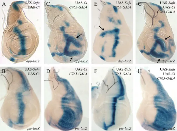
Then, in order to check whether Su(fu) overexpression effects could be modulated by Ci, we overexpressed both ci and Su(fu) simultaneously (Fig. 7), using the C765-GAL4 driver known to be weakly expressed in the entire wing pouch (Méthot and Basler, 1999). This led to different effects in the A and P compartments. In the posterior compartment, overexpressing ci alone led to an
ectopic expression of ptc in the entire compartment and of dpp in two broad posterior stripes (Figs. 7D and 7C, respectively) indicating that the presence of Hh in this compartment leads to a fully activated Ci isoform (Méthot and Basler, 1999). The effects of ci over-expression in this compartment were not modified by simultaneous Su(fu) over-expression (compare Figs. 7G and 7C for dpp and Figs. 7H and 7D for ptc expression), suggesting that Su(fu) overproduction was unable to efficiently counteract Ciact production. In the anterior compartment, ci overexpression alone had no effect (Figs. 7C, D), indicating that the total Ci excess was converted in the repressive Ci75 isoform. In these cells, Su(fu)
overexpression induced ectopic dpp expression associated with Ci155 accumulation; ci expression totally suppressed anterior Su(fu) over-expression effects, as shown by the lack of anterior ectopic dpp expression (compare Figs. 7G and 7E arrow). This suggests that Su(fu) overproduction was unable to counteract Ci75 production.
In conclusion, in both compartments the effects of ci overexpression are totally epistastic over those of Su(fu) overexpression. This suggests that overexpression of Su(fu) does not have any effect on fully activated Ci (Ciact) nor on Ci75.
Effects of Su(fu) over-expression are enhanced in a fu mutant background
It is known that Su(fu) and Fu act antagonistically in the Hh pathway as negative and positive effectors, respectively, (Alves et al., 1998; Préat et al., 1993). To test whether fu could modulate the effects of Su(fu) over-expression, we overexpressed Su(fu) in class I and class II fu mutants, using the fu1 and fuA alleles respectively (see Materials and Methods). We first observed that pupal lethality of fu flies overexpressing Su(fu) was greatly enhanced, even at 21°C or 18°C, when compared to that of fu mutants or to fu+ flies overexpressing Su(fu) (Table 1). These effects were the same with both classes of fu alleles (Table 1). In escapers, the characteristic wing fu mutant phenotype was greatly enhanced : veins 3 and 4 were almost completely fused with a large delta at the margin (Fig. 3D). We also observed anomalies affecting more anterior regions of the wing, namely an enlargement of the domain between vein 2 and the margin, and the more or less complete disappearance of vein 2 (Fig. 3D, asterisk). Both features are reminiscent of an HhMoonrat (HhMrt) phenotype which corresponds to an ectopic hh expression in the anterior compartment (Felsenfeld and Kennison, 1995). Costa duplications were also induced (data not shown). Last, leg anomalies corresponding to anterior duplications, enlargement and fusion of articles were enhanced (Fig. 3H). These effects were seen with both classes of fu alleles.
Correlatively, fuI and fuII wing discs overexpressing Su(fu) displayed changes in dpp and
ptc expression. At the A/P border, dpp and ptc expressions in the wing pouch were further decreased (Figs. 4F and 4H, domains between arrows). These stronger phenotypes conserved certain characteristics of both Su(fu) overexpression, i.e., decrease in expression, and fu loss of function, i.e., an enlargement of the expression domain of dpp and ptc. In the anterior region, the ectopic expression of both dpp (Fig. 4F) and ptc (Fig. 4H) was considerably enhanced, especially for ptc. The effects were the same for both classes of fu alleles (fu1 (Fig. 4), fuA (data not shown)) and are paradoxically reminiscent of previously reported data for fuA Su(fu)- flies (Alves et al., 1998).
In conclusion, the effects of Su(fu) overexpression are enhanced in a fu mutant background. This is consistent with an antagonistic role of Su(fu) and Fu both at the A/P border and in the more anterior regions of imaginal discs. These effects appear similar with both classes of fu alleles thus suggesting that the Fu kinase activity is involved both in cells receiving the Hh signal and in cells that do not.
Discussion
Su(fu) plays a negative role in Hh signalization since it participates both in the cytoplasmic retention of Ci and in the inhibition of the activation of Ci155 (Ingham and McMahon, 2001; Méthot and Basler, 2000; Nybakken and Perrimon, 2002; Ohlmeyer and Kalderon, 1998). Here, we analyzed the effects of Su(fu) over-expression on appendage developpment and on the expression of several Hh target genes in the corresponding discs. In parallel, we studied its accumulation and post-translational modifications during embryonic development in fu+ and fu mutant
backgrounds.
Su(fu) over-expression in imaginal discs leads to opposite effects in cells receiving and not receiving the Hh signal
The effects of Su(fu) over-expression on the Hh pathway were assessed by examining both the adult appendage development and the transcription of well characterized Hh targets (such as dpp and ptc) and accumulation of full-length Ci (Ci155) in the corresponding discs. No effect was detected in the posterior compartment, but two apparently opposite effects were observed in the anterior compartment depending on the distance from the source of Hh.
(i) At the A/P border, there was a decrease in the response to low and high levels of Hh signaling. Indeed, dpp and, to a lesser extent, ptc gene expression was reduced. This result is in agreement with the known inhibitory role of the Su(fu) protein in cells transducing the Hh signal.
(ii) More anteriorly, in cells which do not receive the Hh signal, over-expression of Su(fu) led to anterior duplications in adult appendages. This was correlated with an ectopic expression of dpp in the wing disc or dpp and wg in the leg disc, associated with an accumulation of Ci155. Ectopic ptc expression
was also seen but at a much lower level. These effects phenocopy those of cos2 loss of function mutants (Grau and Simpson, 1987; Simpson and Grau, 1987) or of ectopic hh expression (Felsenfeld and Kennison, 1995). They can be interpreted as a constitutive activation of the pathway. However, the fact that only low levels of ectopic ptc expression are induced shows that the highest levels of Ci activation are not attained.
Anterior ectopic effects of Su(fu) over-expression can occur independently of its effects at the A/P border
High Ptc protein levels at the boundary are known to sequester the Hh protein (Chen and Struhl, 1996). Thus, the anterior ectopic dpp expression observed here in discs overexpressing Su(fu) could be secondary to the deregulation of the Hh pathway at the A/P border: the initial decrease of Ptc at the A/P boundary would result in a further diffusion of Hh to the neighbouring cells in which Ci cleavage would be inhibited, allowing hh and dpp expression. So, step by step, a partial activation of the pathway could be propagated up to the anterior region of the wing pouch. Alternatively, the anterior effects of Su(fu) over-expression could occur independently of events at the A/P border. We favor this latter hypothesis for two reasons: (i) induction of Su(fu) over-expression in the A region, outside the A/P border (using either the vgBE-GAL4 driver or clonal analysis), showed that the ectopic activation of dpp can occur independently of Su(fu) over-expression at the A/P border (Fig. 6), (ii) no significant ectopic hh expression could be detected (Fig. 6H and data not shown).
Su(fu) over-expression modulates Ci states
At least three Ci states have been postulated to exist, depending on the Hh signal gradient: (i) a fully active Ci (Ciact) responsible for high ptc
expression in a stripe 4-5 cells wide close to the A/P border, (ii) a full-length Ci (Ci155) sufficient for dpp
expression 10-15 cell diameters away from the A/P border, (iii) a cleaved Ci form (Ci75) in anterior cells not receiving Hh which represses hh and dpp expression (Aza-Blanc et al., 1997; Dominguez et al., 1996; Méthot and Basler, 1999; Méthot and Basler, 2001; Ohlmeyer and Kalderon, 1998); (for review see Lum and Beachy, 2004; Nybakken and Perrimon, 2002). The balance between these forms of Ci depends on the regulation of non-exclusive processes such as cytoplasmic tethering, protein stability, nuclear shuttling and cleavage (Chen et al., 1999a; Ohlmeyer and Kalderon, 1998; Wang et al., 2000; Wang and Holmgren, 2000). At least two complexes
that contain Ci have been identified: a tetrameric Su(fu)-Ci-Fu-Cos2 complex (complex A) probably present in cells receiving a high level of Hh and a trimeric Ci-Fu-Cos2 complex (complex B) which is devoid of Su(fu) and bound to microtubules in the absence of Hh (Robbins et al., 1997; Sisson et al., 1997; Stegman et al., 2000; Wang and Jiang, 2004). At the molecular level, Su(fu) binds to N-terminal Ci and thus has the capacity to bind both Ci155 and
Ci75 (Monnier et al., 1998; Stegman et al., 2000).
Su(fu) was shown to sequester Ci in the cytoplasm thus controlling the nuclear shuttling of Ci (Méthot and Basler, 2000; Wang et al., 2000; Wang and Jiang, 2004; Wang and Holmgren, 2000). It was also shown to be involved in the stability of Ci155
and Ci75 (Ohlmeyer and Kalderon, 1998).
Here, we show that overexpression of Su(fu) differentially affects the expression of Hh target genes in Hh-receiving and non-receiving cells and that these effects are all reversed by overexpression of Ci. Moreover, the resulting anterior ectopic activation of dpp is associated with an important accumulation of Ci155. To account for
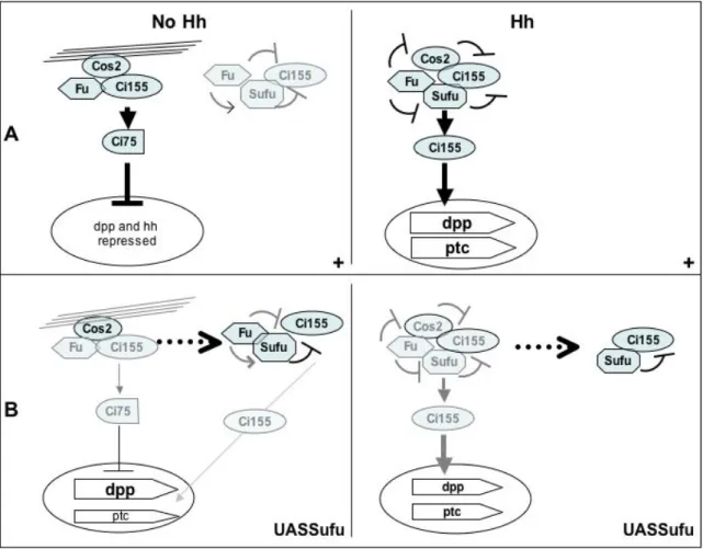
these data, we hypothesize that Su(fu) over-expression disturbs the balance between the different Ci complexes and thus between the different Ci states. We propose a model for Hh signaling in imaginal discs in which the effects of Su(fu) over-expression result mainly from the cytoplasmic retention of Ci155 (Fig. 8). At the A/P
boundary in Hh-receiving cells, Ci155 is normally
present in a tetrameric complex with Su(fu), Fu and Cos2 (complex A). In these cells, Hh signaling via the activation of Fu blocks Cos2 and Su(fu) negative effects in the tetrameric complex, thus preventing Ci cleavage and cytoplasmic retention and favoring the release of Ci, its activation and nuclear access (Fig. 8A [Hh]). Su(fu) over-expression could lead to the recruitment of a significant fraction of endogenous Ci155 into complexes in which Su(fu) is no longer inhibited by Fu. A fraction of Ci is thus sequestered in the cytoplasm as an inactive full-length form (Fig. 8B [Hh]). Co-over-expression of Ci along with Su(fu) would provide enough Ci to buffer the excess of Su(fu), leading to the formation of active Ci155. In
the anterior region where Hh is absent, Ci is present in a microtubule-bound trimeric complex (complex B) containing Fu and Cos2 but not Su(fu), leading to Ci cytoplasmic tethering and favoring its cleavage in the Ci75 repressive form. This complex
would be in equilibrium with a Fu-Su(fu)-Ci complex. In this complex, Su(fu) would act as a safety lock for the cytoplasmic retention of an uncleaved fraction of Ci155 potentially able to yield
some active forms of Ci (Fig. 8A [noHh]). When Su(fu) is overexpressed, extra Su(fu) would bind Ci155, preventing it from joining the
microtubule-bound complex (Fig. 8B [noHh]). Ci would not be effectively processed, leading to the accumulation
of uncleaved Ci155. The reduction in the amount of Ci75 would be sufficient to allow the expression of
dpp but not that of hh, which has been reported to be more sensitive to Ci75 repression than dpp (Méthot and Basler, 1999). There would be an enrichment in the other complex but only a few active Ci forms would be produced in agreement with the almost total absence of ectopic ptc expression.
All effects of Su(fu) over-expression are modulated by Fu
The present data show that all the effects induced by overexpresion of Su(fu) were enhanced in fu mutants, namely pupal lethality, ectopic anterior expression of dpp and ptc genes and their decrease at the antero-posterior border.
At the A/P border, Fu is normally required to antagonize the negative effect of Su(fu) in Hh receiving cells. In fu mutant discs overexpressing Su(fu), the negative effects that Su(fu) exerts on Ci155 cytoplasmic retention in the tetrameric complex would no longer be counteracted by Fu. The shifting of the equilibrium towards the inactive Su(fu)-Ci complex is increased. Less active Ci is available and the reduction in dpp and ptc expression is aggravated. The anterior ectopic activation of the pathway seen in discs overexpressing Su(fu) was greatly enhanced in fu mutants. These unexpected results provide evidence for an inhibitory role of Fu on Ci155 in the absence of the Hh signal. In the
absence of Hh, Fu activity could favor the normal restrictive effect of Su(fu) on Ci155 in the Fu-Su(fu)-Ci complex (Figure 8A [noHh]) In fu- mutants, the
negative effect of Su(fu) on the trapped fraction of Ci155 would be weakened and enough Ci155 would be
active to induce transcription of dpp and of ptc. Strikingly, unlike Su(fu) loss of function mutations, Su(fu) over-expression failed to distinguish between the two classes of fu alleles. Since the regulatory domain is probably necessary for Fu kinase activity, the effects seen are probably all mostly due to a loss of Fu kinase activity which would reduce the level of phosphorylation of Su(fu). As shown here and in several recent reports, the Su(fu) protein is phosphorylated in the embryo (Ho et al., 2005; Lum et al., 2003). We detected multiple levels of phosphorylation, with hyperphosphorylated forms that accumulate at a period in embryonic development when Fu is activated by the Hh signal (Thérond et al., 1996b) and that are significantly reduced in fu mutants. Thus, Fu could modulate Su(fu) activity by controlling, directly or indirectly, its phosphorylation. In the absence of Hh signaling, a low level of Su(fu) phosphorylation by Fu would reinforce the negative effect of Su(fu), whereas a higher phosphorylation level would inactivate Su(fu) in Hh responding cells at the A/P border.
Nevertheless, phosphorylated isoforms were not totally abolished in fu mutants, suggesting that other kinase(s) can phosphorylate Su(fu). In
agreement with this point, numerous putative phosphorylation sites for kinases such as Caseine kinase II or PKC, but not PKA, are present in the Su(fu) protein. However, the biological implications of the Su(fu) isoforms and their modulation by the Hh transduction signal remain to be demonstrated.
Acknowledgments
We thank Isabelle Urbain and Patricia Vandurka for excellent technical assistance, Myriam Barre and Matthieu Sanial for their help in preparing the figures. We also thank the Imaging Facility at the Institut Jacques Monod for precious help in confocal microscopy. We are grateful to Pascal Thérond for critical reading of the manuscript and to Antonia Kropfinger for assistance with the English language. We thank members of Catherine Jessus’s group for technical advice. We thank R. Holmgren for providing us with the 2A1 anti-Ci antibody. This work was supported by grants from the Centre National de la Recherche Scientifique, ACI “Biologie du Développement et Physiologie intégrative”(CR525044) and from the Association pour la Recherche contre le Cancer (4797). F.D.G. and S.F. received fellowships from the MNRT and Ligue contre le Cancer.
References
Alves, G., Limbourg, B. B., Tricoire, H., Brissard, Z. J., Lamour, I. C., and Busson, D. (1998). Modulation of Hedgehog target gene expression by the Fused serine- threonine kinase in wing imaginal discs. Mech Dev 78, 17-31.
Anderson, C. W., Baum, P. R., and Gesteland, R. F. (1973). Processing of adenovirus 2-induced proteins. J Virol 12, 241-52.
Aza-Blanc, P., Ramirez, W. F., Laget, M. P., Schwartz, C., and Kornberg, T. B. (1997). Proteolysis that is inhibited by hedgehog targets Cubitus interruptus protein to the nucleus and converts it to a repressor. Cell 89, 1043-53.
Blackman, R. K., Sanicola, M., Raftery, L. A., Gillevet, T., and Gelbart, W. M. (1991). An extensive 3' cis-regulatory region directs the imaginal disk expression of decapentaplegic, a member of the TGF-beta family in Drosophila. Development 111, 657-66. Brand, A. H., and Perrimon, N. (1993). Targeted
gene expression as a means of altering cell fates and generating dominant phenotypes. Development 118, 401-15.
Busson, D., Limbourg-Bouchon, B., Mariol, M. C., Préat, T., and Lamour Isnard, C. (1988). Genetic analysis of viable and lethal fused mutants of Drosophila melanogaster. Roux's Arch Dev Biol 197, 221-230.
Chen, C. H., von, K. D., Park, W., Wang, B., Ma, Y., and Beachy, P. A. (1999a). Nuclear trafficking of Cubitus interruptus in the transcriptional regulation of Hedgehog target gene expression. Cell 98, 305-16.
Chen, Y., Cardinaux, J. R., Goodman, R. H., and Smolik, S. M. (1999b). Mutants of cubitus interruptus that are independent of PKA regulation are independent of hedgehog signaling. Development 126, 3607-16.
Chen, Y., and Struhl, G. (1996). Dual roles for patched in sequestering and transducing Hedgehog. Cell 87, 553-63.
Delanoue, R., Legent, K., Godefroy, N., Flagiello, D., Dutriaux, A., Vaudin, P., Becker, J. L., and Silber, J. (2004). The Drosophila wing differentiation factor vestigial-scalloped is required for cell proliferation and cell survival at the dorso-ventral boundary of the wing imaginal disc. Cell Death Differ 11, 110-22. Denef, N., Neubuser, D., Perez, L., and Cohen, S. M.
(2000). Hedgehog induces opposite changes in turnover and subcellular localization of patched and smoothened. Cell 102, 521-31. Dominguez, M., Brunner, M., Hafen, E., and Basler,
K. (1996). Sending and receiving the hedgehog signal: control by the Drosophila Gli protein Cubitus interruptus. Science 272, 1621-5.
Felsenfeld, A. L., and Kennison, J. A. (1995). Positional signaling by hedgehog in Drosophila imaginal disc development. Development 121, 1-10.
Grau, Y., and Simpson, P. (1987). The segment polarity gene costal-2 in Drosophila. I. The organization of both primary and secondary embryonic fields may be affected. Dev Biol 122, 186-200.
Guillen, I., Mullor, J. L., Capdevila, J., Sanchez-Herrero, E., Morata, G., and Guerrero, I. (1995). The function of engrailed and the specification of Drosophila wing pattern. Development 121, 3447-56.
Ho, K. S., Suyama, K., Fish, M., and Scott, M. P. (2005). Differential regulation of Hedgehog target gene transcription by Costal2 and Suppressor of Fused. Development 132, 1401-12.
Ingham, P. W., and McMahon, A. P. (2001). Hedgehog signaling in animal development: paradigms and principles. Genes and development 15, 3059-3087.
Laemmli, U. K. (1970). Cleavage of structural proteins during the assembly of the head of bacteriophage T4. Nature 227, 680-5.
Lee, J. J., von Kessler, D. P., Parks, S., and Beachy, P. A. (1992). Secretion and localized transcription suggest a role in positional signaling for products of the segmentation gene hedgehog. Cell 71, 33-50.
Lepage, T., Cohen, S. M., Diaz Benjumea, F. J., and Parkhurst, S. M. (1995). Signal transduction by cAMP-dependent protein kinase A in Drosophila limb patterning [see comments]. Nature 373, 711-5 Issn: 0028-0836.
Lum, L., and Beachy, P. A. (2004). The Hedgehog response network: sensors, switches, and routers. Science 304, 1755-9.
Lum, L., Zhang, C., Oh, S., Mann, R. K., von Kessler, D. P., Taipale, J., Weis-Garcia, F., Gong, R., Wang, B., and Beachy, P. A. (2003). Hedgehog signal transduction via Smoothened association with a cytoplasmic complex scaffolded by the atypical kinesin, Costal-2. Mol Cell 12, 1261-74.
Méthot, N., and Basler, K. (1999). Hedgehog controls limb development by regulating the activities of distinct transcriptional activator and repressor forms of Cubitus interruptus. Cell 96, 819-31.
Méthot, N., and Basler, K. (2000). Suppressor of fused opposes hedgehog signal transduction by impeding nuclear accumulation of the activator form of Cubitus interruptus. Development 127, 4001-10.
Méthot, N., and Basler, K. (2001). An absolute requirement for Cubitus interruptus in Hedgehog signaling. Development 128, 733-42.
Monnier, V., Dussillol, F., Alves, G., Lamour, I. C., and Plessis, A. (1998). Suppressor of fused links fused and Cubitus interruptus on the hedgehog signalling pathway. Curr Biol 8, 583-6.
Monnier, V., Ho, K. S., Sanial, M., Scott, M. P., and Plessis, A. (2002). Hedgehog signal transduction proteins: contacts of the Fused kinase and Ci transcription factor with the Kinesin-related protein Costal2. BMC Dev Biol 2, 4.
Motzny, C. K., and Holmgren, R. (1995). The Drosophila cubitus interruptus protein and its role in the wingless and hedgehog signal transduction pathways. Mech Dev 52, 137-50.
Neufeld, T. P., de la Cruz, A. F., Johnston, L. A., and Edgar, B. A. (1998). Coordination of growth and cell division in the Drosophila wing. Cell 93, 1183-93.
Neumann, C. J., and Cohen, S. M. (1996). A hierarchy of cross-regulation involving Notch, wingless, vestigial and cut organizes the dorsal/ventral axis of the Drosophila wing. Development 122, 3477-85.
Nybakken, K., and Perrimon, N. (2002). Hedgehog signal transduction: recent findings. Curr Opin Genet Dev 12, 503.
Nybakken, K. E., Turck, C. W., Robbins, D. J., and Bishop, J. M. (2002). Hedgehog-stimulated phosphorylation of the kinesin-related protein Costal2 is mediated by the serine/threonine kinase fused. J Biol Chem 277, 24638-47. Ohlmeyer, J. T., and Kalderon, D. (1998). Hedgehog
stimulates maturation of Cubitus interruptus into a labile transcriptional activator. Nature 396, 749-53.
Pham, A., Therond, P., Alves, G., Tournier, F. B., Busson, D., Lamour Isnard, C., Bouchon, B. L., Préat, T., and Tricoire, H. (1995). The Suppressor of fused gene encodes a novel PEST protein involved in Drosophila segment polarity establishment. Genetics 140, 587-598. Préat, T. (1992). Characterization of Suppressor of fused, a complete suppressor of the fused segment polarity gene of Drosophila melanogaster. Genetics 132, 725-736.
Préat, T., Therond, P., Limbourg, B. B., Pham, A., Tricoire, H., Busson, D., and Lamour, I. C. (1993). Segmental polarity in Drosophila melanogaster: genetic dissection of fused in a Suppressor of fused background reveals interaction with costal-2. Genetics 135, 1047-62.
Robbins, D. J., Nybakken, K. E., Kobayashi, R., Sisson, J. C., Bishop, J. M., and Therond, P. P. (1997). Hedgehog elicits signal transduction by means of a large complex containing the kinesin-related protein costal2. Cell 90, 225-34.
Simpson, P., and Grau, Y. (1987). The segment polarity gene costal-2 in Drosophila. II. The origin of imaginal pattern duplications. Dev Biol 122, 201-9.
Sisson, J. C., Ho, K. S., Suyama, K., and Scott, M. P. (1997). Costal2, a novel kinesin-related protein in the Hedgehog signaling pathway. Cell 90, 235-45.
Spradling, A. C., Stern, D., Beaton, A., Rhem, E. J., Laverty, T., Mozden, N., Misra, S., and Rubin, G. M. (1999). The Berkeley Drosophila Genome Project gene disruption project: Single P-element insertions mutating 25% of vital Drosophila genes. Genetics 153, 135-77. Stegman, M., Vallance, J., Elangovan, G., Sosinski,
J., Cheng, Y., and Robbins, D. (2000). Identification of a tetrameric hedgehog signaling complex. J Biol Chem 275, 21809-12.
Stegman, M. A., Goetz, J. A., Ascano, M., Jr., Ogden, S. K., Nybakken, K. E., and Robbins, D. J. (2004). The Kinesin-related protein Costal2 associates with membranes in a Hedgehog-sensitive, Smoothened-independent manner. J Biol Chem 279, 7064-71.
Struhl, G., and Basler, K. (1993). Organizing activity of wingless protein in Drosophila. Cell 72, 527-40.
Thérond, P., Alves, G., Limbourg-Bouchon, B., Tricoire, H., Guillemet, E., Brissard-Zahraoui, J., Lamour-Isnard, C., and Busson, D. (1996a). Functional domains of fused, a serine-threonine kinase required for signaling in Drosophila. Genetics 142, 1181-98.
Thérond, P. P., Knight, J. D., Kornberg, T. B., and Bishop, J. M. (1996b). Phosphorylation of the fused protein kinase in response to signaling from hedgehog. Proc Natl Acad Sci U S A 93, 4224-8.
Vervoort, M. (2000). hedgehog and wing development in Drosophila: a morphogen at work? Bioessays 22, 460-8.
Wang, G., Amanai, K., Wang, B., and Jiang, J. (2000). Interactions with Costal2 and suppressor of fused regulate nuclear translocation and activity of cubitus interruptus. Genes Dev 14, 2893-905. Wang, G., and Jiang, J. (2004). Multiple Cos2/Ci
interactions regulate Ci subcellular localization through microtubule dependent and independent mechanisms. Dev Biol 268, 493-505.
Wang, Q. T., and Holmgren, R. A. (1999). The subcellular localization and activity of Drosophila cubitus interruptus are regulated at multiple levels. Development 126, 5097-106.
Wang, Q. T., and Holmgren, R. A. (2000). Nuclear import of cubitus interruptus is regulated by hedgehog via a mechanism distinct from Ci stabilization and Ci activation. Development 127, 3131-9.
Whittle, J. R. (1976). Clonal analysis of a genetically caused duplication of the anterior wing in Drosophila melanogaster. Dev Biol 51, 257-68.
Wolff, A., de Nechaud, B., Chillet, D., Mazarguil, H., Desbruyeres, E., Audebert, S., Edde, B., Gros, F., and Denoulet, P. (1992). Distribution of glutamylated alpha and beta-tubulin in mouse tissues using a specific monoclonal antibody, GT335. Eur J Cell Biol 59, 425-32.
Fig. 1: Accumulation and post-translational modifications of the Su(fu) protein during embryonic development. A) Immunodetection of the Su(fu) protein in embryonic extracts from 0-2 h, 2-4 h, 4-6 h, 6-8 h Oregon R embryos and from 0-8 h Df(3R)karSZ11/Df(3R)karSZ21 embryos; electrophoresis is performed on a Laemmli type acrylamide gel; upper bands, around 54 kDa, are revealed with our anti-Su(fu) polyclonal antibody, lower bands with an anti- tubulin antibody after stripping of the membrane; note the total absence of immuno-reactive material in embryos deleted for Su(fu); Su(fu) is maternally present in 0-2 h embryos; its level increases in 2-4 h embryos to diminish in 4-6 h and 6-8 h embryos.
B) Su(fu) protein isoforms during Oregon R embryonic development; electrophoresis on an Anderson type gel reveals at least 4 isoforms, a major isoform (1, arrow) of 54kDa, two slower migrating isoforms (2 and 3, arrowheads) and a faster migrating one (4, empty arrowhead); the major isoform 1 does not vary significantly according to the developmental stage; the slower isoforms 2 and 3 appear progressively from 0-2 h (one isoform) to 6-8 h (two isoforms) to decrease from 8 h onward; a reciprocal modulation is seen for the faster isoform 4. C) Bidimensional electrophoresis according to pHi and PM of extracts from 0-16h Oregon R embryos; at least, five isoforms are revealed, one major isoform (a, arrow) and four minor slower migrating acidic isoforms (b, c, d, e, arrowheads).
D) Su(fu) protein phosphorylation; extracts from 0-16 h embryos are incubated, 10, 30, 90 min. at 37°C, without (lanes 1, 3, 5) or with a mix of phosphatase inhibitors (lanes 2, 4, 6) and fractionated on an Anderson type gel; the control corresponds to embryonic extracts not incubated at 37°C (lane 7). In absence of phophatase inhibitors, a progressive disappearance of the higher acidic isoforms (2 and 3, arrowheads) is seen and correlated with an increase of the lower form (4, empty arrowhead). This effect is totally inhibited in the presence of phosphatase inhibitors. No modulation of the 54 kDa major isoform (1, arrow) is observed.
Fig. 2: Modulation of Su(fu) isoforms in a fu mutant background.
Proteic extracts from 0-24 h embryos, wild-type (lane 1) and fu mutants, fuJB3 (lane 2), fuA (lane 3), were
migrated on Anderson type gel and revealed with anti-Su(fu) antibody; extracts from Su(fu)LP (lane 4) and
Df(Su(fu)) (lane 5) embryos are shown as controls. Two exposure times are given. As compared to wild-type, the relative amounts of slower migrating isoforms 2 and 3 are reduced in both class I (fuJB3) and class II (fuA) fu embryonic extracts. Unlike class I fuJB3, class II fuA extracts display a strong increase in faster migrating isoforms 4 and 5.
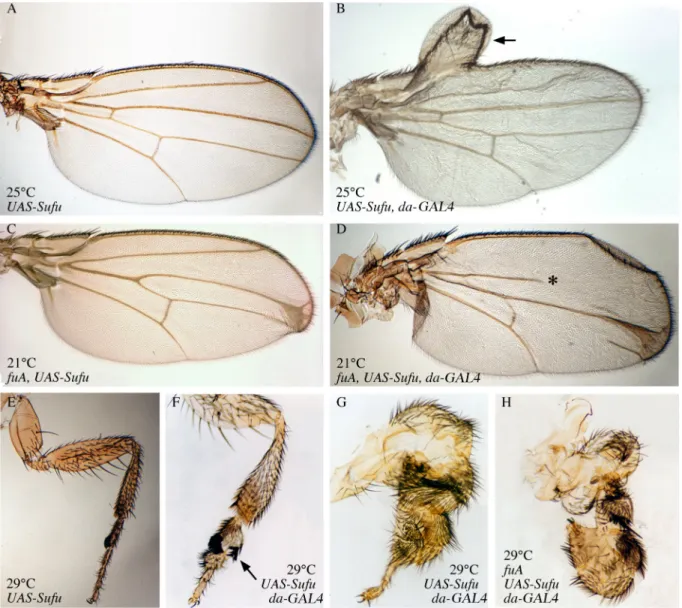
Fig. 3: Effects of Su(fu) overexpression on wing and leg phenotypes in fu+ and fu mutant backgrounds.
(A, B) Wings of UAS-Su(fu) flies (A) and UAS-Su(fu); da-GAL4 flies (B) raised at 25°C. In wings overexpressing Su(fu), the region between veins 3 and 4 is not altered but an anterior duplication on the wing margin is observed (arrow in B). (C, D) Wings of wfuA, UAS-Su(fu) flies (C) and wfuA, UAS-Su(fu); da-GAL4 flies (D) raised at 21°C. In fuA wings overexpressing Su(fu), the region between veins 3 and 4 completely
disappeared; the vein 2 is truncated (* in D) and the domain between vein 2 and the margin is enlarged. Wings in (B) and (D) are observed from rare escapers (see Table 1). Anterior duplications can also be seen (data not shown). (E-G) Legs of UAS-Su(fu) flies (E) and UAS-Su(fu); da-GAL4 flies (F, G) raised at 29°C. In the first pair of legs overexpressing Su(fu), articles are shorter and thicker, and legs present a clear anterior sex comb duplication (arrow in F). Legs of the third pair are extremely deformed, with enlarged and fused articles (G). (H) Legs of UAS-Su(fu), wfuA; da-GAL4 flies raised at 29°C show anterior duplications, enlargment and fusion of
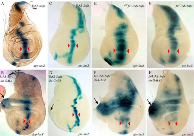
Fig. 4: Effects of ubiquitous Su(fu) overexpression on dpp and ptc expression in wing imaginal disc.
(A-D) Expression of dpp-lacZ (A, B) and ptc-lacZ (C, D) in UAS-Su(fu) wing discs (A, C) and UAS-Su(fu); da-GAL4 discs (B, D) raised at 25°C. When overexpressing Su(fu), the domain of expression of dpp-lacZ and ptc-lacZ along the antero-posterior boundary is reduced (compare B with A, and D with C respectively, width of the domain between red arrowheads ; note that dpp expression is nearly lost at the intersection between A/P and D/V borders). At the same time, dpp-lacZ (arrow in B) and ptc-lacZ (arrow in D) are ectopically expressed in the anterior compartment. (E-H) Expression of dpp-lacZ (E, F) and ptc-lacZ (G, H) in wfu1, UAS-Su(fu) wing discs
(E, G) and wfu1, UAS-Su(fu); da-GAL4 discs (F, H) raised at 25°C. In a fu mutant background, dpp-lacZ and ptc-lacZ expressions are nearly lost in the wing pouch of discs overexpressing Su(fu) (compare F with E, and H with G respectively, width of the domain between red arrowheads). The overexpression of Su(fu) generates also an anterior ectopic expression of dpp-lacZ (arrow in F) and ptc-lacZ (arrow in H). Note that the anterior ectopic expression of dpp-lacZ and ptc-lacZ in a fu mutant background is much stronger than in wild type background (compare F with B and H with D). Overexpression of Su(fu) in a fuA background gives the same kind of
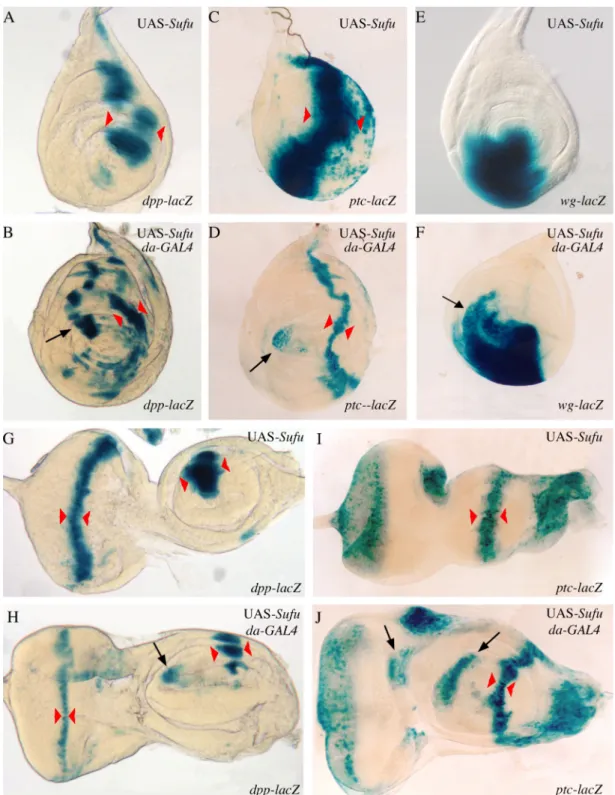
Fig. 5: Effects of ubiquitous Su(fu) overexpression on dpp, wg and ptc expression in leg, antenna and eye imaginal discs.
(A-F) Expression of dpp-lacZ (A, B), ptc-lacZ (C, D) and wg-lacZ (E, F) in UAS-Su(fu) leg discs (A, C, E) and UAS-Su(fu); da-GAL4 leg discs (B, D, F) raised at 25°C. In imaginal discs overexpressing Su(fu), the expression of dpp-lacZ and ptc-lacZ is reduced along the antero-posterior boundary (compare B with A and D with C respectively, width of the domain between red arrowheads), while dpp-lacZ and ptc-lacZ are ectopically expressed in the anterior compartment (arrows respectively in B and D). The expression of wg-lacZ is anteriorly and dorsally extended when overexpressing Su(fu) (compare F with E). (G-J) Expression of dpp-lacZ (G, H) and ptc-lacZ (I, J) in UAS-Su(fu) antenna and eye discs (G, I) and UAS-Su(fu); da-GAL4 discs (H, J) raised at 25°C. Eye-antenna discs overexpressing Su(fu) are highly deformed. In those discs, dpp-lacZ (arrow in H) and ptc-lacZ (arrows in J) are ectopically and anteriorly expressed; in the eye part of the disc, the expression of dpp-lacZ in the furrow is reduced (compare H with G, width of the domain between red arrowheads). In H and J, X-Gal staining has been prolonged to clearly see ectopic expressions, so the expression of dpp-lacZ and ptc-lacZ along the antero-posterior boundary in the antennal part of the discs seems nearly as strong as in normal discs.
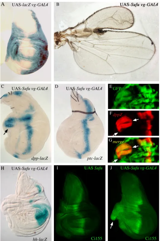
Fig. 6: Overexpression of Su(fu) outside the antero-posterior border in wing imaginal discs also drives ectopic activation of Hh target gene expression.
(A) Expression pattern of the vg-GAL4 driver in vg-GAL4 lacZ imaginal wing disc. (B) wing of UAS-Su(fu); vg-GAL4 fly raised at 25°C. (C, D) Expression of dpp-lacZ (C) and ptc-lacZ (D) in UAS-UAS-Su(fu); vg-GAL4 discs raised at 25°C. Su(fu) overexpression in vg-GAL4 domain leads to an anterior overgrowth and dpp anterior ectopic expression (arrow in C). (E-G) Su(fu) overexpression in clones generated in UAS-Su(fu)/y w hs-flp; act5C>CD2>GAL4, UAS-GFP/ dpp-lacZ flies. GFP (E) and dpp-lacZ (F) expressions are merged in G. dpp-lacZ
expression is detected outside the GFP-expressing clones (arrows in F and G). (H) hh-lacZ expression in UAS-Su(fu); vg-GAL4 discs. (I, J) Ci155 localization in UAS-Su(fu) (I) and UAS-Su(fu); vg-GAL4 (J) discs. In wild type discs, Ci155 is detected in the anterior compartment along the antero-posterior border. When overexpressing
Fig. 7: Epistatic relationship between ci and Su(fu) overexpression effects.
Expression of dpp-lacZ (A, C, E, G) and ptc-lacZ (B, D, F, H), in UAS-Su(fu); UAS-ci control discs (A, B) and UAS-ci; C765-GAL4 (C, D), UAS-Su(fu); C765-GAL4 (E, F) and UAS-Su(fu); UAS-ci; C765-GAL4 (G, H) wing discs, from flies raised at 25°C. Ubiquitous overexpression of ci under the C765-GAL4 driver in the wing disc leads to two stripes of ectopic dpp-lacZ expression in the posterior compartment (arrows in C), and to ectopic ptc-lacZ expression in the whole posterior compartment (D). The overexpression of Su(fu), with the same driver, leads to an anterior ectopic expression of dpp-lacZ (arrow in E), but not of ptc-lacZ (F). Co-over-expression of ci and Su(fu) gives patterns of dpp-lacZ and ptc-lacZ expression similar to those obtained overexpressing ci alone (compare G with C and H with D).