HAL Id: hal-02651928
https://hal-amu.archives-ouvertes.fr/hal-02651928
Submitted on 29 May 2020
HAL is a multi-disciplinary open access
archive for the deposit and dissemination of
sci-entific research documents, whether they are
pub-lished or not. The documents may come from
teaching and research institutions in France or
abroad, or from public or private research centers.
L’archive ouverte pluridisciplinaire HAL, est
destinée au dépôt et à la diffusion de documents
scientifiques de niveau recherche, publiés ou non,
émanant des établissements d’enseignement et de
recherche français ou étrangers, des laboratoires
publics ou privés.
Distributed under a Creative Commons Attribution - NonCommercial| 4.0 International
Mode of delivery in hemophilia: vaginal delivery and
Cesarean section carry similar risks for intracranial
hemorrhages and other major bleeds
Nadine Andersson, Elizabeth Chalmers, Gili Kenet, Rolf Ljung, Anne
Mäkipernaa, Hervé Chambost
To cite this version:
Nadine Andersson, Elizabeth Chalmers, Gili Kenet, Rolf Ljung, Anne Mäkipernaa, et al.. Mode
of delivery in hemophilia: vaginal delivery and Cesarean section carry similar risks for intracranial
hemorrhages and other major bleeds. Haematologica, Ferrata Storti Foundation, 2019, 104 (10),
pp.2100-2106. �10.3324/haematol.2018.209619�. �hal-02651928�
Received: October 18, 2018. Accepted: February 14, 2019. Pre-published: February 21, 2019.
©2019 Ferrata Storti Foundation
Material published in Haematologica is covered by copyright. All rights are reserved to the Ferrata Storti Foundation. Use of published material is allowed under the following terms and conditions:
https://creativecommons.org/licenses/by-nc/4.0/legalcode. Copies of published material are allowed for personal or inter-nal use. Sharing published material for non-commercial pur-poses is subject to the following conditions:
https://creativecommons.org/licenses/by-nc/4.0/legalcode, sect. 3. Reproducing and sharing published material for com-mercial purposes is not allowed without permission in writing from the publisher.
Correspondence:
NADINE G. ANDERSSON nadine.gretenkort_andersson@med.lu.seHaematologica
2019
Volume 104(10):2100-2106
doi:10.3324/haematol.2018.209619Check the online version for the most updated information on this article, online supplements, and information on authorship & disclosures: www.haematologica.org/content/104/10/2100
Ferrata Storti Foundation
T
he optimal mode of delivery for a pregnant hemophilia carrier is
still a matter of debate. The aim of the study was to determine
the incidence of intracranial hemorrhage and other major bleeds
in neonates with moderate and severe hemophilia in relationship to
mode of delivery and known family history. A total of 926 neonates,
786 with severe and 140 with moderate hemophilia were included in
this PedNet multicenter study. Vaginal delivery was performed in
68.3% (n=633) and Cesarean section in 31.6% (n=293). Twenty
intracranial hemorrhages (2.2%) and 44 other major bleeds (4.8%)
occurred. Intracranial hemorrhages occurred in 2.4% of neonates
fol-lowing vaginal delivery compared to 1.7% after Cesarean section
(P=not significant); other major bleeds occurred in 4.2% born by
vagi-nal delivery and in 5.8% after Cesarean section (P=not significant).
Further analysis of subgroups (n=813) identified vaginal delivery with
instruments being a significant risk factor for both intracranial
hemor-rhages and major bleeds (Relative Risk: 4.78-7.39; P<0.01); no other
sig-nificant differences were found between vaginal delivery without
instruments, Cesarean section prior to and during labor. There was no
significant difference in frequency for intracranial hemorrhages and
major bleeds between a planned Cesarean section and a planned
vagi-nal delivery. Children with a family history of hemophilia (n=466)
were more likely to be born by Cesarean section (35.8% vs. 27.6%),
but no difference in the rate of intracranial hemorrhages or major
bleeds was found. In summary, vaginal delivery and Cesarean section
carry similar risks of intracranial hemorrhages and major bleeds. The
‘PedNet Registry’ is registered at clinicaltrials.gov identifier: 02979119.
Mode of delivery in hemophilia: vaginal
delivery and Cesarean section carry similar
risks for intracranial hemorrhages and other
major bleeds
Nadine G. Andersson,1,2Elizabeth A. Chalmers,3Gili Kenet,4Rolf Ljung,2
Anne Mäkipernaa,5Hervé Chambost;6on behalf of the PedNet Haemophilia
Research Foundation
1Centre for Thrombosis and Hemostasis, Skåne University Hospital, Malmö, Sweden; 2Lund University, Department of Clinical Sciences, Lund, Sweden; 3Department of
Haematology, Royal Hospital for Children, Glasgow, UK; 4National Hemophilia Center,
Sheba Medical Center, Tel Hashomer and Sackler School of Medicine, Tel Aviv University, Israel; 5Children’s Hospital and Hematology, Comprehensive Cancer Center, Helsinki
University Hospital, University of Helsinki, Helsinki, Finland and 6Pediatric Haematology
Oncology Department, Children Hospital La Timone, AP-HM, and Aix Marseille Université, INSERM, INRA, C2VN, Faculté de Médecine, Marseille, France
ABSTRACT
Introduction
The optimal mode of delivery for a known hemophilia carrier, i.e. either vaginal delivery (VD) or Cesarean section (CS), is still a matter of debate. A carrier may have an increased bleeding risk herself that might need to be taken into account in the obstetric planning but, from the fetal point of view, the key question is how the mode of delivery may impact on the risk of major bleeds, and in particular intracranial hemorrhages (ICH). These life threatening bleeds can also impair the
require-ment for intensive replacerequire-ment therapy has been identi-fied as a risk factor for inhibitor development in patients
with hemophilia A.3
Several studies have been published with quite a uni-form consensus that around 3-4% of boys with hemophil-ia born in countries with a good standard of obstetric care
have had ICH diagnosed during the neonatal period.2,4-6 It
has also been shown that instrumental delivery using for-ceps or vacuum extraction (VE) is a major risk factor for intra- and extracranial bleeds in neonates with
hemophil-ia.5,7,8 However, figures for bleeds associated with
uncom-plicated VD and CS and, consequently, recommendations on mode of delivery vary between publications and
guide-lines.7,9-11It is undisputed that the risk of a neonatal bleed
in a child with hemophilia is considerably higher than that expected in a non-hemophilia population, although few figures are available. The largest series, among the few studies published on mode of delivery in the normal pop-ulation, suggests an incidence of ICH of 1 per 1,900 in spontaneous VD, 1 per 2,750 in CS with no labor, 1 per 907 delivered by CS during labor, 1 per 860 deliveries with
VE, and 1 per 664 delivered with the use of forceps.12
Using these numbers as a reference, the risk for ICH in hemophilia boys is 60 times higher compared to healthy neonates born by VD.
Knowledge of carrier status in a pregnant woman, or the knowledge of confirmed hemophilia in the fetus follow-ing prenatal diagnosis (PND), may impact on obstetric care, especially when planning the mode of delivery. In recent cohorts, around 50% of all cases of hemophilia are sporadic, i.e. newly diagnosed boys without family histo-ry of hemophilia or without any knowledge of carrier
sta-tus in the mother at birth.13 Many published studies on the
delivery of a hemophilia child have not been able to dis-tinguish between sporadic cases and cases with a known family history of hemophilia. Some studies include neonates with mild hemophilia and, furthermore, it is not always possible to distinguish between CS performed as a result of the baby having hemophilia or for other reasons, and this complicates comparisons between studies.
The PedNet Registry is a prospective, multicenter
data-base that includes all children born since 1stJanuary 2000
diagnosed with hemophilia A (HA) or B (HB) of all sever-ities and treated in the 31 participating hemophilia centers
in Europe, Canada and Israel.14 Baseline data regarding the
neonatal period are collected on mode of delivery, neona-tal events, family history of hemophilia, and gestational age. This longitudinal prospectively collected cohort study makes it possible to address questions of interest on obstetric and neonatal issues.
The aim of this paper was to study the frequency of ICH and other major bleeds in neonates with hemophilia and the association with mode of delivery to improve counseling of pregnant carriers in the future. Furthermore, the results will be stratified according to the presence or absence of a prior knowledge of hemophilia in the family.
Methods
Study group
Data were retrieved from the ‘PedNet Registry’ which is owned and administered by the ‘PedNet Haemophilia Research Foundation’, consisting of 31 international hemophilia treatment centers and registered at clinicaltrials.gov identifier: 02979119. The
purpose of the registry is to promote and facilitate research and healthcare development in children with hemophilia. The PedNet Registry includes all consecutive patients diagnosed and treated in each center born after 1stJanuary 2000. The aim of the PedNet reg-istry is to establish large well-documented birth cohorts of patients with hemophilia enabling studies on side effects and out-come of treatment. Patient data are collected from birth onwards prospectively and consist of all data concerning treatment, side effects and outcome of treatment. Information is collected on mode of delivery and during the first 75 exposure days of treat-ment with factor concentrate; all major bleeds including ICH are registered with detailed information. Approval for data collection was obtained from the institutional review boards of each of the 31 centers taking part in the study, and written informed consent was obtained from the parents or guardians of all participants in accordance with the Declaration of Helsinki. The data quality in the PedNet Registry is monitored regularly and independent audits are carried out in all participating centers.14
Study population
All children included in the Registry by 1stJanuary 2015 with severe (factor VIII/IX activity, < 0.01 IU/mL) or moderate (factor VIII/IX activity, 0.01-0.05 IU/mL) HA and HB and with at least one follow up covering the neonatal period after the initial baseline report were enrolled. This resulted in 926 children born between 1stJanuary 2000 and 1stJanuary 2015 with data on mode of deliv-ery and the neonatal period, defined as 28 days after birth. Prematurity was defined as up to 36 weeks of gestational age.
Data collection
We uniformly collected data on the mode of delivery (including vaginal, vaginal instrumental, CS), and major bleeds including ICH in the neonatal period. Data were also recorded on whether an affected newborn belonged to a family with a known history of hemophilia or was a sporadic case.
Statistical analysis
The primary outcome was ICH and major bleeds, the latter defined as a bleed requiring treatment with factor concentrate and not resolving within 24 hours during the neonatal period. The determinants of outcome were mode of delivery and family histo-ry of hemophilia, either known or unknown. Statistical compar-isons between different groups were made using χ2 test or Fisher´s exact test at a significance level of 0.05. In the comparison of four subgroups on mode of delivery, an overall test was per-formed to compare the frequencies between all groups simultane-ously. If an overall test was significant, it was followed by pair-wise comparisons between the groups. The results of the pairpair-wise comparisons were corrected for multiple testing by Bonferroni correction. Statistical power was shown by the width and magni-tude of the 95% Confidence Interval (95%CI) according to the CONSORT guidelines. All analyses were performed using IBM SPSS Statistics for Windows, Version 24.0. Armonk, NY: IBM Corp., NY, USA, or R: A language and environment for statistical Computing, version 3.4.2. Vienna, Austria, R Foundation for Statistical Computing.
Results
Cohort demography
A total of 926 patients were included, 140 with moder-ate and 786 with severe hemophilia comprising those with HA [n = 803 (86.7%)], and HB [n=123 (13.3%)]. MOD in the 926 patients was vaginal in 633 (68.4%) and
CS in 293 (31.6%). Sixty-two (6.7%) of the included patients were preterm. For more detailed information on cohort demographics see Table 1.
Intracranial hemorrhages and major bleeds
Twenty ICH (2.2%) and 44 other major bleeds (4.8%) were recorded in the 926 children. The majority of major bleeds were soft tissue bleeds (n=14), followed by muscle bleeds (n=7) and mucous membrane bleeds (n=3). One patient each suffered from a shoulder bleed, subgaleal bleed, scalp bleed, cephalohematoma, hematemesis, and a hepatic bleed; in 14 patients the bleeds were not further defined. No significant difference was observed in the fre-quency of bleeds when comparing HA to HB or moderate to severe hemophilia.
In the whole cohort, major bleeds occurred at a frequen-cy of 4.3% (27 of 633) after all vaginal deliveries and 5.8% (17 of 293) after CS (P=0.32). The frequencies of ICH after all vaginal deliveries was 2.4% (15 of 633), compared to 1.7% after CS (5 of 293), with no significance (P=0.631).
Term and preterm neonates
Data on gestational age was available in 849 of 926 neonates. When comparing major bleeds in term and preterm deliveries, major bleeds occurred in 5.2% (41 of 787) of term and in 6.4% (3 of 62) of preterm babies with no significant statistical difference between the groups (P=1.0). The frequency of ICH in the term delivery group was 2.5% (20 of 787) and no cases of ICH was reported in the preterm delivery group (n=62). In the preterm group, neonates were born at a median of 35 gestational weeks (range 26-36 weeks) and only 11 of 62 (17.7%) neonates
were very or extremely preterm (born before the 33rd
ges-tational week). Unfortunately, in 77 cases, gestation at delivery was not recorded, but no major bleeds were reported in this group (Table 2). Because there was no dif-ference between the groups based on gestational age, all further analysis was carried out on the whole cohort.
Mode of delivery
In 813 of 926 patients, more information about the mode of delivery was available and further subgroups could be defined: non-instrumental vaginal delivery (n=541), vaginal with instruments, e.g. forceps or vacuum extraction (n=68), CS prior to labor (n=125), and CS dur-ing labor (n=79). The frequencies for ICH were 1.5% (8 of 541) for non-instrumental vaginal delivery, 10.2% (7 of 68) for instrumental vaginal delivery, 1.6% (2 of 125) CS prior labor, and 2.5% (2 of 79) during labor. Regarding major bleeds, the frequencies showed 2.6% (14 of 541) for non-instrumental vaginal delivery, 19.1% (13 of 68) for instru-mental vaginal delivery, 4.0% (5 of 125) CS prior to labor, and 8.9% (7 of 79) for CS during labor. The results identify vaginal instrumental delivery as a significant risk factor in comparison to vaginal delivery without instruments and CS prior to labor for both major bleeds and ICH: the Relative Risk (RR) for ICH was 6.96 (95%CI: 2.61-18.6;
P=0.0005) and the RR for major bleeds was 7.39 (95%CI:
3.63-15.05; P<0.0001) for comparison with vaginal deliv-ery without instruments; compared to CS prior to labor the RR was 6.43 (95%CI: 1.37-30.12; P=0.010) for ICH and 4.78 (95%CI: 1.78-12.84; P=0.0012) for major bleeds. Regarding major bleeds only at a significance level of
P<0.05, vaginal delivery without instruments was
signifi-cantly safer than CS during labor (P=0.011; RR 3.42, 95%CI: 1.43, 8.22) but no difference for ICH could be seen (P=0.37). All other groups showed no significances in comparison; there was no significant difference in the rate of ICH or major bleeds when comparing instrumental vaginal delivery with CS during labor. For more details see Tables 3 and 4.
A subanalysis of moderate versus severe hemophilia was made showing similar results regarding ICH: 2.14% (3 of 140) ICH for moderate and 2.16% (17 of 786) for severe hemophilia without statistical significance. However, major bleeds were significantly more often reported in severe hemophilia 5.5% (43 of 786) than in moderate
Table 1. Basic characteristics of study group.
n ( %) Type and severity of hemophilia
Moderate hemophilia A 110 (11.9) Severe hemophilia A 693 (74.8) Moderate hemophilia B 30 (3.2) Severe hemophilia B 93 (10.0) Family history of hemophilia
No known family history 445 (48.1) Known family history 466 (50.3) No data on family history 15 (1.6) Mode of delivery Vaginal delivery 633 (68.4) Cesarean section 293 (31.6) Gestational age Born term 787 (85.0) Born preterm 62 (6.7) No data term/preterm 77 (8.3) Total 926
Table 2. Major bleed and intracranial hemorrhage (ICH) in term or preterm delivery in the first 28 days.
All Major bleed n (%) ICH n (%) Term 787 41 (5.2) 20(2.5) Preterm 62 3 (6.4) 0 (0) Missing data term/preterm 77 0 (0) 0 (0)
Total 926 44 20
Table 3. Intracranial hemorrhage (ICH) and major bleeds and mode of delivery.
All ICH Major bleeds n n (%) n (%)
Vaginal delivery without 541 8 (1.5) 14 (2.6) instruments
Vaginal instrumental 68 7 (10.2) 13 (19.1) Cesarean prior to labor 125 2 (1.6) 5 (4.0) Cesarean during labor 79 2 (2.5) 7 (8.9) No detailed information on MOD 113 1 (0.8) 5 (4.4) Total 926 20 44 MOD: mode of delivery.
hemophilia 0.7% (1 of 140) (P=0.009) where only one major bleed in the neonatal period was reported.
The analysis of major bleeds in severe hemophilia only showed the same significant results regarding vaginal instrumental delivery as a risk factor compared to vaginal delivery without instruments (P<0.0001; RR 7.28) and CS prior to labor (P=0.0004; RR 6.05) (Table 3); therefore, these groups were kept together in the further analysis.
Family history of hemophilia
In 466 neonates there was a known family history of hemophilia. In 445 cases, no family history of hemophilia was known; these patients represent sporadic cases where no influence on mode of delivery could be made. The rate of CS differed significantly between the groups (P=0.009): 35.8% (167 of 466) in the group with known family histo-ry were born by CS and 27.6% (123 of 445) of the spo-radic cases (Table 5). The reason for planned CS in the group of known family history was hemophilia carrier sta-tus and/or known hemophilia stasta-tus of the child in 45.2% (38 of 84 cases), in 14.3% (12 of 84 cases) due to a combi-nation of hemophilia status and maternal/fetal issues, and in 29.8% not related to hemophilia (n=16 cases, maternal; n=9 cases, fetal). The reasons recorded for a planned CS in the group with no known family history of hemophilia (n=49) were maternal reasons in 65.3% (n=32), fetal rea-sons in 24.5% (n=12), combined rearea-sons in 4.1% (n=2), and Other in 6.1% (n=3). Vaginal instrumental deliveries occurred less often when a family history was known (18 of 466) than in the group with no known family history (48 of 445) (P=0.00038).
However, there was no significant difference in the fre-quency of major bleeds and ICH between the group with known family history (KFH) and the group with no known family history (NFH) (P=0.87 and P=0.37, respec-tively). In the KFH group, we found an overall frequency of 4.7% (22 of 466) for major bleeds and 1.7% (8 of 466) for ICH. In comparison, in NFH the frequency was of 4.5% (20 of 445) for major bleeds and 2.7% (12 of 445) for ICH. For more detailed information see Table 5.
Prenatal diagnosis
Prenatal diagnosis was performed in 13.7% (62 of 466) of the children with a known family history of
hemophil-ia. In this group, the rate of CS was significantly higher (32 of 62; 51.6% vs. 135 of 404, 33.4%) than in the group with known family history and no PND (P=0.0068).
Mortality
One child with no known family history died after CS in labor due to ICH in the neonatal period. The patient was diagnosed with ICH at six days of age and the diag-nosis of hemophilia A was made on the same day; this child was included in the analysis.
No other deaths related to major bleeds or ICH were reported.
Counseling of a pregnant carrier of hemophilia
In counseling a pregnant carrier, the decision to be taken is between a planned CS (in most cases prior to labor) compared to planned vaginal delivery which can result in vaginal delivery with or without instruments or CS during labor. Patients with planned vaginal delivery (n=703) had a non-instrumental vaginal delivery in 77% (541 of 703), an instrumental delivery in 9.7% (68 of 703), and a CS dur-ing labor in 9.9% (70 of 703); in 24 patients it was unknown if the vaginal delivery was with or without instruments (3.4%). Patients with planned CS (n=134) had a CS prior to labor in 93.2% (125 of 134) and in 9 patients the CS was performed in labor (6.8%) (Table 6). We com-pared planned CS (n=134) to planned vaginal delivery (n=703) for the whole cohort, and no significant difference could be seen for both ICH (P=0.75) and major bleeds (P=0.82). The frequencies for ICH were 1.5% (2 of 134) for planned CS and 2.4% (17 of 703) for planned vaginal delivery. Frequencies for major bleeds were 3.7% (5 of 134) for planned CS and 4.8% (34 of 703) for planned vagi-nal delivery. (See Table 6 for an overview.) We also com-pared the subgroup of patients with known family history of hemophilia and compared the number of major bleeds and ICH in planned CS (n=84) and planned vaginal deliv-ery (n=327); even here, there was no significant difference (P=0.777 for major bleeds, P=1 for ICH).
Discussion
In this multicenter study, no statistical difference was
Mode of delivery and neonatal bleeds in hemophilia
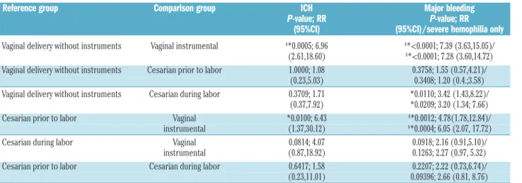
Table 4. Pairwise comparisons between groups on mode of delivery.
Reference group Comparison group ICH Major bleeding P-value; RR P-value; RR (95%CI) (95%CI)/severe hemophilia only
Vaginal delivery without instruments Vaginal instrumental §*0.0005; 6.96 §*<0.0001; 7.39 (3.63,15.05)/
(2.61,18.60) §*<0.0001; 7.28 (3.60,14.72)
Vaginal delivery without instruments Cesarian prior to labor 1.0000; 1.08 0.3758; 1.55 (0.57,4.21)/ (0.23,5.03) 0.3408; 1.20 (0.4.;3.58)
Vaginal delivery without instruments Cesarian during labor 0.3709; 1.71 *0.0110; 3.42 (1.43,8.22)/ (0.37,7.92) *0.0209; 3.20 (1.34; 7.66)
Cesarian prior to labor Vaginal *0.0100; 6.43 §*0.0012; 4.78(1.78,12.84)/
instrumental (1.37,30.12) §*0.0004; 6.05 (2.07, 17.72)
Cesarian during labor Vaginal 0.0814; 4.07 0.0918; 2.16 (0.91,5.10)/ instrumental (0.87,18.92) 0.1263; 2.27 (0.97, 5.32) Cesarian prior to labor Cesarian during labor 0.6417; 1.58 0.2207; 2.22 (0.73,6.74)/ (0.23,11.01) 0.09396; 2.66 (0.81, 8.76) ICH: intracranial hemorrhage; RR. relative risk; CI: Confidence Interval. *Significant at significance level P<0.05; §significant after Bonferroni correction P<0.0083.
found in the rate of major bleeds and intracranial hemor-rhage in neonates with moderate and severe hemophilia between vaginal delivery and Cesarean section: major bleeds occurred in 4.3% neonates born by VD and in 5.8% after CS (P=not significant, ns); ICH in 2.4% follow-ing VD and 1.7% after CS (P=not significat, ns). Further analysis of subgroups by MOD (VD with and without instruments, CS prior to and during labor) revealed instru-mental VD as a risk factor for major bleeds and intracra-nial hemorrhage compared to non-instrumental VD (RR 7.39 major bleed; 6.96 ICH) and CS prior to labor. Neonates with moderate hemophilia had a similar risk regarding ICH compared to severe hemophilia, but a lower risk for other major bleeds. No other significant dif-ferences were found between the subgroups of vaginal delivery without instruments, CS prior and CS during labor. The CS rate was higher in neonates with a known family history and reason was hemophilia in more than half of the cases. However, no significant difference was found between the group with and without a known fam-ily history of hemophilia regarding major bleeds and ICH. The comparison of planned CS (including CS prior to labor in 93.2% but also including some cases during labor) to planned VD (including VD without instruments in 77% and also instrumental VD and CS during labor) also showed no significant difference in the group with known family history
To our knowledge, this study represents the largest prospective, monitored series recording comprehensive data on mode of delivery and neonatal bleeds in patients with moderate and severe hemophilia. The data come from countries with a good and quite uniform standard of health care which should make the results relevant and applicable for these countries, although the results may also be applicable to countries with different health care standards. Due to the inclusion of all consecutive cases in the participating centers, selection bias should be low but cannot be totally excluded. Missing data is a problem for
most registries. Earlier publications of the PedNet registry show high quality data regarding baseline data and first 75 exposure days including bleeds, with only 4% missing
data.14The subanalysis on detailed information on mode
of delivery (vaginal with instruments, CS prior to or dur-ing labor, reason for CS) was available in 813 of 926 (87.8%) patients, which means that in 12.2% of included patients these data are missing, which is still acceptable for analysis. However, the frequencies for both major bleeds and ICH are low, and small differences between the groups analyzed cannot be detected due to the limited number of included patients. For example, to show a dif-ference between ICH in vaginal delivery of 2.4% to CS of 1.7%, over 12,000 patients would have to be included, which is not feasible. We excluded mild hemophilia from our analysis since the diagnosis in these patients is often made at an older age and undiagnosed cases born during the study period have not yet been included in the PedNet Registry.15
Both term and preterm neonates were included in the calculations and one could question if data should have been presented separately since ICH is a well-known complication of, in particular, delivery of an extremely
premature neonate.16 Our series included 849 children
with data on gestational week of birth, of whom 62 (7.3%) were born prematurely, but only 11 children (1.3%) were born before the 33rd gestational week, i.e. very or extremely premature. There were three major bleeds in the premature group (5.2%) and no cases of ICH. In term births, 41 major bleeds in 787 neonates (6.4%) occurred, and 20 ICH (20 of 787; 2.5%). Since there was no significant difference in the frequency of bleeds between the term and preterm groups we considered it justified to merge them together in the calculations. The premature group was still a rather small group, and a much larger group would be needed in order to draw any conclusions between the more extremely premature and less premature on this issue. In another neonatal series,
Table 5. Known and unknown family history of hemophilia.
All Known family history Unknown family history Family of hemophilia of hemophilia history not
known n n (%) n (%) n ICH 20 8 (1.7) 12 (2.7) 0 Major bleeds 44 22 (4.7) 20 (4.5) 2 Vaginal delivery 633 299 (64.2) 322 (72.4) 12 VD without instruments 541 285 274 5 VD instrumental 68 14 48 6
Not known with/without instruments 24 13 10 1
Cesarean section 293 167 (35.8)* 123 (27.6) 3
Planned CS 134 84 49 1
Reason: Hemophilia 37 - 0
Combined hemophilia and maternal 12 - 0
or fetal status Maternal 16 32 1 Fetal 9 12 0 Combined maternal/fetal 0 2 0 Other/unknown 9/1 3/0 0 Total number 926 466 445 15 *Significant at P<0.05 in comparison to unknown family history of hemophilia. n: number; ICH: intracranial hemorrhage; VD: vaginal delivery; CS: Cesarean section.
Richards et al.,8with an overall head bleed rate of 3.5%
and some data on prematurity (29 premature children; 6.0% in the series), had the same issue. It is, however, pos-sible that extreme prematurity is under-represented in the registry due to mortality before diagnosis.
The frequency of major bleeds and ICH for all neonates
was similar to previous studies.9,17,18 When splitting the
group into instrumental VD and non-instrumental VD, and CS prior to and during labor, only instrumental VD was identified as a risk factor. A recent study from the UK on ICH in bleeding disorders had similar findings and identified instrumental delivery as a clear risk factor with
a RR of 10.6.19 This is also known from the normal
popu-lation, but in lower frequencies: Towner et al. reported ICH frequencies of 1 of 860 for VE and 1 of 664 for forceps and VE, which means that the risk for ICH in hemophilic neonates born by instrumental delivery is roughly 80-fold
higher in our series.12In a recent published meta-analysis
from Davies and Kadir, CS was proposed as a safer option for children born with hemophilia, but the comparison
was made with historical cohorts.7 In our prospective
cohort, there was no difference in incidence of major bleeds and ICH in neonates born by vaginal delivery (both instrumental and non-instrumental) compared to CS (prior to and during labor): 5.2% versus 6.4% in major bleeds, and 2.3% versus 1.7% for ICH. A planned CS with the intention to perform CS prior to labor did not prevent neonates from experiencing ICH or major bleeds in com-parison to planned VD. This information is important when counseling a pregnant carrier. It has also been shown in other studies that CS does not prevent neonatal
bleeds.4,8,9 Studies from the normal population with
583,340 births included show that vaginal delivery with-out instruments and CS prior to labor were the safest option but ICH still occurred (1 of 2,750 CS prior to labor,
and 1 of 1,900 delivered spontaneously).12
A known family history of hemophilia (KFH) had some influence on the mode of delivery: 36% (167 of 466) were delivered by CS in the KFH group compared to 28% (123 of 445) in the no known family history group (NFH) (P=0.00038). The group of neonates in the NFH group present the ‘true risk’ of neonatal bleeds in a child with hemophilia, since no precautionary measures had been taken with respect to hemophilia on the obstetric proce-dures. However, there was no significant difference in the rate of major bleeds and ICH between KFH and NFH. Regarding vaginal delivery, 4.5% of the children in the NFH group compared to 4.7% in the KFH had a major bleed and 2.7% had ICH in the NFH-group compared to 1.7% in the KFH-group. This may be explained by our findings, that the mode of delivery (CS vs. vaginal deliv-ery) did not impact the rate of major bleeds and ICH. Similar findings were shown in a recent study of Kulkarni
et al. in which 547 neonates with all severities of
hemo-philia were included.20The reason to choose a planned CS
was hemophilia in 45%; but in 55%, other maternal and fetal reasons played a role illustrating that the planning of a delivery is a complicated and multifactorial task.
Prenatal diagnosis was performed in 14% (62 of 466) of known carriers, and in these cases it was definitely known that they were carrying a child with hemophilia. In this subgroup, 51.6% were delivered by CS, demonstrating a statistically significant impact on the decision to choose a CS delivery. We do not have data on how many carriers terminated pregnancy after PND with an affected fetus, so
the numbers on PND might be underestimated. However, recent figures from Sweden indicate that carriers today often choose PND in order to prepare for having a hemo-philia child and not for termination of the pregnancy; but
this may not be the case in all participating centers.21
One child died from ICH, representing 1 of 786 boys with severe hemophilia (0.13%), and 1 of 20 of all ICH (5%). These numbers are low compared to historical data, but they are in line with a recent CDC report that included
all ICH with a mortality rate of around 2.5%.22 These
numbers may be underestimated due to undiagnosed or unreported cases.
It would have been of interest to analyze the risks and outcomes for the carrier mothers who gave birth to a child with hemophilia according to delivery mode, but our reg-istry is limited to pediatric data.
In summary, vaginal delivery and Cesarean section carry similar risks of ICH and major bleeds in neonates with severe and moderate hemophilia, and pregnant carriers of hemophilia should be informed about different options for mode of delivery and their potential risks.
Acknowledgments
The authors thank study coordinator Ella van Hardeveld and statistician Susann Ullén for their help with this study. We would like to acknowledge unrestricted funding from Bayer AG, Baxter/Baxalta/ Shire, CSL Behring, Pfizer, Novonordisk, Sobi and Grifols to the PedNet group that were partly used for this study.
Appendix
PedNet Haemophilia Research Foundation contributors to this study.
Europe
C Altisent Roca, Unitat Hemofilia, Hospital Vall d’Hebron, Barcelona, Spain; MT Alvarez Romàn, Unidad de Coagulopatías, Hospital Universitario La Paz, Madrid, Spain
M Bührlen, Gesundheit Nord, Klinikum Bremen Mitte, Prof.-Hess-Kinderklinik, Bremen, Germany; HM van den Berg, PedNet Haemophilia Research Foundation, Baarn, The Netherlands; E Chalmers, Department of Haematology, Royal Hospital for Children, Glasgow, UK; H Chambost, Pediatric Haematology Oncology Department, Children Hospital La
Mode of delivery and neonatal bleeds in hemophilia
Table 6. Planned vaginal delivery versus planned Cesarean section – major bleeds and intracranial hemorrhage (ICH).
n ICH Major bleeds n (%) n (%) Planned VD 703 17 (2.4) 34 (4.8)
VD without instruments 541 10 18
VD instrumental 68 7 13
CS during labor 70 0 3
VD not known with 24 0 0
or without instruments Planned CS 134 2 (1.5) 5 (3.7) CS prior to labor 125 2 5
CS during labor 9 0 0
Planning not known 89 1 5
P-value; RR (CI) 0.753; 1.62 0.822; 1.30
(0.38,6.93) (0.52,3.25) VD: vaginal delivery; CS: Cesarean section; RR: relative risk.
Timone, AP-HM, and INSERM, INRA, C2VN, Faculté de Médecine, Aix-Marseille University, Marseille, France; A Rosa Cid, Unidad de Hemostasia y Trombosis, Hospital Universitario y Politécnico La Fe, Valencia, Spain; S Claeyssens, Centre Regional d’Hemophilie, Centre Hospitalo Universitaire, Toulouse, France; C Escuriola, HZRM Hämophilie Zentrum Rhein Main GmbH, Mörfelden-Walldorf, Germany; K Fischer, Van Creveld Kliniek, University Medical Center Utrecht, Utrecht, The Netherlands; C Van Geet, Catholic University of Leuven, Campus Gasthuisberg, Service of Pediatric Haematology, Leuven, Belgium; R Kobelt, Hämophiliezentrum, Wabern and Children's Hospital of the University of Berne, Switzerland; C Königs, J .W. Goethe University Hospital, Department of Pediatrics, Frankfurt, Germany; K Kurnik, Dr. V. Haunersches Kinderspital, University of Munich, Munich, Germany; R Liesner, Hemophilia Center, Department of Haematology, Great Ormond Street Hospital for Children, London, UK; R Ljung, Department of Clinical Sciences, Lund University, Lund; Department of Pediatrics and Malmö Centre for Thrombosis and Haemostasis, Skåne University Hospital, Malmö, Sweden; A Mäkipernaa, Children’s Hospital , Helsinki University Central Hospital and University of Helsinki, Helsinki, Finland; A Molinari, Dipartimento di Ematologia ed Oncologia, Unità Trombosi ed Emostasi, Ospedale Pediatrico Giannina Gaslini, Genova, Italy; W Muntean, Universitäts-Klinik für Kinder- und Jugendheilkunde, Graz, Austria; B Nolan, Department of Paediatric Haematology, Our Lady’s
Children’s Hospital for Sick Children, Crumlin, Dublin, Ireland; J Oldenburg, Institut für Experimentelle Hämatologie und Transfusionsmedizin, Universitätsklinikum Bonn, Germany; R Pérez Garrido, Hospital General Unidad de Hemofilia, Hospitales Universitarios Virgen del Rocio, Sevilla, Spain; H Platokouki, Haemophilia-Haemostasis Unit, St. Sophia Children’s Hospital, Athens, Greece; A Rafowicz, Centre de Référence pour le Traitement des Maladies Hémorragiques (CRTH), Hôpital Bicêtre, Kremlin Bicêtre AP-HP , France; S Ranta, Department of Pediatrics, Clinic of Coagulation Disorders, Karolinska Hospital, Stockholm,Sweden; E Santagostino, ME Mancuso, Angelo Bianchi Bonomi Hemophilia and Thrombosis Center, Fondazione, IRCCS Ca’ Granda, Ospedale Maggiore Policlinico, Milan, Italy; T Stamm Mikkelsen, Department of Pediatrics, University Hospital of Aarhus at Skejby, Aarhus, Denmark; A Thomas, Royal Hospital for Sick Children, Edinburgh, UK; M Williams, Department of Haematology, The Children’s Hospital, Birmingham, UK.
Israel
G Kenet, National Hemophilia Center, Ministry of Health, Sheba Medical Center, Tel Hashomer, Israel
Canada
M Carcao, Division of Haematology/Oncology, Hospital for Sick Children, Toronto, Canada; G Rivard, Division of Hematology/Oncology, Hôpital St Justine, Montréal, Canada.
References
1. Stieltjes N, Calvez T, Demiguel V, et al. Intracranial haemorrhages in French haemophilia patients (1991-2001): clinical presentation, management and prognosis factors for death. Haemophilia. 2005;11 (5):452-458.
2. Klinge J, Auberger K, Auerswald G, Brackmann HH, Mauz-Korholz C, Kreuz W. Prevalence and outcome of intracranial haemorrhage in haemophiliacs--a survey of the paediatric group of the German Society of Thrombosis and Haemostasis (GTH). Eur J Pediatr. 1999;158 Suppl 3:S162-165. 3. Gouw SC, van der Bom JG, Marijke van
den Berg H. Treatment-related risk factors of inhibitor development in previously untreated patients with hemophilia A: the CANAL cohort study. Blood. 2007;109 (11):4648-4654.
4. Kulkarni R, Lusher JM. Intracranial and extracranial hemorrhages in newborns with hemophilia: a review of the literature. J Pediatr Hematol Oncol. 1999;21(4):289-295. 5. Ljung R, Lindgren AC, Petrini P, Tengborn L. Normal vaginal delivery is to be recom-mended for haemophilia carrier gravidae. Acta Paediatr. 1994;83(6):609-611. 6. Revel-Vilk S, Golomb MR, Achonu C, et al.
Effect of intracranial bleeds on the health and quality of life of boys with hemophilia. J Pediatr. 2004;144(4):490-495.
7. Davies J, Kadir RA. Mode of delivery and cranial bleeding in newborns with
haemophilia: a systematic review and meta-analysis of the literature. Haemophilia. 2016;22(1):32-38.
8. Richards M, Lavigne Lissalde G, Combescure C, et al. Neonatal bleeding in haemophilia: a European cohort study. Br J Haematol. 2012;156(3):374-382.
9. Nazir HF, Al Lawati T, Beshlawi I, et al. Mode of delivery and risk of intracranial haemorrhage in newborns with severe haemophilia A: a multicentre study in Gulf region. Haemophilia. 2016;22(3):e134-138. 10. Ljung R. The optimal mode of delivery for
the haemophilia carrier expecting an affect-ed infant is vaginal delivery. Haemophilia. 2010;16(3):415-419.
11. James AH, Hoots K. The optimal mode of delivery for the haemophilia carrier expect-ing an affected infant is caesarean delivery. Haemophilia. 2010;16(3):420-424. 12. Towner D, Castro MA, Eby-Wilkens E,
Gilbert WM. Effect of mode of delivery in nulliparous women on neonatal intracra-nial injury. N Engl J Med. 1999;341 (23):1709-1714.
13. Kasper CK, Lin JC. Prevalence of sporadic and familial haemophilia. Haemophilia. 2007;13(1):90-92.
14. Fischer K, Ljung R, Platokouki H, et al. Prospective observational cohort studies for studying rare diseases: the European PedNet Haemophilia Registry. Haemophilia. 2014;20(4):e280-286. 15. Chambost H, Gaboulaud V, Coatmelec B,
Rafowicz A, Schneider P, Calvez T. What factors influence the age at diagnosis of
hemophilia? Results of the French hemo-philia cohort. J Pediatr. 2002;141(4):548-552.
16. Stoll BJ, Hansen NI, Bell EF, et al. Trends in Care Practices, Morbidity, and Mortality of Extremely Preterm Neonates, 1993-2012. Jama. 2015;314(10):1039-1051.
17. Ljung RC. Intracranial haemorrhage in haemophilia A and B. Br J Haematol. 2008;140(4):378-384.
18. Palomo Bravo A, Nunez R, Gutierrez Pimentel MJ, Nieto MD, Cos C, Perez R. Haemophilia neonates: mode of delivery and perinatal complications. Haemophilia. 2016;22(3):e225-228.
19. Chalmers EA, Alamelu J, Collins PW, et al. Intracranial haemorrhage in children with inherited bleeding disorders in the UK 2003-2015: A national cohort study. Haemophilia. 2018;24(4):641-647. 20. Kulkarni R, Presley RJ, Lusher JM, et al.
Complications of haemophilia in babies (first two years of life): a report from the Centers for Disease Control and Prevention Universal Data Collection System. Haemophilia. 2017;23(2):207-214. 21. Martensson A, Tedgard U, Ljung R.
Prenatal diagnosis of haemophilia in Sweden now more commonly used for psychological preparation than termination of pregnancy. Haemophilia. 2014;20 (6):854-858.
22. Witmer CM. Low mortality from intracra-nial haemorrhage in paediatric patients with haemophilia. Haemophilia. 2015;21 (5):e359-363.