HAL Id: inserm-01285612
https://www.hal.inserm.fr/inserm-01285612
Submitted on 9 Mar 2016
HAL is a multi-disciplinary open access
archive for the deposit and dissemination of
sci-entific research documents, whether they are
pub-lished or not. The documents may come from
teaching and research institutions in France or
abroad, or from public or private research centers.
L’archive ouverte pluridisciplinaire HAL, est
destinée au dépôt et à la diffusion de documents
scientifiques de niveau recherche, publiés ou non,
émanant des établissements d’enseignement et de
recherche français ou étrangers, des laboratoires
publics ou privés.
Molecular keys of the tropism of integration of the
cholera toxin phage
Bhabatosh Das, Julien Bischerour, Marie-Eve Val, François-Xavier Barre
To cite this version:
Bhabatosh Das, Julien Bischerour, Marie-Eve Val, François-Xavier Barre. Molecular keys of the
tropism of integration of the cholera toxin phage. Proceedings of the National Academy of Sciences
of the United States of America , National Academy of Sciences, 2009, �10.1073/pnas.0910212107�.
�inserm-01285612�
Molecular keys of the tropism of integration of the
cholera toxin phage
Bhabatosh Dasa,b,1, Julien Bischeroura,b,1, Marie-Eve Vala,b, and François-Xavier Barrea,b,2
aCentre de Génétique Moléculaire, Centre National de la Recherche Scientifique, 91198 Gif-sur-Yvette, France; andbUniversité Paris-Sud, 91405 Orsay,
France
Edited by G. Balakrish Nair, National Institute of Cholera and Enteric Diseases, Kolkata, India, and approved December 22, 2009 (received for review September 7, 2009)
Cholera toxin is encoded in the genome of CTXϕ, a lysogenic fila-mentous phage of Vibrio cholerae. CTXϕ variants contribute to the genetic diversity of cholera epidemic strains. It has been shown that the El Tor variant of CTXϕ hijacks XerC and XerD, two host-encoded tyrosine recombinases that normally function to resolve chromosome dimers, to integrate at dif1, the dimer resolution site of the larger of the two V. cholerae chromosomes. However, the exact mechanism of integration of CTXϕ and the rules governing its integration remained puzzling, with phage variants integrated at either or both dimer resolution sites of the two V. cholerae chromosomes. We designed a genetic system to determine exper-imentally the tropism of integration of CTXϕ and thus define rules of compatibility between phage variants and dimer resolution sites. We then showed in vitro how these rules are explained by the direct integration of the single-stranded phage genome into the double-stranded bacterial genome. Finally, we showed how the evolution of phage attachment and chromosome dimer reso-lution sites contributes to the generation of genetic diversity among cholera epidemic strains.
lysogenic conversion
|
site-specific recombinationC
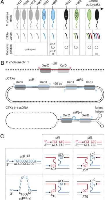
holera toxin, which is responsible for the deadly diarrhea associated with the disease of the same name, is one of the most significant virulence factors of Vibrio cholerae (1). It is encoded in the genome of a lysogenicfilamentous bacteriophage, CTXϕ (2). Different CTXϕ variants exist, the two main ones being classified as “classical” and “El Tor,” according to the biotype of the hosts in which they originally were identified (Fig. 1A) (3). The existence of several different CTXϕ variants and their integration in variable copy numbers on thefirst, the sec-ond, or bothV. cholerae chromosomes contribute to the genetic diversity of cholera epidemic strains (Fig. 1A) (4).In contrast to most other lysogenic phages, such as phageλ, CTXϕ does not encode its own integration machinery. Instead, it has been shown that the El Tor variant of CTXϕ hijacks XerC and XerD, two host-encoded tyrosine recombinases that nor-mally function to resolve chromosome dimers (5), to integrate at dif1, the dimer resolution site of the larger of the two V. cholerae chromosomes (6). Xer recombination sites consist of binding sites for XerC and XerD, separated by a 6-bp to 8-bp overlap region; strand exchanges occur at the border of this region (7). The replicative form of CTXϕ harbors two putative Xer recombination sites in inverted orientations, attP1 and attP2 (pCTXϕ; Fig. 1B). This observation led to the proposal of two integration models (8, 9). Thefirst model predicts that recom-bination occurs between dif1 and attP1 as the result of an unknown architectural function played by attP2. The second model relies on the formation of a forked hairpin by the∼150-bp region encompassingattP1 and attP2 in the (+) ssDNA genome of the phage, which unmasks a putative phage attachment site, attP(+) [CTXϕ (+) ssDNA; Fig. 1B]. Both models rely on the exchange of a single pair of strands catalyzed by XerC and on the conversion of the resulting Holliday junction into product by repair and/or replication.
According to both models, no integration should occur atdif2, the dimer resolution site of the secondV. cholerae chromosome, because no Watson–Crick bp interactions could stabilize the exchange of strands catalyzed by XerC (Fig. 1C). Nevertheless, the classical variant of CTXϕ was found integrated at dif2 in classical strains (3) and in recent El Tor isolates (4, 10–14). The mode of dissemination of this variant is of particular interest, because it allows the production of an elevated amount of cholera toxin (15, 16) that seems to be implicated in a high proportion of severe infections associated with classical strains (17). It was proposed that this variant recently invaded the genome of El Tor strains through chitin-induced competence and homologous recombination (18). However, the trans-formation efficiency was very low, in the order of 10−6to 10−4. In addition, such a mechanism does not explain how CTXϕ initially achieveddif2 integration or the diversity of combinations of the different genetic elements that were found integrated at the two chromosome dimer resolution sites ofV. cholerae strains (4).
In this study, we designed a sensitive assay to monitor the effi-ciency with which CTXϕ integrates at dif1 and dif2 in V. cholerae. Using this assay, we demonstrated the specificity of integration of the El Tor variant of CTXϕ harbored by strain N16961. In con-trast, the classical variant of CTXϕ harbored by strain 569B effi-ciently integrated at bothdif1 and dif2. We found that the altered integration behavior of the classical phage is caused by two base changes in the overlap region of attP2 that allow V. cholerae XerCD to recombine the classical ssDNAattP(+) region with dif1 anddif2 in vitro. These results further support the ssDNA inte-gration model and allow the definition of rules of compatibility between phage attachment and dimer resolution sites that explain the tropism of integration of the different CTXϕ variants. Based on these rules, we designed a phage attachment site that exclu-sively targetsdif2. We also explained how O1 and O139 V. cholerae strains with altered dimer resolution sites can escape lysogenic conversion by the most common variants of CTXϕ and showed how a new variant of CTXϕ has evolved that can integrate into the genome of these particular strains. Taken together, these results suggest that lysogenic conversion by CTXϕ is the primary mode of acquisition of the cholera toxin genes, which, along with the evo-lution of phage attachment and chromosome dimer resoevo-lution sites, contributes to the generation of genetic diversity among cholera epidemic strains.
Author contributions: B.D., J.B. and F.-X.B. designed research; B.D. and J.B. performed research; M.-E.V. contributed new reagents/analytic tools; B.D., J.B. and F.-X.B. analyzed data and wrote the paper.
The authors declare no conflict of interest. This article is a PNAS Direct Submission. See Commentary on page 3951.
1B.D. and J.B. contributed equally to this work.
2To whom correspondence should be addressed. E-mail: barre@cgm.cnrs-gif.fr.
This article contains supporting information online awww.pnas.org/cgi/content/full/ 0910212107/DCSupplemental.
www.pnas.org/cgi/doi/10.1073/pnas.0910212107 PNAS | March 2, 2010 | vol. 107 | no. 9 | 4377–4382
MICRO
BIOLOGY
SEE
COM
Results
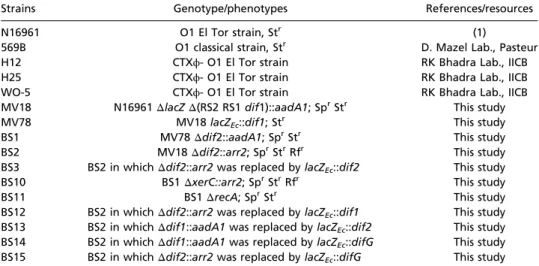
Design of a Sensitive Assay to Monitor the Efficiency and Specificity of CTXϕ Integration.Previous studies that addressed the mecha-nism of integration of CTXϕ used Southern and PCR techniques to determine the location of each integration event. These techniques are robust. However, because the integration of CTXϕ at dif2 might be a very rare event, we sought to design a time- and cost-effective method to monitor its frequency of occurrence. To this aim, we inserteddif2 in the coding region of the Escherichia coli lacZ gene in such a manner that the pro-duced peptide might retain itsβ-galactosidase activity. We then engineered an N16961 El Tor strain in which the endogenous lacZ gene was deleted and in which dif2 was replaced by the lacZ::dif2 allele (Table 1). These cells turn blue in the presence of X-gal because theβ-galactosidase they produce is active. dif2 integration events disrupt the lacZ ORF, thereby abolishing β-galactosidase production. Thus we could screen for such events simply by plating cells on X-gal media.
The two chromosomes harbored by classical strains possess the same dif2 dimer resolution site (CP000626.1). This situation could have favoreddif2 integration because of the absence of the preferential attachment site of CTXϕ in the bacterial genome. To mimic this situation, we used as a background for our assays an N16961 El Tor strain in whichdif1 and the integrated copies of CTXϕ surrounding it had been deleted (Table S1). Finally, we engineered alacZ−/dif2−N16961 El Tor strain in whichdif1 was replaced by a functionalE. coli lacZ::dif1 gene to monitor the efficiency with which CTXϕ integrates at dif1 (Table S1).
The Variant of CTXϕ Found in the 569B Classical Strain Integrates at Both dif1 and dif2.Studies of the mechanism of integration of the classical variant of CTXϕ were complicated because no phage production was observed in any of the strains harboring it (3, 11). To circumvent this difficulty, we used conjugation to deliver circular DNA molecules carrying the replication and integration region of the phage found in the 569B classical strain, hereafter referred to as“RSCl,” directly into V. cholerae cells. To this aim, we cloned RSClin a conjugative vector that carries a chloramphenical resistance gene but cannot replicate autonomously in V. cholerae (Table S2). We also cloned the replication and integration region of RS1, a truncated deriva-tive of CTXϕ found in the N16961 El Tor strain (19), hereafter referred to as“RSET” (Table S2). The attachment site of RS1 is identical to the attachment site of the El Tor variants of CTXϕ. In addition, RS1 efficiently and specifically integrates at dif1, making it a perfect control for our experiments. Conjugation of the RSETand RSClvectors gave rise to similar numbers of CmRcolonies. RSETintegrated in 100% of the colonies ofdif1+/ dif2−cells and in no colonies ofdif1−/dif2+cells, confirming its highly specific integration at dif1 (Table 1). In contrast, RSCl integrated in 36.4% of the colonies ofdif1+/dif2−cells and in 4% of the colonies ofdif1−/dif2+cells, indicating that it efficiently targets bothdif1 and dif2 (Table 1). The integration specificity was not linked to the genomic context of the dimer resolution sites, because RSET integrated as efficiently when dif1 was located on chromosome 2 as when it was on chromosome 1 (Table 1). Finally, integration was suppressed entirely inxerC− cells and did not require homologous recombination (Table S3).
Two Bases Determine the Capacity of RSClto Integrate at Both dif1
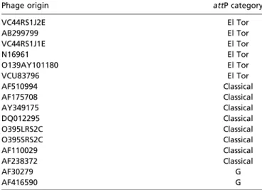
and dif2.We next investigated which differences in the sequences of RSCland RSETwere involved in their different integration behaviors. Because these differences were numerous, we decided to restrict the number of positions tested by searching for CTXϕ residues that would be specifically conserved among classical or El Tor isolates in the available genomic sequences of toxigenic V. cholerae strains. During this search, we identified three cate-gories of CTXϕ attachment regions, two of which seemed
spe-Fig. 1. Lysogenic conversion of V. cholerae strains by CTXϕ. (A) CTXϕ var-iants found in cholera epidemic strains. V. cholerae strains: gray, unknown; blue, classical; black, El Tor; green, O139. CTXϕ variants: gray, unknown; blue, classical; black, El Tor; green, O139; red, G; black and blue, El Tor and classical hybrid. (B) Scheme of Xer recombination sites. dif1, dimer resolution site of the N16961 chromosome 1; attP1 and attP2, Xer sites found in the replicative form of CTXϕ (pCTXϕ); attP(+), the recombination site created by the folding of the (+) ssDNA genome of CTXϕ. The two DNA strands of each site are drawn. The strand cleaved by XerC is shown in color. Vertical bars indicate bases present in the overlap region of each site. (C) Schemes of the Watson–Crick bp interactions that could stabilize the strand exchange cat-alyzed by XerC between the overlap regions of the two chromosome dimer resolution sites of N16961 and the two putative attachment sites of El Tor variants of CTXϕ. dif1, chromosome 1 dimer resolution site; dif2, chromo-some 2 dimer resolution site; attP1ET, attP1 found in CTXϕ El Tor variants;
attP(+)ET, attachment site unmasked by the folding of the (+) ssDNA genome
of El Tor variants of CTXϕ. The strands cleaved by XerC on dimer resolution and phage attachment sites are shown in red and in blue, respectively. Pairing interactions are indicated by the proximity of the bases.
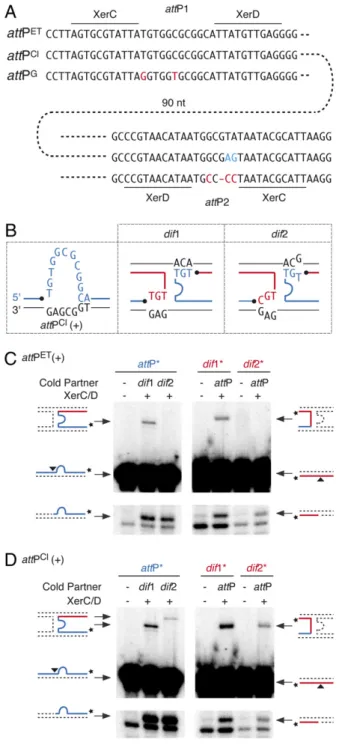
cifically linked to classical and El Tor isolates (Fig. 2A andTable S4). Indeed, the attachment region of classical variants of CTXϕ,
attPCl, differs from the attachment region of El Tor and O139 variants,attPET, by two residues in the overlap region between the XerC- and XerD-binding sites ofattP2 (Fig. 2A, blue resi-dues). Introduction of the classical residues in the attachment region of RSETled to its efficient integration at both dif1 and dif2 (Table 1), and introduction of the El Tor residues in the attachment region of RSClabolisheddif2 integration (Table 1), demonstrating that the respective relaxation and specific inte-gration behaviors of RSCland RSETare determined solely by a difference of sequence at these two positions.
XerCD Recombines attPCl(+) with dif1 and dif2 in Vitro.The T-to-G transversion found in the overlap region of classicalattP2 allows the recovery of one Watson–Crick bp interaction on one side of the reaction between the (+) ssDNA ofattPClanddif2 (Fig. 2B, red strand). Perfect Watson–Crick pairing is not recovered on the other side but is replaced by a TG wobble bp (Fig. 2B, blue strand). Likewise, one Watson–Crick bp is replaced by a TG wobble bp on one side of the reaction between the stem of the hairpin formed by the (+) ssDNA ofattPClanddif1 (Fig. 2C, red strand). Thisfinding prompted us to check whether V. cholerae XerC and XerD could recombine the (+) ssDNA ofattPClwith dif1 and dif2 in vitro. We did so using purified V. cholerae XerC and XerD proteins and annealed synthetic oligonucleotides that mimicdif1, dif2, and the stems of the hairpins created by attPCl andattPET(Table S5).
Three steps can be defined in the strand exchange reaction performed by tyrosine recombinases:first, a single strand in each of the two recombining sites is cleaved by one recombinase, generating two 3′-phosphorotyrosyl recombinase/DNA covalent intermediates; the liberated 5′-hydroxyl extremities then are exchanged;finally, they attack the phosphorotyrosyl bond of the partner site to form phosphodiester bonds. Cleavage of each of the two recombining strands and their subsequent ligation to the opposite partner strand can be monitored when the strand is labeled at its 3′ extremity: Strand cleavage leads to the appari-tion of a shorter migraappari-tion product on a sequencing gel; ligaappari-tion to a partner strand harboring a longer extension on the 5′ side of the XerC-binding site leads to the creation of a longer recombinant product. Ligation products were detected when attPET(+) was reacted against dif1 (Fig. 2C, Upper) and when attPCl(+) was reacted againstdif1 or dif2 (Fig. 2D, Upper). Thus, in the absence of any other host or phage factors, XerC and XerD can promote the exchange of one pair of strands when one Watson–Crick bp is replaced by a TG wobble bp on one side of the recombining complex. However, no ligation product was detected between attPET(+) anddif2; Watson–Crick pairing is lost on both sides of this reaction (Fig. 2C, Upper).
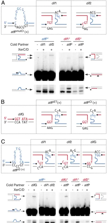
Design of a dif2-Specific Phage Variant.To demonstrate further that the specificity of integration of the different variants of CTXϕ is governed by the ability to establish bp interactions that stabilize strand exchanges, we designed a phage that, based on the ssDNA integration model, should integrate specifically at dif2. To this aim, the thymine immediately downstream of the site of cleavage of XerC was replaced by a cytosine inattPCl(+) [attPmutCl(+); Fig. 3A]. Because of this substitution, perfect Watson–Crick pairing is lost on both sides of the reaction withdif1 (Fig. 3A). However, it is re-established on both sides of the reaction withdif2 (Fig. 3A). As expected, we observed a normal number of XerC-mediated strand exchanges between attPmutCl(+) and dif2 in vitro, whereas recombination withdif1 was barely detectable (Fig. 3A). In vivo, this process resulted in the fully specific integration of a vector carrying the attPmutlCl(+) attachment site at dif2 (attPmutCl; Table 2).
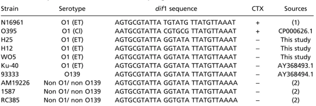
Rules of Compatibility Drive the Evolution Between Phage Attachment and Dimer Resolution Sites.We next investigated the sequence of the dimer resolution sites of natural CTXϕ -V. cholerae strains. We found that the first chromosome of many recently isolated O1 El Tor, O139 and non-O1 non-O139 strains harbors a dimer resolution site with an overlap region different from that ofdif1 and dif2; we called this site “difG” (Fig. 3B and
Table S6). RSETand RSCldid not integrate atdifG in vivo (Table 2). This result was expected, because no bp interactions could stabilize strand exchanges between difG and attPET(+) or attPCl(+) (Fig. 3B). However, the stem of the hairpin created by the folding of the third category of attachment sites that was identified during our database searches (attPG
; Fig. 2A) seemed suitable for integration withdifG (Fig. 3C). In contrast, attPG(+) did not seem suitable for integration at dif1 or dif2. Corre-spondingly,V. cholerae XerCD specifically recombined attPG(+) with difG in vitro (Fig. 3C), and a phage harboring attPG spe-cifically integrated at difG in vivo (Table 2) independently of the chromosomal context of the site (Table S7).
Discussion
Previous work on the El Tor variant of CTXϕ suggested that it specifically integrates at dif1 (6). In this context, the integration of CTXϕ at dif2 in classical strains (3) and in recent El Tor isolates (4, 10–14) was puzzling. It was observed recently that El Tor strains can acquire the dif2-integrated classical copy of CTXϕ via chitin-induced competence and homologous recom-bination (18). However, such a mechanism left open the ques-tions of howdif2 integration could be achieved initially and the frequency at which this integration could occur. In addition, it did not explain the diversity of combinations of the different genetic elements that were found integrated at the chromosome dimer resolution sites loci ofV. cholerae strains (4). To address these points, we designed a sensitive assay to monitor the
spe-Table 1. In vivo integration of RSETand RSCl
Phage machinery attP dif Chromosome % integration Screened colonies
Classical Classical dif1 1 36.4 466
Classical Classical dif2 2 4.0 451
El Tor El Tor dif1 1 100.0 454
El Tor El Tor dif2 2 <0.1 1099
El Tor Classical dif1 1 100.0 675
El Tor Classical dif2 2 36.3 1556
Classical El Tor dif1 1 91.7 743
Classical El Tor dif2 2 <0.1 883
El Tor El Tor dif1 2 100.0 875
El Tor El Tor dif2 1 <0.1 1070
Data were obtained from at least three independent experiments.
Das et al. PNAS | March 2, 2010 | vol. 107 | no. 9 | 4379
MICRO
BIOLOGY
SEE
COM
cificity and efficiency of dif1 and dif2 integration events. We then cloned the replication and integration region of the var-iant of CTXϕ found in the 569B classical strain into a vector harboring a conditional origin of replication; this vector was delivered into N16961 El Tor cells by conjugation. Surprisingly, we observed that this vector efficiently integrated at both dif1 anddif2 independently of the chromosomal context of the sites (Table 1). This result explains how the classical variant of CTXϕ could integrate in the genome of classical strains even if their two chromosomes share the same dif2 dimer resolution site (CP000626.1) and why it was found integrated atdif1 on thefirst chromosome of the El Tor BX330286 strain although such a configuration had not been observed previously in any other strain (4). In contrast, the N16961 El Tor variant of CTXϕ specifically targeted dif1 (Table 1). Likewise, we observed that classical and El Tor variants of CTXϕ could not integrate at difG, the dimer resolution site on the larger chromosome of many nontoxigenic strains, but that difG is targeted specifically by a third type of phage that was isolated recently (Table 2).
CTXϕ integration is irreversible (6, 9), and the secretion of new phage particles relies on the production of its ssDNA genome by rolling circle replication across tandemly integrated copies (20, 21). However, we observed that RSClcan integrate in tandem copies that should ensure such a production (Fig. S1). Correspondingly, the sequencing of the genome of several V. cholerae strains revealed the presence of tandem copies of the classical phage (El Tor strains BX330286 and B33) (4). In addition, it was observed that classical copies of the phage coexist with intact El Tor copies and/or with the RS1 element in other strains (4, 14); such coexistence also could ensure the production of classical CTXϕ virions. Interestingly, the conjunct production of El Tor and classical phages could favor the emergence of hybrid phages, a phenomenon that has been observed recently (Fig. 1A) (4, 13). Taken together, these observations suggest that lysogenic conversion by CTXϕ is the primary mode of acquisition of the cholera toxin genes; this mode of acquisition, along with the evolution of phage attachment and chromosome dimer resolution sites, contrib-utes to the generation of genetic diversity among cholera epidemic strains.
Our results further indicate that the relaxation and specific integration behaviors of the classical and the El Tor variants of CTXϕ are determined solely by a difference in sequence at two positions in the seven-bp overlap region ofattP2, immediately 3′ to the position at which XerC should cleave (Fig. 1B). Two models have been proposed for CTXϕ integration at dif1 (8, 9). In the first model, XerCD would catalyze the formation of a Holliday junc-tion between the host dimer resolujunc-tion sites and the dsDNA form ofattP1, which is found on the replicative form of the phage (8). In this model,attP2 is thought to play a structural role in stabilizing the synapse and/or the exchange; this assumption makes it diffi-cult to explain how mutations in its overlap region could influence the specificity of integration. In contrast, the base immediately 3′ to the XerC cleavage site in the overlap region ofattP2 contributes to the formation of bp interactions during the strand exchange predicted by the ssDNA integration model (Fig. 1C) (9). Here, we show that all of the combinations between (+) ssDNA phage attachment sites and chromosome dimer resolution sites in which Watson–Crick bp interactions could stabilize the exchange of strands catalyzed by XerC [attPET(+)xdif1, attPmutCl(+)xdif2, and attPG(+)xdifG] were recombined in vitro and promoted integra-tion in vivo. No in vitro recombinaintegra-tion and no in vivo integraintegra-tion were detected for combinations in which no Watson–Crick bp interactions could be formed [attPET(+)xdif2, attPmutCl(+)xdif1, attPG(+)xdif1, and attPG(+)xdif2]. In the remaining two combi-nations we tested [attPCl(+)xdif1 and attPCl(+)xdif2], proper Watson–Crick bps could form only on one side of the
recombi-Fig. 2. The (+) ssDNA of classical variants of CTXϕ recombines with both dif1 and dif2. (A) The three categories of phage attachment regions found in CTXϕ variants. Residues specifically conserved in classical and G variants of the phage are shown in blue and red, respectively. (B) Schemes of Watson– Crick bp interactions that could stabilize the strand exchange catalyzed by XerC between the overlap regions of the two dimer resolution sites of N16961 and the attachment site found in the (+) ssDNA genome of classical variants of CTXϕ. The legend is as in Fig. 1C. (C) V. cholerae XerCD-mediated recombination of attPET(+) with dif1 and dif2. A short radioactively labeled
attP substrate was reacted with a longer cold dimer resolution substrate (Left Panels), and a short radioactively labeled dimer resolution substrate was reacted with a longer cold attP substrate (Right Panels). Schemes of substrate and products are indicated on the side of each panel. A black triangle indicates the position of cleavage of V. cholerae XerC. A star indi-cates the position of the radioactive label of the probe. (D) V. cholerae XerCD-mediated recombination of attPCl(+) with dif1 and dif2. Schemes of
nation complex, but nevertheless in vitro recombination and in vivo integration were observed. To stabilize the exchange of strands, tyrosine recombinases normally require one Watson– Crick bp interaction on each side of the recombination complex immediately after the position of cleavage of the recombinases (22–31). The only notable exception to this rule is the integrase of some bacteroides conjugative transposons (32). In this context, it does not seem fortuitous that a TG wobble bp replaces the missing Watson–Crick bp in reactions between attPCl(+) anddif1
ordif2 (Fig. 2B). Taken together, these results give considerable support to the ssDNA integration model and allow the definition of rules of compatibility between phage attachment and dimer resolution sites that dictate the possibility for lysogenic conversion. Finally, in four of the five effective combinations of attach-ment and dimer resolution sites we tested [attPET(+)xdif1, attPCl(+)xdif1, attPCl(+)xdif2, and attPmutCl(+)xdif2], the efficiency of integration correlated with the efficiency of recombination observed in vitro. Furthermore, the in vitro recombination efficiency of these four combinations (Fig. 2 C and D and Fig. 3A, Upper) correlated with the stability and/ or frequency of formation of the two cleaved substrates involved in the strand exchange (Fig. 2C and D and Fig. 3A, Lower). For instance, the lower efficiency of the recombination of attPmutCl(+)xdif2 when compared with attPET(+)xdif1, attPCl(+) xdif1, and attPCl(+)xdif2 is explained by the lower stability of the dif2/XerC and attPmutCl/XerC covalent complexes compared with the dif1/XerC, attPET/XerC, and attPCl/XerC covalent complexes (with respective mean frequencies of 0.15± 0.08%, 0.16± 0.08%, 0.46 ± 0.18%, 0.41 ± 0.12%, and 1.19 ± 0.65% out of at least four independent experiments). The relatively low number of dif2/XerC covalent intermediates fits with previous results that indicated that the control of recombination is more stringent at dif2 than at dif1 (5). The relatively low number of attPmutCl/XerC covalent intermediates further suggests that the presence of a cytosine immediately 5′ to the XerC cleavage site is detrimental to XerC cleavage and/or to the stability of the cleaved complex. In contrast, the frequency with which a vector carrying theattPGattachment region integrated atdifG was low when compared with the efficiency of the attPG
(+)xdifG recombination reaction in vitro (Table 2 and Fig. 3B). This finding suggests that factors other than the efficiency with which strand exchanges are performed govern CTXϕ lysogeny. Such factors also could explain why integration was less efficient for vectors harboring the classical replication machinery than for vectors harboring the El Tor replication machinery (Tables 1 and 2). Some factors could play a role in the production and/or sta-bilization of the (+) ssDNA genome of the phage. Others might play a role in the conversion of the Holliday junction inter-mediate into fully recombinant products by repair and/or repli-cation. The observation thatattPGand RSClare detrimental to some of these factors should help unravel these other important aspects of CTXϕ integration.
Methods
Strains and Plasmids. Relevant strains and plasmids are described inTables S1
andS2, respectively. Both E. coli and V. cholerae cells were grown in LB at 37 °C with shaking (220 rpm). Unless otherwise indicated, cognate antibiotics were used at the following concentrations: streptomycin, 100μg/mL; spec-tinomycin, 100μg/mL; chloramphenicol, 34 μg/mL for E. coli and 3 μg/mL for V. cholerae; and rifampicin, 100μg/mL for E. coli and 2 μg/mL for V. cholerae. All V. cholerae reporter strains were constructed by allele exchange methods
Table 2. Integration compatibility of CTXϕ attP(+) sites and of dif sites
Phage machinery attP dif % integration Screened colonies
El Tor mutCl dif1* <0.1 1256
El Tor mutCl dif2† 30.4 1449
El Tor El Tor difG* <0.1 1335
Classical Classical difG* <0.1 1399
El Tor G difG* 1.1 2110
El Tor G dif1* <0.3 307
El Tor G dif2† <0.4 271
Data were obtained from at least three independent experiments. *On chromosome 1.
†On chromosome 2.
Fig. 3. Homology determinants implicated in lysogenic conversion. (A) Design of a variant of CTXϕ specifically integrating at dif2. The legend is as in Figs. 1C and 2C. (B) Scheme showing the possible pairing interactions between difG and attPET(+) or attPCl(+). The legend is as in Fig. 1C. (C) V.
cholerae XerCD-mediated recombination of attPG(+) with dif1, dif2, and
difG. A scheme of the possible pairing interactions is shown above the gels. The legend is as in Figs. 1C and 2C.
Das et al. PNAS | March 2, 2010 | vol. 107 | no. 9 | 4381
MICRO
BIOLOGY
SEE
COM
using derivatives of suicide vectors carrying either sacB or rpsL as a counter selectable marker (33, 34). Engineered strains were confirmed by PCR and sequencing. For long storage, cells were maintained at–70 °C in LB con-taining 20% glycerol. Classical and El Tor phage replication and integration machinery (RS elements) were amplified using genomic DNA of V. cholerae strains 569B or N16961, respectively, as templates. The amplicons were cloned into the suicide vector pSW23T (30). Plasmids carrying hybrid phage variants were engineered by inverse PCR. The recombinant suicide vectors carrying the functional lacZ::dif alleleflanking by the chromosomal frag-ments of V. cholerae were constructed by cloning the 28-bp dif site in the natural ClaI site of the E. coli lacZ gene.
In Vivo Integration Assay. For conjugation both donor [diaminopimelic acid (DAP) auxotroph E. coli)] and recipient (V. cholerae) strains were grown separately in LB to an OD of∼0.3 at 600 nm. Bacteria were pelleted by centrifugation, washed, and mixed in 25% of the initial volume in fresh LB supplemented with 0.3 mM DAP. The mixture then was deposited on a sterilefilter paper covering an LB-agar plate supplemented with DAP. After 4 h of incubation at 37 °C, bacterial cells were resuspended and plated on LB plates containing X-gal isopropyl β-D-1-thiogalactopyranoside (IPTG) and cognate antibiotics. Transconjugants carrying integrated or replicative forms of phage machinery were monitored after 36 h of growth at 37 °C. Protein Purification. The XerC and XerD ORFs were amplified by PCR from N16961 genome and cloned into the pTYB-11 (New England Biolabs) expression vector using SapI and PstI restriction sites. Proteins were produced at 30 °C in BL21Gold cells (Stratagene). XerD-producing cells were grown for
2 h in the presence of 0.1 mM IPTG. XerC-producing cells were grown for 2 h in the presence of 0.2% glucose and 0.5 mM IPTG. Cells were collected and resuspended in buffer A (25 mM TrisHCl, pH8/1 M NaCl/10% glycerol), frozen in dry ice, and lysed with a Carver press. Lysates were centrifuged for 1 h at 25,000× g. The supernatants were loaded on chitin bead columns and washed extensively with buffer A. Intein tag cleavage was performed in buffer A adjusted to 0.5 M NaCl and supplemented with 50 mM DTT at 7 °C for 16 h. Untagged XerC and XerD were eluted, and small aliquots were frozen and stored at−70 °C. Protein concentrations were evaluated by the Bradford methods using BSA as standard.
In Vitro Recombination Assays. Synthetic oligos used to mimic dif1, dif2, difG, attPET, attPCl, attPmutCl, and attPGare shown inTable S5. Recombination
reactions were performed in a 20-μL volume, in the presence of 25 mM Tris-HCl (pH 7.4), 100 mM NaCl, 1 mM EDTA, 0.1μg/mL BSA, 40% glycerol, and 5 nM each of the cold and radioactively labeled recombination substrates. XerC and XerD were used at 150 nM and 100 nMfinal concentrations, respectively. Reactions were incubated for 3 h at 37 °C, ethanol precipitated, and analyzed by PAGE using a 10% acrylamide-urea gel. Dried gels were exposed to phosphor screen. Signals were detected using a Typhoon instrument and quantitated using the IQT 7.0 software (GE Healthcare). ACKNOWLEDGMENTS. We thank R.K. Bhadra and D. Mazel for the kind gift of V. cholerae strains and C. Possoz for helpful discussions. This work was supported by the Fondation pour la Recherche Médicale (Equipe 2007), the European Molecular Biology Organization (YIP 2006), and the Centre National pour la Recherche Scientifique (ATIP+).
1. De SN (1959) Enterotoxicity of bacteria-free culture-filtrate of Vibrio cholerae. Nature 183:1533–1534.
2. Waldor MK, Mekalanos JJ (1996) Lysogenic conversion by afilamentous phage encoding cholera toxin. Science 272:1910–1914.
3. Davis BM, Moyer KE, Boyd EF, Waldor MK (2000) CTX prophages in classical biotype Vibrio cholerae: Functional phage genes but dysfunctional phage genomes. J Bacteriol 182:6992–6998.
4. Chun J, et al. (2009) Comparative genomics reveals mechanism for short-term and long-term clonal transitions in pandemic Vibrio cholerae. Proc Natl Acad Sci USA 106: 15442–15447.
5. Val M-E, et al. (2008) FtsK-dependent dimer resolution on multiple chromosomes in the pathogen Vibrio cholerae. PLoS Genet 4:e1000201.
6. Huber KE, Waldor MK (2002) Filamentous phage integration requires the host recombinases XerC and XerD. Nature 417:656–659.
7. Barre F-X, Sherratt DJS (2002) Xer site-specific recombination: Promoting chromosome segregation. Mobile DNA II, eds Craig NL, Craigie R, Gellert M, Lambowitz A (ASM Press, Washington, D.C.), Vol 1, pp 149–161.
8. McLeod SM, Waldor MK (2004) Characterization of XerC- and XerD-dependent CTX phage integration in Vibrio cholerae. Mol Microbiol 54:935–947.
9. Val M-E, et al. (2005) The single-stranded genome of phage CTX is the form used for integration into the genome of Vibrio cholerae. Mol Cell 19:559–566.
10. Das B, Halder K, Pal P, Bhadra RK (2007) Small chromosomal integration site of classical CTX prophage in Mozambique Vibrio cholerae O1 biotype El Tor strain. Arch Microbiol 188:677–683.
11. Faruque SM, et al. (2007) Genomic analysis of the Mozambique strain of Vibrio cholerae O1 reveals the origin of El Tor strains carrying classical CTX prophage. Proc Natl Acad Sci USA 104:5151–5156.
12. Ledón T, et al. (2008) El Tor and Calcutta CTXPhi precursors coexisting with intact CTXPhi copies in Vibrio cholerae O139 isolates. Res Microbiol 159:81–87. 13. Minh NB, et al. (2009) Cholera outbreaks caused by an altered Vibrio cholerae O1 El
Tor biotype strain producing classical type cholera toxin B in Vietnam 2007-2008. J Clin Microbiol .
14. Raychoudhuri A, et al. (2009) Classical ctxB in Vibrio cholerae O1, Kolkata, India. Emerg Infect Dis 15:131–132.
15. Hung DT, Mekalanos JJ (2005) Bile acids induce cholera toxin expression in Vibrio cholerae in a ToxT-independent manner. Proc Natl Acad Sci USA 102:3028–3033. 16. Mekalanos JJ (1983) Duplication and amplification of toxin genes in Vibrio cholerae.
Cell 35:253–263.
17. Kaper JB, Morris JG, Jr, Levine MM (1995) Cholera. Clin Microbiol Rev 8:48–86. 18. Udden SM, et al. (2008) Acquisition of classical CTX prophage from Vibrio cholerae
O141 by El Tor strains aided by lytic phages and chitin-induced competence. Proc Natl Acad Sci USA 105:11951–11956.
19. Waldor MK, Rubin EJ, Pearson GD, Kimsey H, Mekalanos JJ (1997) Regulation, replication, and integration functions of the Vibrio cholerae CTXphi are encoded by region RS2. Mol Microbiol 24:917–926.
20. Davis BM, Waldor MK (2000) CTXphi contains a hybrid genome derived from tandemly integrated elements. Proc Natl Acad Sci USA 97:8572–8577.
21. Moyer KE, Kimsey HH, Waldor MK (2001) Evidence for a rolling-circle mechanism of phage DNA synthesis from both replicative and integrated forms of CTXphi. Mol Microbiol 41:311–323.
22. Arciszewska L, Grainge I, Sherratt D (1995) Effects of Holliday junction position on Xer-mediated recombination in vitro. EMBO J 14:2651–2660.
23. Nunes-Düby SE, Yu D, Landy A (1997) Sensing homology at the strand-swapping step in lambda excisive recombination. J Mol Biol 272:493–508.
24. Nunes-Düby SE, Azaro MA, Landy A (1995) Swapping DNA strands and sensing homology without branch migration in lambda site-specific recombination. Curr Biol 5:139–148.
25. Lee SY, Landy A (2004) The efficiency of mispaired ligations by lambda integrase is extremely sensitive to context. J Mol Biol 342:1647–1658.
26. Zhu XD, Pan G, Luetke K, Sadowski PD (1995) Homology requirements for ligation and strand exchange by the FLP recombinase. J Biol Chem 270:11646–11653. 27. Lee J, Jayaram M (1995) Role of partner homology in DNA recombination.
Complementary base pairing orients the 5′-hydroxyl for strand joining during Flp site-specific recombination. J Biol Chem 270:4042–4052.
28. Hoess RH, Wierzbicki A, Abremski K (1986) The role of the loxP spacer region in P1 site-specific recombination. Nucleic Acids Res 14:2287–2300.
29. MacDonald D, Demarre G, Bouvier M, Mazel D, Gopaul DN (2006) Structural basis for broad DNA-specificity in integron recombination. Nature 440:1157–1162. 30. Demarre G, et al. (2005) A new family of mobilizable suicide plasmids based on broad
host range R388 plasmid (IncW) and RP4 plasmid (IncPalpha) conjugative machineries and their cognate Escherichia coli host strains. Res Microbiol 156:245–255. 31. Barabas O, et al. (2008) Mechanism of IS200/IS605 family DNA transposases:
Activation and transposon-directed target site selection. Cell 132:208–220. 32. Malanowska K, Yoneji S, Salyers AA, Gardner JF (2007) CTnDOT integrase performs
ordered homology-dependent and homology-independent strand exchanges. Nucleic Acids Res 35:5861–5873.
33. Philippe N, Alcaraz JP, Coursange E, Geiselmann J, Schneider D (2004) Improvement of pCVD442, a suicide plasmid for gene allele exchange in bacteria. Plasmid 51: 246–255.
34. Skorupski K, Taylor RK (1996) Positive selection vectors for allelic exchange. Gene 169: 47–52.
Supporting Information
Das et al. 10.1073/pnas.0910212107
Fig. S1. Tandem integration of the classical variant of CTXϕ. The genomic DNA from BS1 bacteria in which RSClhad integrated successfully was subjected to
Hpa1 restriction digest. Restriction fragments were separated by agarose gel electrophoresis and blotted on a PVDF membrane. The dif1-containing fragment was revealed by hybridization with a radioactive probe made from the Hpa1 lacZ fragment.
Table S1. Strain list.
Strains Genotype/phenotypes References/resources
N16961 O1 El Tor strain, Str (1)
569B O1 classical strain, Str D. Mazel Lab., Pasteur
H12 CTXϕ- O1 El Tor strain RK Bhadra Lab., IICB
H25 CTXϕ- O1 El Tor strain RK Bhadra Lab., IICB
WO-5 CTXϕ- O1 El Tor strain RK Bhadra Lab., IICB
MV18 N16961ΔlacZ Δ(RS2 RS1 dif1)::aadA1; SprStr This study
MV78 MV18 lacZEc::dif1; Str This study
BS1 MV78Δdif2::aadA1; SprStr This study
BS2 MV18Δdif2::arr2; SprStrRfr This study
BS3 BS2 in whichΔdif2::arr2 was replaced by lacZEc::dif2 This study
BS10 BS1ΔxerC::arr2; SprStrRfr This study
BS11 BS1ΔrecA; SprStr This study
BS12 BS2 in whichΔdif2::arr2 was replaced by lacZEc::dif1 This study
BS13 BS2 in whichΔdif1::aadA1 was replaced by lacZEc::dif2 This study
BS14 BS2 in whichΔdif1::aadA1 was replaced by lacZEc::difG This study
BS15 BS2 in whichΔdif2::arr2 was replaced by lacZEc::difG This study
1. Heidelberg JF, et al. (2000) DNA sequence of both chromosomes of the cholera pathogen Vibrio cholerae. Nature 406:477–483.
Table S2. Plasmid list.
Name Description References/resources
pSW23T pSW23::oriTRP4; oriVR6Kγ; Cmr (1)
pDS132 pCVD442 derivative carrying the sacB counter selectable marker; Cmr (2) pKAS32 pGP704 derivative carrying the rpsL counter selectable marker; Apr (3) pBS22 RSCl; pSW23T harboring the replication and integration machinery of the CTXϕ prophage of
V. cholerae strain 569B; Cmr
This study
pBS39 pBS22 where attPClwas modified into attPET; Cmr This study
pMEV30 RSET; pSW23T harboring the replication and integration machinery of RS1, a CTXϕ satellite phage of
V. cholerae strain N16961; Cmr
This study
pBS4 pMEV30 in which attPETwas modified into attPCl; Cmr This study
pBS15 pBS4 in which attPClwas modified into attPmutCl; Cmr This study
pBS35 pMEV30 in which attPETwas modified into attPG; Cmr This study
pMEV136 pDS132 carrying an aad1 cassetteflanked by the upstream and downstream regions of dif1; Cmr, Spr This study
pBS8 pKAS32 carrying an aad1 cassetteflanked by the upstream and downstream regions of dif2; Apr, Spr This study
pMEV235 pDS132 carrying an arr2 cassetteflanked by the upstream and downstream regions of dif2; Cmr, Rfr This study
pMEV78 pDS132 carrying the lacZ::dif1 alleleflanked by the upstream and downstream regions of dif1; Cmr This study
pBS3 pDS132 carrying the lacZ::dif2 alleleflanked by the upstream and downstream regions of dif2; Cmr This study
pBS24 pDS132 carrying the lacZ::dif2 alleleflanked by the upstream and downstream regions of dif1; Cmr This study
pMEV184 pDS132 carrying the lacZ::dif1 alleleflanked by the upstream and downstream regions of dif2; Cmr This study
pBS42 pDS132 carrying the lacZ::difG alleleflanked by the upstream and downstream regions of dif1; Cmr This study
pBS38 pDS132 carrying the lacZ::difG alleleflanked by the upstream and downstream regions of dif2; Cmr This study pMEV245 pDS132 carrying an arr2 cassetteflanked by the upstream and downstream region of V. cholerae
xerC; Cmr, Rfr
This study pMEV68 pDS132 carrying the upstream and downstream regions of V. cholerae recA; Cmr (4)
1. Demarre G, et al. (2005) A new family of mobilizable suicide plasmids based on broad host range R388 plasmid (IncW) and RP4 plasmid (IncPalpha) conjugative machineries and their cognate Escherichia coli host strains. Res Microbiol 156:245–255.
2. Philippe N, Alcaraz JP, Coursange E, Geiselmann J, Schneider D (2004) Improvement of pCVD442, a suicide plasmid for gene allele exchange in bacteria. Plasmid 51:246–255. 3. Skorupski K, Taylor RK (1996) Positive selection vectors for allelic exchange. Gene 169:47–52.
4. Val M-E, et al. (2008) FtsK-dependent dimer resolution on multiple chromosomes in the pathogen Vibrio cholerae. PLoS Genet 4:e1000201.
Table S3. dif1 integration of CTXφ in recA−and xerC−strains
Phage machinery attP sequence Host machinery % Integration Screened colonies
El Tor El Tor recA− 100.0 180
El Tor El Tor xerC− <0.5 183
Classical Classical recA− 47.4 76
Classical Classical xerC− <0.2 550
Table S4. The category of the attP attachment region of various CTXϕ variants
Phage origin attP category
VC44RS1J2E El Tor AB299799 El Tor VC44RS1J1E El Tor N16961 El Tor O139AY101180 El Tor VCU83796 El Tor AF510994 Classical AF175708 Classical AY349175 Classical DQ012295 Classical O395LRS2C Classical O395SRS2C Classical AF110029 Classical AF238372 Classical AF30279 G AF416590 G
Table S5. Oligonucleotides used in this study Name Sequence Dif1top 5′ATCAGTGCGCATTATGTATGTTATGTTAAATGGA Dif1bot 5′CTGTCCATTTAACATAACATACATAATGCGCACTGAT Dif2top 5′ATCAATGCGCATTACGTGCGTTATGTTAAATGGA Dif2bot 5′CTGTCCATTTAACATAACGCACGTAATGCGCATTGAT DifGtop 5′ATCAGTGCGCATTAGGTATATTATGTTAAATGGA DifGbot 5′CTGTCCATTTAACATAATATACCTAATGCGCACTGAT AttPdif1top 5′TACGCCCTTAGTGCGTATTATGTGGCGCGGCATTATGTTGAGGGTTCCG AttPdif1bot 5′CTGCGGAACCCGTAACATAATGGCGTATAATACGCATTAAGGGCGTA AttPmutClbot 5′CTGCGGAACCCGTAACATAATGGCGAGTAATACGCATTAAGGGCGTA AttPmuCltop 5′TACGCCCTTAGTGCGTATTACGTGGCGCGGCATTATGTTGAGGGTTCCG AttPGtop 5′TACGCCCTTAGTGCGTATTAGGTGGTGCGGCATTATGTTGAGGGTTCCG AttPGbot 5′CTGCGGAACCCGTAACATAATGCCCCTAATACGCATTAAGGGCGTA Dif2topextended 5′TAATCTAGATTATGCCTTAATTTAACATAACGCACGTAATGCGCATTAAGTGTTCGTAGGTCGACGAT Dif2botextended 5′ATCGTCGACCTACGAACACTTAATGCGCATTACGTGCGTTATGTTAAATTAAGGCATAATCTAGATTA DifGtopextended 5′CAATCTAGACCGCCGCCTTAGTGCGCATTAGGTATATTATGTTAAATTAAGGCATAATGTCGACAA DifGbotextended 5′TTGTCGACATTATGCCTTAATTTAACATAATATACCTAATGCGCACTAAGGCGGCGGTCTAGATTG Dif1topextended 5′CAATCTAGACCGCCGCCTTAGTGCGCATTATGTATGTTATGTTAAATTAAGGCATAATGTCGACAA Dif1botextended 5′TTGTCGACATTATGCCTTAATTTAACATAACATACATAATGCGCACTAAGGCGGCGGTCTAGATTG
Table S6. Sequence of dif1 in different CTXϕ-negative strains
Strain Serotype dif1 sequence CTX Sources
N16961 O1 (ET) AGTGCGTATTA TGTATG TTATGTTAAAT + (1)
O395 O1 (Cl) AATGCGTATTA CGTGCG TTATGTTAAAT + CP000626.1 H25 O1 (ET) AGTGCGTATTA GGTATA TTATGTTAAAT − This study H12 O1 (ET) AGTGCGTATTA GGTATA TTATGTTAAAT − This study WO5 O1 (ET) AGTGCGTATTA GGTATA TTATGTTAAAT − This study Ku-40 O1 (ET) AGTGCGTATTA GGTATA TTATGTTAAAT − AY368493.1
93333 O139 AGTGCGTATTA GGTATA TTATGTTAAAT − AY368494.1
AM19226 Non O1/ non O139 AGTGCGTATTA GGTATA TTATGTTAAAA − (2) 1587 Non O1/ non O139 AGTGCGTATTA GGTATA TTATGTTAAAT − (2) RC385 Non O1/ non O139 AGTGCGTATTA GGTGTA TTATGTTAAAA − (2)
Table S7. difG integration of CTXϕ on the second chromosome of V. cholerae
Phage machinery attP sequence dif sequence % integration Number of screened colonies
El Tor El Tor difG <0.2 625
Classic Classic difG <0.1 1526
El Tor G difG 2.2 1598
1. Val M-E, et al. (2008) FtsK-dependent dimer resolution on multiple chromosomes in the pathogen Vibrio cholerae. PLoS Genet 4:e1000201.
2. Faruque SM, et al. (2007) Genomic analysis of the Mozambique strain of Vibrio cholerae O1 reveals the origin of El Tor strains carrying classical CTX prophage. Proc Natl Acad Sci USA 104:5151–5156.
Supporting Information
Das et al. 10.1073/pnas.0910212107
Fig. S1. Tandem integration of the classical variant of CTXϕ. The genomic DNA from BS1 bacteria in which RSClhad integrated successfully was subjected to
Hpa1 restriction digest. Restriction fragments were separated by agarose gel electrophoresis and blotted on a PVDF membrane. The dif1-containing fragment was revealed by hybridization with a radioactive probe made from the Hpa1 lacZ fragment.
Table S1. Strain list.
Strains Genotype/phenotypes References/resources
N16961 O1 El Tor strain, Str (1)
569B O1 classical strain, Str D. Mazel Lab., Pasteur
H12 CTXϕ- O1 El Tor strain RK Bhadra Lab., IICB
H25 CTXϕ- O1 El Tor strain RK Bhadra Lab., IICB
WO-5 CTXϕ- O1 El Tor strain RK Bhadra Lab., IICB
MV18 N16961ΔlacZ Δ(RS2 RS1 dif1)::aadA1; SprStr This study
MV78 MV18 lacZEc::dif1; Str This study
BS1 MV78Δdif2::aadA1; SprStr This study
BS2 MV18Δdif2::arr2; SprStrRfr This study
BS3 BS2 in whichΔdif2::arr2 was replaced by lacZEc::dif2 This study
BS10 BS1ΔxerC::arr2; SprStrRfr This study
BS11 BS1ΔrecA; SprStr This study
BS12 BS2 in whichΔdif2::arr2 was replaced by lacZEc::dif1 This study
BS13 BS2 in whichΔdif1::aadA1 was replaced by lacZEc::dif2 This study
BS14 BS2 in whichΔdif1::aadA1 was replaced by lacZEc::difG This study
BS15 BS2 in whichΔdif2::arr2 was replaced by lacZEc::difG This study
Table S2. Plasmid list.
Name Description References/resources
pSW23T pSW23::oriTRP4; oriVR6Kγ; Cmr (1)
pDS132 pCVD442 derivative carrying the sacB counter selectable marker; Cmr (2) pKAS32 pGP704 derivative carrying the rpsL counter selectable marker; Apr (3) pBS22 RSCl; pSW23T harboring the replication and integration machinery of the CTXϕ prophage of
V. cholerae strain 569B; Cmr
This study
pBS39 pBS22 where attPClwas modified into attPET; Cmr This study
pMEV30 RSET; pSW23T harboring the replication and integration machinery of RS1, a CTXϕ satellite phage of
V. cholerae strain N16961; Cmr
This study
pBS4 pMEV30 in which attPETwas modified into attPCl; Cmr This study
pBS15 pBS4 in which attPClwas modified into attPmutCl; Cmr This study
pBS35 pMEV30 in which attPETwas modified into attPG; Cmr This study
pMEV136 pDS132 carrying an aad1 cassetteflanked by the upstream and downstream regions of dif1; Cmr, Spr This study
pBS8 pKAS32 carrying an aad1 cassetteflanked by the upstream and downstream regions of dif2; Apr, Spr This study
pMEV235 pDS132 carrying an arr2 cassetteflanked by the upstream and downstream regions of dif2; Cmr, Rfr This study
pMEV78 pDS132 carrying the lacZ::dif1 alleleflanked by the upstream and downstream regions of dif1; Cmr This study
pBS3 pDS132 carrying the lacZ::dif2 alleleflanked by the upstream and downstream regions of dif2; Cmr This study
pBS24 pDS132 carrying the lacZ::dif2 alleleflanked by the upstream and downstream regions of dif1; Cmr This study
pMEV184 pDS132 carrying the lacZ::dif1 alleleflanked by the upstream and downstream regions of dif2; Cmr This study
pBS42 pDS132 carrying the lacZ::difG alleleflanked by the upstream and downstream regions of dif1; Cmr This study
pBS38 pDS132 carrying the lacZ::difG alleleflanked by the upstream and downstream regions of dif2; Cmr This study pMEV245 pDS132 carrying an arr2 cassetteflanked by the upstream and downstream region of V. cholerae
xerC; Cmr, Rfr
This study pMEV68 pDS132 carrying the upstream and downstream regions of V. cholerae recA; Cmr (4)
1. Demarre G, et al. (2005) A new family of mobilizable suicide plasmids based on broad host range R388 plasmid (IncW) and RP4 plasmid (IncPalpha) conjugative machineries and their cognate Escherichia coli host strains. Res Microbiol 156:245–255.
2. Philippe N, Alcaraz JP, Coursange E, Geiselmann J, Schneider D (2004) Improvement of pCVD442, a suicide plasmid for gene allele exchange in bacteria. Plasmid 51:246–255. 3. Skorupski K, Taylor RK (1996) Positive selection vectors for allelic exchange. Gene 169:47–52.
4. Val M-E, et al. (2008) FtsK-dependent dimer resolution on multiple chromosomes in the pathogen Vibrio cholerae. PLoS Genet 4:e1000201.
Table S3. dif1 integration of CTXφ in recA−and xerC−strains
Phage machinery attP sequence Host machinery % Integration Screened colonies
El Tor El Tor recA− 100.0 180
El Tor El Tor xerC− <0.5 183
Classical Classical recA− 47.4 76
Classical Classical xerC− <0.2 550
Table S4. The category of the attP attachment region of various CTXϕ variants
Phage origin attP category
VC44RS1J2E El Tor AB299799 El Tor VC44RS1J1E El Tor N16961 El Tor O139AY101180 El Tor VCU83796 El Tor AF510994 Classical AF175708 Classical AY349175 Classical DQ012295 Classical O395LRS2C Classical O395SRS2C Classical AF110029 Classical AF238372 Classical AF30279 G AF416590 G
Table S5. Oligonucleotides used in this study Name Sequence Dif1top 5′ATCAGTGCGCATTATGTATGTTATGTTAAATGGA Dif1bot 5′CTGTCCATTTAACATAACATACATAATGCGCACTGAT Dif2top 5′ATCAATGCGCATTACGTGCGTTATGTTAAATGGA Dif2bot 5′CTGTCCATTTAACATAACGCACGTAATGCGCATTGAT DifGtop 5′ATCAGTGCGCATTAGGTATATTATGTTAAATGGA DifGbot 5′CTGTCCATTTAACATAATATACCTAATGCGCACTGAT AttPdif1top 5′TACGCCCTTAGTGCGTATTATGTGGCGCGGCATTATGTTGAGGGTTCCG AttPdif1bot 5′CTGCGGAACCCGTAACATAATGGCGTATAATACGCATTAAGGGCGTA AttPmutClbot 5′CTGCGGAACCCGTAACATAATGGCGAGTAATACGCATTAAGGGCGTA AttPmuCltop 5′TACGCCCTTAGTGCGTATTACGTGGCGCGGCATTATGTTGAGGGTTCCG AttPGtop 5′TACGCCCTTAGTGCGTATTAGGTGGTGCGGCATTATGTTGAGGGTTCCG AttPGbot 5′CTGCGGAACCCGTAACATAATGCCCCTAATACGCATTAAGGGCGTA Dif2topextended 5′TAATCTAGATTATGCCTTAATTTAACATAACGCACGTAATGCGCATTAAGTGTTCGTAGGTCGACGAT Dif2botextended 5′ATCGTCGACCTACGAACACTTAATGCGCATTACGTGCGTTATGTTAAATTAAGGCATAATCTAGATTA DifGtopextended 5′CAATCTAGACCGCCGCCTTAGTGCGCATTAGGTATATTATGTTAAATTAAGGCATAATGTCGACAA DifGbotextended 5′TTGTCGACATTATGCCTTAATTTAACATAATATACCTAATGCGCACTAAGGCGGCGGTCTAGATTG Dif1topextended 5′CAATCTAGACCGCCGCCTTAGTGCGCATTATGTATGTTATGTTAAATTAAGGCATAATGTCGACAA Dif1botextended 5′TTGTCGACATTATGCCTTAATTTAACATAACATACATAATGCGCACTAAGGCGGCGGTCTAGATTG
Table S6. Sequence of dif1 in different CTXϕ-negative strains
Strain Serotype dif1 sequence CTX Sources
N16961 O1 (ET) AGTGCGTATTA TGTATG TTATGTTAAAT + (1)
O395 O1 (Cl) AATGCGTATTA CGTGCG TTATGTTAAAT + CP000626.1 H25 O1 (ET) AGTGCGTATTA GGTATA TTATGTTAAAT − This study H12 O1 (ET) AGTGCGTATTA GGTATA TTATGTTAAAT − This study WO5 O1 (ET) AGTGCGTATTA GGTATA TTATGTTAAAT − This study Ku-40 O1 (ET) AGTGCGTATTA GGTATA TTATGTTAAAT − AY368493.1
93333 O139 AGTGCGTATTA GGTATA TTATGTTAAAT − AY368494.1
AM19226 Non O1/ non O139 AGTGCGTATTA GGTATA TTATGTTAAAA − (2) 1587 Non O1/ non O139 AGTGCGTATTA GGTATA TTATGTTAAAT − (2) RC385 Non O1/ non O139 AGTGCGTATTA GGTGTA TTATGTTAAAA − (2)
Table S7. difG integration of CTXϕ on the second chromosome of V. cholerae
Phage machinery attP sequence dif sequence % integration Number of screened colonies
El Tor El Tor difG <0.2 625
Classic Classic difG <0.1 1526
El Tor G difG 2.2 1598
1. Val M-E, et al. (2008) FtsK-dependent dimer resolution on multiple chromosomes in the pathogen Vibrio cholerae. PLoS Genet 4:e1000201.
2. Faruque SM, et al. (2007) Genomic analysis of the Mozambique strain of Vibrio cholerae O1 reveals the origin of El Tor strains carrying classical CTX prophage. Proc Natl Acad Sci USA 104:5151–5156.