HAL Id: hal-01560898
https://hal-amu.archives-ouvertes.fr/hal-01560898
Submitted on 12 Jul 2017
HAL is a multi-disciplinary open access
archive for the deposit and dissemination of
sci-entific research documents, whether they are
pub-lished or not. The documents may come from
teaching and research institutions in France or
abroad, or from public or private research centers.
L’archive ouverte pluridisciplinaire HAL, est
destinée au dépôt et à la diffusion de documents
scientifiques de niveau recherche, publiés ou non,
émanant des établissements d’enseignement et de
recherche français ou étrangers, des laboratoires
publics ou privés.
Assembly of XcpR in the Cytoplasmic Membrane Is
Required for Extracellular Protein Secretion in
Pseudomonas aeruginosa
Genevieve Ball, Virginie Chapon, Sophie Bleves, Gérard Michel, Marc Bally
To cite this version:
Genevieve Ball, Virginie Chapon, Sophie Bleves, Gérard Michel, Marc Bally. Assembly of XcpR in the
Cytoplasmic Membrane Is Required for Extracellular Protein Secretion in Pseudomonas aeruginosa.
Journal of Bacteriology, American Society for Microbiology, 1999, 181 (2), pp.382-388. �hal-01560898�
Copyright © 1999, American Society for Microbiology. All Rights Reserved.
Assembly of XcpR in the Cytoplasmic Membrane Is Required
for Extracellular Protein Secretion in Pseudomonas aeruginosa
GENEVIE`VE BALL, VIRGINIE CHAPON-HERVE´, SOPHIE BLEVES,† GE´RARD MICHEL,
ANDMARC BALLY*
Laboratoire d’Inge´nierie des Syste`mes Macromole´culaires, Centre National de la Recherche Scientifique, 13402 Marseille Cedex 20, France
Received 19 June 1998/Accepted 26 October 1998
A broad range of extracellular proteins secreted by Pseudomonas aeruginosa use the type II or general secretory pathway (GSP) to reach the medium. This pathway requires the expression of at least 12 xcp gene products. XcpR, a putative nucleotide-binding protein, is essential for the secretion process across the outer membrane even though the protein contains no hydrophobic sequence that could target or anchor it to the bacterial envelope. For a better understanding of the relationship between XcpR and the other Xcp proteins which are located in the envelope, we have studied its subcellular localization. In a wild-type P. aeruginosa strain, XcpR was found associated with the cytoplasmic membrane. This association depends on the presence of the XcpY protein, which also appears to be necessary for XcpR stability. Functional complementation of an xcpY mutant required the XcpY protein to be expressed at a low level. Higher expression precluded the complementing activity of XcpY, although membrane association of XcpR was restored. This behavior sug-gested that an excess of free XcpY might interfere with the secretion by formation of inactive XcpR-XcpY complexes which cannot properly interact with their natural partners in the secretion machinery. These data show that a precise stoichiometric ratio between several components may be crucial for the functioning of the GSP.
Pseudomonas aeruginosa is an opportunistic pathogen caus-ing chronic and acute infections in humans. Like many other bacterial pathogens of plants and animals, the virulence char-acter of P. aeruginosa is multifactorial and associated with the elaboration of a large number of extracellular proteins with toxic or hydrolytic activities (25). During chronic infections in patients with cystic fibrosis, these enzymes have been impli-cated as important factors contributing directly or indirectly to the lung diseases (46). To be targeted to the surrounding medium, the secreted proteins must cross the two membranes enveloping this gram-negative bacterium. At least three dis-tinct secretion pathways, each specialized for different sub-strate proteins, coexist in P. aeruginosa (14, 18, 48).
A majority of P. aeruginosa exoproteins are secreted by means of a two-step pathway (43), known as the type II or general secretory pathway (GSP) (34, 37). The first step of secretion, through the cytoplasmic membrane, is promoted by the presence of an N-terminal signal sequence and apparently occurs via a classical sec-dependent pathway (11). The second step, from the periplasm to the surrounding medium, requires the products of the xcp genes in P. aeruginosa. Genetic analysis of mutants defective for the secretion of proteases, lipase, and toxin A led to the identification of 12 genes (xcpP to -Z and xcpA/pilD) that are essential for protein translocation across the outer membrane (1, 5, 6, 14). Nucleotide sequence data revealed that these genes are homologous to the pul genes
involved in pullulanase secretion by Klebsiella oxytoca, the first organism in which this pathway was identified (36). In recent years, similar sets of secretion genes have been found in a wide variety of gram-negative bacteria (20, 24, 31, 35).
The GSP mechanism involved in the outer membrane trans-location step has not been determined, and there is little in-formation about the structure or function of the majority of the secretion factors. XcpQ is an integral outer membrane protein that forms large ring-shaped homomultimers which may func-tion as specialized pores (7). XcpT, -U, -V, and -W are struc-turally related to the type IV pilin subunits and are processed on the cytoplasmic face of the membrane by XcpA/PilD, a peptidase/methylase also required for pilus assembly (5, 30). An additional protein, XcpX, was recently shown to belong to the same family (the pseudopilins) and to also be processed by XcpA/PilD, although it contains an atypical N-terminal region (9). Four other proteins are located in the cytoplasmic mem-brane. XcpS is a polytopic protein with three transmembrane domains (3, 42); XcpP, -Y, and -Z span the membrane once, with their N termini in the cytoplasm (8). In XcpP and XcpZ, the transmembrane domain is close to the N terminus, so most of the protein is exposed to the periplasm. In contrast, the majority of XcpY is located in the cytoplasm, with a smaller domain extending into the periplasm. XcpR and its homo-logues have conserved sequences commonly found in nucleo-tide-binding proteins (Walker box A [45]). Mutations in these motifs result in defects in protein secretion (32, 33, 44), sug-gesting the involvement of these proteins in an energy-depen-dent step of the mechanism. Although one might expect such an activity to be performed by a membrane protein, a partic-ularity of XcpR is an overall hydrophilic character suggesting a cytoplasmic location (5).
In this work, we investigated the cellular localization of XcpR and found that the protein is associated with the cyto-plasmic membrane through an interaction with XcpY. These
* Corresponding author. Mailing address: Laboratoire d’Inge´nierie des Syste`mes Macromole´culaires, Centre National de la Recherche Scientifique, 31 Chemin Joseph-Aiguier, 13402 Marseille Cedex 20, France. Phone: 33 (0)491-164487. Fax: 33 (0)491-712124. E-mail: bally @ibsm.cnrs-mrs.fr.
† Present address: International Institute of Cellular and Molecular Pathology and Faculte´ de Medecine, Universite´ Catholique de Lou-vain, UCL 74-49, B-1200 Brussels, Belgium.
findings corroborate and expand those previously obtained with EpsE and EpsL, the XcpR and XcpY homologues, re-spectively, in Vibrio cholerae (38). In addition, we present data indicating a stoichiometric relationship between XcpR and one or several additional components of the secretion system. These results are consistent with the idea of a multiprotein secretion complex organized within the envelope of gram-neg-ative bacteria.
MATERIALS AND METHODS
Bacterial strains and plasmids.The strains and plasmids used in this study are listed in Table 1. The Escherichia coli K-12 strain TG1 was used as a host for cloning experiments and for expression of constructs carrying xcpR and xcpY. The
xcp-deleted strain DZQ40 is a derivative of P. aeruginosa wild-type strain PAO1
constructed as follows. A 8.5-kb deletion between the ScaI-1 site in xcpZ and the
SalI-6 site in xcpQ was created by coligation of the 1.1-kb EcoRI-1–ScaI-1 and
the 1.1-kb SalI-6–SalI-7 DNA fragments into pUC19 (Fig. 1). Cohesive ends between the two DNA fragments were generated by previous cloning in the pUC19 polylinker. This construction was introduced into P. aeruginosa PAO1 by electroporation (39). Since pUC19 cannot replicate autonomously in P.
aerugi-nosa, its maintenance is dependent solely on its ability to integrate into the
chromosome. Clones with plasmid integration were selected for on plates con-taining carbenicillin. The second homologous recombination event resulting in loss of the integrated plasmid and the xcp chromosomal region was generated during serial subcultures in the absence of antibiotic. Several carbenicillin-sen-sitive clones were obtained and tested for their secretion phenotype on tryptic soy agar plates containing either 1.5% skim milk (Difco Laboratories) or elastine (15). The 8.5-kb chromosomal deletion in clone DZQ40 was verified by Southern blot analysis (not shown).
The conjugative properties of pRK2013 (13) were used to transfer recombi-nant plasmids from E. coli to P. aeruginosa by triparental mating. Bacterial cells were grown at 37°C in Luria-Bertani broth for E. coli and in tryptic soy broth (TSB; Difco) for P. aeruginosa. When necessary, isopropyl-b-D
-thiogalactopyr-TABLE 1. Bacterial strains and plasmids used
Strain or plasmid Relevant characteristics Reference or source
E. coli strains
TG1 supED(lac-proAB) thi hsdRD5 (F9 traD36 proA1B1lacIqZDM15) 26
pop2136 endA thi hsdR malT::cI857 O. Raibaud, Institut Pasteur
P. aeruginosa strains
PAO1 Prototroph, chl-2 Holloway collection
DZQ40 PAO1 with xcpP to -Z chromosomal deletion This study
KS910-503 xcpY51 met-9011 47
Plasmids
pUC19 ColE1 replicon, lacIf80dlacZ Apr 49
pYZ4 ColE1 replicon, oriF1 placUV5Kmr 50
pRK2013 ColE1 replicon, Tra1Mob1(RK2) Kmr 13
pLAFR3 pLAFR1 derivative, IncP1 oriT cos Tcr 16
pMMB67HE RSF1010 replicon, ptaclacIqApr 17
pMMB190 RSF1010 replicon, ptacplacUV5lacIq, Apr 29
pAX24 pLAFR3 with xcpP to -Z on 20-kb DNA insert 15
pLFR4 1.8-kb PstI-AsuII DNA insert carrying xcpR cloned in pLAFR3 This study
pMR1 Same insert as pLFR4 cloned in pMMB67HE This study
pSB31 1.4-kb SalI-SphI insert carrying xcpY cloned in pMMB67HE This study pSB34 1.35-kb SalI-PstI insert carrying xcpY cloned in pYZ4 This study
pMYS xcpY9 (Met13Pro234) cloned in pMMB190 This study
pSB72 lasBSP-9xcpY (Arg2593stop) cloned in pMMB190 28
pMYR 1.35-kb SalI-PstI and 1.8-kb PstI-AsuII DNA inserts carrying xcpY and xcpR cloned
together in pMMB67HE This study
FIG. 1. Genetic organization and restriction map of the P. aeruginosa xcp region at 40 min. The 10.3-kb DNA fragment carrying the xcpP to -Z genes is shown by a double line; the 8.5-kb chromosomal sequence deleted in strain DZQ40 is shown by a dotted line; locations of plasmid subclones are indicated by bold lines. Restriction sites: A, AsuII; B, BamHI; Ba, BalI; E, EcoRI; N, NotI; S, SalI; Sc, ScaI; Sp, SphI; P, PstI. Only the relevant positions are indicated for the AsuII, PstI, and NotI restriction sites.
anoside (IPTG) was added in early exponential-phase cultures. Antibiotics used were tetracycline (100mg/ml) and carbenicillin (300 mg/ml) for P. aeruginosa and tetracycline (20mg/ml) and ampicillin (50 mg/ml) for E. coli.
Plasmid and DNA procedures.Plasmid DNA isolation and manipulations were as described by Maniatis et al. (26). The transmembrane and C-terminal domains of XcpY were deleted by blunt-end ligation between the filled-in NotI site in xcpY cloned in pSB34 and the filled-in XmaI site in the polylinker of pYZ4 vector. Insertion of an in-frame stop codon following the xcpY9 sequences was generated by the procedure of Fellay et al. (12), using theV element carried on pH45V. The truncated xcpY was subsequently transferred into pMMB190 as a 0.7-kb SalI-HindIII fragment by using restriction sites provided by pYZ4 and the V element.
Antisera production.XcpR antiserum against a fusion protein expressed un-der control of the bacteriophage lambda pRpromoter in the plasmid vector
pEX3 was raised in a rabbit (41). The 0.6-kb SalI-AsuII DNA fragment contain-ing the 39 region of xcpR was cloned in frame into the pEX3 polylinker sequence, resulting in a cro9-lacI9-lacZ9-9xcpR gene fusion. E. coli pop2136, which carries the gene coding for the cIts857 repressor, was transformed with the recombinant plasmid. To isolate the fusion protein, cells were grown to log phase at 30°C and transferred at 42°C for 2 h. Subsequently, cells were pelleted and lysed as described by Kusters et al. (23). The hybrid protein was recovered under an aggregated form after lysate centrifugation and loaded onto a preparative so-dium dodecyl sulfate (SDS)-polyacrylamide gel. After polyacrylamide gel elec-trophoresis (PAGE), the protein band was stained with 1 M potassium acetate, cut out, and electroeluted in an ISCO sample concentrator cup. Purified hybrid protein (approximately 0.2 mg) was emulsified with Freund’s adjuvant (Sigma) and injected subcutaneously into an adult rabbit. Booster injections were given at 3-week intervals.
Antibodies directed against XcpY were obtained by using a glutathione S-transferase (GST) fusion protein. A construction coding for a GST-XcpY pro-tein fusion was generated by cloning a DNA fragment containing entire xcpY into pGEX-2T (40). PCR was used to isolate the coding sequence of xcpY and create in-frame gene fusion. The hybrid protein was purified by affinity chromatography on glutathione-agarose beads as described elsewhere (9).
Subcellular fractionation and membrane analysis.P. aeruginosa cells were
grown until the transition between the late exponential phase and the beginning of stationary phase. Expression of the xcp genes increases during this period of growth, thereby facilitating immunodetection of XcpR (10). E. coli was grown to mid-exponential phase. Approximately 23 109cells were harvested by low-speed
centrifugation, and pellets were washed and resuspended in 10 mM Tris-HCl (pH 8). The cells were then disrupted by sonication (three pulses of 10 s each) in the presence of 1 mM phenylmethylsulfonyl fluoride and 1 mM EDTA to inhibit proteolytic activities. Unbroken cells and cellular debris were removed by low-speed centrifugation, and soluble and membrane fractions were separated by ultracentrifugation for 60 min at 100,0003 g (Beckman TLA-45 rotor). Cyto-plasmic and periCyto-plasmic proteins in the supernatant were precipitated with 10% (wt/vol) trichloroacetic acid. Pelleted proteins were washed with ice-cold ace-tone, resuspended in sample buffer, and examined by SDS-PAGE and immuno-blotting.
Protein extractions were carried out by using membranes resuspended in 10 mM Tris-HCl (pH 8)–0.4 mM phenylmethylsulfonyl fluoride. Incubation in the presence of 1.5 or 5 M urea–1 M NaCl–0.1 M NaOH–0.1 M Na2CO3(pH
11)–2% Triton X-100 containing 1 mM MgCl2or 2% sarcosyl
(N-lauroylsar-cosine) was performed on ice for 30 min. Samples were centrifuged at 100,0003
g for 1 h. Proteins in the pellet were solubilized in SDS-PAGE sample buffer. The
supernatants were fivefold diluted; proteins were precipitated with trichloroace-tic acid and resuspended in sample buffer.
P. aeruginosa inner and outer membranes were separated as described
previ-ously (4). Briefly, cells were grown in TSB, harvested, and disrupted in a French pressure cell (40,000 lb/in2). Unbroken cells were removed, and the lysate was
applied to a discontinuous sucrose gradient. After centrifugation for 16 h in a Beckman SW41Ti rotor at 183,0003 g, fractions were collected from the top.
SDS-PAGE and immunoblotting.Samples were solubilized by heating for 5 min at 95°C in sample buffer (2% SDS, 0.75 Mb-mercaptoethanol, 10% glycerol, 60 mM Tris-HCl [pH 6.8], 0.02% bromophenol blue) prior to electrophoresis on 11% acrylamide gels. Proteins were electrophoretically transferred onto nitro-cellulose membranes (Transblot SD apparatus; Bio-Rad). Filters were blocked with 5% nonfat milk in Tris-buffered saline and then incubated with appropriate antisera. Immunodetection of XcpR and XcpZ in P. aeruginosa was performed in the presence of concentrated cell extracts of the xcpP to -Z deletion strain DZQ40 in order to reduce the immunoreaction background. Antiserum against elastase was obtained as described elsewhere (21). Reactions with antisera were developed with secondary antibodies conjugated to horseradish peroxidase and visualized by chemiluminescence (Pierce).
RESULTS
Subcellular localization of XcpR in P. aeruginosa. Among the 12 identified Xcp components (XcpP to -Z encoded by the 40-min locus [Fig. 1] and XcpA/PilD [4]), XcpR is the only protein that lacks a hydrophobic domain and exhibits the
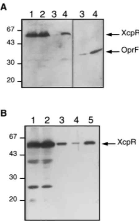
gen-eral characteristics of a cytoplasmic protein (5). To determine its cellular localization, soluble and membrane fractions of P. aeruginosa wild-type strain PAO1 were analyzed by SDS-PAGE followed by immunodetection with XcpR anti-body. As shown in Fig. 2A, XcpR was confined to the mem-brane fraction (lane 4) and was not detected in the fraction containing cytoplasmic and periplasmic proteins (lane 3). Sep-aration of membrane fractions by centrifugation through a sucrose gradient showed that XcpR is associated with the low-density fractions corresponding to the cytoplasmic membrane. The nature of the association was further examined by incu-bating corresponding fractions in the presence of various re-agents and studying the susceptibility of XcpR to extraction. Alkali treatments (0.1 N NaOH, 0.1 M Na2CO3[pH 11]), high
salt (1 M NaCl), or protein denaturant (5 M urea) released almost all XcpR, suggesting that it is peripherally bound to the cytoplasmic membrane. Treatment of membranes with the nonionic detergent Triton X-100 resulted in complete solubi-lization (data not shown).
Localization of XcpR in the absence of other Xcp proteins.
Anticipating that XcpR might be membrane associated in P. aeruginosa through interaction with one or several Xcp pro-teins, we sought to determine its localization in the absence of the components encoded by the xcpP to -Z gene cluster. The 1.8-kb PstI-AsuII DNA fragment that carries the xcpR gene was cloned into pMMB67HE to give pMR1 (Fig. 1). In this construction, xcpR is expressed under the control of the ptac promoter. Plasmid pMR1 was introduced by triparental mating into strain DZQ40, a PAO1 derivative which is chromosomally
FIG. 2. Subcellular localization of XcpR in P. aeruginosa. Soluble and mem-brane fractions were prepared as described in Materials and Methods. Proteins were separated by SDS-PAGE, transferred to nitrocellulose, and immunode-tected with anti-XcpR serum. (A) Fractionation of wild-type PAO1 cells. Lanes: 1, total cells; 2, cell lysate; 3, soluble fraction; 4, membrane fraction. Immuno-detection of the outer membrane protein OprF is shown as a fractionation control (right panel). Positions of protein molecular mass markers are indicated on the left in kilodaltons. (B) Fractionation of DZQ40/pMR1(xcpR) cells grown in the presence of 2 mM IPTG. The membrane pellet was extracted with 2% Triton X-100 in the presence of 1 mM MgCl2and centrifuged again. Lanes: 1,
total cell lysate; 2, soluble fraction; 3, membrane fraction; 4, soluble material after Triton X-100 extraction of the membrane fraction; 5, insoluble material after Triton X-100 extraction. Positions of molecular mass markers are indicated on the left in kilodaltons.
deleted for the xcpP to -Z genes (see Fig. 1 and Materials and Methods for details on strain construction). Following DZQ40/ pMR1 cell fractionation and immunodetection, the XcpR pro-tein was detected mainly in the soluble fraction (Fig. 2B, lanes 2 and 3). The low amount of protein found in the particulate fraction was not membrane associated because it was only weakly soluble in 2% Triton X-100 (Fig. 2B, lanes 4 and 5). Such behavior suggested that upon overproduction, a minor fraction of XcpR could form aggregates sedimenting with membrane vesicles, as suggested earlier for the homologous PulE protein of K. oxytoca (32). These results indicate that the association of XcpR to the cytoplasmic membrane of P. aerugi-nosa requires the expression of other Xcp proteins encoded by the 40-min locus.
Role of other Xcp proteins in the association of XcpR to inner membrane.The previous identification of an interaction between EpsL and EpsE, the XcpY and XcpR homologues in V. cholerae (38), was suggestive of a possible role of the XcpY protein in the membrane localization of XcpR. To test this hypothesis, the cloned xcpR and xcpY genes were expressed in the genetic background of the deletion strain DZQ40. Plasmid pLFR4, which carries xcpR under control of the native pro-moter, was mobilized into DZQ40. Figure 3A shows that XcpR was detected in cells of DZQ40/pLFR4 but was not recovered after the cell fractionation procedure (lane set 1). In contrast, the results in Fig. 2B show that XcpR overexpressed under ptac promoter control in DZQ40 was stable during cell fraction-ation and recovered mainly in the soluble fraction. Possibly, a high expression level causes the accumulation of XcpR under a state, different from the native one, that increases the stabil-ity of the protein. Introduction of a second plasmid (pSB31, carrying xcpY) into DZQ40/pLFR4 led to increase in the level of XcpR. Under these conditions, XcpR was found to cofrac-tionate mainly with the pellet (Fig. 3A, lane set 2), showing that XcpY alone can promote its association with the mem-brane of P. aeruginosa.
Although it seems clear that XcpY is required for the sub-cellular location of XcpR, the effect of XcpY could still be indirect, with some other, unidentified component providing a link between XcpY and XcpR. To further address this ques-tion, we examined the localization of XcpR in the heterol-ogous genetic background of E. coli. When expressed alone in strain TG1/pMR1, XcpR was detected exclusively in the solu-ble fraction (Fig. 3B, lane set 1). The xcpY gene cloned on the compatible plasmid pSB34 was introduced into TG1/pMR1. Immunodetection after cell fractionation showed that approx-imately half of XcpR was membrane associated (Fig. 3B, lane set 2). The dual localization of XcpR in membrane and soluble fractions observed here might be related to some limitation in the number of XcpY molecules inserted into the cytoplasmic membrane.
Dosage-dependent complementation and interference in the xcpY51 mutant.Previous studies have shown that introduction of a wild-type xcpY gene in the xcpY51 mutant strain restores the secretion phenotype (14). To analyze the effect of the xcpY51 complementation on the localization of XcpR, we mo-bilized plasmid pSB31, carrying xcpY under ptaccontrol, into strain KS910-503 (xcpY51). As shown in Fig. 4B, XcpY was not detected in extracts of the control strain KS910-503/ pMMB67HE (lane set 1). Sequencing of chromosomal DNA from xcpY51 identified a frameshift mutation that is responsi-ble for the synthesis of a modified protein with a basic C-terminal domain that probably impairs membrane insertion and causes a rapid degradation (28). The presence of the wild-type xcpY gene in trans allowed production of a detectable amount of XcpY in KS910-503/pSB31 without IPTG induction (Fig. 4B, lanes 2). Protease plate assay (not shown) and
im-FIG. 3. Membrane association of XcpR in the presence of XcpY. (A) Ex-pression of XcpR or of XcpR and XcpY in P. aeruginosa. Lane sets: 1, strain DZQ40/pLFR4(xcpR)/pMMB67HE; 2, strain DZQ40/pLFR4(xcpR)/pSB31-(xcpY). (B) Expression of XcpR in E. coli in the absence or presence of XcpY. Lane sets: 1, strains TG1/pMR1(xcpR)/pYZ4; 2, TG1/pMR1(xcpR)/pSB34(xcpY). Cells were grown in the presence of IPTG (2 mM for P. aeruginosa; 0.1 mM for
E. coli) for specific expression of plasmid-encoded genes, and total cells (t) were
fractionated into soluble (s) and membrane (m) fractions. Samples were ana-lyzed by SDS-PAGE and immunoblotting with anti-XcpR serum. The band of lower molecular size corresponds to the pMR1-encoded LacI repressor (38.6 kDa) which is recognized by the serum. Positions of molecular mass markers are indicated on the left in kilodaltons.
FIG. 4. Dosage-dependent complementation and interference in a P.
ae-ruginosa xcpY51 mutant. Strain KS910-503 (xcpY51) containing plasmid
pMMB67HE (control; lane set 1) or pSB31(xcpY) (lane sets 2 to 4) was grown in TSB in the absence (lane sets 1 and 2) or presence of 0.1 (lane set 3) or 2 (lane set 4) mM IPTG. Cells and extracellular medium were separated by centrifuga-tion. (A) Whole cells (i) and proteins in culture supernatants (o) were analyzed by SDS-PAGE and immunoblotting with anti-LasB serum. (B) Cells were dis-rupted by sonication and fractionated by centrifugation as described in Materials and Methods. Samples of soluble (s) and membrane (m) fractions corresponding to equivalent amounts of cells were analyzed by SDS-PAGE followed by immu-noblotting with anti-XcpR and anti-XcpY sera. Immunodetection of the cytoso-lic RpoS sigma factor was performed as a fractionation control.
munodetection of LasB elastase, the major protease secreted by the Xcp pathway (43), showed that the secretion defect of xcpY51 was complemented (Fig. 4A, lane set 2). It is known that LacI repression of the ptacpromoter is incomplete in P. aeruginosa (2), and the basal expression level of xcpY from uninduced ptacon pSB31 thus appears sufficient for phenotypic complementation of the xcpY51 mutation. This complementa-tion was correlated with the presence of XcpR in the mem-brane fraction, whereas trace amounts of XcpR were detected only in the soluble compartment of the control strain (Fig. 4B, lane sets 1 and 2).
To examine the effects of an increased expression of xcpY, we repeated the experiment in the presence of inducer. Addi-tion of 0.1 mM IPTG during the growth of KS910-503/pSB31 resulted in a higher level of XcpY (Fig. 4B, lane set 3) and in a partial secretion defect, as shown by the accumulation of mature-sized LasB in total cell fraction (Fig. 4A, lane set 3). This result suggested that the overproduced XcpY might in-terfere with normal extracellular protein secretion. A small amount of XcpY, possibly representing molecules that failed to enter the export pathway to be inserted into the cytoplasmic membrane, was detected in the soluble fraction of induced cells. Immunodetection of XcpR showed that induced cells contained reduced amounts of the protein but did not reveal a marked difference in its subcellular distribution, indicating that the overexpressed XcpY efficiently binds XcpR to the mem-brane. The secretion defect was not the result merely of a decreased level of XcpR, because a stronger induction by 2 mM IPTG resulted in a complete block of elastase secretion (Fig. 4A, lane set 4) whereas the amount of XcpR was un-changed (Fig. 4B, lane set 4).
Since the XcpR-XcpY complexes were formed, we assumed that one or several other components of the secretion machin-ery were becoming limiting for interaction or coassembly with XcpY, XcpR, or both, so that the chromosomally encoded XcpR was displaced from the functional machinery by the excess of membrane-bound XcpY located outside the active secretion complexes. To test this hypothesis, we reasoned that if the negative effect is brought about by sequestration of XcpR from functional complexes, it should be overcome by the con-comitant overexpression of XcpR. To this end, the xcpR and xcpY genes were cloned together under ptac control into pMMB67, to give pMYR. This construct was introduced into PAO1, and the effects of IPTG induction were analyzed in comparison to PAO1 carrying xcpY alone on pSB31. In the absence of induction, both strains secreted LasB into the ex-tracellular medium (Fig. 5A, lane sets 1 and 3). In the presence of 2 mM IPTG, the LasB elastase produced by PAO1/pSB31 was accumulated inside the cells (lane set 2), while the protein was secreted by PAO1/pMYR and not detected in the cellular compartment (lane set 4). Immunodetection of XcpY and XcpR showed that the two proteins were efficiently overpro-duced from pMYR (Fig. 5B). We thus concluded that the increased amount of XcpR allows the restoration of a number of active secretion complexes because it can saturate the excess of XcpY responsible for the secretion interference.
Competitive inhibition by the soluble domains of XcpY.
XcpY is an inner membrane protein with a single transmem-brane segment which connects two hydrophilic domains (8). This topology is consistent with an interaction between XcpR and the N-terminal cytoplasmic domain of XcpY. To deter-mine if this was the case, we constructed a 39-truncated xcpY gene that lacks the sequences for the transmembrane and the C-terminal periplasmic regions. We introduced the truncated allele carried on pMYS in PAO1 and examined whether pro-duction of the cytoplasmic portion of XcpY (XcpYN) could
compete for the XcpR-XcpY interaction. Data in Fig. 6B show that upon increasing synthesis of the XcpYNproduct by
induc-tion of ptacon pMYS, the majority (lane set 2) or the totality (lane set 3) of XcpR partitioned with the soluble fraction. Correlated with the displacement of XcpR from the mem-brane, secretion of extracellular LasB was impaired or abol-ished (Fig. 6A, lane sets 2 and 3). These results strongly
indi-FIG. 5. Phenotypic suppression of interference by XcpR overexpression. Wild-type strain PAO1 containing plasmid pSB31 (xcpY; lane sets 1 and 2) or pMYR (xcpR xcpY; lane sets 3 and 4) was grown in the absence (lane sets 1 and 3) or presence (lane sets 2 and 4) of 2 mM IPTG. (A) Proteins in cell lysates (i) or culture supernatants (o) were separated by SDS-PAGE, blotted, and probed with anti-LasB serum. (B) Equivalent amounts of cellular proteins were analyzed by SDS-PAGE and detected with anti-XcpY (left) or anti-XcpR (right) serum. Positions of molecular mass markers are indicated on the left in kilodaltons.
FIG. 6. Dominant negative effect of truncated XcpY protein. Strain PAO1 carrying pMYS (xcpY9) was grown in TSB in the absence (lanes 1) or presence of 10 (lane set 2) or 100mM (lane set 3) IPTG. (A) Same as for Fig. 5A. (B) Cellular proteins were separated by SDS-PAGE and immunodetected with anti-XcpR (top) or anti-XcpY (bottom) serum. The truncated form of XcpY (XcpYN) is indicated. Positions of molecular mass markers are indicated on the
cate that the truncated XcpY exerts a dominant negative effect on secretion by competing with chromosomally encoded XcpY for the formation of XcpR-XcpY active complexes. Remark-ably, the low level of XcpYN produced in the absence of
in-ducer was nevertheless sufficient to displace approximately half of XcpR from the membrane, while secretion was apparently not impaired (Fig. 6A, lane set 1). Thus, the secretory pathway can tolerate a large decrease in the number of functional XcpR molecules without effect on the efficiency of extracellular re-lease, suggesting that the step involving XcpR is not limiting under the conditions used.
The accumulation of soluble forms of XcpR in the presence of XcpYNindicates that XcpR has no permanent interaction
with another membrane protein than XcpY. However, as the effects of XcpY overproduction (Fig. 4) support the existence of at least one additional partner interacting with the XcpR-XcpY complex, we considered the possibility that the C-termi-nal domain of XcpY is involved in interactions with some other component(s) in the periplasm. Plasmid pSB72 carries an in-frame gene fusion between the sequences encoding the LasB signal peptide (LasBSP) and those corresponding to the
C-terminal periplasmic domain of XcpY (XcpYC; residues 259 to
382) (28). When the LasBSP-XcpYC hybrid was expressed in
PAO1, the LasB signal peptide was cleaved off and the XcpYC
domain was immunodetected in the periplasm. However, LasB secretion was unaffected by the accumulation of this periplas-mic form of XcpY (not shown).
DISCUSSION
In this report we show that XcpR, despite its hydrophilic character, is associated with the cytoplasmic membrane in P. aeruginosa. Membrane association is not an intrinsic property of XcpR but requires the xcpY gene product. XcpY spans the membrane once (8) and has a large N-terminal domain that faces the cytoplasm and likely interacts with XcpR. Consistent with this inference, the expression in trans of the soluble N-terminal region of XcpY resulted in a cytoplasmic location of XcpR and was inhibitory to protein secretion.
Previous studies by Sandkvist et al. (38) established that EpsE, the V. cholerae XcpR homologue, is associated to the cytoplasmic membrane through interaction with EpsL, the XcpY homologue in this bacteria. Our data obtained with the P. aeruginosa system support the prediction that the pattern of interactions taking place during the secretion process must be conserved in the GSP from different bacteria. However, it is notable that the K. oxytoca XcpR homologue, PulE, has been found associated with the particulate fraction in a reconsti-tuted system in E. coli regardless of whether the other Pul functions were expressed. As suggested by Possot and Pugsley (32), this discrepancy could be related to a peculiar propensity of PulE to form small aggregates that associate with membrane vesicles when the protein is produced in the absence of the complete set of Pul secretion proteins.
Beside anchoring the XcpR protein to the cytoplasmic mem-brane, XcpY also appears to be required for its accumulation in P. aeruginosa. The cellular level of plasmid-expressed XcpR in the xcpP to -Z chromosomal deletion strain is considerably increased by the expression of a wild-type xcpY allele in trans. Similarly, the level of XcpR is very low in the xcpY-defective KS910-503 strain but restored to a wild-type level in the pres-ence of XcpY. The reason for the apparent instability is un-clear but may be due to an altered conformation in the absence of XcpY resulting in susceptibility to cellular proteases. In the V. cholerae system, the expression of EpsL also resulted in stabilization of the membrane-associated EpsE (38).
The relative stoichiometry between XcpR and XcpY seems to be crucial for the functioning of the secretion machinery. Overexpression of XcpY causes a negative secretion pheno-type, although the membrane localization of XcpR is appar-ently not altered. Our interpretation of these observations is that the amount of chromosomally encoded XcpR is seques-tered by the large majority of XcpY molecules that have failed to appropriately interact with the other component(s) of the secretory apparatus. The excess of XcpY might thus interfere with the assembly of the system because limiting amounts of other interacting Xcp proteins may not be able to provide final active secretion complexes. Concomitant overexpression of XcpR can suppress the inhibitory effect very likely by reestab-lishing a stoichiometric equilibrium with XcpY, so that the population of XcpY molecules correctly located can be satu-rated by XcpR. Consistent with the notion of at least a second protein-protein interaction involving XcpY, recent work of this laboratory indicates that XcpY has additional interactions with XcpZ (28). Furthermore, interference by XcpY overproduc-tion appears to be partially relieved by an increased level of XcpZ, suggesting that this protein could be required for activ-ity of the XcpR-XcpY binary complexes or their targeting to secretion sites. Of course, the observed relief of interference could be related to more indirect events; further work is needed to determine the primary effect of XcpZ overexpres-sion that might lead to the phenotypic suppresoverexpres-sion effect.
Current data are insufficient to propose a definitive model for the function of XcpR family members in GSP. These pro-teins have a typical Walker box A which is essential for their activity (32, 38, 44). Although attempts to demonstrate ATPase activity were not successful (27, 38), these proteins could carry out functions related to an energetic step of the secretory pathway. By analogy with the role of the homologous protein PilB in type IV pilus assembly, it has been proposed that XcpR participates in the membrane organization of the five so-called pseudopilins XcpT, -U, -V, -W, and -X into a polymeric struc-ture spanning the periplasm (5, 19, 34). The recent isolation of a mutation of xcpR that is capable of suppressing a tempera-ture-sensitive allele of xcpT supports the possibility of an in-teraction between XcpR and pseudopilins (22). It might be also that XcpR participates in a gatekeeping function and somehow regulates the opening of the outer membrane pore constituted by XcpQ (7), possibly by modulating the polymeric state of the pseudopilins that could act as a plug in the absence of exoprotein movement through the channel. Whether it is required for steps in the assembly of a macromolecular Xcp complex or for the regulated activity of the secretory appara-tus, XcpR probably does not act directly in the cytoplasm. On the basis of the association between XcpR and XcpY reported here, one appealing possibility is that XcpY contributes to coupling an energy-dependent activity of XcpR to the process of outer membrane translocation, maybe by transmitting a conformational change to upper components of the system. Further understanding of XcpR and XcpY interactions is re-quired before such functional aspects can be addressed.
ACKNOWLEDGMENTS
We are grateful to A. Lazdunski for support throughout the course of this work. We thank R. E. W. Hancock for providing antibodies against the protein OprF and K. Tanaka for RpoS antisera.
This work was supported in part by the Ministe`re de la Recherche et de la Technologie and by a grant from the Association Franc¸aise de Lutte contre la Mucoviscidose.
REFERENCES
1. Akrim, M., M. Bally, G. Ball, J. Tommassen, H. Teerink, A. Filloux, and A. Lazdunski.1993. Xcp-mediated protein secretion in Pseudomonas
nosa: identification of two additional genes and evidence for regulation of xcp gene expression. Mol. Microbiol. 10:431–443.
2. Bagdasarian, M. M., E. Amann, R. Lurz, B. Ru¨ckert, and M. Bagdasarian. 1983. Activity of the hybrid trp-lac (tac) promoter of Escherichia coli in
Pseudomonas putida. Construction of broad-host-range,
controlled-expres-sion vectors. Gene 26:273–282. 3. Ball, G., and M. Bally. Unpublished data.
4. Bally, M., G. Ball, A. Bade`re, and A. Lazdunski. 1991. Protein secretion in
Pseudomonas aeruginosa: the xcpA gene encodes an integral inner membrane
protein homologous to Klebsiella pneumoniae secretion function PulO. J. Bacteriol. 173:479–486.
5. Bally, M., A. Filloux, M. Akrim, G. Ball, A. Lazdunski, and J. Tommassen. 1992. Protein secretion in Pseudomonas aeruginosa: characterization of seven
xcp genes and processing of secretory apparatus components by prepilin
peptidase. Mol. Microbiol. 6:1121–1131.
6. Bally, M., B. Wretlind, and A. Lazdunski. 1989. Protein secretion in
Pseudo-monas aeruginosa: molecular cloning and characterization of the xcp-1 gene.
J. Bacteriol. 171:4342–4348.
7. Bitter, W., M. Koster, M. Latijnhouwers, H. de Cock, and J. Tommassen. 1998. Formation of oligomeric rings by XcpQ and PilQ, which are involved in protein transport across the outer membrane of Pseudomonas aeruginosa. Mol. Microbiol. 27:209–219.
8. Bleves, S., A. Lazdunski, and A. Filloux. 1996. Membrane topology of three Xcp proteins involved in exoprotein transport by Pseudomonas aeruginosa. J. Bacteriol. 178:4297–4300.
9. Bleves, S., R. Voulhoux, G. Michel, A. Lazdunski, J. Tommassen, and A. Filloux.1998. The secretion apparatus of Pseudomonas aeruginosa: identifi-cation of a fifth pseudopilin, XcpX (GspK family). Mol. Microbiol. 27:31–40. 10. Chapon-Herve´, V., M. Akrim, A. Latifi, P. Williams, A. Lazdunski, and M. Bally.1997. Regulation of the xcp secretion pathway by multiple quorum-sensing modulons in Pseudomonas aeruginosa. Mol. Microbiol. 24:1169– 1178.
11. Duong, F., J. Eichler, A. Price, M. Rice Leonard, and W. Wickner. 1997. Biogenesis of the gram-negative bacterial envelope. Cell 91:567–573. 12. Fellay, R., J. Frey, and H. Krisch. 1987. Interposon mutagenesis of soil and
water bacteria: a family of DNA fragments designed for in vitro insertional mutagenesis of Gram-negative bacteria. Gene 52:147–154.
13. Figurski, D. H., and D. R. Helinski. 1979. Replication of an origin-containing derivative of plasmid RK2 dependent on a plasmid function provided in
trans. Proc. Natl. Acad. Sci. USA 79:1648–1652.
14. Filloux, A., M. Bally, G. Ball, M. Akrim, J. Tommassen, and A. Lazdunski. 1990. Protein secretion in Gram-negative bacteria: transport across the outer membrane involves common mechanisms in different bacteria. EMBO J. 9:4323–4329.
15. Filloux, A., M. Bally, M. Murgier, B. Wretlind, and A. Lazdunski. 1989. Cloning of xcp genes located at the 55 min region of the chromosome and involved in protein secretion in Pseudomonas aeruginosa. Mol. Microbiol. 3:261–265.
16. Friedman, A. M., S. R. Long, S. E. Brown, W. J. Buikema, and F. M. Ausubel. 1982. Construction of a broad host range cosmid cloning vector and its use in the genetic analysis of Rhizobium mutants. Gene 18:289–296.
17. Fu¨rste, J. P., W. Pansegrau, R. Frank, H. Blo¨cker, P. Scholz, M. Bagdasar-ian, and E. Lanka.1986. Molecular cloning of the plasmid RP4 primase region in a multi-host-range tacP expression vector. Gene 48:119–131. 18. Guzzo, J., F. Duong, C. Wandersman, M. Murgier, and A. Lazdunski. 1991.
The secretion genes of Pseudomonas aeruginosa alkaline protease are func-tionally related to those of Erwinia chrysanthemi proteases and Escherichia
colia-haemolysin. Mol. Microbiol. 5:447–453.
19. Hobbs, M., and J. S. Mattick. 1993. Common components in the assembly of type 4 fimbriae, DNA transfer systems, filamentous phage and protein-secretion apparatus: a general system for the formation of surface-associated protein complexes. Mol. Microbiol. 10:233–243.
20. Howard, S. P., J. Critch, and A. Bedi. 1993. Isolation and analysis of eight exe genes and their involvement in extracellular protein secretion and outer membrane assembly in Aeromonas hydrophila. J. Bacteriol. 175:6695–6703. 21. Jaffar-Bandjee, M.-C., A. Lazdunski, M. Bally, J. Carre`re, J.-P. Chazalette, and C. Galabert.1995. Production of elastase, exotoxin A, and alkaline protease in sputa during pulmonary exacerbation of cystic fibrosis in patients chronically infected by Pseudomonas aeruginosa. J. Clin. Microbiol. 33:924– 929.
22. Kagami, Y., M. Ratliff, M. Surber, A. Martinez, and D. N. Nunn. 1998. Type II protein secretion by Pseudomonas aeruginosa: genetic suppression of a conditional mutation in the pilin-like component XcpT by the cytoplasmic component XcpR. Mol. Microbiol. 27:221–233.
23. Kusters, J. G., E. J. Jager, and B. A. M. van der Zeijst. 1989. Improvement of the cloning linker of the bacterial expression vector pEX. Nucleic Acids Res. 17:8007.
24. Lindeberg, M., and A. Collmer. 1992. Analysis of eight out genes in a cluster
required for pectic enzyme secretion by Erwinia chrysanthemi: sequence comparison with secretion genes from other gram-negative bacteria. J. Bac-teriol. 174:7385–7397.
25. Liu, P. V. 1974. Extracellular toxins of Pseudomonas aeruginosa. J. Infect. Dis. 130:94–99.
26. Maniatis, T., E. F. Fritsch, and J. Sambrook. 1982. Molecular cloning: a laboratory manual. Cold Spring Harbor Laboratory Press, Cold Spring Har-bor, N.Y.
27. Michel, G. Unpublished data.
28. Michel, G., S. Bleves, G. Ball, A. Lazdunski, and A. Filloux. Mutual stabi-lization of the XcpZ and XcpY components of the secretory apparatus in
Pseudomonas aeruginosa. Microbiology, in press.
29. Morales, V. M., A. Ba¨ckman, and M. Bagdasarian. 1991. A series of wide-host-range low-copy-number vectors that allow direct screening for recom-binants. Gene 97:39–47.
30. Nunn, D. N., and S. Lory. 1993. Cleavage, methylation and localization of the
Pseudomonas aeruginosa export proteins XcpT, -U, -V, and -W. J. Bacteriol.
175:4375–4382.
31. Overbye, L. J., M. Sandkvist, and M. Bagdasarian. 1993. Genes required for extracellular secretion of enterotoxin are clustered in Vibrio cholerae. Gene 132:101–106.
32. Possot, O., and A. P. Pugsley. 1994. Molecular characterisation of PulE, a protein required for pullulanase secretion. Mol. Microbiol. 12:287–299. 33. Pugsley, A. P. 1992. Superfamilies of bacterial transport systems with
nucle-otide binding components. Symp. Soc. Gen. Microbiol. 47:223–248. 34. Pugsley, A. P. 1993. The complete general secretory pathway in
gram-neg-ative bacteria. Microbiol. Rev. 57:50–108.
35. Reeves, P. J., D. Whitcombe, S. Wharam, M. Gibson, G. Allison, N. Bunce, R. Barallon, P. Douglas, V. Mulholland, S. Stevens, D. Walker, and G. P. C. Salmond.1993. Molecular cloning and characterization of 13 out genes from
Erwinia carotovora subspecies carotovora: genes encoding members of a
general secretion pathway (GSP) widespread in Gram-negative bacteria. Mol. Microbiol. 8:443–456.
36. Reyss, I., and A. P. Pugsley. 1990. Five additional genes in the pulC-O operon of the gram-negative bacterium Klebsiella oxytoca UNF5023 which are required for pullulanase secretion. Mol. Gen. Genet. 222:176–184. 37. Salmond, G. P. C., and P. J. Reeves. 1993. Membrane traffic wardens and
protein secretion in Gram-negative bacteria. Trends Biochem. Sci. 18:7–12. 38. Sandkvist, M., M. Bagdasarian, S. P. Howard, and V. J. Di Rita. 1995. Interaction between the autokinase EpsE and EpsL in the cytoplasmic mem-brane is required for extracellular secretion in Vibrio cholerae. EMBO J. 14:1664–1673.
39. Smith, A. W., and B. H. Iglewski. 1989. Transformation of Pseudomonas
aeruginosa by electroporation. Nucleic Acids Res. 17:10509.
40. Smith, D. B., and K. S. Johnson. 1988. Single-step purification of polypep-tides expressed in Escherichia coli as fusions with glutathione S-transferase. Gene 67:31–40.
41. Stanley, K. K., and J. P. Luzio. 1984. Construction of a new family of high efficiency bacterial expression vectors: identification of cDNA clones coding for human liver proteins. EMBO J. 3:1429–1434.
42. Thomas, J. D., P. J. Reeves, and G. P. C. Salmond. 1997. The general secretion pathway of Erwinia carotovora subsp. carotovora: analysis of the membrane topology of OutC and OutF. Microbiology 143:713–720. 43. Tommassen, J., A. Filloux, M. Bally, M. Murgier, and A. Lazdunski. 1992.
Protein secretion in Pseudomonas aeruginosa. FEMS Microbiol. Rev. 103: 73–90.
44. Turner, L. R., J. Cano Lara, D. N. Nunn, and S. Lory. 1993. Mutations in the consensus ATP-binding sites of XcpR and PilB eliminate extracellular pro-tein secretion and pilus biogenesis in Pseudomonas aeruginosa. J. Bacteriol. 175:4962–4969.
45. Walker, J. E., M. Saraste, M. J. Runswick, and N. J. Gay. 1982. Distantly related sequences in thea- and b-subunits of ATP synthase, myosin, kinases and other ATP-requiring enzymes and a common nucleotide binding fold. EMBO J. 8:945–951.
46. Woods, D. E., and P. A. Sokol. 1986. Role of Pseudomonas aeruginosa extracellular enzymes in lung disease. Clin. Investig. Med. 9:108–112. 47. Wretlind, B., and O. Pavlovskis. 1984. Genetic mapping and characterization
of Pseudomonas aeruginosa mutants defective in the formation of extracel-lular proteins. J. Bacteriol. 158:801–808.
48. Yahr, T. L., L. M. Mende-Mueller, M. B. Friese, and D. W. Frank. 1997. Identification of type III secreted products of the Pseudomonas aeruginosa exoenzyme S regulon. J. Bacteriol. 179:7165–7168.
49. Yanisch-Perron, C., J. Vieira, and J. Messing. 1985. Improved M13 cloning vectors and host strains: nucleotide sequences of the M13mp18 and pUC19 vectors. Gene 33:103–119.
50. Zhang, Y., and J. K. Broome-Smith. 1990. Correct insertion of a simple eukaryotic plasma-membrane protein into the cytoplasmic membrane of