HAL Id: inserm-00189847
https://www.hal.inserm.fr/inserm-00189847
Submitted on 22 Nov 2007
HAL is a multi-disciplinary open access archive for the deposit and dissemination of sci-entific research documents, whether they are pub-lished or not. The documents may come from teaching and research institutions in France or abroad, or from public or private research centers.
L’archive ouverte pluridisciplinaire HAL, est destinée au dépôt et à la diffusion de documents scientifiques de niveau recherche, publiés ou non, émanant des établissements d’enseignement et de recherche français ou étrangers, des laboratoires publics ou privés.
Toric HIFU Transducer for Large Thermal Ablation.
David Melodelima, William Apoutou N’Djin, Hubert Parmentier, Michel
Rivoire, Jean Yves Chapelon
To cite this version:
David Melodelima, William Apoutou N’Djin, Hubert Parmentier, Michel Rivoire, Jean Yves Chapelon. Toric HIFU Transducer for Large Thermal Ablation.. Conference proceedings : .. Annual International Conference of the IEEE Engineering in Medicine and Biology Society. IEEE Engineering in Medicine and Biology Society. Annual Conference, Institute of Electrical and Electronics Engineers (IEEE), 2007, 1, pp.230-3. �10.1109/IEMBS.2007.4352265�. �inserm-00189847�
Abstract— A new geometry of High Intensity Focused Ultrasound (HIFU) transducer is described to enlarge the coagulated volume and decrease treatments time. Eight transducer elements and their quarter-wave plate were diced out of a single toric piezocomposite element. Each transducer operates at a frequency of 3 MHz. The focal zone is conic and located at 70 mm from the transducer. A 7.5 MHz ultrasound imaging probe is placed in the centre of the device for guiding the treatment. Optimal exposure parameters were determined from numerical simulations. This new geometry allows achieving a thermal ablation of 7.5 cm3 when each of the eight transducers has performed a 5-s ultrasound exposure alternatively and consecutively. In vivo trials have been performed on five pigs to demonstrate this new principle. 33 elementary lesions have been performed. All lesions were reproducible and homogeneous. The average diameter of an elementary lesion obtained in 40 seconds was 19.5 ± 3.8 mm (min 10 – max 29 mm). The coagulated volume obtained in 40 seconds was on average 9.1 ± 4.6 cm3 (min 1.5 – max 17.6 cm3).
I. INTRODUCTION
ethods for local tumor destruction, such as the use of physical agents, have raised interest in the last decade. For example, radiofrequency, laser, cryotherapy, microwaves or high intensity focused ultrasound are widespread [1-3]. Thermal damages induce by these methods are irreversible but generally limited to a volume of 3 to 4 centimeters in diameter [3]. New therapeutic modalities that can induce large lesions (>3 cm in diameter) rapidly are needed especially for tumors like liver metastases that can measured up to several centimeters in diameter. HIFU is a new treatment modality which presents many advantages over the above local treatments. The focused energy, after being absorbed by tissues for only a few seconds, can induce high temperatures (typically >60°C) and generate irreversible tissue necrosis at the target region while not damaging surrounding tissues. Excellent results have been obtained (both experimentally and clinically) at inducing homogeneous and reproducible tumor destruction by thermal coagulation necrosis [4-7]. The main technical limitation to widespread clinical use of HIFU has been the long time required to treat tumors of several cubic
Manuscript received April 2, 2007. This work was supported in part by the Cancérpôle Lyon Auvergne Rhône Alpes, France
D. Melodelima is with the French Institute of Health and Medical Research, Lyon, F-69003, France (phone: 681-930; fax: +33-472-681-930; e-mail: [email protected]).
A. N’Djin, H. Parmentier and JY Chapelon are with the French Institute of Health and Medical Research, Lyon, F-69003, France (e-mail:
[email protected], [email protected],
M. Rivoire is with the Centre Leon Berard, Lyon, F-69008, France (e-mail: [email protected]).
centimeter volumes. Until now, the clinical application of HIFU for liver tumors treatment in humans was limited to studies on hepatocellular carcinoma [8]. Our group has developed an intra operative HIFU prototype to treat liver metastases. The new HIFU applicator described in this paper can induce large lesions rapidly. An ultrasound imaging probe that is located in the centre of the device is for providing guidance of the treatment. The feasibility and efficiency of this new HIFU therapy has been tested in vivo in pigs. Trials were conducted in five animals to demonstrate that large elementary lesions (2 cm in diameter) can be produced in 40 seconds.
II. MATERIAL AND METHODS
A. Ultrasound equipment
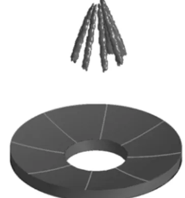
The HIFU transmitter is composed of eight ultrasound transducers operating at a frequency of 3 MHz and distributed according to a toric geometry with a radius of curvature of 7 centimeters (Figure 1). Each transducer has a focal area spatially located next to the focal area of its adjacent transducers. The focal zone of all transducers was distributed over a cone of 10 mm in diameter at 70 mm from the transducers. The absorption of the ultrasound energy creates heat deposition in the focal area of each transducer and induces a thermal lesion in 5 seconds. Each transducer is activated alternatively and consecutively. This allows creating a broad heating pattern without forming unwanted secondary foci either in front of or behind the focal plane. Therefore, each exposure benefits from the heat deposited by the other sectors. A 7.5 MHz ultrasound imaging probe is placed in the centre of the device for guiding the treatment.
Figure 1: Schematic diagram of the applicator.
The HIFU device can be sterilized in order to be used during surgery. The emitting focused ultrasound transducers
Toric HIFU Transducer for Large Thermal Ablation
David Melodelima, William A. N’Djin, Hubert Parmentier, Michel Rivoire and Jean-Yves Chapelon
M
This material is presented to ensure timely dissemination of scholarly and technical work. Copyright and all rights therein are retained by authors or by other copyright holders.
All persons copying this information are expected to adhere to the terms and constraints invoked by each author's copyright. In most cases, these works may not be reposted without the explicit permission of the copyright holder.
HAL author manuscript inserm-00189847, version 1
HAL author manuscript
will be put into acoustic contact with the liver using a sterile ultrasound coupling fluid (Ablasonic®, EDAP, France) which is contained in a sterile polyurethane envelope. This envelope attenuated the ultrasound pressure by about 2%. During treatments, the transducer was cooled using a continuous flow of degassed water at 15°C at a rate of 0.3 L/min. A peristaltic Masterflex pump (L/S model 7518-60, Cole-Parmer Instruments Co., Chicago, IL, USA) drove the water around a closed cooling circuit.
Electrical power was provided by 8 AHF 855 power amplifiers (Adece, Artannes, France) which converted the TTL signals generated by a 8-channels pattern generator (PG1050 Acute, Hsin Chuang City, Taiwan) into a sine wave [9]. 0-10 V digital-analogue output cards were used to adjust the gain on each amplifier. Input cards captured analogue voltages which are directly proportional to direct and reflected power measured using a directional coupler inside each amplifier. A Pentium 4 PC controlled both input-output cards and the pattern generator. An I/O card management program developed under Dynamic C was used to capture and store the values of gain and power provided during each ultrasound exposure. A significant increase in reflected power indicates a problem (vaporization of cooling water, incorrect acoustic coupling, bubble formation or overheating of the transducer) which can damage the ultrasound emitters. For this reason, if the reflected power was 10% greater than the incident power, the program automatically cut signal generation.
B. Animals
Trials were conducted in vivo on five healthy pigs liver because, to our knowledge, there is no pig tumor model. Animals with an average weight of 32.6 ± 2.6 kg (min 29.0 – max 35.0 kg) were used. All procedures were performed in
the laboratory for experimental surgery (DSV693880501) of
the Centre Leon Berard in Lyon (F-69008, France). Animal experiments were performed under an approved research protocol, in accordance with the World Medical Assembly Declaration of Helsinki. These experiments conformed to the requirements of the local Office of Animal Experimentation and were in accordance with the legal conditions of the National Commission on Animal Experimentation.
C. HIFU Treatments
Premedication was performed using an intramuscular injection of ketamine (15 mg/kg) 30 minutes before anaesthesia which was achieved using a 15 mL intravenous injection of Propofol (Diprivan®, Zeneca). Oxygenation was supplied from an assisted ventilation system at a rate of 10 L/min. Anaesthesia was maintained using a slow intravenous injection of Propofol (20 mL/h) and Sufentanil (Sufenta®, Jansen) at a rate of 5 mL/h in a continuous perfusion of physiological salt solution. Hydration was provided by an isotonic perfusion of physiological salt solution at 9%. Animals were in a dorsal decubitus position.
A 25 cm median laparotomy was performed from the xyphoïd process after a classical surgical asepsis. Treatments were performed under sterile conditions and the HIFU device was hold by hand. The region to be treated was located using the integrated ultrasound imaging probe. Each HIFU exposure was performed during apnea to avoid liver movements. Apnea periods always began 5 seconds before the sonication and last 60 seconds. Between phases of apnea, two minutes of controlled mechanical ventilation periods were used. The ultrasound exposures were performed using an acoustic power of 60 watts. The exposure time was set at 5 seconds for all transducers. Each of the eight transducers was activated alternatively and consecutively. Therefore the total exposure time for one elementary lesion was 40 seconds. These exposure conditions come from preliminary numerical studies performed using the software described by Curiel et al. [10]. According to the anatomy of pig livers, between 6 and 8 elementary lesions can be performed per animal. When the treatment was completed, all the lesions were observed using the ultrasound imaging probe. The animals were monitored continuously, with evaluation of hemodynamic status and blood oxygen saturation.
Figure 2: Typical lesion induced in 40 seconds with 60 watt of acoustic power.
Pigs were euthanized under general anesthesia four days after the treatment using an intravenous injection of 20 mL of potassium chloride at 10%. The delay between the treatment and euthanasia was chosen since the dimensions of thermal lesions do not change after four days [11-12]. Autopsy was performed to allow observation of the thermal damage administered. The entire peritoneal cavity, including the liver and adjacent organs was examined during autopsy. The liver was removed and sliced to inspect ultrasound effects visually. Each hepatic lesion was sectioned transversally and then sliced into samples of 5 mm thick to determine if thermal damages were homogeneous. The dimensions of the necroses were measured using a caliper. The volume of each lesion was calculated using equation (1) where a is the maximal diameter, b is the diameter at the extremity of the conical lesion and c is the depth.
3
)
(
a
2b
2ab
c
V
=
+
+
π
×
(1)Representative portions of treated tissue were also frozen, sectioned in a cryostat at five microns in thickness, and stained using routine hematoxylin and eosin (H&E) methods and a histochemical technique for tissue oxidative enzymes using nicotinamide adenine dinucleotide (NADH). This staining method permits the evaluation of tissue ablation based on cell viability rather than on histological characteristics. Data are given as mean values ± SEM.
III. RESULTS
Thirty-three homogeneous elementary lesions were created. On average 7.0 ± 0.7 (min 6 – max 8) lesions were created per animal. Figure 2 shows a typical elementary lesion. Edges of the coagulated area are well-delimited. The coagulated tissues were clearly distinguishable from untreated tissue; the lesion has an off-white colour, sometimes dark at the most heated points. The lesions were surrounded by a fibrous response of tissues. Each lesion was obtained within 40 seconds of treatment. The dimensions of the coagulated volumes were an average diameter of 19.5 ± 3.8 mm (min 10 – max 29 mm), an average depth of 28.6 ± 9.3 mm (min 10 – max 50 mm) and an average volume of
9.1 ± 4.6 cm3 (min 1.5 – max 17.6 cm3). Figure 3 shows the
diameter of elementary lesions as a function of the lesion number.
Figure 3: Coagulated diameter as a function of the lesion number.
Unlike HIFU lesions in the prostate, 90% of the HIFU lesions in the liver were seen as a hypoechoic area on sonograms (Figure 4) due to different changes in ultrasound backscatters [13]. In 10% of the cases, lesions were masked by boiling and a hyperechoic area was visible. The correlation between the dimensions of the lesions measured on sonograms with the dimensions of the lesions observed macroscopically was 90%. The difference was 2,4 ± 2,1 mm (0,0 - 11,4 mm), or 10,9 ± 7,6 % (0,0 - 36,0 %).
Figure 4: Sonogram of a HIFU lesion.
The local and biological tolerance of the treatment were excellent. The pigs remain hemodynamically stable during the procedures. They recovered from the procedures within two hours after termination of anaesthesia, and resumed eating and normal behavior. Two secondary lesions (6% of the total number of lesions) were noted due to initial use of the device. These lesions were avoided since adjacent organs were carefully isolated using pads.
Figure 4: Section stained with hematoxylin and eosin identifies damaged cells by the HIFU treatment. * Untreated liver. O Coagulated tissues.
Using standard hematoxylin and eosin staining, histological evaluation of formalin fixed, paraffin-embedded sections of excised HIFU ablation in liver tissues revealed evidence of tissue necrosis and cell death (Figure 4). To more accurately evaluate local cell necrosis, we performed NADH diaphorase staining, a vital staining [14]. In this study, a clear, non-stained zone was observed within the boundary of the ablation lesion, indicating a loss of cell viability in all the treated region (figure 5).
Figure 5: NADH stained liver tissue, cell viability represented by dark blue area and dead cells by white area.
IV. DISCUSSION
The use of HIFU thermal ablation has been established for decades [15]. The most distinctive characteristic of HIFU compared with other ablation techniques (radiofrequency, laser …) is its demonstrated ability to destroy a preselected target located deep within tissue, without damage to overlying or intervening structures. The main technical limitation to widespread clinical use of HIFU has been the long time required to treat tumors of typically several cubic centimeter volumes. In order to fill this therapeutic gap, we have designed a new geometry of HIFU device and developed a new method of treatment that can induce large lesions rapidly. Irreversible thermal lesions of 2 centimeters in diameter can be induced in 40 seconds in the liver. Therefore, treatment duration, known to be long for HIFU treatment is significantly shortened using this new geometry of transducers. The dimensions of the treatment zone can be enlarged by juxtaposing these elementary lesions.
In conventional HIFU treatments [4], a duty cycle of typically 50% is applied between exposures (5 seconds) and cooling (5 seconds). The new geometry of transducer described in this study allows performing ultrasound exposure continuously since each transducer is excited alternatively. The cooling of each transducer is performed during the exposures of all other transducers. This new principle allows creating a broad heating pattern since each exposure benefits from the heat and bubble activity produced by previous sonications. In addition, when conventional HIFU devices were used and even if the energy deposited between the transducer and the focal zone is very low, repeated sonications can produce side effects such as skin or rectal burns. The new geometry described in the present paper allows decreasing the ultrasound energy in surrounding tissues since each transducer is excited only once to produce a lesion of 2 centimeters in diameter.
In conclusion, a new geometry of HIFU transducers has been described and validated in vivo in pig livers. Large and irreversible coagulated volumes can be obtained in short times using this new HIFU device.
ACKNOWLEDGMENT
The authors wish to thank the staff of the laboratory for experimental surgery for their aid in the animal study.
REFERENCES
[1] E.W. Ramsey, “Benign prostatic hyperplasia: a review,” Can J Urol, vol. 7, pp. 1135-1143, 2000.
[2] Y. Ni, S. Mulier, Y. Miao, L. Michel, and G. Marchal, “A review of the general aspects of radiofrequency ablation,” Abdom Imaging, vol. 30, pp. 381-400, 2005
[3] G. Garcea, T.D. Lloyd, C. Aylott, G. Maddern, and D.P. Berry, “The emergent role of focal liver ablation techniques in the treatment of primary and secondary liver tumours,” Eur J Cancer, vol. 39, pp. 2150-2164, 2003.
[4] A. Gelet, J.Y. Chapelon, L. Poissonnier, R. Bouvier, O. Rouviere, L. Curiel, M. Janier. and G. Vallancien, “Local recurrence of prostate cancer after external beam radiotherapy: early experience of salvage therapy using high-intensity focused ultrasonography,” Urology, vol. 63, pp. 625-629, 2004.
[5] R.O. Illing, J.E. Kennedy, F. Wu, G.R. ter Haar, A.S. Protheroe, P.J. Friend, F.V. Gleeson, D.W. Cranston, R.R. Phillips and M. Middleton, “The safety and feasibility of extracorporeal high-intensity focused ultrasound (HIFU) for the treatment of liver and kidney tumours in a Western population,” Br J Cancer, vol. 93,pp. 890-895, 2005. [6] F.A. Jolesz, K. Hynynen, N. McDannold and C. Tempany, “MR
imaging-controlled focused ultrasound ablation: a noninvasive image-guided surgery,” Magn Reson Imaging Clin N Am, vol. 13, pp. 545-560, 2005.
[7] A.B. Ross, C.J. Diederich, W.H. Nau, V. Rieke, R.K. Butts, G. Sommer, H. Gill and D.M. Bouley, “Curvilinear transurethral ultrasound applicator for selective prostate thermal therapy,” Med
Phys, vol. 32, pp. 1555-1565, 2005.
[8] F. Wu, Z.B. Wang, W.Z. Chen, et al. “Extracorporeal High Intensity Focused Ultrasound Ablation in the Treatment of Patients with Large Hepatocellular Carcinoma,” Annals of Surgical Oncology, vol. 11, pp. 1061-1069, 2004.
[9] D. Melodelima, R. Salomir, C. Mougenot, C. Moonen and D. Cathignol D, “64-Element intraluminal ultrasound cylindrical phased array applicator compatible with magnetic resonance imaging for transesophageal thermal ablation,” Medical Physics, vol. 33, pp. 2926-2934, 2006.
[10] L. Curiel, F. Chavrier, B. Gignoux, S. Pichardo, S. Chesnais and J.Y. Chapelon, “Experimental evaluation of lesion prediction modeling in the presence of cavitation bubbles: intended for high-intensity focused ultrasound prostate treatment,” Med Biol Eng Comput, vol. 42, pp. 44-54, 2004.
[11] D. Melodelima, C. Lafon, F. Prat, Y. Theillère, A. Arefiev and D. Cathignol, “Transesophageal ultrasound applicator for sector-based thermal ablation: First in vivo experiments,” Ultrasound in Med. and
Biol, vol. 29, pp. 285-291, 2003.
[12] M. Nikfarjam, C. Malcontenti-Wilson and C. Christophi, “Progressive microvascular injury in liver and colorectal liver metastases following laser induced focal hyperthermia therapy”, Lasers Surg Med, vol. 37, pp. 64-73, 2005.
[13] A.E. Worthington, J. Trachtenberg and M.D. SherarMD, “Ultrasound properties of human prostate tissue during heating,” Ultrasound in
Med. and Biol, vol. 28, pp. 1311-1318, 2002.
[14] R.A. Neumann, R.M. Knobler, F. Pieczkowski and W. Gebhart, “Enzyme histochemical analysis of cell viability after argon laser-induced coagulation necrosis of the skin,” J Am Acad Dermatol, vol. 25, pp. 991–998, 1991.
[15] J.G. Lynn, R.L. Zwemer, A.J. Chick and A.E. Miller, “A new method for the generation and use of focused ultrasound in experimental biology,” J Gen Physiol, vol. 26, pp. 179-193, 1942.