HAL Id: hal-00990333
https://hal-univ-rennes1.archives-ouvertes.fr/hal-00990333
Submitted on 13 May 2014HAL is a multi-disciplinary open access archive for the deposit and dissemination of sci-entific research documents, whether they are pub-lished or not. The documents may come from teaching and research institutions in France or abroad, or from public or private research centers.
L’archive ouverte pluridisciplinaire HAL, est destinée au dépôt et à la diffusion de documents scientifiques de niveau recherche, publiés ou non, émanant des établissements d’enseignement et de recherche français ou étrangers, des laboratoires publics ou privés.
Impact of isomalathion on malathion cytotoxicity and
genotoxicity in human HepaRG cells.
Rozenn Josse, Ahmad Sharanek, Camille C Savary, André Guillouzo
To cite this version:
Rozenn Josse, Ahmad Sharanek, Camille C Savary, André Guillouzo. Impact of isomalathion on malathion cytotoxicity and genotoxicity in human HepaRG cells.. Chemico-Biological Interactions, Elsevier, 2014, 209, pp.68-76. �10.1016/j.cbi.2013.12.002�. �hal-00990333�
1 Impact of isomalathion on malathion cytotoxicity and genotoxicity in human HepaRG cells
Rozenn JOSSE†1,2, Ahmad SHARANEK†1,2, Camille SAVARY1,2 and Andre GUILLOUZO1,2.
1. Inserm, UMR991, Liver Metabolisms and Cancer, Faculty of Pharmacy, Rennes, France.
2. University of Rennes1, Rennes, France
† These authors contributed equally to this work.
Corresponding author :
Professeur Andre GUILLOUZO
INSERM U991 Faculté de Pharmacie, Université de Rennes 1 2 avenue L. Bernard, 35043 RENNES Cedex, France
Tel:33 (0)2 23 23 4791 ; Fax: 33 (0)2 23 23 53 85 andre.guillouzo@univ-rennes1.fr
2 ABSTRACT
Isomalathion is a major impurity of technical grade malathion, one of the most abundantly applied insecticides; however little is known about its hepatotoxicity. In the present study, cytotoxicity and genotoxicity of malathion and isomalathion either individually or in combination, were assessed using the metabolically competent human liver HepaRG cell line. Isomalathion reduced cell viability starting at a 100µM concentration after a 24h exposure. It also significantly induced caspase-3 activity in a dose-dependent manner starting at 5 µM. On the contrary, even at concentrations as high as 500µM malathion affected neither cell viability nor caspase-3 activity. Moreover, co-exposure of both compounds resulted in decreased toxicity of isomalathion. By contrast, malathion and isomalathion either separately or in combination, slightly induced micronuclei formation at low concentrations and had additive genotoxic effects when combined at 25µM. Individually or combined isomalathion directly inhibited activity of carboxyesterases which are involved in detoxication of malathion. In addition, transcripts of CYP2B6 and CYP3A4, two CYPs responsible for malathion phase I metabolism, were strongly induced by the mixture while isomalathion alone only moderately decreased CYP1A2 and increased CYP2B6 transcripts. However, these CYPs were not altered at the protein or activity levels. Taken altogether, our results showed that isomalathion was much more cytotoxic than malathion while both compounds had comparable genotoxic effects in HepaRG hepatocytes at low concentrations and brought further support to the importance of considering impurities and interactions during evaluation of health risks of pesticides.
3 INTRODUCTION
Extensive use of pesticides has caused both environmental and public health concerns. Because of its low mammalian toxicity and high selectivity toward insects [1], malathion has become one of the most commonly used organophosphate insecticides for both domestic and commercial agricultural purposes; it has been employed in malaria eradication programs in Africa and Central America and in wide-scale pest-control in the United States. Exposure of workers who applied the insecticide or harvested the crops was estimated to be 1 to 3 mg/kg/day and 1 to 270 µg/kg/day, respectively which might give rise to a range of plasma concentrations between 0.03 and 80 µM [2]. Its rapid degradation by carboxylesterases (CEs) competes with the cytochrome P450 (CYP)-catalyzed formation of the toxic metabolite, malaoxon [3].In human liver, malaoxon formation is mainly catalyzed by CYP1A2 and, to a lesser extent, CYP2B6 at low malathion concentrations (<50 µM), whereas at high levels the role of CYP3A4 becomes relevant [4]. Alterations or individual variations in both CYP and CE activities could result in increased malaoxon formation, enhancing malathion toxicity. Technical grade malathion (90-95%) contains several impurities, such as isomalathion and various trimethyl phosphorothioate esters, formed during production and/or storage, that can potentiate malathion-induced toxicity up to 10-fold [5, 6]. The presence of isomalathion, a toxic degradation product, in commercial formulations was implicated in the 1976 epidemic malathion poisoning of 2800 spraymen (including 5 deaths) among 7500 field workers, during the Pakistan malaria control program. The greatest toxicity was observed with the formulation containing the highest level of isomalathion [7]. Isomalathion has been found to inhibit CEs in a non-competitive manner, shifting the metabolic pathway toward the bioactivation reaction [8-10]. However, little is known about isomalathion toxicity.
Malathion has been found to be genotoxic in several in vivo and in vitro studies. However, data were usually obtained with the technical grade and high doses of the compound [11, 12]. No increase in the frequency of micronucleated cells and no inhibition of proliferation in lymphocyte cultures from two cohorts of applicators intermittently exposed to relatively low doses of malathion were observed [13].On occupational settings, pesticide applicators exposed to technical grade malathion and
4 other insecticides were reported to exhibit higher levels of chromosome aberrations and sister chromatid exchanges [14]. Weak or negative results have been reported with pure-grade malathion [15]. Noticeably, most genotoxic studies have been performed in the absence of a bioactivation system.
In the present study, mixtures containing both malathion and its major impurity, isomalathion, were used to better understand their toxic and genotoxic effects in the metabolically competent human liver HepaRG cell line [16]. Since in vivo hepatic concentrations are usually higher than circulating ones, malathion concentrations around 50 µM could be easily achieved in exposed individuals, and therefore, a 50 µM maximal concentration was used in vitro to be representative of the in vivo situation. Moreover, the presence of isomalathion levels as high as 0.2% is documented in actual commercial formulations, and it is known that additional amounts can be formed during storage [6]. This study showed for the first time, the potent toxic and genotoxic effects of isomalathion in an in vitro metabolically competent cell model and highlighted the necessity to take into consideration the impurities present in technical pesticide grade.
Materials and methods
Chemicals
Malathion, isomalathion, methylthiazoletetrazolium (MTT), 6β-hydroxytestosterone, 16β-hydroxytestosterone and p-nitrophenyl acetate (PNPA) were purchased from Sigma (St. Quentin Fallavier, France). 2’,7’-dichlorodihydrofluorescein (H2DCFDA) was from Invitrogen-Molecular Probe (Cergy-Pontoise, France).
Cell cultures
HepaRG cells are derived from a human cholangiohepatocarcinoma; they are untransformed cells and were used before passage 18. Briefly, they were seeded at a density of 2.6×10⁴cells/cm² as described previously [17, 18]. They were first incubated in the Williams' E medium supplemented with 10% fetal calf serum (FCS), 100 units/ml penicillin, 100 µg/ml streptomycin, 5 µg/ml insulin, 2 mM glutamine, and 5 × 10⁻⁷M hydrocortisone hemisuccinate for 2 weeks. Then, HepaRG cells were shifted to the same medium supplemented with 2% dimethylsulfoxide (DMSO) for two
5 further weeks in order to obtain confluent differentiated cultures with maximum functional activities. At this time, these cultures contained hepatocyte-like and progenitors/primitive biliary-like cells at around 50% each [19]. The medium was renewed every 2 or 3 days. During treatment, the cells were exposed to the compounds in a medium supplemented by 2%FCS and 1% DMSO.
Preparation of microsomal fractions
Human liver tissue samples were homogenized in 50mM Tris-HCl buffer (pH 7.4) containing 0.25M sucrose and 1mM EDTA. Microsomal fractions were the sediment and supernatant, respectively, from the last of three successive centrifugations at 4 °C (3000 g, 10 min; 8000 g, 20 min; and 30000 g, 60 min).
Cell viability
Cytotoxicity of malathion and isomalathion, was evaluated by the MTT spectrometry assay [17]. Briefly, after treatment, medium was removed and serum-free medium containing MTT (0.5 mg/ml) was added to each well and incubated for 2 h at 37°C. After removal of the incubation solution, water-insoluble formazan was dissolved in DMSO and absorbance was measured at 550 nm. Data were expressed as the mean of three independent experiments.
Caspase-3 activity
After treatment with pesticides, differentiated HepaRG cells were harvested and stored as pellets at −80°C. After cell lysis, 40µg of protein were incubated with 80µM Ac-DEVD-AMC in caspase-3 activity buffer (20 mM PIPES pH 7.2, 100 mM NaCl, 10mM dithiotreitol, 1mM EDTA, 0.1% CHAPS and 10% sucrose) at 37°C for 1 h. Caspase 3-mediated cleavage of Ac-DEVD-AMC peptide was continuously measured by spectrofluorimetry using excitation/emission wavelengths of 380/440 nm [20].
Evaluation of oxidative stress
Cells were incubated for 2 hours at 37°C with 5 µM H2DCFDA; then they were washed with cold phosphate buffered saline (PBS), and scraped in a solution containing an equal volume of potassium buffer (10 mM, pH 7.4) and methanol (v/v), supplemented with 0.1 % Triton X-100. Fluorescence intensity of cell extracts was determined by spectrofluorimetry using excitation/emission wavelengths of 498/520 nm.
6 Real time - quantitative polymerase chain reaction (RT-qPCR) analysis
Total RNA was extracted from HepaRG cells with the SV total RNA isolation system (Promega, Madison, WI). Five hundred nanograms of total RNA were reversed-transcribed into cDNA using the High-Capacity cDNA Archive kit (Applied Biosystems, Foster City, CA). RT-qPCR was performed by the fluorescent dye SYBR Green methodology using the SYBR Green PCR Master Mix and the ABI Prism 7000 (Applied Biosystems). Primers pairs for each transcript are described in Table 1. Amplification curves were read with the ABI Prism 7000 SDS Software using the comparative cycle threshold method and the relative quantification of the steady-state mRNA levels was normalized against 18S RNA.
Micronucleus assay
Genotoxic effects of malathion and isomalathion either individually or combined, were evaluated by the micronucleus assay as previously described [21]. Briefly, after 24 h of treatment with pesticides, followed by a 72h epidermal growth factor (EGF) stimulation corresponding to the recovery time, HepaRG cells were fixed for 10 min in 4% paraformaldehyde at 4°C and then permeabilized for 45 min in 1X phosphate-buffered saline containing 10% donkey serum and 0.1% saponin. F-actin, a cytoskeletal component, was labelled with 1/400 phalloidin conjugated to sulforhodamine for 20 min and nuclei were stained with 1/5000 Hoechst for 5 min. Images were taken with an upright microscope (Axioimager M1) and were a mosaic of six pictures per condition. All experiments were performed in triplicate. Mono-micronucleated, multi-Mono-micronucleated, apoptotic and mitotic cells were scored manually and blindly with Image Tool software from micrographs obtained with a Axioimager microscope. Criteria used for identifying micronuclei were as follows: area less than one-third the main nucleus area, no overlap with the nucleus (distinct borders) and same aspect as the chromatin. Cells containing two to five and more than five micronuclei were scored as multi-micronucleated and apoptotic cells, respectively. Cytotoxicity was also evaluated by calculation of relative population doubling (RPD) as follows: RPD = 100 × number of population doublings in treated cultures/number of population doublings in control cultures; where population doubling = [log (post-treatment cell number)/initial cell number)]/log2.
7 For the determination of the effects of pesticides on CYP3A4-, CYP2B6- and CYP1A2-related activities HepaRG cells were incubated with 200 µM testosterone for 2h for CYP3A4 and CYP2B6 analysis or with 200 µM phenacetin for 20h for measurement of CYP1A2 activity in phenol red-free medium deprived of both FCS and DMSO. Rifampicin at 50 µM and 3-methylcholanthrene at 5 µM were used as positive controls and added to the cell cultures for 48 h before incubation with the specific substrates. Culture media were then collected and centrifuged for 2 min at 10,000 rpm; 50µl of sample was injected in the high-performance liquid chromatography system for quantification of the specific metabolites, 6β- and 16β -hydroxytestosterone and acetaminophen generated by CYP3A4, CYP2B6 and CYP1A2 respectively, resulting from 6β- and 16β-hydroxylation of testosterone and phenacetin deethylation activities, respectively. A nucleosil C18 column (150 × 4.6 mm, 5 µm) was used for chromatographic separation of testosterone metabolites at 254 nm. The mobile phase consisted of two solvents, A (0.1% acetic acid) and B (acetonitrile), with the following gradient: 0 min, 20% B; and 20 min, 33% B. Chromatographic separation of phenacetin metabolites was carried out with an Interchrom C18 column (250 × 4.0 mm, 5 µm) at 250 nm. The mobile phase consisted of two solvents, A (2 mM ammonium acetate, pH 2.6) and B (acetonitrile) with a linear gradient (20 min, 3% B).The high-performance liquid chromatography apparatus consisted of an Agilent 1100 Series high-performance liquid chromatograph equipped with an autosampler and Agilent 1100 Series fluorescence and UV detectors (Agilent Technologies, Palo Alto, CA). A computer running Agilent 1100 software (ChemStation) was used to integrate and calculate the separated peak area and to plot metabolite patterns. Metabolites were identified by comparison of retention times, and quantifications were estimated from calibration.
Carboxylesterase activity was determined by measuring hydrolysis of PNPA, an esterase substrate, to p-nitrophenol in HepaRG cells and for comparison in human liver microsomes as previously described [22]. One, 4, 24h and 48h after pesticide treatment, HepaRG cells were incubated with 5mM PNPA in phenol red-free medium deprived of both FCS and DMSO during 15 min. In microsomes, after a 2-min preincubation at 37°C with either pesticides or DMSO (vehicle) the reaction was initiated by addition of PNPA 1 mM (final concentration) to 200 µl solution
8 containing100 mM K2HPO4 buffer, pH 7.4, containing 5 mM MgCl2 and 20 µg of microsomal proteins in 96 multi-well plates as final concentrations.
Hydrolysis of PNPA to p-nitrophenol was determined spectrophotometrically by measuring increase in absorbance determined at 405 nm, after a 15 min exposure. Because p-nitrophenol exists as a contaminant in commercially available PNPA, its content in the mixture incubated without cells or without microsomes was substracted from that measured in the cells or microsomes respectively to correct the activity level. Data are expressed relative to absorbance found in untreated cells arbitrarily set at 1.
Western blot analysis
HepaRG cells were treated for 48 hours with malathion and isomalathion either individually or with various combinations, then they were scrapped with CytoBuster Protein Extraction Reagent (Novagen, USA) supplemented with 1X complete protease inhibitors and 1X Phosphatase Inhibitor (Roche, Mannheim, Germany). Twenty micrograms of protein were separated by electrophoresis on 4 to 12% gradient Bis-Tris gels (Invitrogen), transferred to Hybond ECL nitrocellulose membranes (GE Healthcare, Chalfont St. Giles, Buckinghamshire, UK) and immunoblotted with antibodies against CYP3A4 or CYP2B6 (Santa Cruz Biotechnology, Inc., Santa Cruz, CA) and heat shock cognate protein 70 (HSC70) (Tebu-bio, Le Perray en Yvelines, France). Blots were then incubated with appropriate secondary antibodies, and protein bands were revealed by enhanced chemiluminescence in a Chemi-smart imager (Thermo Fisher Scientific, Ilkirch, France). HSC70 was used to normalize protein loadings, and quantification was performed with BIO-1D software (Vilber Lourmat, Marne la Vallée, France).
Statistical analysis
The Mann-Whitney U test was applied to compare data between treated cells and corresponding control cultures. For the micronucleus assay, a chi-square one-tailed test associated with a Yates correction was performed. Data were considered significantly different when P <0.05.
RESULTS
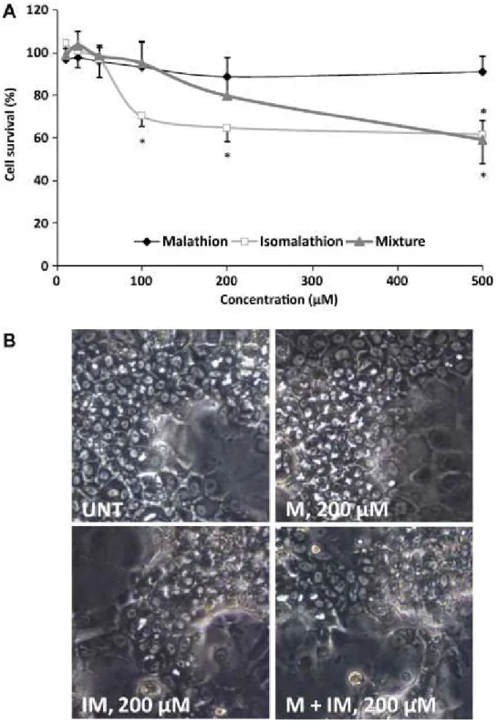
9 First, cytotoxicity induced by malathion (M), isomalathion (IM) and an equimolar mixture (M+IM) was assessed in HepaRG cells after a 24 h exposure to varying concentrations up to 500 µM using the MTT assay. Similar results were obtained with the ATP assay (not shown). Malathion did not affect cell viability contrary to isomalathion which reduced cell viability at 100 µM (Figure 1A). A decrease in cell viability occurred with the (M + IM) mixture from 200 µM. Importantly, as evidenced by light microscopic examination, morphological alterations of HepaRG cells were restricted to hepatocyte-like cells (Figure 1B). No change in cell viability was observed in HepG2 cells using the same range of concentrations (data not shown).
Cytotoxic effects of isomalathion were confirmed by measuring caspase-3 activity. Isomalathion significantly induced caspase-3activity in a dose-dependent manner at concentrations that did not affect cell viability (Figure 2). A slight production of ROS was observed after treatment with 50 µM isomalathion and the equimolar (M25 + IM 25) mixture (Figure 3A). In order to evaluate modulation of the antioxidant system in response to the compounds, mRNA expression levels of the transcription factor Nrf2, hemeoxygenase 1 (HO-1) and superoxide dismutase (MnSOD) were measured. In parallel to the oxidative stress induced by isomalathion, a two-fold increase of HO-1 mRNA expression was observed but no effect was noticed on Nrf2 and MnSOD level expression after a 24h exposure to either malathion or isomalathion (Figure 3B).
Genotoxicity of malathion and isomalathion individually and in combination In order to evaluate malathion and isomalathion genotoxic effects, the micronucleus assay was performed in HepaRG cells as previously described [21]. Two positive controls were used: MMS, a classical DNA damaging agent and AFB1, a metabolism-requiring genotoxic compound (Table 2). At the tested concentrations, none of the compounds alone or in combination induced toxicity as shown by the calculation of the RPD. Both malathion and isomalathion slightly increased the number of mono-micronucleated cells compared to untreated cells but this number did not vary with concentrations up to 25 µM and no variation was observed in the number of multi-micronucleated or apoptotic cells. Similarly, the number of mono-micronucleated cells induced by the (M + IM) mixture was not significantly different from that induced by either malathion or isomalathion, except for the equimolar (M 25 + IM 25) mixture which had additive effects compared to either compound at 25 µM.
10 Effect of malathion and isomalathion on their metabolic-required enzymes To better understand malathion and isomalathion effects in HepaRG cells, we analyzed gene expression and activities of the main CYPs as well as the two detoxifying enzymes CE1 and CE2 (Figures4 and 5). Malathion alone did not modify expression of any of the main CYPs while isomalathion decreased CYP1A2 mRNA and increased CYP2B6 mRNA. All the mixture ratios except the (M25 + IM 0.05) ratio, strongly induced CYP2B6 and CYP3A4 expression (Figure 4A). Despite a change in CYP expression levels caused by isomalathion alone or in mixture, CYP activities and protein levels were not altered (Figure 4B-D). Nevertheless, neither alone nor in mixture did malathion or isomalathion modify the transcript levels of the two CEs (CE1 and CE2) (Figure 5A) except IM at 50 µM which slightly down-regulated CE1 and CE2. However, isomalathion was found to strongly inhibit CEs activity in HepaRG cells. As early as one hour of pesticide treatment, this non-competitive inhibition was nearly complete with 5 µM isomalathion and persisted after 48h (Figure 5B-C). Malathional one had no any modulatory effect on CEs activity even at high concentration. In agreement, all the mixture ratios had the same inhibitory effect as isomalathion alone. Similar results were obtained with human liver microsomes (Figure 5D).
Noticeably, transcripts of other genes related to drug metabolism (UGT1A1, MRP2), the p53 pathway (p21,FDXR, BTG2)or previously found to be deregulated by genotoxic compounds (FHIT, BCAS3, SMYD3) [23], were also measured; none of them was found to be significantly deregulated after exposure to malathion and isomalathion individually or in combination (not shown).
DISCUSSION
In the present study, we investigated the effects of malathion and its impurity, isomalathion, individually or in combination, in HepaRG cells, an in vitro metabolically competent liver cell model [16]. Up to now, most of the literature concerns malathion toxicity and only few studies have been performed on human liver cells. We found that malathion alone was much less cytotoxic than isomalathion and exerted cytotoxic effects at similar concentrations as in primary human hepatocytes [24]. It was also previously reported to significantly decrease cell viability in HepG2 cells
11 after a 48 h exposure only at very high concentrations close to the insolubility, i.e. at 18 mM [25]. In agreement, we observed that malathion and isomalathion did not cause any cell loss in HepG2 cells when using the same range of concentrationsas those used for HepaRG cells. These results support our previous data showing that HepaRG cells are much more sensitive to compounds requiring liver metabolism to exert their toxic effects than HepG2 cells that did not express most of the major cytochromes P450 [21, 26].
Malathion concentrations used in the present study were relevant to an human in vivo exposure [2]. Indeed, malathion concentrations ≤50 µM could be very likely present in the blood of exposed individuals. Even higher concentrations can be reached in the liver, where most of malathion bioactivation takes place [27, 28]. For agricultural workers, exposure to malathion was estimated to be 1–270 µg/kg/day [2], which may give rise to plasma concentrations up to 80 µM, considering an almost complete absorption. It has been reported that malathion induced complex structural chromosomal aberrations in whole human blood cells in the presence of S9 mix [29]. In a review, Flessel et al. [11] concluded that technical grade malathion and malaoxon caused an increase in sister chromatid exchange frequency, indicating that this pesticide, its impurities or metabolites, can affect DNA. Moreover, human lymphocytes exposed to malaoxon and isomalathion showed a positive comet assay from 25 µM and 75 µM, respectively. DNA damage induced by isomalathion was efficiently and quickly repaired as soon as 15 min while malaoxon induced more persistent DNA damage [15] but these data were obtained in the absence of a bioactivation system. To our knowledge, no report is available in the literature regarding the level of cytotoxicity and DNA damage when human liver cells are exposed to a combination of both compounds. According to the degree of contamination of technical malathion by isomalathion, different toxic effects could be expected. In the metabolically competent HepaRG liver cell model, malathion and isomalathion were able to slightly increase micronuclei at a concentration as low as 10 µM or less but no further increase was evidenced with concentrations up to 25µM and, in combination they showed a limited additive effect only at the equimolar concentration of 25 µM. An in vivo study has also shown that three common organosphosphate pesticides, i.e. malathion, chlorpyrifos and methyl parathion, given in mixture to rats were not able to potentiate the toxicity of each other [30].
12 In our study, the interaction between malathion and isomalathion was shown to be dependent on the endpoint measured and their concentration. Noticeably, malathion antagonized cytotoxic and apoptotic effects of isomalathion. A concentration-dependent induction of caspase-3 activity by isomalathion was evidenced at concentrations that did not alter cell viability. To our knowledge, these results represent the first demonstration that isomalathion is directly hepatotoxic. No synergistic toxic effects between malathion and isomalathion were observed, suggesting occurrence of a more complex balance between the bioactivation and detoxication/DNA repair pathways in a metabolically competent system such as the HepaRG cell line. In order to better understand the underlying toxic mechanisms, we focused on the effects of malathion and isomalathion on the phase I metabolism enzymes. Formation of metabolites could be at least partially responsible for genotoxicity of the compounds in HepaRG cells as shown by using the micronucleus assay. As previously described by Buratti et al. [4], CYP1A2>CYP2B6>CYP3A4 play a pivotal role in malaoxon production while CE, mainly CE1 in liver, also detoxifies malathion. However, as previously reported [9] we found that isomalathion inhibited CEs in human microsomes and similarly in HepaRG cells in a non-competitive manner, suggesting that malathion in the presence of isomalathion, might shift its metabolic pathway toward a bioactivation reaction and be more toxic [10]. Noticeably, even at high concentration malathion did not alter CEs activity. In addition, while malathion and isomalathion combination did not change CYP1A2 expression, it strongly induced expression of CYP2B6 and CYP3A4 without any induction of corresponding protein or activity levels. In agreement, in a recent study using the HepaRG cell line but in different treatment conditions, malathion was found to activate the nuclear receptor constitutive androstane receptor (CAR) and some of its target genes, including CYP2B6 and CYP3A4, without induction of their corresponding activities [31]. Using bupropion as another specific CYP2B6 substrate, the same lack of induction was observed after malathion exposure (data not shown). Presently, such differences between transcripts and protein activities response remain unclear and require further investigation.
In conclusion, our findings show that although chemically similar, malathion and its impurity isomalathion, either individually or in combination, might exert different toxic effects in the metabolically competent HepaRG cells. We clearly evidence that isomalathion by itself induced caspase-3 activation and can be a DNA-damaging
13 agent. The combination of malathion with isomalathion had additive effects on genotoxicity. However, isomalathion-induced apoptosis was abolished by a co-exposure with malathion. This study highlights the importance of considering possible impurities and interactions during evaluation of health risks of pesticides.
Funding
This work was supported by grants from the Ligue 35 contre le Cancer and ANR (contract NISTEC 09-CESA-003-002).
14 References
[1] R.D. Wauchope, T.M. Buttler, A.G. Hornsby, P.W. Augustijn-Beckers, J.P. Burt, The
SCS/ARS/CES pesticide properties database for environmental decision-making, Rev Environ Contam Toxicol 123 (1992) 1-155.
[2] R.I. Krieger, T.M. Dinoff, Malathion deposition, metabolite clearance, and cholinesterase status of date dusters and harvesters in California, Arch Environ Contam Toxicol 38(4) (2000) 546-553.
[3] L.G. Sultatos, Mammalian toxicology of organophosphorus pesticides, J Toxicol Environ Health 43(3) (1994) 271-289.
[4] F.M. Buratti, A. D'Aniello, M.T. Volpe, A. Meneguz, E. Testai, Malathion bioactivation in the human liver: the contribution of different cytochrome p450 isoforms, Drug Metab Dispos 33(3) (2005) 295-302.
[5] G. Pellegrini, R. Santi, Potentiation of toxicity of organophosphorus compounds containing carboxylic ester functions toward warm-blooded animals by some organophosphorus impurities, J Agric Food Chem 20(5) (1972) 944-950.
[6] W.N. Aldridge, J.W. Miles, D.L. Mount, R.D. Verschoyle, The toxicological properties of impurities in malathion, Arch Toxicol 42(2) (1979) 95-106.
[7] E.L. Baker, Jr., M. Warren, M. Zack, R.D. Dobbin, J.W. Miles, S. Miller, L. Alderman, W.R. Teeters, Epidemic malathion poisoning in Pakistan malaria workers, Lancet 1(8054) (1978) 31-34. [8] R.E. Talcott, H. Denk, N.M. Mallipudi, Malathion carboxylesterase activity in human liver and its inactivation by isomalathion, Toxicol Appl Pharmacol 49(2) (1979) 373-376.
[9] F.M. Buratti, E. Testai, Malathion detoxification by human hepatic carboxylesterases and its inhibition by isomalathion and other pesticides, J Biochem Mol Toxicol 19(6) (2005) 406-414. [10] A.F. Hernandez, T. Parron, A.M. Tsatsakis, M. Requena, R. Alarcon, O. Lopez-Guarnido, Toxic effects of pesticide mixtures at a molecular level: their relevance to human health, Toxicology 307 (2013) 136-145.
[11] P. Flessel, P.J. Quintana, K. Hooper, Genetic toxicity of malathion: a review, Environ Mol Mutagen 22(1) (1993) 7-17.
[12] J.M. Pluth, J.A. Nicklas, J.P. O'Neill, R.J. Albertini, Increased frequency of specific genomic deletions resulting from in vitro malathion exposure, Cancer Res 56(10) (1996) 2393-2399.
[13] N. Titenko-Holland, G. Windham, P. Kolachana, F. Reinisch, S. Parvatham, A.M. Osorio, M.T. Smith, Genotoxicity of malathion in human lymphocytes assessed using the micronucleus assay in vitro and in vivo: a study of malathion-exposed workers, Mutat Res 388(1) (1997) 85-95.
[14] D.S. Rupa, P.P. Reddy, O.S. Reddi, Clastogenic effect of pesticides in peripheral lymphocytes of cotton-field workers, Mutat Res 261(3) (1991) 177-180.
[15] J. Blasiak, P. Jaloszynski, A. Trzeciak, K. Szyfter, In vitro studies on the genotoxicity of the organophosphorus insecticide malathion and its two analogues, Mutation research 445(2) (1999) 275-283.
[16] A. Guillouzo, A. Corlu, C. Aninat, D. Glaise, F. Morel, C. Guguen-Guillouzo, The human hepatoma HepaRG cells: a highly differentiated model for studies of liver metabolism and toxicity of xenobiotics, Chem Biol Interact 168(1) (2007) 66-73.
[17] C. Aninat, A. Piton, D. Glaise, T. Le Charpentier, S. Langouet, F. Morel, C. Guguen-Guillouzo, A. Guillouzo, Expression of cytochromes P450, conjugating enzymes and nuclear receptors in human hepatoma HepaRG cells, Drug Metab Dispos 34(1) (2006) 75-83.
[18] P. Gripon, S. Rumin, S. Urban, J. Le Seyec, D. Glaise, I. Cannie, C. Guyomard, J. Lucas, C. Trepo, C. Guguen-Guillouzo, Infection of a human hepatoma cell line by hepatitis B virus, Proc Natl Acad Sci U S A 99(24) (2002) 15655-15660.
15 [19] V. Cerec, D. Glaise, D. Garnier, S. Morosan, B. Turlin, B. Drenou, P. Gripon, D. Kremsdorf, C. Guguen-Guillouzo, A. Corlu, Transdifferentiation of hepatocyte-like cells from the human hepatoma HepaRG cell line through bipotent progenitor, Hepatology 45(4) (2007) 957-967.
[20] J. Dumont, R. Josse, C. Lambert, S. Antherieu, L. Le Hegarat, C. Aninat, M.A. Robin, C. Guguen-Guillouzo, A. Guguen-Guillouzo, Differential toxicity of heterocyclic aromatic amines and their mixture in metabolically competent HepaRG cells, Toxicology and applied pharmacology 245(2) (2010) 256-263. [21] R. Josse, A. Rogue, E. Lorge, A. Guillouzo, An adaptation of the human HepaRG cells to the in vitro micronucleus assay, Mutagenesis 27(3) (2012) 295-304.
[22] E.W. Morgan, B. Yan, D. Greenway, D.R. Petersen, A. Parkinson, Purification and
characterization of two rat liver microsomal carboxylesterases (hydrolase A and B), Arch Biochem Biophys 315(2) (1994) 495-512.
[23] R. Josse, J. Dumont, A. Fautrel, M.A. Robin, A. Guillouzo, Identification of early target genes of aflatoxin B1 in human hepatocytes, inter-individual variability and comparison with other genotoxic compounds, Toxicol Appl Pharmacol 258(2) (2012) 176-187.
[24] G. de Sousa, F. Fontaine, M. Pralavorio, D. Botta-Fridlund, Y. Letreut, R. Rahmani, Insecticide cytotoxicity and CYP1A1/2 induction in primary human and rat hepatocyte cultures, Toxicol In Vitro 11(5) (1997) 451-457.
[25] P.D. Moore, C.G. Yedjou, P.B. Tchounwou, Malathion-induced oxidative stress, cytotoxicity, and genotoxicity in human liver carcinoma (HepG2) cells, Environ Toxicol 25(3) (2010) 221-226. [26] R. Josse, C. Aninat, D. Glaise, J. Dumont, V. Fessard, F. Morel, J.M. Poul, C. Guguen-Guillouzo, A. Guillouzo, Long-term functional stability of human HepaRG hepatocytes and use for chronic toxicity and genotoxicity studies, Drug metabolism and disposition: the biological fate of chemicals 36(6) (2008) 1111-1118.
[27] F.W. Kutz, B.T. Cook, O.D. Carter-Pokras, D. Brody, R.S. Murphy, Selected pesticide residues and metabolites in urine from a survey of the U.S. general population, J Toxicol Environ Health 37(2) (1992) 277-291.
[28] J.L. Adgate, D.B. Barr, C.A. Clayton, L.E. Eberly, N.C. Freeman, P.J. Lioy, L.L. Needham, E.D. Pellizzari, J.J. Quackenboss, A. Roy, K. Sexton, Measurement of children's exposure to pesticides: analysis of urinary metabolite levels in a probability-based sample, Environ Health Perspect 109(6) (2001) 583-590.
[29] V.F. Garry, R.L. Nelson, J. Griffith, M. Harkins, Preparation for human study of pesticide applicators: sister chromatid exchanges and chromosome aberrations in cultured human lymphocytes exposed to selected fumigants, Teratog Carcinog Mutagen 10(1) (1990) 21-29.
[30] A. Ojha, S.K. Yaduvanshi, S.C. Pant, V. Lomash, N. Srivastava, Evaluation of DNA damage and cytotoxicity induced by three commonly used organophosphate pesticides individually and in mixture, in rat tissues, Environ Toxicol (2011).
[31] K. Abass, V. Lamsa, P. Reponen, J. Kublbeck, P. Honkakoski, S. Mattila, O. Pelkonen, J. Hakkola, Characterization of human cytochrome P450 induction by pesticides, Toxicology 294(1) (2012) 17-26.
16 Table 1. List of primer sequences used in RT-qPCR
Gene Name Forward Primer Reverse Primer
CE1 carboxylesterase 1 AGGTCCTGGGGAAGTATGCC TGCATCTTGGGAGCACATAGG CE2 carboxylesterase 2 GGAGTGGTGTGAGAGATGCG CAGGTTAGAGCCCTCACGG CYP1A2 cytochrome P450, family 1,
subfamily A, polypeptide 2
TGGAGACCTTCCGACACTCCT CGTTGTGTCCTTTGTTGTGC CYP2B6 cytochrome P450, family 2,
subfamily B, polypeptide 6
TTCCTACTGCTTCCGTCTATCAAA GTGCAGATTCCCACAGCTCA CYP3A4 cytochrome P450, family 3,
subfamily A, polypeptide 4
CTTCATCCAATGGACTGCATA TCCCAAGTATAACAGCACTCTA CACAGAC
HO1 hemeoxygenase 1 ACTTTCAGAAGGGCCAGGT TTGTTGCGCTCAATCTCCT MnSOD manganese superoxide dismutase GGGTTGGCTTGGTTTCAATA CTGATTTGGACAAGCAGCAA Nrf2 NF-E2-related factor 2 TCAGCATGCTACGTGATGAAG TTTGCTGCAGGGAGTATTCA
17 Table 2. Numbers of mono-micro- nucleated , multi-micronucleated, apoptotic and metaphasic cells induced by malathion (M) and isomalathion (IM) separately or in combination (M + IM) in HepaRG cells after a 24 h exposure.
Results are expressed as mean number per 1000 cells ± SD of three independent cultures. Chi-square test associated with a Yates correction: *P< 0.05; **P< 0.01; ***P< 0.001 from untreated (UNT) cells. AFB1 (aflatoxinB1) and MMS (methyl methanesulfonate) were used as positive controls. RPD (Relative population doubling) has been calculated.
Compound (µM) Mono-micro- nucleated Multi- micro-nucleated Apoptotic Metaphasic RPD (%) UNT 7 ± 1 0.7 ± 1.2 1.7 ± 0.6 19.7 ± 5.7 100 AFB1 0.5 49 ± 2.6*** 11 ± 2.6 10 ± 4 28 ± 5.3 88.2 MMS 270 50 ± 4.4*** 9.3 ± 2.5 5.3 ± 2.1 33.7 ± 8.3 95.4 Malathion 10 17.7 ± 3.1* 1 ± 0 2 ± 1 24 ± 6.2 99.7 25 17.3 ± 3.2* 2.7 ± 2.1 3 ± 2.6 22.7 ± 4.5 100.2 50 21 ± 4.6** 0.7 ± 0.6 3.7 ± 1.2 22.7 ± 6.7 100.2 Isomalathion 0.05 13.7 ± 5.5 1.7 ± 1.5 2.7 ± 1.5 21.7 ± 3.1 99.6 5 11.3 ± 2.1 1 ± 1 1.3 ± 0.6 18.7 ± 1.2 100.6 10 16 ± 1.7 1 ± 1 1 ± 0 20.7 ± 7 99.8 25 17.7 ± 5.5* 2.3 ± 2.5 2.3 ± 1.2 22.7 ± 6.4 99.7 50 26.7 ± 2.3*** 2 ± 1.0 1.3 ± 0.6 23.7 ± 5.7 100.5 Mixture M25 + IM 0.05 15.3 ± 2.1 1 ± 1 2.3 ± 0.6 19.3 ± 3.8 99.6 M25 + IM 5 15.7 ± 2.3 0.7 ± 0.6 2.7 ± 2.1 21.7 ± 5.7 99.5 M10 + IM 10 18.7 ± 4* 2 ± 1 3.3 ± 2.1 23 ± 7.8 99.6 M25 + IM 25 27 ± 8.9*** 1.7 ± 0.6 2 ± 2 23.3 ± 4 100.3
18 Legends to Figures
Figure 1. Cytotoxic effects of malathion (M), isomalathion (IM) and their mixture (M + IM) in HepaRG cells (A). Cells were exposed to M, IM or the mixture (M + IM) for 24 h. Cytotoxicity was assayed using the MTT assay. Results are expressed as % of the value found in control cells arbitrarily set at 100%. Data are means ± SD of three independent experiments. (B) Morphological changes after exposure of HepaRG cells to 0.2 % DMSO (UNT), M, IM or their mixture (M + IM) for 24 h.
19
Figure 2. Pro-apoptotic effects of malathion (M), isomalathion (IM) and their mixture (M+IM) in HepaRG cells. Cells were exposed to 0.2 % DMSO (UNT), M, IM, the mixture (M + IM) or 2 µM staurosporine (positive control of apoptosis induction) for 24 h. Apoptosis was assayed by measuring caspase-3 activation. Results are expressed as % of the value found in control cells arbitrarily set at 100%. Data are means ± SD of three independent experiments. *p<0.05 compared to untreated cells.
20
Figure 3. Oxidative stress induced by malathion (M), isomalathion (IM) and their mixture (M + IM) in HepaRG cells. (A) Oxidative stress was assessed by ROS formation. Cells were exposed to 0.2 % DMSO (UNT), M, IM or the mixture (M + IM) and 100 mM H2O2 (positive
control) for 24 h. Data are means ± SD of three independent experiments. *p<0.05 vs control cells. (B) Nrf2, HO-1 and MnSOD mRNA levels were estimated by RT-qPCR. Data were expressed relative to mRNA levels found in untreated cells arbitrarily set at 1.
21
Figure 4. Effect of malathion (M), isomalathion (IM) and their mixture (M + IM) on expression and activities of CYPs involved in malathion metabolism. (A) CYP mRNAs were analyzed by RT-qPCR. HepaRG cells were exposed to 0.2 % DMSO (UNT) or incubated with different concentrations of M, IM, or their mixture (M + IM) for 24 h. Results are expressed as fold of the value found in untreated cells arbitrarily set at 1. Data are means ± SD of three independent experiments. *p<0.05 compared to untreated cells. Activity of CYP3A4, CYP2B6 (B) and CYP1A2(C) and protein levels (D) after treatment of HepaRG cells with malathion (M), isomalathion (IM) and their mixture (M + IM).(B-C) Cells were exposed to 0.2 % DMSO (UNT), M, IM, the mixture (M + IM) or positive control (25 µM rifampicin or 5 µM 3-methylcholanthrene) for 48 h. CYP activities were determined directly into the cells as pmoles/min/mg protein. Data are expressed as fold induction of the value found in untreated cells arbitrarily set at 1; they are means ± SD of three independent experiments. *p<0.05 compared to untreated cells. (D) Western blotting of CYP3A4 and CYP2B6 proteins after a 48 h exposure.
22
Figure 5. Effect of malathion (M), isomalathion (IM) and their mixture (M + IM) on expression and activities of CEs involved in malathion metabolism. (A) CE mRNAs were analyzed by RT-qPCR. HepaRG cells were exposed to 0.2 % DMSO (UNT) or incubated with different concentrations of M, IM, or their mixture (M + IM) for 24 h. Results are expressed as fold of the value found in untreated cells arbitrarily set at 1. Data are means ± SD of three independent experiments. *p<0.05 compared to untreated cells. CE activities in HepaRG cells after 1 (B) and 48 h (C) and in human liver microsomes (D) after treatment with malathion (M), isomalathion (IM) and their mixture (M + IM). Data are expressed as fold induction of the value found in untreated cells arbitrarily set at 1; they are means ± SD of three independent experiments. *p<0.05 compared to untreated cells.