HAL Id: hal-03033165
https://hal.archives-ouvertes.fr/hal-03033165
Submitted on 1 Dec 2020HAL is a multi-disciplinary open access archive for the deposit and dissemination of sci-entific research documents, whether they are pub-lished or not. The documents may come from teaching and research institutions in France or abroad, or from public or private research centers.
L’archive ouverte pluridisciplinaire HAL, est destinée au dépôt et à la diffusion de documents scientifiques de niveau recherche, publiés ou non, émanant des établissements d’enseignement et de recherche français ou étrangers, des laboratoires publics ou privés.
Judith Luckman, Sylvie Chokron, Shalom Michowiz, Eugenia Belenky, Helen
Toledano, Alon Zahavi, Nitza Goldenberg-Cohen
To cite this version:
Judith Luckman, Sylvie Chokron, Shalom Michowiz, Eugenia Belenky, Helen Toledano, et al.. The need to look for deficit after stroke in children. Frontiers in Neurology, Frontiers, 2020, 11, pp.617. �10.3389/fneur.2020.00617�. �hal-03033165�
---1Present address: Department of Ophthalmology, Bnai Zion Medical Center, Haifa 3339419; affiliated to The Technion, Israel Institute of Technology, Faculty of Medicine, Haifa 3525433, Israel
The need to look for deficit after stroke in children
Judith Luckmana, Sylvie Chokronb, Shalom Michowizc,d, Eugenia Belenkyc,d, Helen
Toledanoe, Alon Zahavie , Nitza Goldenberg-Cohenf
a Department of Radiology, Rabin Medical Center – Beilinson Hospital, Petach Tikva 4941492,
Israel
bFondation Ophtalmologique A. de Rothschild et CNRS UMR 8002, Paris, France c Kaplan Medical Center, Rehovot 76100, Israel
dHebrew University Hadassah Medical Center, Jerusalem 91120, Israel
ePediatric Oncology, Schneider Children’s Medical Center of Israel, Petach Tikva 4920235,
Israel and Neurosurgery Unit, Schneider Children’s Medical Center of Israel, Petach Tikva 4920235, Israel and Department of Ophthalmology,
fKrieger Eye Research Laboratory, Felsenstein Medical Research Center, Rabin Medical Center,
Petach Tikva 4941492, Israel
Technion – Israel Institute of Technology, Haifa, 3200003
Running title: Stroke in children and eye exam E-mail addresses
J. Luckman: [email protected]
S. Chokron: [email protected]
E. Belenky: [email protected]
H. Toledano: [email protected]
*Correspondence to: N Goldenberg-Cohen, MD, Ophthalmology, Bnai Zion Medical Center, Haifa 3339419, Israel. Tel: +972-4-835 9554; Fax: +972-4-835 9556;
E-mail: [email protected]
ABSTRACT
PURPOSE: To evaluate the role of the ophthalmologist at presentation and during
follow-up of children with arterial stroke.
METHODS: This retrospective case series comprised 26 children with arterial stroke
who were followed for at least 12 months in our tertiary pediatric medical center in 2005-2015. Demographic data and findings on radiological neuroimaging, ophthalmological and neurological examination were retrieved from the patient medical files.
RESULTS: Of the 26 children identified with stroke, 5 (20%) had a metabolic syndrome,
3 (11%) cardiac anomaly, 3 (11%) recent major surgery for repair of cardiac anomaly, 3 (11%) vascular anomaly, 3 (11%), head trauma with traumatic dissection, 1 (4%) had hypercoagulability and in 8 patients (31%), no apparent cause was found (idiopathic). Only 9 (35%) had an eye exam during the follow-up. Eleven patients had a
non-ophthalmological neurological deficit as a result of the stroke. Positive non-ophthalmological manifestations included hemianopic visual field defect in 2 patients (8%), complete blindness and visual acuity deficit in one patient each (4%). At the last visit, no change in visual function was detected in those with ophthalmological deficit and 5 (20%) of the patients with other neurologic manifestations still had residual neurological deficit.
CONCLUSION: Arterial stroke in children is a rare event and only a few are referred
for an eye examination. In view of the significant ophthalmological manifestations revealed in the current study, children with arterial stroke should be referred for early ophthalmological evaluation.
Introduction
Stroke is relatively rare in children, but it can lead to significant morbidity and mortality [1, 2]. International incidence rates for childhood arterial stroke (from age 30 days to 18 years) range from 1.3 per 100,000 to 13 per 100,000 [3]. In children, arterial brain stroke differs from venous stroke in terms of clinical presentation, signs and symptoms, outcome, and consequences. Risk factors are numerous and vary by age; many are unique to children [4-6]. The main risk factors for pediatric arterial stroke are congenital heart disease, hematologic disorders, and traumatic arterial dissection. Congenital as well as acquired heart diseases are the most common causes of repeated stroke in children [7].
The reported failure to identify risk factors in about one-third of children with stroke implies that many cases may go undiagnosed or misdiagnosed [1]. The diagnosis is confirmed by neuroimaging, either computed tomography or magnetic resonance imaging with venographic sequencing. Treatment requires a combination of medical and surgical approaches in a multidisciplinary pediatric hospital setting. Prognosis depends on the extent of vessel and brain parenchymal involvement and the timeliness of diagnosis and therapy.
Ophthalmological symptoms and signs may be present in the course of arterial stroke but the rate of referral for ophthalmological evaluation is not clear. The aim of this study was to explore the role of the ophthalmologist at presentation and during follow-up of pediatric arterial stroke based on the 10-year experience of a major pediatric
Materials and methods
The study was designed as a retrospective case series. The study protocol was approved by the local Institutional Review Board Committee. Data was collected by reviewing the computerized hospital admission databases as well as the database from the radiology and ophthalmology services of a tertiary, university-affiliated, pediatric medical center.
Inclusion criteria were diagnosis of arterial brain stroke on imaging, children aged 2 to18 years and a follow-up for at least 2 months after the acute event. The files of all patients with arterial stroke between January 2005 and February 2016 were reviewed (n=53, incidence of 4.81 per year). Children with venous stroke (n=3) or with perinatal stroke and its sequelae (n=22) were excluded from the study. Also were excluded 2 patients with stroke and pernicious anemia due to incomplete information and lack of timely imaging. Data on demographics and findings on ophthalmological and
neurological examination and neuroimaging were collected from the medical files.
Ophthalmological and neurological symptoms and signs were summarized at presentation and at last follow-up.
Results
Twenty-six children met the inclusion criteria. Their clinical data are summarized in Table 1. Median age was 9.13 years (IQR 6.13-13), 18 were males (69%) and 8 were females (31%).
Clinical data of the arterial-stroke group (Table 1, Figures 1-4)
Twenty one (81%) children with arterial stroke had a disturbance in the anterior circulation territory while 5 (19%) had stroke in the posterior circulation territory. Of the 26 children with stroke, 5 (19%) had congenital metabolic syndrome, 3 (12%) cardiac anomaly, 3 (12%) recent major surgery for repair of cardiac anomaly, 3 (12%) vascular anomaly, 2 (8%) suffered stroke immediately following intracranial tumor resection, 2 (8%) head trauma with traumatic dissection, 1 (4%) had hypercoagulability, and in 7 patients (27%), no apparent cause was found (idiopathic). Nine (35%) of the patients who presented with arterial stroke underwent ophthalmological evaluation at presentation, of whom one patient (4%) suffered complete blindness, 4 (15%) had visual field defect (hemianopia); 3 of them had reduced visual acuity and 2 (8%) had gaze paralysis. The child with bilateral occipital stroke had multiple uneventful cardiac catheterizations, but was rendered completely blind following Fontan repair of cardiac anomalies. Twenty children (77%) had a non-visual/non-ophthalmological neurological deficit, manifesting as hemiparesis/plegia with or without central VII nerve palsy.
During the follow-up, one patient with Marfan syndrome had recurrent episodes of transient ischemic attacks, and 2 patients with hypoplastic left heart, including 2 with Moya-Moya syndrome (part of Schimke syndrome and alagille syndrome), one had recurrent stroke and one underwent arterial bypass with no recurrent stroke over 15 years of follow-up. The visual acuity and gaze paralysis in one child (stroke after resection of suprasellar tumor) improved while no change in the ophthalmological symptoms was noted during the follow-up period in the others.
Discussion
This study describes the findings of ophthalmological evaluation in children with arterial brain stroke. Only 35% were referred for ophthalmological
evaluation, which revealed significant findings in 56% of patients examined (5 out of 9). The visual defects (visual loss or visual field defect) persisted in these patients during the follow-up period and improved in one. This is not surprising, since 40% of the brain involves the visual pathway. Visual impairment due to stroke usually occur secondary to damage to the posterior circulation. Occipital stroke is a rare event, especially if is bilateral. Only one child had complete blindness in this study, as a complication of cardiac surgery and bilateral occipital stroke. In line with our results [8], the location of the ischemic strokes in children involved mainly the territory of the anterior cerebral circulation (73%), and only 14% the posterior circulation.
The recent consensus and evidence-based guidelines for the treatment of stroke in children [9-11] found that children with cardiac sources of stroke need different
management. Dowling et al. [11] noted that one-quarter of the children with stroke and cardiac disorders (31% of the total children with stroke) had procedure-related strokes. Children without cardiac disorders were older, but showed similar complications (focal deficits, seizures, or recent infection). None of the children in their cohort underwent ophthalmology exam. These guidelines regarding treatment of children with stroke did not mention visual complications, although the posterior circulation was commonly involved. Bilateral strokes and hemorrhagic conversion were more prevalent in children with cardiac disorders [11].
The International Pediatric Stroke Study Group reported that among the presenting signs of ischemic stroke summarizing 557 children, papilledema was detected in only 13 children (2%). [12]
Visual field defects are more common in post-chiasmal ischemic damage. When only unilateral involvement of the visual pathway occurs in children, a hemi-field defect may be "asymptomatic", unnoticed or not reported [13]. Children rarely complain that they cannot see things to their right or left. In some cases, it may be detected only during targeted exam when it is specifically sought, for example head turning - to determine the whole field of vision, since the variable etiology and presentation may mask specific ophthalmological signs.
The presenting clinical features and outcome of pediatric stroke differ from adult stroke due to the greater brain plasticity observed in younger patients. Similar to previous reports [1, 4-6, 8-16], the most common etiologies in children were metabolic and cardiac congenital anomalies. Etiological data available for 667 children with arterial ischemic stroke, aged 29 days to 19 years [11], showed cardiac disorders in 31%. Of those, congenital defects in 59%, acquired 20%, and isolated patent foramen ovale in 15%. These data are in line with our small cohort of 24 children with arterial stroke, but in contrast to another report by Tsze & Valente [1] that included patients with sickle cell anemia, infection, or vasculitis. This is probably because of the variance in population between the studies and the different nature of reporting. Nintey children were included in another study with ischemic etiology of stroke, mainly arterial stroke [8].
Risk factors for stroke in adults include hypertension, diabetes, and smoking, whereas in children, they are mainly congenital syndromes and hypercoagulability.
Similar to previous reports [1, 4-6, 9-16], in the present study, the children had cardiac anomalies, prothrombotic states, vascular anomalies, and trauma/surgery for brain tumor or cardiac anomaly as well as metabolic disorders. In about one-third of the patients, the etiology was not identified (idiopathic stroke).
Kieslich et al. [17] suggested that a detailed medical history of the days before stroke manifestation may identify more traumatic events, especially in idiopathic stroke. In our study, 2 children had a history of trauma prior to the development of an arterial stroke. One of them presented to the emergency room with anisocoria and was diagnosed with Horner syndrome due to carotid dissection. On follow-up, traumatic optic
neuropathy associated with monocular visual loss was diagnosed. The other patient had minor trauma and was also diagnosed with carotid dissection, but without visual
complications.
Our extensive search of the literature did not reveal data on the diagnosis, follow-up, or treatment of children with stroke in terms of ophthalmological pathologies. This might be due to a failure of physicians to refer children with arterial stroke for
ophthalmological examination at presentation, or because no change in visual deficits occurs during follow-up. In our series, an eye examination was conducted in only 9 children (35%), mainly during follow-up and rehabilitation efforts, while few were referred because of a suspected relationship between the stroke and a background syndrome. We are convinced that an eye exam is vital to the rehabilitation process.
In the United States, the mortality rate of stroke in children has decreased dramatically over the last 20 years [18, 19]. Most stroke-related deaths involve the middle cerebral artery and not the occipital area. In our study, none of the children
diagnosed with arterial stroke died during the study period. Only 3 had obstruction in the posterior circulation, while one suffered hemorrhagic thalamic stroke following resection of a suprasellar tumor.
The outcome in children aged 3 months to 15 years at the time of stroke (median age 5 years) with median follow up duration of 3 year showed that only 13 (14%) of 90 had no residual impairments [8]. Poor outcome with impairment in daily life activity was reported in 53 (60%) children, but surprisingly no attention was paid to the visual
outcome. Parents and therapist were not asked regarding vision impairment. Even when visual acuity is normal, children can experience impaired visual cognition. The central visual functions of visual acuity, contrast sensitivity, and color vision, as well as the peripheral visual fields can be afflicted by lesions from the chiasm towards the primary visual cortices [20, 21].Cerebral visual impairment (CVI) arises as a consequence of damage or disorder of the brain. CVI may cause very low visual acuity in both eyes, or alternatively, significant visual difficulties may be evident in the context of normal visual acuities. Homonymous visual field impairment may present. A definitive diagnosis is made from the overall clinical picture supported by imaging and
electrophysiological investigations.
Vision has a cardinal role in a child’s visual development and CVI can
compromise learning, behavioral development, and interaction with the outside world. CVI is invisible and often goes unnoticed by the child and the physicians. Therefore, there is an urgent need for greater understanding of these impairments in events of arterial brain stroke, to enable better and earlier diagnosis and treatment, and optimal differentiation visual impairment from stroke-induced neurological impairments [20, 21].
The main limitation of the present study is its retrospective nature which results in potential bias. Although we searched all data of children with arterial stroke during a long period, only 53 were identified in our large referral center, and 26 included to the study. We assume these numbers reflect the real incidence, as only one child was excluded due to missing data and none were lost to follow-up.
The study is important because it provides essential insights into the incidence of arterial stroke, etiologies, visual involvement and the need for a multidisciplinary
approach in the diagnosis and management of stroke in children, with emphasis on the role of the ophthalmologist. Not much data have been collected since the report of Ganesan et al in 2000 [8]. The personal and economic sequelae of childhood stroke in adult life yet remained unexplored. Exam of the visual function of these children may help professionals involved in the rehabilitation of children having stroke.
Conclusion
For children with arterial stroke, the variable etiology and presentation may mask specific ophthalmological signs. Therefore, visual function, if examined at all, is usually examined only on follow-up, after diagnosis and disease stabilization. We believe that eye exam including visualization of the retinal arteries, evaluation of the visual fields and visual acuity is important for rehabilitation and should be an essential part of the initial exam and follow-up of all children with an arterial ischemic event to the brain.
Author Contributions
Design of the study (NG-C, EB and JL); collection of data (NG-C, EB and DR); analysis and interpretation (NG-C, HL, EB, JL and SM); preparation of the manuscript (NG-C, EB, DR and SM); review and approval of the manuscript (all authors)
Conflict of interest: The authors declare no conflict of interest.
Financial Disclosures: This study was supported in part by the Zanvyl and Isabelle
Krieger Fund, Baltimore, MD (NGC). The funding organization had no role in the design or conduct of this research.
References
[1] Tsze DS, Valente JH. Pediatric stroke: a review. Emerg Med Int 2011;2011:734506.
[2] Mirabelli-Badenier M, Braunersreuther V, Lenglet S, et al. Pathophysiological role of inflammatory molecules in paediatric ischaemic brain injury. Eur J Clin Invest 2012;42:784-94.
[3] Lynch JK. Cerebrovascular disorders in children. Curr Neurol Neurosci Rep 2004;4:129-38.
[4] deVeber G, Roach ES, Riela AR, Wiznitzer M. Stroke in children: recognition, treatment, and future directions. Semin Pediatr Neurol 2000;7:309-17.
[5] Roach ES. Etiology of stroke in children. Semin Pediatr Neurol 2000;7:244-60. [6] Roach ES. Stroke in children. Curr Treat Options Neurol 2000;2:295-304. [7] Brankovic-Sreckovic V, Milic-Rasic V, Jovic N, Milic N, Todorovic S. The
recurrence risk of ischemic stroke in childhood. Med Princ Pract 2004;13:153-8. [8] Ganesan V, Hogan A, Shack N, Gordon A, Isaacs E, KirkhamFJ. Outcome after
ischaemic stroke in childhoodDevelopmental Medicine & Child Neurology 2000; 42: 455–61.
[9] Monagle P, Chalmers E, Chan A, et al. Antithrombotic therapy in neonates and children: American College of Chest Physicians Evidence-Based Clinical Practice Guidelines (8th edition). Chest 2008;133(6 Suppl):887S-968S.
[10] Roach ES, Golomb MR, Adams R, et al; American Heart Association Stroke Council; Council on Cardiovascular Disease in the Young. Management of stroke in infants and children: a scientific statement from a Special Writing Group of the
American Heart Association Stroke Council and the Council on Cardiovascular Disease in the Young. Stroke 2008;39:2644-91.
[11] Dowling MM, Hynan LS, Lo W, et al; International Paediatric Stroke Study Group. International Paediatric Stroke Study: stroke associated with cardiac disorders. Int J Stroke 2013;8(Suppl A100):39-44.
[12] Mackay MT, Wiznitzer M, Benedict SL, Lee KJ, deVeber GA, Ganesan V, on behalf of the International Pediatric Stroke Study Group. Arterial Ischemic Stroke Risk Factors: The International Pediatric Stroke Study.Ann Neurol 2011;69:130– 140.
[13] Harbert MJ, Yeh-Nayre LA, O'Halloran HS, Levy ML, Crawford JR.
Unrecognized visual field deficit in children with primary central nervous system brain tumors. J Neurooncol 2012;107:545-9.
[14] Ganesan V, Prengler M, McShane MA, Wade AM, Kirkham FJ. Investigation of risk factors in children with arterial ischemic stroke. Ann Neurol 2003;53:167-73. [15] Kirkham FJ, Hogan AM. Risk factors for arterial ischemic stroke in childhood.
CNS Spectr 2004;9:451-64.
[16] Dai AL. Paediatric cerebral venous thrombosis. J Pak Med Assoc 2006;56:531-5. [17] Kieslich M, Fiedler A, Heller C, Kreuz W, Jacobi G. Minor head injury as cause
and co-factor in the aetiology of stroke in childhood: a report of eight cases. J Neurol Neurosurg Psychiatry 2002;73:13-6.
[18] Fullerton HJ, Chetkovich DM, Wu YW, Smith WS, Johnston SC. Deaths from stroke in US children, 1979 to 1998. Neurology 2002;59:34-9.
[19] Fullerton HJ, Elkins JS, Johnston SC. Pediatric stroke belt: geographic variation in stroke mortality in US children. Stroke 2004;35:1570-3.
[20] Chokron S. Evaluation of visuo-spatial abilities (EVA): a simple and rapid battery to screen for CVI in young children. In: Lueck AH, Dutton GN, editors.
Impairment of vision due to disorders of the visual brain in childhood: a practical approach. Huntington: American Foundation for the Blind (AFB) Press; 2015. [21] Chokron S, Perez C, Peyrin C. Behavioral consequences and cortical
Legends to Figures
Figure 1. Acute right basal ganglia infarct – magnetic resonance (MR) findings (patient 7, Table 1).
A. FLAIR B. DWI C. T2 imaging. Right acute basal ganglia infarct
with right M2 segment pseudoaneurysm visualized on the T2 weighted imaging, consistent with the patient history of Marfan disease.
Figure 2. Acute left basal ganglia infarct – magnetic resonance (MR) findings (patient 10 or 16, Table 1).
A. Diffusion weighted image. B. MR FLAIR,C. MR angiogram
(3D-TOF). Left basal ganglia and perisylvian acute stroke visualized mostly on diffusion weighted imaging. No mass effect. The
angiogram shows an atretic left A1 segment (anatomic variant). No evidence of arterial occlusion. No etiology identified on imaging studies.
Figure 3. Bilateral Lt>Rt occipital stroke in a patient after recent Fontan procedure due to a thromboembolic event (patient 24, Table 1) A. FLAIR B. T2 C. STIR D. DWI axial studies, showing old
bilateral occipital infarct (PCA distribution), No restriction on DWI.
Figure 4. Subacute stroke, left MCA territory (patient 17, Table 1) A. T2 B. FLAIR C. DWI - axial studies, subacute fronto-parietal
stroke with restriction on DWI. A left sylvian MCA acute infarct is visualized with restricted diffusion. The finding is visualized on a T2, FLAIR and DWI images. D (right eye) and E (Left eye)
demonstrate by ocular coherent tomography-angiography (OCT-A). Showing Segmentation of different vascular layers using OCTA: left to right: superficial plexus, deep plexus of the inner retina, outer retina (shows absence of vasculature) and choriocapillaris layer . Severe attenuation of the retinal arteries in the superficial layer is demonstrated in both eyes, Lower line: superficial retina and OCT b-scan image of the retina, showing normal retinal layers.
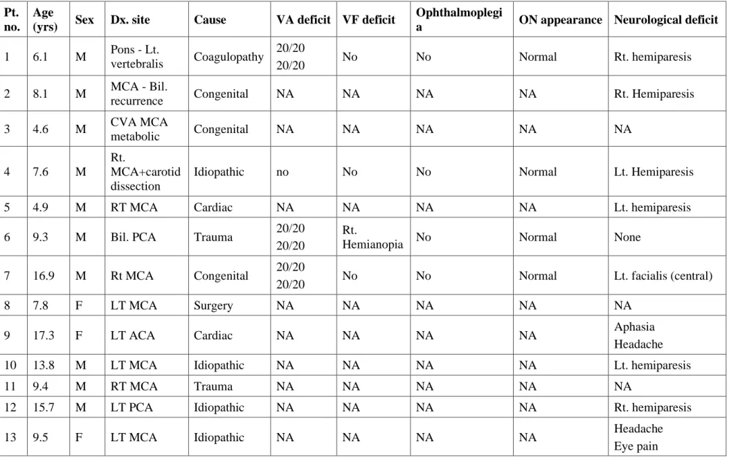
Table 1 Data for 26 patients with cerebral arterial stroke Pt.
no. Age
(yrs) Sex Dx. site Cause VA deficit VF deficit
Ophthalmoplegi
a ON appearance Neurological deficit
1 6.1 M Pons - Lt.
vertebralis Coagulopathy
20/20
20/20 No No Normal Rt. hemiparesis
2 8.1 M MCA - Bil.
recurrence Congenital NA NA NA NA Rt. Hemiparesis 3 4.6 M CVA MCA metabolic Congenital NA NA NA NA NA 4 7.6 M Rt. MCA+carotid dissection
Idiopathic no No No Normal Lt. Hemiparesis
5 4.9 M RT MCA Cardiac NA NA NA NA Lt. hemiparesis
6 9.3 M Bil. PCA Trauma 20/20 20/20
Rt.
Hemianopia No Normal None
7 16.9 M Rt MCA Congenital 20/20
20/20 No No Normal Lt. facialis (central)
8 7.8 F LT MCA Surgery NA NA NA NA NA
9 17.3 F LT ACA Cardiac NA NA NA NA Aphasia
Headache
10 13.8 M LT MCA Idiopathic NA NA NA NA Lt. hemiparesis
11 9.4 M RT MCA Trauma NA NA NA NA NA
12 15.7 M LT PCA Idiopathic NA NA NA NA Rt. hemiparesis
13 9.5 F LT MCA Idiopathic NA NA NA NA Headache
Facial asymmetry 14 10.7 F RT MCA Idiopathic 20/20 20/20 No No Swollen, structural (no papilledema) Rt. hemiparesis+Lt. facialis (central)
15 8.0 F LT MCA Idiopathic NA NA NA NA Rt. hemiparesis
16 7.0 M Lt. MCA Congenital NA NA NA NA Rt. hemiparesis
17 13.1 M LT MCA Surgery for aortic coarctation and aneurysm 20/133 20/33 Rt. Hemianopia Full motility, reduced Rt. saccades Normal disc with severe attenuated retinal arteries Rt. hemiparesis+Rt. facialis (central) Hypertension
18 10.5 F LT MCA Idiopathic NA NA NA NA Rt. hemiparesis
19 5.7 M LT MCA Cardiac NA NA NA NA Rt. hemiparesis
20 3.2 M LT MCA Congenital NA NA NA NA Focal seizure
21 7.4 M RT MCA Congenital NA NA NA NA Lt. hemiparesis
22 3.7 M RT MCA Congenital Alagille syndrome 20/20 20/20 NA NA PTC with papilledema, resolved None s/p superficial temporal artery to middle artery bypass
23 9.4 M LT MCA Congenital NA NA NA NA Rt. hemiparesis
24 5.7 F Bil. PCA Surgery for cardiac anomaly NLP NLP no fields No Normal No neurological deficit 25 5.7 M Lt. MCA Intracranial surgery chiasmal glioma 20/300 20/300 Rt. Hemianopia Rt. gaze palsy+exotropia (resolved)
Bil. optic nerve atrophy
(temporal)
Rt. hemiparesis+Rt. neglect
thalamus surgery hypothalamic / chiasmal glioma (PXA) CF Hemianopia exotropia+Lt. hypertropia Mutism
ACA, anterior cerebral artery; Bil., bilateral; Dx, diagnosis; Lt.- left; MCA, middle cerebral artery; NA, not applicable [did not have an eye exam]; NLP, no light perception; ON, optic nerve; PCA, posterior cerebral artery; PXA, pleomorphic xanthoastrocytoma; Rt.- right; VA, visual acuity; VF, visual field