Cytomegalovirus (CMV)–Specific T Cell Immunity after Renal Transplantation
Mediates Protection from CMV Disease by Limiting the Systemic Virus Load
Pierre Reusser,1,2Gieri Cathomas,3,aRudolf Attenhofer,2Michael Tamm,1and Gilbert Thiel1
1Departments of Medicine and2Research and3Institute of Pathology, University Hospital, Basel, Switzerland
The role of cytomegalovirus (CMV)–specific cytotoxic T lymphocytes (CTLs) and T helper cells (Th) in controlling CMV infection, as detected by antigenemia assay and polymerase chain reaction (PCR) in blood leukocytes, and CMV disease was investigated in 20 renal transplant recipients. Within 3 months after transplant, CMV-specific CTL and Th responses were demonstrable in 11 (55%) and 15 (75%) patients, respectively; CMV infection was detected by antigenemia and PCR in 19 (95%) patients each. During the month of first CMV detection, there was an inverse correlation between CTL response and antigenemia at>20 positive cells/ 105
leukocytes (P5 .007) but no association with lower antigenemia levels or PCR positivity. CMV disease developed in 7 (35%) patients and was associated with high-level antigenemia but was inversely correlated with detection of CTLs (P5 .04). After renal transplantation, CMV-specific CTLs limit the systemic virus load as reflected by antigenemia levels and thereby mediate protection from CMV disease.
Renal transplant recipients are at increased risk for cyto-megalovirus (CMV) infection and disease during the posttrans-plant period, when they require intensive immunosuppressive regimens for prevention of and therapy for graft rejection [1, 2]. The highest rates of CMV infection are observed among pretransplant CMV-seropositive and -seronegative patients with a seropositive kidney donor, and primary CMV disease is generally more frequent and severe than disease due to reac-tivation or reinfection [1–4].
The nature of the specific immunologic defects predisposing organ transplant recipients to CMV infection and CMV disease have been partially elucidated. Studies among patients after bone marrow or peripheral blood stem cell transplantation doc-umented a protective effect of CMV-specific cytotoxic T lym-phocytes (CTLs) obtained from peripheral blood [5–8]. Among autograft recipients, the presence of a CMV-specific major his-tocompatibility complex (MHC) class I–restricted CTL re-sponse was associated with prevention of CMV infection,
Received 13 November 1998; revised 24 March 1999; electronically pub-lished 9 July 1999.
Presented in part: 36th Interscience Conference on Antimicrobial Agents and Chemotherapy, New Orleans, September 1996 (abstract H6); 8th Eu-ropean Congress of Clinical Microbiology and Infectious Diseases, Lau-sanne, Switzerland, May 1997 (abstract P1041).
Written informed consent was obtained from each patient. The study protocol was approved by the local institutional ethical committee.
Grant support: Swiss National Research Foundation 32-31314.91 (to P.R.).
a Present affiliation: Institute of Pathology, University Hospital, Zurich, Switzerland.
Reprints or correspondence: Dr. Pierre Reusser, Dept. of Medicine, University Hospital, Petersgraben 4, CH-4031 Basel, Switzerland (reusser @ubaclu.unibas.ch).
The Journal of Infectious Diseases 1999; 180:247–53
q 1999 by the Infectious Diseases Society of America. All rights reserved.
0022-1899/99/8002-0001$02.00
whereas in the more profoundly immunodeficient allograft re-cipients, this CTL response did not correlate with suppression of CMV infection but protected against serious CMV disease [6, 7]. In renal transplant recipients, data on cytolytic T cell immunity to CMV are limited and were generated before the introduction of aggressive immunosuppressive regimens, which include cyclosporine and antithymocyte globulin (ATG) [9].
In recent years, rapid and sensitive methods for the detection of CMV in peripheral blood polymorphonuclear leukocytes (PMNL) by antigenemia assay or by polymerase chain reaction (PCR) were introduced and shown to recognize CMV infection in blood at an early stage, when the systemic virus load is still low [10–15]. The role of the specific T cell immunity during this early phase of CMV infection is unknown. The present study characterizes the MHC-restricted T cell immunity in renal transplant recipients and evaluates the association of CMV-specific CTL and T helper cell (Th) responses with the presence of CMV in peripheral blood PMNL as detected by antigenemia assay and PCR. These immune functions were also correlated with CMV disease that occurs in the first 3 months after renal transplantation.
Patients and Methods
Patient population. The investigation was conducted prospec-tively among 20 recipients of cadaveric donor or living–related donor kidney transplants at University Hospital, Basel, Switzer-land. Patients were selected for study if they were seropositive for CMV IgG antibody before transplantation or were seronegative with a seropositive kidney donor. Characteristics of the study pop-ulation are summarized in table 1. After renal transplantation, all patients received combined immunosuppressive induction treat-ment with cyclosporine, azathioprine, prednisolone, and ATG. Three patients required additional ATG for therapy of
steroid-Table 1. Characteristics of the 20 renal transplant recipients in whom cytomegalovirus (CMV)–specific T cell immunity and occur-rence of CMV infection and disease were studied within 3 months after transplantation.
Characteristic No. (range)
Median age in years 48 (19–69)
Sex (male/female) 9/11 Type of transplant Cadaveric donor 15 Living–related donor 5 Pretransplant CMV serology Donor1/recipient1 11 Donor2/recipient1 7 Donor1/recipient2 2
Patients alive 3 months after transplantation 20
resistant graft rejection. CMV-seronegative blood products were used for transfusion. No subject received prophylactic or preemp-tive treatment of CMV infection with acyclovir, ganciclovir, or foscarnet.
Generation of CMV-specific CTLs in vitro. Patients were eval-uated for the presence of CMV-specific CTLs in peripheral blood immediately before transplantation and at 1, 2, and 3 months after transplantation. CMV-specific CTLs were cultured and expanded in vitro as described [6, 7]. In brief, skin biopsies were obtained from each patient, to establish fibroblast lines for use as both stim-ulator and target cells. Fibroblast lines were grown in Waymouth’s medium (Gibco BRL/Life Technologies, Basel, Switzerland) sup-plemented with 20% heat-inactivated fetal calf serum (FCS), 2 mmol/L ofL-glutamine, 50 U/mL of penicillin, and 50 mg/mL of streptomycin. Autologous fibroblasts were plated in 6-well plates at 6cells/well and infected for 2 h with the human CMV
0.53 10
AD169 strain (American Type Culture Collection, Rockville, MD) at an MOI of 5 before initiation of lymphocyte culture.
Peripheral blood mononuclear cells (PBMC) obtained by His-topaque (Sigma, Buchs, Switzerland) gradient centrifugation were resuspended in RPMI-HEPES (Gibco BRL) supplemented with 10% CMV-seronegative human AB serum,2.53 1025mol/L of 2-mercaptoethanol, 2 mmol/L ofL-glutamine, 50 U/mL of penicillin, and 50 mg/mL of streptomycin and were dispensed at 107
cells/well in the 6-well plates containing autologous CMV-infected fibroblast stimulators. After 7 days of incubation at 377C in a humidified 5% CO2atmosphere, the cultured cells were harvested, washed, and
recultured at a ratio of 20:1 with fresh CMV-infected fibroblast stimulators and supplemented with autologous irradiated (30500
cGy) PBMC as filler cells. Two days later, recombinant interleukin-2 (Becton Dickinson, Bedford, MA) was added to the cultures, to achieve a final concentration of 2 U/mL. As demonstrated in our earlier studies [6, 7] and by others [8, 16–19], this cell culture system results in preferential activation and expansion of CMV-specific MHC class I–restricted CTLs with a CD31, CD81, CD42 phenotype.
Cytotoxicity assay. Two weeks after initiation of lymphocyte cultures, the cytotoxicity of the effector cells was assessed by 4-h
51
Cr release assay. The panel of targets used for each assay included autologous and MHC class I–mismatched CMV-infected and mock-infected fibroblasts as reported [6, 7]. Fibroblast targets were incubated before use for 48 h with recombinant interferon-g (Sche-ring, Kenilworth, NJ) at 800 U/106
cells to enhance MHC class I
expression and thereby the sensitivity of the cytotoxicity assay [17]. The targets were then labeled overnight with51
Cr at 100 mCi/106
cells (Amersham Laboratories, Amersham, UK), and an aliquot was infected with CMV AD169 at an MOI of 5. Labeled targets were harvested and suspended at 105
cells/mL in RPMI with 10% FCS, and 100 mL (104
cells) was dispensed in triplicate into 96-well round-bottom plates, together with 100 mL of effector cell suspen-sion at an effector-to-target ratio of 15:1. Cytotoxicity was also simultaneously assayed against targets preincubated with the anti–class I monoclonal antibody W6/32 (provided by G. De Lib-ero, Department of Research, University Hospital, Basel) to con-firm MHC class I restriction of target cell lysis [6, 18]. After in-cubation for 4 h, 100 mL of supernatant was harvested from each well, and radioactivity was measured in a gamma counter.
Specific cytotoxicity was calculated by the standard formula, with maximum release reflecting counts per minute (cpm) from incubation of target cells with 1% Nonidet P40-solution and spon-taneous release, which never exceeded 30% of maximum release, reflecting cpm following incubation of targets with medium alone. Investigations in 5 CMV-seropositive healthy volunteers showed a median specific lysis of autologous CMV-infected fibroblasts of 34% (range, 28%–53%; data not shown). Based on our previous results, a CMV-specific CTL response was considered positive if lysis of autologous CMV-infected fibroblast targets was15% above
the level of lysis obtained with autologous mock-infected and MHC class I–mismatched CMV-infected and mock-infected targets [6, 7].
Lymphoproliferative assay. The proliferative response to sol-uble CMV antigen, which reflects the CMV-specific MHC class II–restricted CD41Th response [8, 20], and to phytohemagglutinin
(PHA) was assessed each time a CTL culture was initiated as de-scribed [6, 7]. In brief, PBMC were suspended at 106cells/mL in
lymphocyte culture medium, and 100 mL was dispensed in triplicate into wells of 96-well round-bottom plates. Soluble CMV antigen or PHA (Murex Diagnostics Benelux, Schaffhausen, Switzerland) was added at final concentrations of 1:100 and 10 mg/mL, respec-tively, and the plates were incubated at 377C in a humidified 5% CO2atmosphere for 96 h. The cells were pulsed with 1 mCi/well of
[3
H]thymidine (Amersham Laboratories) 16 h before harvest. The wells were then harvested and samples measured in a b-scintillation counter. Results were expressed as a stimulation index calculated by dividing the mean cpm of cells exposed to CMV antigen or to PHA by the mean cpm of cells incubated with medium alone. A stimulation index>4 indicated a positive lymphoproliferative re-sponse [7].
Detection of CMV in blood by antigenemia assay and by PCR.
Patients were monitored for detection of CMV in PMNL by both antigenemia assay and nested PCR once before transplantation and at weekly intervals during the first 3 months after transplantation. While the antigenemia assay was done on the day of blood sam-pling, the specimens for PCR were cryopreserved and processed after study completion. For the antigenemia assay, 105
PMNL were cytocentrifuged in triplicate on microscopic glass slides and incu-bated at room temperature for 1 h with the anti-pp65 monoclonal antibodies C-10 and C-11 (Clonab; Biotest, Dreieich, Germany) [10]. The slides were then incubated with biotinylated horse anti-mouse serum for 30 min. We used the avidin-biotin complex tech-nique for visualization of the product using the ABC-elite kit ac-cording to the protocol of the manufacturer (Vector, Burlingame,
CA). The immunoperoxidase reaction was visualized using 3-amino-9-ethylcarbazole (Sigma) and was counterstained with he-matoxylin. Slides were screened by light microscopy, and the result was expressed as the number of antigen-positive cells/105
stained PMNL.
For the nested PCR, DNA was extracted from PMNL by di-gesting the cell pellet overnight in a buffer containing 1% SDS and 1 mg/mL of proteinase K, which was followed by phenol/chloro-form extraction and precipitation of the DNA by ethanol. The pellet was then dissolved in H2O. A DNA equivalent of 10
5
cells measured by fluorometer (Hoefer Scientific Instruments, San Fran-cisco) was used in the first round of the nested PCR assay, which contained 10 mM Tris-HCl (pH 8.3), 50 mM KCl, 1.5 mM MgCl2,
1 mM sense and antisense primer, 200 mM of each dNTP, and 1.25 U of AmpliTaq DNA polymerase (Perkin Elmer, Ueberlingen, Ger-many). Primers for the major immediate early gene region of CMV were used. In the first PCR round, the upstream sense primer MIE2783 (50-CGCCGCATTGAGGAGATCTGC) and the anti-sense downstream primer MIE-5 (50
-CACCACCATCCTCCT-CTTCCTCTGG) were applied [21]. One percent of the product of the first round was transferred to the second PCR reaction using the sense upstream primer IE-1 (50 -CCACCCGTGGTG-CCAGCTCC) and the antisense downstream primer MIE3114 (50 -GACTTGACAGACACAGTG), leading to a final PCR product of 183 bp [21]. Thirty cycles were performed in both PCR rounds, with the following cycle conditions: denaturation for 1 min at 947C, reanealing for 1 min at 557C, and extension for 55 s at 727C, with a final extension step in the last round of 7 min at 727C. All PCR products were analyzed on a 2% agarose gel and visualized by ethidium bromide stain in a UV illuminator.
Definition of CMV infection and CMV disease. CMV infection was defined as positive CMV antigenemia or detection of CMV by viral cultures or histology in clinical specimens collected at the discretion of the primary care physicians. Since the PCR results were not available for clinical decision-making, they were not used to define CMV infection. The definition of CMV disease included both a CMV syndrome and CMV organ disease. The CMV syn-drome was considered present if CMV infection was associated with unexplained fever1387C for >3 days with one of the following
factors: leukopenia!33 103/mL, thrombocytopenia!105/mL, serum
alanine aminotransferase >2.5 times the upper limit of normal, atypical lymphocytosis120%, or interstitial infiltrates on chest
ra-diograph [22–24]. CMV organ disease required the evidence of CMV in tissue specimens or for CMV pneumonia in bronchoal-veolar lavage fluid, with associated symptoms and signs.
Statistical analyses. Comparisons between multiple groups were done by one-way analysis of variance on ranks and the Krus-kal-Wallis test. For comparison of two groups, the Wilcoxon rank sum test was used for continuous variables, and Fisher’s exact test was used for dichotomous variables. P!.05 was considered significant.
Results
CMV-specific CTL response. Before transplantation, a CMV-specific CTL activity was detectable in 12 (67%) of the 18 CMV-seropositive patients and was undetectable in the 2
seronegative patients. Within 3 months after renal transplan-tation, the presence of a CMV-specific CTL response was de-monstrable in 11 (55%) of 20 patients. Of the 12 patients with pretransplant CTL activity, 8 had a detectable CMV-specific CTL response in the first 3 months after transplantation, and 4 did not. Among the 8 patients without a CTL response specific for CMV before transplantation, 3 developed this response dur-ing the posttransplant course (includdur-ing 1 of the 2 pretransplant CMV-seronegative patients), and 5 did not.
The lytic activity against autologous CMV-infected target cells was significantly higher than the level of cytotoxicity against autologous mock-infected or MHC class I–mismatched CMV-infected and mock-infected targets before transplantation (P!.001) and at 1 (P5 .03), 2 (P5 .01), and 3 months (P! ) after transplantation (figure 1). The preferential lysis of .001
autologous CMV-infected fibroblast targets over MHC-mis-matched infected targets at all time points indicates that the culture system used generated classical MHC class I–restricted CTL specific for CMV. This was further supported by the effect of the anti–class I monoclonal antibody W6/32, which reduced lysis of autologous CMV-infected fibroblast targets on average by 59% (P!.001; data not shown). Compared with the pre-transplant results, the magnitude of CMV-specific CTL activity was significantly decreased at 1 month (P5 .002) and 2 months (P5 .01) after renal transplantation but was similar 3 months after transplant (figure 1).
Lymphoproliferative response to CMV and to PHA. Before transplantation, lymphoproliferation to CMV antigen, which reflects the specific CD41 Th function, was detectable in 14 (70%) of 20 patients, and a proliferative response to PHA was present in 19 (95%) patients (figure 2). Within 3 months after transplantation, a lymphoproliferative response to CMV an-tigen was demonstrable in 15 (75%) patients, and lymphopro-liferation to PHA was detectable in all patients (figure 2). The proliferative response to CMV antigen was significantly de-pressed at 1 (P5 .007) and 2 months (P5 .01) but not at 3 months after transplantation, compared with pretransplant val-ues (figure 2). Lymphoproliferation to PHA was significantly weaker at 1 (P5 .01), 2 (P5 .03) and 3 months (P5 .008) after transplantation, compared with the pretransplantation re-sponse (figure 2). All patients who had a demonstrable CMV-specific CTL activity after transplantation had a positive lym-phoproliferative response to CMV antigen by the time of first CTL detection. There was a significant correlation between si-multaneous presence or absence of these two immune functions (P!.001).
CMV antigenemia and PCR-based detection of CMV DNA in blood. CMV antigenemia occurred in 19 (95%) of 20 pa-tients within the first 3 months after renal transplantation. Me-dian (range) time to first detection of CMV antigenemia was 26 days (10–48) after transplant. Sixteen (80%) patients had antigenemia levels>10 cells, 13 (65%) had >20 cells, and 10 (50%) had>50 cells. All patients in whom CMV was
docu-Figure 1. Cytotoxicity of cells from renal transplant recipients with detectable cytomegalovirus (CMV)–specific major histocompatibility complex (MHC) class I–restricted cytotoxic T lymphocyte (CTL) response. This CTL response was demonstrable in 12 patients before and in 11 within 3 months after transplantation. Cytotoxicity was assayed at effector-to-target ratio of 15:1 against autologous CMV-infected (A) and mock-infected (B) fibroblast targets and against MHC class I–mismatched CMV-mock-infected (C) and mock-mock-infected (D) fibroblast targets. Horizontal bar, median.
mented at sites other than blood had previous positive CMV antigenemia. During the posttransplant course, CMV DNA in PMNL was detected by PCR in 19 (95%) of 20 patients at a median (range) of 23 (8–46) days after transplantation, which was not significantly earlier than the occurrence of the first positive CMV antigenemia.
CMV antigenemia and CMV disease. During the post-transplant study period, 9 episodes of CMV disease occurred in 7 (35%) of 20 patients. Eight of these 9 episodes were di-agnosed as CMV syndrome and 1 as CMV pneumonia. Median (range) onset of the first episode of CMV disease was on day 29 (23–70) after transplantation. There was no association be-tween occurrence of a first episode of CMV disease and CMV antigenemia at any number of positive cells. However, a first episode of CMV disease developed in 7/13 patients with CMV antigenemia at>20 positive cells/105PMNL but in none of the
7 patients with lower antigenemia levels (P5 .04).
CMV-specific T cell immunity and CMV infection and dis-ease. When the association of CMV-specific CTL response with detection of CMV in blood by antigenemia assay or by PCR was evaluated independently from the timing of these events within 3 months after transplantation, no correlation between these variables was found. Because the degree of im-munosuppression may vary during the posttransplant course, a time-dependent analysis was done in which the occurrence of CMV antigenemia and of PCR positivity was correlated with the specific CTL response at the end of the month when CMV
infection was first detected by these diagnostic assays. During the month of first positive CMV antigenemia, there was no association between CMV-specific CTL response and anti-genemia at any number of positive cells. In fact, there was an inverse correlation between specific CTL activity and CMV antigenemia at >20 positive cells/105PMNL (P5 .007; table
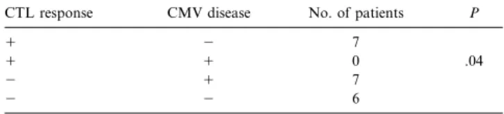
2). During the month of the first positive PCR in blood, no statistically significant association was found between CTL re-sponse and PCR positivity. Of importance, during the month of a first episode of CMV disease, the CMV-specific CTL re-sponse was inversely correlated with the occurrence of CMV disease (P5 .04; table 3).
Similar analyses were performed to evaluate the association of a CMV-specific Th response with the occurrence of CMV antigenemia or PCR positivity for CMV in blood and with the development CMV disease after transplantation. There was no statistically significant correlation between these variables whether the timing of events was considered or not (data not shown).
Discussion
This study characterizes the CMV-specific CTL and Th re-sponses in renal transplant recipients and defines the role of these effector cells in controlling both CMV infection, as de-tected by antigenemia and PCR in peripheral blood, and CMV disease. A CMV-specific MHC class I–restricted CTL activity
Table 2. Cytomegalovirus (CMV)–specific major histocompatibility complex class I–restricted CTL response at the end of the month during which CMV antigenemia was first detected at>20 positive cells/105
PMNL within 3 months after renal transplantation.
CTL response CMV antigenemia No. of patients P
1 2 5
1 1 1 .007
2 1 12
2 2 2
NOTE. CTL, cytotoxic T lymphocyte; PMNL, polymorphonuclear leukocytes.
Figure 2. Lymphoproliferative responses to cytomegalovirus (CMV) antigen and to phytohemagglutinin (PHA) among 20 renal transplant recipients. Stimulation index (SI) was calculated as described under Patients and Methods; SI> 4.0 was considered positive. Horizontal bar, median.
was demonstrable in 55% of patients in the first 3 months after transplantation and was associated with protection from high-level CMV antigenemia and from CMV disease.
Within 3 months after transplantation, CMV antigenemia developed in 95% of our patients. The antigenemia assay used for early detection of CMV infection is based on recognition of the lower matrix protein pp65 of CMV, which is present in blood leukocytes during active CMV infection [10, 14, 15, 25]. The number of antigen-positive cells in blood furthermore re-flects the systemic virus load, and high CMV antigenemia levels were shown to predict CMV disease in renal transplant recip-ients [12, 14, 15]. Investigations of the fine specificity of T cell responses to human CMV identified the CMV pp65 antigen as a major target for both CTL [26, 27] and Th cells [28–31]. Thus, the evaluation of the relationship between T cell responses spe-cific for CMV in blood and CMV antigenemia provides infor-mation on the effects of an immunodominant part of T cell immunity on the occurrence of CMV infection and on the in-crease in systemic virus load up to levels at which patients carry an elevated risk for CMV disease.
During the month of the first positive CMV antigenemia, we found no correlation between CMV-specific CTL response and antigenemia at any number of positive cells. Thus, CTLs spe-cific for CMV did not prevent CMV infection that usually results from reactivation of latent virus in the host or from acquisition of exogenous virus strains from the organ donor [3, 4]. The CMV-specific CTL response, however, was associated with protection from high-level antigenemia at >20 positive cells/105PMNL. Thus, in the period in which renal transplant
recipients require intensive immunosuppression for prophylaxis or therapy of graft rejection, these effector cells appear to con-trol CMV infection by limiting the systemic virus load.
The lack of correlation in our study between PCR-based detection of CMV DNA in blood PMNL and the presence of CTLs specific for CMV is consistent with the results observed at low levels of antigenemia. The median times to first positive CMV antigenemia and PCR in PMNL were similar, which suggests that both methods had a comparable sensitivity in detecting early CMV infection in blood. No attempt was made to quantify the virus load by PCR. Our results indicate that the occurrence of CMV infection as diagnosed by qualitative PCR in peripheral blood PMNL is not prevented by CMV-specific CTLs within 3 months after renal transplantation.
An increased systemic virus load as reflected by the antige-nemia assay predisposes to CMV disease after renal transplan-tation. Our data confirm the earlier observation that a cutoff level of 20 antigen-positive cells/105PMNL differentiates
be-Table 3. Cytomegalovirus (CMV)–specific major histocompatibility complex class I–restricted CTL response at the end of the month of a first episode of CMV disease within 3 months after renal transplantation.
CTL response CMV disease No. of patients P
1 2 7
1 1 0 .04
2 1 7
2 2 6
NOTE. CTL, cytotoxic T lymphocyte.
tween low and high risk for CMV disease [12, 14]. Most im-portantly, CMV-specific CTLs in our patients mediated pro-tection from CMV disease in the first 3 months after transplantation. The control of systemic virus load by this arm of T cell immunity thus appears to be pivotal in the prevention of CMV disease in renal transplant recipients.
In contrast, the presence of a Th response to CMV was neither associated with protection against high-level CMV anti-genemia or PCR positivity in blood nor from CMV disease, although this immune function was demonstrable in 75% of our patients during the posttransplant course. However, anti-gen-specific Th cells seemed to play an important role in the generation of a CMV-specific CTL activity, which is consistent with similar findings among recipients of allogeneic or autol-ogous bone marrow and peripheral blood stem cell transplants [6, 7, 32].
With the cell culture system used in the present investigation, MHC class I–restricted CTL specific for CMV are readily de-tectable in healthy CMV-seropositive persons who show no evidence of viral reactivation [6, 16, 18, 19]. Only 67% of our CMV-seropositive patients had a demonstrable CMV-specific CTL response before transplantation. In the pretransplant pe-riod, all patients required long-term dialysis. Impaired Th func-tions have been observed in persons with chronic renal failure and were not improved by hemodialysis [33, 34]. Studies of the effects of chronic renal failure on CMV-specific CTL activity have not been reported to date. Our results indicate that pa-tients with chronic renal failure who receive long-term dialysis have a deficient CTL immunity to CMV. The fact that CMV infection nevertheless occurs in most cases after renal trans-plantation suggests that allogeneic stimulation or more pro-found immunosuppression are necessary to reactivate CMV.
In conclusion, our data indicate that the CMV-specific MHC class I–restricted CTL response plays a crucial role in protecting renal transplant recipients from serious CMV infection by lim-iting the systemic virus load and thereby reducing the risk for CMV disease. By contrast, Th cell immunity to CMV does not seem directly involved in suppressing CMV infection but is essential for the generation of a specific CTL activity in these patients. Our results could have implications for the adoptive immunotherapy with virus-specific CTL clones, which is a promising approach to the prevention of viral infections in im-munodeficient hosts [32, 35]. The CTL response to CMV in
our patients afforded protection from high-level CMV anti-genemia, which infers that an important proportion of these effector cells in peripheral blood recognizes the pp65 antigen of CMV. Adoptive immunotherapy with CMV-specific T cell clones would be greatly facilitated if future investigations dem-onstrate that the exclusive transfer of clones against this CMV antigen mediates sufficient protection from CMV infection and CMV disease in immunocompromised patients.
Acknowledgments
We thank P. Jordan for statistical advice and consultation; C. McGandy, M. Kasper, and A. Stalder for performing the antigenemia and PCR assays; and the staff of the renal transplant outpatient unit for help in collecting the clinical specimens.
References
1. Hibberd PL, Tolkoff-Rubin NE, Cosimi AB, et al. Symptomatic cytomeg-alovirus disease in the cytomegcytomeg-alovirus antibody seropositive renal trans-plant recipient treated with OKT3. Transtrans-plantation 1992; 53:68–72. 2. Pillay D, Ali AA, Liu SF, Kops E, Sweny P, Griffiths PD. The prognostic
significance of positive CMV cultures during surveillance of renal trans-plant recipients. Transtrans-plantation 1993; 56:103–8.
3. Grundy JE, Lui SF, Super M, et al. Symptomatic cytomegalovirus infection in seropositive kidney recipients: reinfection with donor virus rather than reactivation of recipient virus. Lancet 1988; 2:132–5.
4. Reusser P. Human cytomegalovirus infection and disease after bone marrow and solid organ transplantation. Bailliere’s Clin Infect Dis 1996; 3:357–71. 5. Quinnan GV, Kirmani N, Rook AH, et al. Cytotoxic T cells in cytomega-lovirus infection: HLA-resticted T-lymphocyte and non–T-lymphocyte cy-totoxic responses correlate with recovery from cytomegalovirus infection in bone-marrow-transplant recipients. N Engl J Med 1982; 307:6–13. 6. Reusser P, Riddell SR, Meyers JD, Greenberg PD. Cytotoxic T lymphocyte
response to cytomegalovirus following human allogeneic bone marrow transplantation: pattern of recovery and correlation with cytomegalovirus infection and disease. Blood 1991; 78:1373–80.
7. Reusser P, Attenhofer R, Hebart H, Helg C, Chapuis B, Einsele H. Cyto-megalovirus-specific T-cell immunity in recipients of autologous peripheral blood stem cell or bone marrow transplants. Blood 1997; 89:3873–9. 8. Li CR, Greenberg PD, Gilbert MJ, Goodrich JM, Riddell SR. Recovery of
HLA-restricted cytomegalovirus (CMV)-specific T-cell responses after al-logeneic bone marrow transplant: correlation with CMV disease and effect of ganciclovir prophylaxis. Blood 1994; 83:1971–9.
9. Rook AH, Quinnan GV, Frederick, et al. Importance of cytotoxic lympho-cytes during cytomegalovirus infection in renal transplant recipients. Am J Med 1984; 76:385–92.
10. van der Bij W, Torensma R, van Son WJ, Schirm J, Tegzess AM, The TH. Rapid immunodiagnosis of active cytomegalovirus infection by mono-clonal antibody staining of blood leukocytes. J Med Virol 1988; 25:179–88. 11. Jiwa NM, van Gemert GW, Raap AK, et al. Rapid detection of human cytomegalovirus DNA in peripheral blood leukocytes of viremic trans-plant recipients by the polymerase chain reaction. Transtrans-plantation 1989; 48:72–6.
12. van den Berg AP, van der Bij W, van Son WJ. Cytomegalovirus antigenemia as a useful marker of symptomatic cytomegalovirus infection after renal transplantation—a report of 130 consecutive patients. Transplantation 1989; 48:991–5.
13. Einsele H, Ehninger G, Hebart H, et al. Polymerase chain reaction monitoring reduces the incidence of cytomegalovirus disease and the duration and
side effects of antiviral therapy after bone marrow transplantation. Blood 1995; 86:2815–20.
14. The TH, van der Ploeg M, van den Berg AP, Vlieger AM, van der Giessen M, van Son WJ. Direct detection of cytomegalovirus in peripheral blood leukocytes: a review of the antigenemia assay and polymerase chain re-action. Transplantation 1992; 54:193–8.
15. Meyer-Ko¨nig U, Serr A, von Laer D. Human cytomegalovirus immediate early and late transcripts in peripheral blood leukocytes: diagnostic value in renal transplant recipients. J Infect Dis 1995; 171:705–9.
16. Borysiewicz LK, Morris S, Page JD, Patrick Sissons JG. Human cytomeg-alovirus-specific cytotoxic T lymphocytes: requirements for in vitro gen-eration and specificity. Eur J Immunol 1983; 13:804–9.
17. Laubscher A, Bluestein HG, Spector SA, Zvaifler NJ. Generation of human cytomegalovirus-specific cytotoxic T lymphocytes in a short-term culture. J Immunol Methods 1988; 110:69–77.
18. Riddell SR, Greenberg PD. The use of anti-CD3 and anti-CD28 monoclonal antibodies to clone and expand human antigen-specific T cells. J Immunol Methods 1990; 128:189–201.
19. Riddell SR, Rabin M, Geballe AP, Britt WJ, Greenberg PD. Class I MHC-restricted cytotoxic T lymphocyte recognition of cells infected with human cytomegalovirus does not require endogenous viral gene expression. J Immunol 1991; 146:2795–804.
20. Gehrz RC, Fuad S, Liu YNC, Bach FH. HLA class II restriction of T helper cell response to cytomegalovirus. J Immunol 1987; 138:3145–51. 21. Chou S. Effect of interstrain variation on diagnostic DNA amplification of
the cytomegalovirus major immediate-early gene region. J Clin Microbiol 1992; 30:2307–10.
22. Snydman DR, Werner BG, Heinze-Lacey B, et al. Use of cytomegalovirus immune globulin to prevent cytomegalovirus disease in renal-transplant recipients. N Engl J Med 1987; 317:1049–54.
23. Balfour HH Jr, Chace BA, Stapleton JT, Simmons RL, Fryd DS. A ran-domized, placebo-controlled trial of oral acyclovir for the prevention of cytomegalovirus disease in recipients of renal allografts. N Engl J Med 1989; 320:1381–7.
24. Hibberd PL, Tolkoff-Rubin NE, Conti D, et al. Preemptive ganciclovir ther-apy to prevent cytomegalovirus disease in cytomegalovirus antibody-pos-itive renal transplant recipients: a randomized controlled trial. Ann Intern Med 1995; 123:18–26.
25. Grefte JMM, van der Gun BTF, Schmolke S, et al. The lower matrix protein pp65 is the principal viral antigen present in peripheral blood leukocytes during an active cytomegalovirus infection. J Gen Virol 1992; 73:2923–32. 26. Gilbert MJ, Riddell SR, Li CR, Greenberg PD. Selective interference with class I major histocompatibility complex presentation of the major im-mediate-early protein following infection with human cytomegalovirus. J Virol 1993; 67:3461–9.
27. McLaughlin-Taylor E, Pande H, Forman SJ, et al. Identification of the major late human cytomegalovirus matrix protein pp65 as a target antigen for CD81 virus-specific cytotoxic T lymphocytes. J Med Virol 1994; 43:
103–10.
28. van Zanten J, Harmsen MC, van der Meer P, et al. Proliferative T cell responses to four human cytomegalovirus-specific proteins in healthy sub-jects and solid organ transplant recipients. J Infect Dis 1995; 172:879–82. 29. Forman SJ, Zaia JA, Clark BR, et al. A 64,000 dalton matrix protein of human cytomegalovirus induces in vitro immune responses similar to those of whole viral antigen. J Immunol 1985; 134:3391–5.
30. Beninga J, Kropff B, Mach M. Comparative analysis of fourteen individual human cytomegalovirus proteins for helper T cell response. J Gen Virol 1995; 76:153–60.
31. Khattab BAM, Lindenmaier W, Frank R, Link H. Three T-cell epitopes within the C-terminal 265 amino acids of the matrix protein pp65 of human cytomegalovirus recognized by human lymphocytes. J Med Virol 1997; 52:68–76.
32. Walter EA, Greenberg PD, Gilbert MJ, et al. Reconstitution of cellular im-munity against cytomegalovirus in recipients of allogeneic bone marrow by transfer of T-cell clones from the donor. N Engl J Med 1995; 333: 1038–44.
33. Daniels JC, Sakai H, Remmers AR Jr, et al. In vitro reactivity of human lymphocytes in chronic uraemia: analysis and interpretation. Clin Exp Immunol 1971; 8:213–27.
34. Newberry WM, Sanford JP. Defective cellular immunity in renal failure: depression of reactivity of lymphocytes to phytohemagglutinin by renal failure serum. J Clin Invest 1971; 50:1262–71.
35. Riddell SR, Watanabe KS, Goodrich JM, Li CR, Agha ME, Greenberg PD. Restoration of viral immunity in immunodeficient humans by the adoptive transfer of T cell clones. Science 1992; 257:238–41.