Real-time visualization of ultrasound-guided retrobulbar
blockade: an imaging study
C. Luyet
1, U. Eichenberger
1*, B. Moriggl
3, L. Remonda
2and R. Greif
11
Department of Anaesthesiology and Pain Therapy and2Department of Diagnostic and Interventional Neuroradiology, Bern University Hospital and University of Bern, Inselspital, CH-3010 Bern, Switzerland.
3
Department of Anatomy, Histology, and Embryology, Innsbruck Medical University, A-6020 Innsbruck, Austria
*Corresponding author. E-mail: [email protected]
Background. Retrobulbar anaesthesia allows eye surgery in awake patients. Severe compli-cations of the blind techniques are reported. Ultrasound-guided needle introduction and direct visualization of the spread of local anaesthetic may improve quality and safety of retrobulbar anaesthesia. Therefore, we developed a new ultrasound-guided technique using human cadavers.
Methods. In total, 20 blocks on both sides in 10 embalmed human cadavers were performed. Using a small curved array transducer and a long-axis approach, a 22 G short bevel needle was introduced under ultrasound guidance lateral and caudal of the eyeball until the needle tip was seen 2 mm away from the optic nerve. At this point, 2 ml of contrast dye as a substitute for local anaesthetic was injected. Immediately after the injection, the spread of the contrast dye was documented by means of CT scans performed in each cadaver.
Results. The CT scans showed the distribution of the contrast dye in the muscle cone and behind the posterior sclera in all but one case. No contrast dye was found inside the optic nerve or inside the eyeball. In one case, there could be an additional trace of contrast dye behind the orbita.
Conclusions. Our new ultrasound-guided technique has the potential to improve safety and efficacy of the procedure by direct visualization of the needle placement and the distribution of the injected fluid. Furthermore, the precise injection near the optic nerve could lead to a reduction of the amount of the local anaesthetic needed with fewer related complications. Br J Anaesth 2008; 101: 855–9
Keywords: anaesthetic techniques, regional; anaesthetic techniques, regional, retrobulbar; anatomy; monitoring, ultrasound
Accepted for publication: September 5, 2008
Retrobulbar local anaesthesia for ocular surgery was devel-oped by Atkinson1 in 1961. The needle is blindly intro-duced into the part of the orbital cavity behind the globe, formed by the four recti muscles (muscle cone) and the superior and inferior oblique muscles, for close injection of local anaesthetics near the optic nerve.
Complications of this blind needle passage are rare but often devastating. Perforations of the globe,2 3 damage to the optic nerve,4persistent diplopia by direct injection of the anaesthetic into the muscle,5 – 7 and potentially fatal consequences of local anaesthetic agents on the central nervous system8 – 10are described in the literature. Analysis
of the closed claims database of the American Society of Anesthesiologists shows that eye blocks (retrobulbar and peribulbar) are the leading cause of claims compared with other peripheral nerve blocks, especially for closed claims with permanent disabling complications.11
Therefore, to reduce the risk of intraorbital or intraocular damage, other techniques have become popular.12 13 For peribulbar anaesthesia, the local anaesthetic is injected around the equator of the globe.12However, even if the risk of globe perforation is lower than the reported incidences in retrobulbar blocks, there are still a certain number of globe perforations with this technique.2 14 15 Moreover, the
peribulbar block has been shown to be less effective than the retrobulbar block.16 On the other hand, sub-Tenon’s block, also known as parabulbar or episcleral block, has been reported to be very effective for eye surgery.13 But with this block, complete akinesia of the eye cannot be achieved which is a disadvantage for operations close to the retina.17 18Moreover, severe side-effects such as globe per-forations, optic nerve damage, and brain stem anaesthesia have also been reported with the sub-Tenon’s block.19 – 23
As in other areas of regional anaesthesia,24 ultrasound-guided injection of local anaesthesia might improve the quality and safety of retrobulbar anaesthesia by visualiza-tion of the needle placement.
Therefore, we undertook this feasibility study on human cadavers with the aim of evaluating the performance of real-time ultrasound-guided visualization of the needle placement and the injection of contrast dye as a substitute for local anaesthetic. The accuracy of the ultrasound-guided technique was assessed by a CT imaging study after each retrobulbar injection. The hypothesis of the study was that the needle can be visualized and safely advanced using ultrasound guidance. The distribution of the injected contrast medium in the retrobulbar area was controlled by CT scan.
Methods
We undertook the procedure in 10 cadavers in legal custody of the Department of Anatomy, Histology and
Embryology of the Medical University, Innsbruck,
Austria, with institutional approval. A special embalming procedure, with an embalming fluid composed of ethanol, glycerol, and phenol, was performed so that the cadavers could further be used within operating courses. With this special embalming procedure, the tissue of the cadavers corresponds well to live patients and their use for sono-graphic studies has been demonstrated previously.25 – 27We restored the intraocular pressure by injection of alcohol into the globe in the case of collapsed eyes. The injection was performed with a 23 gauge needle (MicrolanceTM 3,
BD Drogheda, Ireland) 4 mm posterior to the limbus, in the inferotemporal quadrant until the globe felt plump to palpation. There were no tonometric measurements for that purpose.
Two examiners (R.G. and U. E.) performed ultrasound-guided retrobulbar blockade. In each cadaver, the right and the left eyes were randomly assigned to the examiners. The ultrasound device used was a SonoSitew
MicroMaxx (SonoSite Inc., Bothell, WA, USA) with an 11 MHz curved array transducer (C11e, 8 – 5 MHz, 11 mm broad-band curved array, SonoSite Inc.). The ultrasound trans-ducer was placed in the upper and inner quadrant of the orbita (Fig. 1) and transverse sonogram obtained showing the bulbus and the optic nerve (Fig. 2).
A 22 gauge, 5 cm ultrasound needle (Polymedic USC050-22, TE ME SA SAS, Carrie`res sur Seine, France) was introduced at the lower outer quadrant of the orbita inline to the transducer (Fig. 1) and advanced vertically until the needle tip reached the equator of the eyeball. At this point, it was swept in the direction of the optic nerve and advanced close to the nerve (distance needle – nerve: 2 mm beside the optic nerve; Fig. 2).
Once the needle was placed in the correct position, a bolus of 2 ml of diluted contrast dye (Jopamiro; 300 mg J ml21; Bracco, Milano, Italy) 1:10 in normal saline was injected through the needle, which was removed afterwards. The spread of the contrast dye was documented by means of 3.5 mm CT scans (Synergy; GE Medical Systems, Milwaukee, WI, USA) of the orbita.
Needle Optic nerve5,1
Eyeball
Fig 2The transverse sonogram shows the bulbus and the optic nerve. By advancing the needle inline with the transducer, the entire needle can be visualized and guided close to the nerve without penetrating any important structures. During injection, the spread of the fluid and the vis a tergo of the eye can be observed by real-time sonography (not shown here).
Fig 1 The curved array ultrasound transducer is placed at the upper and inner quadrant of the orbita. The needle penetrates the skin at the lower outer quadrant inline to the transducer. (Picture taken on a model instead of a cadaver).
The control CT scans were evaluated after termination of the practical part of the study by a neuroradiologist who was not otherwise involved in the study.
Results
We performed a total of 20 ultrasound-guided retrobulbar blocks in 10 cadavers (seven males and three females). The cadavers’ mean age at death was 71.2 yr (range 51– 91), mean body weight 67.9 kg (SD6.3), mean height 171.4 cm
(SD 6.3), and mean body mass index 24 (median 25, range
17–33). Because no traumatic head injury or eye injury was reported before death, we could perform the study on both eyes in all cadavers. They were placed in supine position and in all cases of alcohol had to be injected into the bulbus to restore globe pressure [1.6 (1.1) ml].
In all 20 cases, the placement of the needle could be easily visualized during the whole procedure and the final position of the needle was documented by a frozen ultra-sound image. In one case, the placement of the needle was difficult. The spread of the contrast dye around the optic nerve and an impressive anterior lifting of the eye (the so-called vis a tergo) during the injection of the 2 ml of contrast dye could be observed in all cases by real-time sonograms.
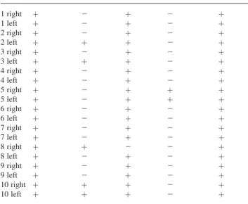
The exact distribution of the contrast dye around all 20 examined eyes is shown in Table 1. The distribution of the contrast dye was in the muscle cone and behind the pos-terior sclera in all cadavers except one. This was the cadaver where the placement of the needle had been described as difficult. In this case, traces of contrast dye
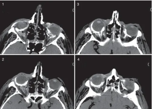
were found in the retro-orbital region but only between bone and muscle cone, without intraconal spread. The radiologist identified a hypertrophic bone close to the maxillary sinus as evidence of chronic sinusitis with small-sized retro-orbital space, which could explain the difficulty in needle placement. No contrast dye was found in the optic nerves. In another case, the radiologist could not exclude contrast dye in the eyeball. Since this eyeball contained air bubbles as a sign of insufficient firmness and the contrast dye was found predominantly in the medial area opposite the needle placement, this was interpreted as extrabulbar contrast dye. In all other cases, intrabulbar contrast dye could be excluded. In another case, a trace of contrast dye seemed to be in the retro-orbital area of the planum sphenoidale region with a slight predominance to the left side only on two CT images. Movement of con-trast dye across the superior orbital fissure was possible in cadavers. In all other cases, there was no contrast dye behind the orbita. Figure 3 shows an example of a CT scan after injection of contrast dye.
Discussion
For the first time, we have demonstrated with this study that retrobulbar block can be performed safely and with high accuracy using ultrasound guidance. The verification of the distribution of the injected contrast dye by CT scan showed a perfect distribution around the optic nerve in all but one case. Real-time ultrasound guidance provides the operator visualization of the eye and the optic nerve before and during the insertion of the needle, and during injection.
Ultrasound imaging for regional anaesthesia has had a huge impact on the technique and performance in the clinical practice of our speciality in the last few years. Up to now, there have been no studies evaluating imaging of needle placement and injection of local anaesthetics during regional anaesthetic procedures for eye surgery.
Even if blindly performed, retrobulbar anaesthesia is
known to be safe with a complication rate ,1%,28 but
visualization of the needle with ultrasound can further improve safety and efficacy. The risk of ocular perforation is particularly high in eyes with high myopia because of the increased axial length.3 14 This should no longer be a problem when using ultrasound guidance because the needle tip can be tracked and guided along the sclera.
We injected only 2 ml which resulted in a consistent spread into the muscle cone and a perfect distribution of the contrast dye around the targeted structure (optic nerve). Compared with the amount of local anaesthetics recommended in the literature, of 3 – 10 ml8 16 and our usual practice of 4 ml, this is a reduction of about 50%. Even with this reduced amount of contrast dye, there was an additional spreading of the contrast dye in the prebulbar area in all eyes (Fig. 3). The contrast dye probably spreads Table 1Distribution of contrast dye in each block. In all cases, we found
retrobulbar contrast dye but also contrast dye in the prebulbar region, suggesting backwards spread along the needle track. In one cadaver, the muscle cone was missed and in one cadaver there is a trace of contrast dye into the retro-orbital area (region of the planum sphenoidale)
Eye no. Retrobulbar Extraconal Intraconal Retro-orbital Prebulbar
1 right þ 2 þ 2 þ 1 left þ 2 þ 2 þ 2 right þ 2 þ 2 þ 2 left þ þ þ 2 þ 3 right þ 2 þ 2 þ 3 left þ þ þ 2 þ 4 right þ 2 þ 2 þ 4 left þ 2 þ 2 þ 5 right þ 2 þ þ þ 5 left þ 2 þ þ þ 6 right þ 2 þ 2 þ 6 left þ 2 þ 2 þ 7 right þ 2 þ 2 þ 7 left þ 2 þ 2 þ 8 right þ þ 2 2 þ 8 left þ 2 þ 2 þ 9 right þ 2 þ 2 þ 9 left þ 2 þ 2 þ 10 right þ þ þ 2 þ 10 left þ þ þ 2 þ
backwards along the needle track as identified by real-time sonographic observation of the dye spread during the injection. If this precise, close, and safe application of local anaesthetic to the optic nerve results in a clinically important reduction of the injected volume, then this must be proven in further investigations. Since this is a study on cadavers without clinical substrate, we do not know whether only injecting the local anaesthetics around the nerve would lead to the required akinesia and analgesia. On the other hand, reducing the volume might also reduce the risk of toxic systemic complications with grand-mal seizures or brain stem anaesthesia.8 – 10
One limitation of the study is the quality of the CT scans and the relatively small number of CT scans for interpret-ation of the retrobulbar area. Moreover, even when the con-trast dye is diluted, its density is high and hinders a more precise interpretation of the contrast dye spread. Because of these limitations, our radiologist could not definitely rule out an intrabulbar injection in one case and a retro-orbital spread of dye in another case. Moreover, the reason for retro-orbital spread could also be due to the destruction of the barriers after death. It is also not possible to exclude an additional episcleral spread (spread between sclera and vit-reous body) in all cases, although this is improbable.
Obviously, our results in cadavers have to be confirmed in patients including clinical effect, and satisfaction with the method from the new patients, the surgeons, and anaesthetists.
However, as ultrasound-guided regional anaesthesia tech-niques are becoming more popular, retrobulbar anaesthesia, with real-time visualization may improve the incidence of nerve or eye.
Funding
Only departmental research grants were spent on this research project. The ultrasound device was generously provided by SONOSITE AUSTRIA, Sittendorf, Austria.
Acknowledgements
We thank the staff of the Department of Anatomy, Histology and Embryology of the Medical University of Innsbruck for providing us with the cadavers and the locations for the study. Rupert Gstrein (anatomy technician) helped with the cadavers throughout the study and Bernd Kofler (radiology technician) operated the CT scanner. Special thanks to Jeff Crowder for proofreading the English of this manuscript.
References
1 Atkinson WS. The development of ophthalmic anesthesia. Am J Ophthalmol 1961; 51: 1 – 14
2 Wearne MJ, Flaxel CJ, Gray P, Sullivan PM, Cooling RJ. Vitreoretinal surgery after inadvertent globe penetration during local ocular anesthesia. Ophthalmology 1998; 105: 371 – 6 3 Ramsay RC, Knobloch WH. Ocular perforation following
retro-bulbar anesthesia for retinal detachment surgery. Am J Ophthalmol 1978; 86: 61 – 4
4 Pautler SE, Grizzard WS, Thompson LN, Wing GL. Blindness from retrobulbar injection into the optic nerve. Ophthalmic Surg 1986; 17: 334 – 7
5 Gomez-Arnau JI, Yanguela J, Gonzalez A, et al. Anaesthesia-related diplopia after cataract surgery. Br J Anaesth 2003; 90: 189 – 93
6 Golnik KC, West CE, Kaye E, Corcoran KT, Cionni RJ. Incidence of ocular misalignment and diplopia after uneventful cataract surgery. J Cataract Refract Surg 2000; 26: 1205 – 9
1 3
2 4
Fig 3 On the four images of this representative CT scan, the spread of the contrast dye can be seen. The optic nerve of the left eye is surrounded by contrast dye on image 2. This is also visible for the right eye on image 3, where additional spread around the eye up to the prebulbar area is seen.
7 Johnson DA. Persistent vertical binocular diplopia after cataract surgery. Am J Ophthalmol 2001; 132: 831 – 5
8 Javitt JC, Addiego R, Friedberg HL, Libonati MM, Leahy JJ. Brain stem anesthesia after retrobulbar block. Ophthalmology 1987; 94: 718 – 24
9 Brookshire GL, Gleitsmann KY, Schenk EC. Life-threatening com-plication of retrobulbar block. A hypothesis. Ophthalmology 1986; 93: 1476 – 8
10 Rosenblatt RM, May DR, Barsoumian K. Cardiopulmonary arrest after retrobular block. Am J Ophthalmol 1980; 90: 425 – 7 11 Lee LA, Domino KB. Complications associated with peripheral
nerve blocks: lessons from the ASA closed claims project. Int Anesthesiol Clin 2005; 43: 111 – 8
12 Davis DB, 2nd, Mandel MR. Posterior peribulbar anesthesia: an alternative to retrobulbar anesthesia. J Cataract Refract Surg 1986; 12: 182 – 4
13 Stevens JD. A new local anesthesia technique for cataract extrac-tion by one quadrant sub-Tenon’s infiltraextrac-tion. Br J Ophthalmol 1992; 76: 670 – 4
14 Hay A, Flynn HW Jr, Hoffman JI, Rivera AH. Needle penetration of the globe during retrobulbar and peribulbar injections. Ophthalmology 1991; 98: 1017 – 24
15 Wadood AC, Dhillon B, Singh J. Inadvertent ocular perforation and intravitreal injection of an anesthetic agent during retro-bulbar injection. J Cataract Refract Surg 2002; 28: 562 – 5
16 Hamilton RC, Gimbel HV, Strunin L. Regional anaesthesia for 12,000 cataract extraction and intraocular lens implantation pro-cedures. Can J Anaesth 1988; 35: 615 – 23
17 Kumar CM, Dodds C, McLure H, Chabria R. A comparison of three sub-Tenon’s cannulae. Eye 2004; 18: 873 – 6
18 Kumar CM, Dodds C. Evaluation of the Greenbaum sub-Tenon’s block. Br J Anaesth 2001; 87: 631 – 3
19 Kumar CM, Dowd TC. Complications of ophthalmic regional blocks: their treatment and prevention. Ophthalmologica 2006; 220: 73 – 82
20 Frieman BJ, Friedberg MA. Globe perforation associated with subtenon’s anesthesia. Am J Ophthalmol 2001; 131: 520 – 1 21 Ruschen H, Bremner FD, Carr C. Complications after sub-Tenon’s
eye block. Anesth Analg 2003; 96: 273 – 7, table of contents 22 Kim SK, Andreoli CM, Rizzo JF, 3rd, Golden MA, Bradbury MJ.
Optic neuropathy secondary to sub-tenon anesthetic injection in cataract surgery. Arch Ophthalmol 2003; 121: 907 – 9
23 Quantock CL, Goswami T. Death potentially secondary to sub-Tenon’s block. Anaesthesia 2007; 62: 175 – 7
24 Marhofer P, Chan VW. Ultrasound-guided regional anesthesia: current concepts and future trends. Anesth Analg 2007; 104: 1265 – 9, tables of contents
25 Greher M, Kirchmair L, Enna B, et al. Ultrasound-guided lumbar facet nerve block: accuracy of a new technique confirmed by computed tomography. Anesthesiology 2004; 101: 1195 – 200 26 Eichenberger U, Greher M, Kirchmair L, Curatolo M, Moriggl B.
Ultrasound-guided blocks of the ilioinguinal and iliohypogastric nerve: accuracy of a selective new technique confirmed by ana-tomical dissection. Br J Anaesth 2006; 97: 238 – 43
27 Kirchmair L, Entner T, Kapral S, Mitterschiffthaler G. Ultrasound guidance for the psoas compartment block: an imaging study. Anesth Analg 2002; 94: 706 – 10, table of contents
28 Davis DB, 2nd, Mandel MR. Efficacy and complication rate of 16,224 consecutive peribulbar blocks. A prospective multicenter study. J Cataract Refract Surg 1994; 20: 327 – 37