971
Differential Regulation of Inducible Nitric Oxide Synthase Production in Bovine
and Caprine Macrophages
Heiko Adler,
*
Barbara Adler,*
Paola Peveri,*
Ernst R. Werner, Helmut Wachter, Ernst Peterhans, and Thomas W. JungiInstitute of Veterinary Virology, University of Berne, Berne, Switzerland; Institute for Medical Chemistry and Biochemistry, University of Innsbruck, Innsbruck, Austria.
Inducible nitric oxide synthase (iNOS) regulation in human and murine macrophages in vitro differs considerably. In this study, expression of macrophage iNOS in ruminants was addressed. Nitric oxide (NO) output by cattle and goat macrophages was as different as that by human and mouse macrophages. Bovine macrophages activated by heatedSalmonella dublinor lipopolysaccha-ride (LPS) expressed high levels of iNOS mRNA, protein, and enzyme activity. Analogously cultured caprine macrophages did not respond to these and other activators by NO generation and iNOS expression. The lack of response was not due to general unresponsiveness to stimuli. Caprine iNOS mRNA was induced by stimulation of caprine macrophages with LPS, as shown by reverse transcription polymerase chain reaction. The level of mRNA expression in activated goat macro-phages was lower than in resting bovine macromacro-phages. A caprine 372-bp iNOS mRNA fragment that was sequenced closely resembled the bovine counterpart. This points to species-specific iNOS gene regulation.
The term "activated macrophages," as originally defined, describes a state in which macrophages express enhanced mi-crobicidal activity toward intracellular pathogens and tumori-static and tumoricidal activity [1, 2]. This cellular state is char-acterized by induction and up-regulation of transcription, alterations at the posttranscriptional level, and activation of enzyme cascades. Studies in rodents showed that the key bio-chemical pathway for macrophage activation is the induction of a high-output inducible nitric oxide synthase (iNOS) that is induced by activation with lipopolysaccharide (LPS) and interferon-y (IFN-y), either alone or combined [3]. The en-zyme iNOS catalyzes the conversion of arginine to citrulline and nitric oxide (NO) [4]. NO, a highly reactive nonpolar gas, is a versatile biologic mediator [5, 6]. The production of high concentrations of NO by activated macrophages leads to an array of biochemical reactions culminating in the inactivation of key enzymes of target pathogens or tumor cells [3, 7].
The above concept was questioned when it was realized that human macrophages do not generate NO under conventional
Received 14 September 1995; revised 27 November 1995.
Presented in part: spring meeting, Society ofImmunology, Heidelberg, Ger-many, 10 March 1994; 30th national meeting, Society for Leukocyte Biology, Tucson, Arizona, 23 September 1994; 8th annual conference, European Macro-phage Study Group, Lausanne, Switzerland, 28 September 1994.
H.A. and B.A. equally contributed to this work.
Financial support: Swiss National Foundation (32-32.450.92 and 3100-039733.9311), Swiss Federal Veterinary Office and Austrian "Fonds zur F6rder-ung der wissenschaftlichen ForschF6rder-ung" (project 9685), and Deutscher Akade-mischer Austauschdienst (H.A.).
Reprints or correspondence: Dr. T. W. Jungi, Institute of Veterinary Virol-ogy, Laenggass-Strasse 122, CH-3012 Berne, Switzerland.
*Present affiliations: Dana Farber Cancer Institute, Boston (H.A. and B.A.); Institute of Animal Breeding, University of Berne, Switzerland (P.P.). The Journal of Infectious Diseases 1996; 173:971-8
©1996 by The University of Chicago. All rights reserved. 0022-1899/96/7304-0026$01.00
activation conditions. Although a stimulatory regime leading to enhanced antimicrobial or tumoricidal activity was very sim-ilar for murine and human mononuclear phagocytes, activation was associated with NO generation in murine but not human macrophages [8, 9]. Some studies reported iNOS induction, NO induction, or both in human monocytes or macrophages [10-14], but the activation conditions reported could not be duplicated by others or were much more restricted than those reported for rodent macrophages. This marked species differ-ence raised the possibility that in evolution, rodent and primate macrophages acquired distinct biochemical pathways mediat-ing antimicrobial and antitumoral activity. Itwas of interest, therefore, to study the iNOS pathway in other species. We recently showed that bovine bone marrow-derived macro-phages and monocyte-derived macromacro-phages produce NO upon stimulation with bacterial constituents, such as LPS [15]. A study on cytokine control of bovine and murine macrophage iNOS showed that iNOS induction is more restricted in bovine macrophages [16]. In the present study, we compared iNOS expression and NO generation in activated macrophages from 2 closely related ruminant species, cattle and goat.
Materials and Methods
Reagents. Recombinant bovine (rbo) IFN-y and rbo tumor necrosis factor-a (TNF-a)were provided by Ciba-Geigy (Basel, Switzerland). The bioactivity of these cytokines was ascertained as described [16]. rbo granulocyte-macrophage colony-stimulating factor (GM-CSF) was from American Cyanamid (Princeton, NJ). Fetal calf serum (FCS) oflow endotoxin content was from Biologi-cal Industries (Kibbutz Beth Haemek, Israel), and goat serum was from Sigma (no. 6898; St. Louis). Cell culture media and additives were purchased from Seromed Biochrom (Munich) or from Life Technologies (Basel). Medium with low endotoxin content
«
10pg/mL LPS bioactivity) was selected. LPS (Escherichia coli 055:B5) was obtained from Sigma (no. L2637). Salmonella dublin and Listeria monocytogenes were provided by the Institute of Vet-erinary Bacteriology (University of Berne) and were prepared and heat-inactivated (120 min at 60°C) as described [15]. The nitrate reductase (NAD[P]H) from Aspergillus species was obtained from Sigma (no. 7265). Monoclonal and polyclonal antibodies specific for mouse macrophage iNOS were from Transduction Laboratories (no. 32020 and 32030; Lexington, KY). In lysates of activated bovine macrophages, monoclonal anti-iNOS stained a band repre-senting iNOS. Polyclonal anti-iNOS strongly stained several other LPS-induced bands but reacted only weakly with bovine iNOS and therefore was not used. D,L-kynurenine and L-tryptophan for calibration of high pressure liquid chromatography (HPLC) were from Serva (Heidelberg, Germany), and acetonitrile and potassium phosphates for preparation of the eluent were from Merck (Darms-tadt, Germany).
Culture of bovine and caprine macrophages. Bovine periph-eral blood monocyte-derived macrophages (MDMs) were generated by isolation of peripheral blood mononuclear cells by centrifugation over ficoll-hypaque and by selective adherence to tissue-culture flasks. The adherent cells were dislodged after an overnight culture by replacing the medium with PBS and chilling (4°C, 30 min) and vortexing the flasks. Recovered cells were washed once, resuspended in fresh medium, and put into Teflon bags for 6 or 7 days, at which time they had matured to macro-phages. The cells were cultured in Iscove's medium without phenol red but containing HEPES (10mM,pH 7.4), L-glutamine (2mM), nonessential amino acids (Life Technologies; 1% vol/vol), MEM vitamin solution (Life Technologies; 1% vol/vol), penicillin (100 U/mL), streptomycin (100 ttg/mL), amphotericin B (2.5 ttg/mL), ,B-mercaptoethanol (50ttM),and 20% FCS.
Caprine MDMs were prepared and cultured either as described above for bovine MDMs or as follows: Goat mononuclear cells were prepared from buffy coats of citrated blood and separated from neutrophils by a ficoll-hypaque gradient. Contaminating erythrocytes were lysed using a solution of 155 mMN~Cl,10 mMKHC03,and 0.1 mMEDTA, pH 7.4. The mononuclear cells were washed three times with PBS, and macrophages were derived by culturing the cells for 10-12 days in Teflon bags. The culture medium was RPMI 1640 containing HEPES (10mM, pH 7.4), L-glutamine (2 mM),penicillin (100 U/mL), streptomycin (100ttg/
mL), ,B-mercaptoethanol (50ttM), and 10% goat serum. Parallel experiments assured that differences in isolation methods did not influence NO generation by goat or bovine cells.
Bovine and caprine cells that had been cultured in suspension were subcultured in 96- or 24-well culture plates or 25-cm2
flasks, in which they were exposed to cytokines or bacterial agents (or both). Iscove's medium supplemented as described above and con-taining 2% FCS was used for subculture.
Nitrite determination. After MDMs (105cells/well in culture medium with 2% FCS) were subcultured for the times indicated, 100 ttL of cell-free supernatants or sodium nitrite standard dilu-tions were mixed with 50 ttL of Griess reagent (1 % sulfanilamide, 0.1 % naphthyl ethylenediamine dihydrochloride, and 1.25% H3P04) and incubated for 10 min at room temperature before N02 concentrations were determined by measuring optical density at 550 nm in an ELISA reader. The detection limit of the test was
2 ttMN02.
Conversion of nitrate into nitrite. Nitrate was converted into nitrite by use of nitrate reductase (Sigma; 0.4 U/well for 18 h in the presence of 500ttM ,B-NADPH)and then assayed as nitrite. Using these conditions, the detection limit of the test was ~4ttM
NO) as determined by the conversion of sodium nitrate standard dilutions, which were included in the test.
Detection of iNOS by immunoblotting. Cells were washed in PBS and then disrupted by sonication in a sample buffer containing 6% SDS and 10% mercaptoethanol. The proteins were separated on polyacrylamide gels and electrophoretically transferred to a Hybond C membrane (Amersham, Arlington Heights, IL). The blot was incubated with a monoclonal anti-murine iNOS antibody (Transduction Laboratories) and stained using a peroxidase-conju-gated sheep anti-mouse antibody and a chemiluminescence protein detection method (Amersham). Total cellular protein was deter-mined by measuring the optical density at 280 nm.
RNA isolation. For analysis of iNOS mRNA, bovine and cap-rine MDMs were cultured in 25-cm2
flasks, and total cellular RNA was isolated from the cell monolayers using a protocol optimized for TRIzol (Life Technologies).
Analysis of mRNA by reverse transcription-polymerase chain reaction (RT-PCR) and Southern blotting. First-strand cDNAs were synthesized by incubating 5ttgof total RNA from bovine or caprine MDMs with 10nMof the appropriate antisense primer in a 50-ttL reaction volume containing 50 mM TRIS-HCL (pH 8.3), 1 mM MgCh, 75 mM KCI, 1O.mM dithiothreitol, 40 U of RNasin (Promega, Madison, WI), 0.5 mM dNTPs, and 8 U of AMV (avian myeloblastosis virus) reverse transcriptase (Pro-mega). The mixtures were incubated for 1.5 h at 42°C. The PCR reaction contained 10 mM TRIS-HCI (pH 9.0), 50 mM KCI, 1.5
mM MgCh, 0.1% (wt/vol) gelatin, 1% (vol/vol) Triton X-100, 0.25 mM dNTPs, 0.2 mM sense and antisense primer, 1 U of Taq
polymerase (Stehelin, Basel), and 5 mL of RT reaction in a 100-mL volume. A temperature profile of 30 s at 94°C, 1 min at 56°C, and 1 min at 74°C was used for 30 cycles. Bovine iNOS primers
(372-bp product) were sense
5'-TAGAGGAACATCTGG-CCAGG-3' and antisense 5'-TGGCAGGGTCCCCTCTG ATG-3'-[16]. Human glyceraldehY,de-3-phosphate dehydrogenase (GAPDH) primers (356-bp product) were sense 5'-GAGATG-ATGACCCTT TTGGC-3' and antisense 5'-GTGAAGGTCGGA-GTCAACG-3' [17].
The PCR products were fractionated on agarose gels and blotted onto a positively charged nylon membrane (Boehringer, Mann-heim, Germany), and the blot was processed with a digoxigenin-labeled iNOS-specific RNA probe [16] and a digoxigenin-Iabeled GAPDH-specific RNA probe using the digoxigenin detection kit (Boehringer).
DNA sequencing. The 372-bp iNOS RT-PCR amplification product was cloned into pBluescript SK
+
(Stratagene, La Jolla, CA) and was sequenced on both strands by use of the dideoxy-nucleotide chain termination method with T7 DNA polymerase (Sequenase, United States Biochemicals, Cleveland) and synthetic oligonucleotide primers.Cytokine assays. CaprineTNF-a activity was determined by a recently described cytotoxicity assay, using porcine PK(15) cells [18]. Cytotoxicity of the PK(15) cells by supernatants of caprine macrophage cultures could be prevented in vitro by a murine monoclonal anti-bovine TNF-a antibody, confirming that this assay is specific forTNF-a [19].
JID 1996; 173 (April) NO Synthase of Ruminant Macrophages 973
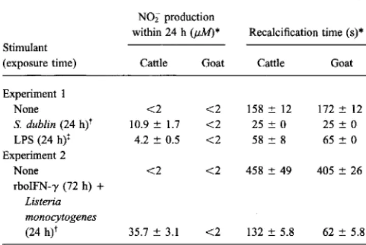
Table1. Generation ofN02"by bovine and caprine monocyte-de-rived macrophages stimulated by Salmonella dublin or lipopoly-saccharide (LPS).
NOTE. Bovine or caprine monocyte-derived macrophages were harvested from Teflon bags and subcultured in microtiter plates (l05 cells/well), in which they were exposed for indicated time to various activating stimuli. Next, nitrite was measured in supernatants, and recalcification time was determined for cell monolayers.
* Means of triplicates±SD of 2 representative experiments of 19 for goats and >40 for cattle.
t200jLg/mL.
t1 jLg/mL.
stimulated and LPS-stimulated cells displayed enhanced peA, as determined by a recalcification-time assay (table 1), and similar results were obtained after stimulation with rboIFN-y, rboTNF-O',L.monocytogenes(data not shown), andL.
monocy-togenesand rboIFN-y combined (table 1). Enhanced peA is a hallmark of activated macrophages.
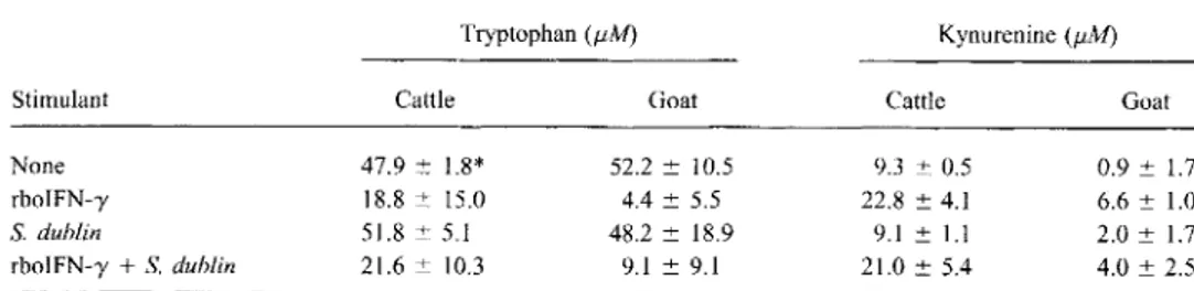
We also tested for the presence of an enzyme known to be induced by IFN-y, indoleamine 2,3-dioxygenase (IDO), which cleaves thepyrrole ring from tryptophan and other indole-amines, thereby generating variOl,ls tryptophan metabolites of the kynurenine pathway [22]. The IDO pathway appears to be involved in the destruction of some intracellular parasites [23]. IDO activity was evidenced by determining the levels of kyn-urenine in the supernatants of stimulated and unstimulated mac-rophages. Decreases in tryptophan and increases in kynurenine levels were noted in supernatants of stimulated but not unstimu-lated macrophages of either species (table 2). Thus, rboIFN-y could activate both bovine and caprine macrophages.
Conversion of nitrate into nitrite using nitrate reductase.
Since NO is an unstable product converted to either nitrite or nitrate, depending on local conditions, the possibility was considered that in cultures of goat macrophages, NO is con-verted to nitrate rather than nitrite. Supernatants of activated macrophages from both cattle and goats were exposed to nitrate reductase followed by the Griess reaction. In cattle, nitrite was increased 2- to 4-fold by nitrate reductase treatment (data not shown). No increase was obtained with goat supernatants, dis-counting the possibility that NO was generated but went unde-tected in simple nitrite determination experiments.
Cattle Goat Cattle Goat
<2 <2 158± 12 172±12 10.9± 1.7 <2 25±0 25±0 4.2±0.5 <2 58±8 65±0 <2 <2 458±49 405±26 62±5.8 Recalcification time (s)* 132±5.8 <2 N02 production within 24 h(J.lM)* 35.7±3.1 Experiment1 None S.dublin (24 h)t LPS (24 h)t Experiment 2 None rboIFN-y (72 h)+ Listeria monocytogenes (24 h)t Stimulant (exposure time)
Measurement ofprocoagulant activity. Expression ofprocoag-ulant activity (PCA) by intact macrophages was measured in mi-crotiter plates, using the turbidimetric, kinetic plasma recalcifica-tion time assay as described [20].
Detection of tryptophan and kynurenine in cell-free superna-tants. The presence of tryptophan and kynurenine was deter-mined by reversed-phase HPLC by use of 1050 HPLC equipment (Hewlett Packard, Vienna) and a 125-mm-long, 4-mm-diameter, 5-mm particle size lichrospher RP-18 column (Merck 50943) equipped with a 4X 4-mm precolumn of the same material. Cell-free supernatant (50 mL) was injected and eluted isocratically at a flow rate of 0.8 mL/min with 15 mMpotassium phosphate, pH 6.4, containing 1.8% (vol/vol) acetonitrile. Tryptophan was detected by UV absorption at 280 nm (detection limit, 0.5 mM), and kynurenine was detected at 360 nm (detection limit, 0.3mM).
Infection with bovine viral diarrhea virus (BVDV) and staining for viral antigen. BVDV infections of bovine or caprine MDMs and detection of virus-infected cells were performed as previously described [21].
Limulus amebocyte lysate test. All reagents to which macro-phages were exposed were subjected to a kinetic limulus amebocyte lysate assay. When LPS from E. coli 055:B5 (Sigma) was used as a standard, the test had a sensitivity of 2 pg/mL. Sensitivity was lower for sera and complex media (threshold of sensitivity, 10-40 pg/mL, depending on the agent). Samples were measured at various concentrations with or without the addition of a 50-pg LPS spike; a spike recovery between 75% and 125% was considered acceptable.
Results
Bovine mononuclear phagocytes produce nitrite upon bacte-rial stimulation. Bovine and caprine monocytes were allowed to differentiate into macrophages in vitro by use of a nonadher-ent (Teflon-foil based) culture system. Macrophages were har-vested and subcultured under identical conditions in microtiter plates. Bovine and caprine MDMs were either mock-stimulated or stimulated with heat-killedS.dublinor purified LPS(E. coli
055:B5). Nitrite was determined in 24-h supernatants by use of Griess reagent. BothS. dublinand LPS induced high levels of nitrite in supernatants of bovine MDMs, but neither did so in caprine MDMs (table 1, 1st experiment). Nitrite production by bovine MDMs was suppressed by coincubation with N°-monomethyl-L-arginine (data not shown). Even after prolonged stimulation withS. dublin, nitrite was detected in supernatants of bovine but not of caprine MDMs (data not shown).
Unresponsiveness of goat macrophages is restricted to NO generation. The inability of caprine cells to produce nitrite was not due to a general lack of activation by the above-mentioned stimuli, as shown by a variety of functions measured in addition to nitrite generation. Thus, although stimulated cap-rine macrophages failed to produce NO, they produced copious amounts of TNF-a. On average, supernatants ofS.
dublin-exposed goat cells caused 85% ± 4% cell lysis in a PK(15) cytotoxicity assay (n
=
3), whereas supernatants of unstimu-lated cells failed to lyse these target cells. Both S.dublin-Table 2. Induction of indoleamine 2,3-dioxygenase in cattle and goat macrophages stimulated by recombinant bovine interferon-y (rboIFN-y) and Salmonella dublin.
Tryptophan(J.1,M) Kynurenine(f.lM)
Stimulant Cattle Goat Cattle Goat
None rboIFN-y S.dublin rboIFN-y+S.dublin 47.9 ::t: 1.8* 18.8 ::t: 15.0 51.8 ::t: 5.1 2\.6 ::t: 10.3 52.2 ::t: 10.5 4.4 ::t: 5.5 48.2 ::t: 18.9 9.1 ::t: 9.1 9.3 ::t: 0.5 22.8 ::t: 4.1 9.1 ::t: 1.1 21.0 ::t: 5.4 0.9 ::t:1.7 6.6 ::t: 1.0 2.0 ::t: 1.7 4.0::t: 2.5 NOTE. Bovine or caprine monocyte-derived macrophages were cultured in Teflon bags (l06 cells/mL) and stimulated with rboIFN-y (1000 UlmL for 72 h) and with heat-killedS.dublin (200 J.1g/mL for last 24 h). Tryptophan and kynurenine were determined in cell-free supernatants as described in Materials and Methods.
* Values represent means ::t: SD(n= 2 for cattle and 4 for goats).
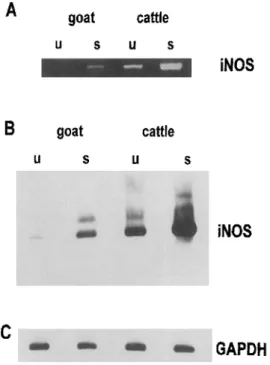
Figure 1. Detection of iNOS by immunoblotting. Monoclonal anti-body to murine macrophage iNOS was used for detection in cattle lysates and goat monocyte-derived macrophages. Cells were unstim-ulated (u) or stimunstim-ulated (s) with heat-inactivated Salmonella dublin
(200 j.Lg/mLfor 48 h). Celllysates (10 j.Lg/lane) were fractionated by PAGE, transferred to nitrocellulose, and stained. Antigen was detected by enhanced chemiluminescence. Arrow, position of iNOS (~130 kDa). Protein molecular mass markers are indicated in kDa. Positive control for iNOS (lysate of stimulated mouse macrophages) was loaded in lane c.
body (MAb). iNOS protein was detected by the MAb in stimu-lated bovine but not in unstimustimu-lated bovine macrophages or caprine MDMs, regardless of the state of activation (figure 1).
Detection of iNOS mRNA. Using iNOS-specific primers, a PCR signal was detected in unstimulated bovine but not caprine macrophages. After induction by S. dublin, a clear increase in the iNOS mRNA was detected by RT-PCR in bovine macro-phages, and a faint signal was visible also in S. dublin-stimu-lated caprine MDMs (figure 2A). To verify the RT-PCR prod-ucts, the bands were stained with a specific bovine iNOS probe [16] in a Southern blot (figure 2B). This confirmed that the iNOS-specific signal in activated caprine macrophages was stronger than in resting counterparts but weaker than in resting bovine MDMs. Of note, in the latter, NO production could not be detected by the Griess assay.
Cloning and sequencing of a caprine iNOS fragment. The previous experiment suggested that an iNOS transcript was
cattle
s
goatu
s
u
c
87 199-120-Assessment of various conditions activating goat and cattle mononuclear phagocytes. Since macrophage iNOS induction may not depend solely on LPS but may need additional priming signals, goat macrophages were pretreated with various cyto-kines (rboIFN-y, rboTNF-a, and rboGM-CSF) either alone or followed by stimulation with LPS. None of these regimes in-duced NO generation in goat macrophages (data not shown). Another treatment that induced high levels of NO in bovine macrophages, the combination of rboIFN-y and L.
monocyto-genes, failed to trigger NO production in goat MDMs (table 1, 2nd experiment).
Infection of bovine macrophages by noncytopathic BVDV primed these cells for enhanced iNOS response upon bacterial stimulation [21]. To test whether a similar priming rendered caprine macrophages responsive to bacterial stimuli, caprine MDMs were infected with noncytopathic BVDV, and virus infection was checked by staining for viral antigen. Although goat cells were readily infected by BVDV, as detected by immunoenzymatic staining of cells for viral antigen, no nitrite was detected in supernatants of virus-infected, S. dublin-stim-ulated goat cells (data not shown).
Because of our earlier observation that the absolute amount of nitrite produced may be dependent on the culture conditions of macrophages and particularly on the serum source used [16], bovine and caprine MDMs were cultured under identical conditions using either FCS or goat serum. Bovine MDMs produced nitrite regardless of the serum source used. In con-trast, caprine MDMs did not produce nitrite under either condi-tion (data not shown).
Since iNOS regulation differed in bovine monocytes and macrophages [23a], freshly isolated caprine blood monocytes were also tested for iNOS induction. These cells also failed to produce nitrite in response to rboIFN-y, rboTNF-a, and rboGM-CSF either alone or followed by stimulation with LPS (data not shown).
Detection of iNOS by immunoblotting. To demonstrate iNOS protein induction, lysates of bovine and caprine MDMs either unstimulated or stimulated with S. dublin were immu-noblotted by use of a murine iNOS-specific monoclonal
anti-JID 1996; 173 (April) NO SynthaseofRuminant Macrophages 975
Discussion
The arginine-dependent NO pathway is considered to be an essential part of the antimicrobial and antitumoral arsenal of activated macrophages. This assumption rests on the following lines of evidence generated in rodents: The activation condi-tions of mouse macrophages leading to antimicrobial and anti-tumoral activity closely resemble those leading to the genera-tion of NO [24, 25]; arginine analogs serving as metabolic inhibitors of iNOS not only prevent the formation of NO but
also antimicrobial and antitumoral effector functions [24-26]; chemical NO generators have potent antimicrobial and antitu-moral activity [27, 28]; and iNOS-deficient mice show ineffi-cient antimicrobial resistance [29, 30].
This solid body of evidence contrasts with the controversy as to the role of the iNOS-NO pathway in effector functions of human macrophages. The investigation of macrophage iNOS in other species is therefore of high interest. We previously demonstrated that bovine macrophages can be activated to gen-erate iNOS mRNA and to produce arginine-derived nitrite [15, 16]. The current study shows that there are significant differ-ences in the high-output iNOS pathway between bovine and caprine macrophages. Thus, whereas bovine macrophages could be triggered to express iNOS mRNA and activity byS.
dublin, LPS, or bacterial constituents combined with IFN-y,
goat macrophages did not generate detectable levels of NO after activation by a broad array of activating agents, including those providing a positive response in bovine counterpart cells. An analysis ofmRNA expression by RT-PCR and Southern blotting showed that levels of mRNA of activated goat macro-phages were lower than those of resting bovine macromacro-phages but higher than those of resting goat macrophages. Thus, al-though the level of mRNA expression increased following acti-vation, the amount of transcript generated was insufficient to maintain generation of enzyme in amounts sufficient to allow detection of nitrite by the Griess assay. This notion was sup-ported by the failure to detect iNOS in activated goat cells by Western blotting. An alternative interpretation is that the MAb, which was raised against murine iNOS, recognizes cattle iNOS but not goat iNOS. We think this unlikely, since the same antibody recognizes mouse, cattle, and human iNOS, and there is a high degree of similarity between species throughout the iNOS genome. Moreover, the absent Western blot signal in goats paralleled the lack of generated nitrite and nitrate, as determined by the Griess assay. .
Our observations raise the possibility that macrophages from closely related species differ in iNOS activity when conven-tionally activated in vitro. Among the species reported, rat [24], mouse [31, 32], cattle [15], and chicken [33] are high responders as regards macrophage iNOS, whereas goat (this study), rabbit [8], pig [34], dog (unpublished data), and human [8] are low responders. This suggests that differences in macro-phage iNOS regulation, as manifested in vitro, may have devel-oped more than once, including at times of speciation within ruminants. The mechanism(s) causing these differences is un-known. One possibility is that iNOS is differently regulated at the transcription level. In keeping with this hypothesis, the promoter of murine (macrophage) iNOS and human (hepato-cyte) iNOS is clearly distinct. Reporter constructs using ele-ments of the human iNOS promoter appear not to be readily inducible. Similar constructs containing elements of murine iNOS promoters are readily induced by IFN-y and other agents (Billiar TR, personal communication). The promoters of rumi-nant iNOS are not known; however, it is unlikely that promoters
iNOS
iNOS
GAPDH
s
s
cattle
cattle
u
us
goat
s
goat
uu
A
B
c
Figure 2. Detection of iNOS mRNA. Total RNA was isolated from unstimulated (u) and stimulated (s) (Salmonella dublin; 200 ILg/mL) goat and cattle macrophages 6 h after stimulation, reverse transcribed, amplified by polymerase chain reaction (PCR) using iNOS-specific primer pair, and evaluated by PAGE followed by ethidium bromide staining (A). iNOS PCR product is 372-bp long. PCR products were blotted onto positively charged nylon membrane, and blot was pro-cessed as described in Materials and Methods (B). 10 ILL of iNOS reverse transcription reaction was used to amplify housekeeping gene, glyceraldehyde-3-phosphate dehydrogenase (GAPDH), using human GAPDH primers (C).
expressed in caprine macrophages, although at a low level only. The 372-bp RT-PCR fragment of the caprine macrophage iNOS was cloned into pBluescript SK
+
(Stratagene) and sequenced as described in Materials and Methods to determine the degree of similarity to iNOS of other species. This sequence has been deposited in GenBank (accession no. U29085). The sequence has a 97% similarity with the nucleotide sequence of bovine iNOS [16]. At the amino acid level, only one mismatch was found. An alignment of the bovine iNOS with the cloned and sequenced caprine iNOS fragment is shown in figure 3.CGCTATGCCGGCTACCAGATGCCAGATGGCAG GTGGAAGCAGTAACAAAGGAGATAGAAACAACAGGAACCTACCAGCTGAC
---G---GCTGCATCGGAAGGATCCAGTGGTCGAACCl'GCAGGTCTTTGACGCCCGG
---G---AGCTGTTCCACGGCCCAGGAAATGTTCGAACACATCTGCAGACACGTGCG
---T---GGGAGATGAGCTCATCTTCGCCACCAAGCAGGCCTGGCGCAACGCCCCCC ---T---T---TTATGCCACCAGCAACGGCAACATCAGGTCGGCCATCACl'G'l'GTTCCCCC ---C---A---T---C---AGCGGAGCGATGGGAAGCATGACTTCCGGGTCTGGAACGCCCAGCTCATC
---T---bovine
iROS
1)caprine iROS
1)bovine
iROS
51)caprine iROS
51)bovine
iROS
(101)caprine iROS
(101)bovine
iROS
(151)caprine iROS
(151)bovine
iROS
(201)caprine iROS
(201)bovine
iROS
(251)caprine iROS
(251)bovine
iROS
(301)caprine iROS
(301)Figure 3. Alignment of nucleotide sequence of caprine-iNOS fragment with bovine iNOS sequence [16].
of caprine and bovine iNOS are as different as those of murine and human iNOS. This does not exclude that subtle differences in the promoter region are responsible for the low levels of iNOS transcript found in caprine cells. In humans, even subtle allelic differences in the TNF-apromoter code for differences in TNF-aexpression upon infection withPlasmodium Jalcipa-rum, the high responders being at risk for contracting cerebral malaria more readily than the low responders [35].
A primary concern regarding the different iNOS activity in caprine macrophages was the ability of stimuli to actually acti-vate caprine MDMs (e.g., by induction of other functions). Three functions characteristic for activated macrophages were expressed by goat macrophages in the absence of iNOS expres-sion: up-regulation of PCA, expression ofTNF-a, and up-regulation of IDO. The latter two have been implicated in protection against a number of microorganisms [36, 37] and tumors [38]. Of interest, although species variation has been shown for expression of IDO [38-41], goat and cattle do not appear to differ in this respect. Thus, the inability of caprine macrophages to respond to activating stimuli, such as bacterial constituents, cytokines, or a combination of the two, is specifi-cally restricted to iNOS expression, as also reported for human macrophages.
Another possible explanation for different iNOS activity in bovine and caprine macrophages is that cofactors (e.g., tetrahy-drobiopterin levels) differ in the two respective cell types. We regard this to be unlikely for two reasons: The difference in iNOS activity is manifest also at the level ofmRNA and protein expression, as determined by immunoblotting, and preliminary experiments aimed at determining the levels of various biop-terin metabolites did not reveal conspicuous differences be-tween goat and cattle macrophages.
Yet another interpretation of the reported results is that the differences observed in vitro differ from macrophage effector function in vivo. This would imply that under in vitro
condi-tions, important aspects of macrophage functions cannot be reproduced in cells of some species but easily can be repro-duced in cells of closely related species, even when adhering to identical in vitro conditions. In a similar vein, iNOS expres-sion is observed in bovine bone marrow- derived macrophages under more restricted activation conditions than in murine counterparts [16, 42], and mononuclear phagocytes of distinct differentiation stages show differences in iNOS regulation [23a]. The reasons for species- and differentiation stage-spe-cific iNOS induction in macrophages are not known.Itwill be of interest to compare macrophage iNOS expression or activity in vivo in tissues harboring intracellular pathogens.
Regardless of whether nonresponder macrophages can or cannot be induced to express iNOS and produce NO under the "right" conditions, an intriguing question is how activated macrophages of nonrespondyr species promote antimicrobial and antitumoral activity under conventional activation condi-tions in vitro (i.e., in the absence of iNOS expression). Antimi-crobial activity of macrophages may rest on more than one pathway, depending on the microorganism, the type of macro-phage being infected, and other conditions. In several models of infection by intracellular pathogens, arginine- or NO-dependent pathways are essential or not essential, depending on the cir-cumstances. These models include pathogens such asL. mono-cytogenes [43-46], Francisella tularensis [47, 48], and Tox-oplasma gondii[36, 49,50]. For example, human glioblastoma cells infected by T gondiicontrol this parasite in an IFN-y-and tryptophan-dependent but arginine-independent fashion [23].
Much of the evidence for an essential role of iNOS rests on studies with metabolic inhibitors (arginine analogues). Inhibi-tors of any kind are prone to have side effects, and since arginine is an essential amino acid, arginine analogues might compete with arginine in vital cellular processes unrelated to macrophage effector function as such. Having at our disposition
JID 1996; 173 (April) NO Synthase of Ruminant Macrophages 977
macrophages from 2 closely related species, which express distinct levels of iNOS when cultivated under identical culture conditions, it will now be interesting to compare antimicrobial effector functions in an inhibitor-free setting in these two in vitro models.
Acknowledgments
We thank Ciba-Geigy (Basel, Switzerland) and American Cya-namid (Princeton, NJ) for gifts of bovine cytokines, H. R. Vogt (Berne) for obtaining blood samples from goats, Marija Brcic and Redi Pfister (Berne) and Bettina Fritz (Innsbruck) for expert tech-nical assistance, and 1. Nicolet and Franziska Lechner (Institute of Veterinary Virology, University of Berne) for providing bacteria and a GAPDR-specific RNA probe, respectively.
References
1. Mackaness GB. Cellular resistance to infection. J Exp Med 1962; 116: 381-406.
2. North RJ. The concept of the activated macrophage. J Immunol 1978; 121:806-9.
3. Nathan C. Nitric oxide as a secretory product of mammalian cells. FASEB J1992; 6:3051-64.
4. Hibbs JBJ, Taintor RR, Vavrin Z, Rachlin EM. Nitric oxide: a cytotoxic activated macrophage effector molecule. Biochem Biophys Res Com-mun 1988; 157:87-94.
5. Moncada S, Palmer RMJ, Higgs EA. Nitric oxide: physiology, pathophysi-ology, and pharmacology. Pharmacol Rev 1991; 43: 109-42. 6. Schmidt HHHW, Walter U. NO at work. Cell 1994;78:919-25. 7. Green SJ, Nacy CA, Meltzer MS. Cytokine-induced synthesis of nitrogen
oxides in macrophages: a protective host response toLeishmania and other intracellular pathogens. J Leukoc BioI 1991;50:93-103. 8. Schneemann M, Schoedon G, Hofer S, Blau N, Guerrero L, Schaffner A.
Nitric oxide synthase is not a constituent of the antimicrobial armature of human mononuclear phagocytes. J Infect Dis 1993; 167: 1358-63. 9. Keller R, Keist R, Joller P, Groscurth P. Mononuclear phagocytes from
human bone marrow progenitor cells; morphology, surface phenotype, and functional properties of resting and activated cells. Clin Exp Immu-nol 1993; 91: 176-82.
10. Zembala M, Siedlar M, Marcinkiewicz J, Pryjma J. Human monocytes are stimulated for nitric oxide release in vitro by some tumor cells but not by cytokines and lipopolysaccharide. Eur J Immunol 1994; 24: 435-9.
11. Bukrinsky MI, Nottet HS, Schmidtmayerova H, et al. Regulation of nitric oxide synthase activity in human immunodeficiency virus type 1 (HIV-I)-infected monocytes: implications for HIV-associated neurological disease. J Exp Med 1995; 181:735-45.
12. De Maria R, Cifone MG, Trotta R, et al. Triggering of human monocyte activation through CD69, a member of the natural killer cell gene com-plex family of signal transducing receptors. J Exp Med 1994;l80:l999~
2004.
13. Martin JHJ, Edwards SW. Changes in mechanisms of monocyte/macro-phage-mediated cytotoxicity during culture. J Immunol 1993; 150: 3478-86.
14. Paul Eugene N, Kolb JP, Sarfati M, et al. Ligation of CD23 activates soluble guanylate cyclase in human monocytes via an L-arginine-de-pendent mechanism. J Leukoc Bioi 1995;57:160-7.
15. Adler H, Peterhans E, Nicolet J, Jungi TW. Inducible L-arginine-depen-dent nitric oxide synthase activity in bovine bone marrow-derived mac-rophages. Biochem Biophys Res Commun 1994; 198:510-5.
16. Adler H, Frech B, ThOny M, Pfister H, Peterhans E, Jungi TW. Inducible nitric oxide synthase in cattle. Differential cytokine regulation of nitric oxide synthase in bovine and murine macrophages. J Immunol 1995; 154:4710-8.
17. Tso JY, Sun XH, Kao TH, Reece KS, Wu R. Isolation and characterization of rat and human glyceraldehyde-3-phosphate dehydrogenase cDNAs: genomic complexity and molecular evolution of the gene. Nucleic Acids Res 1995; 13:2485-502.
18. Bertoni G, Kuhnert P, Peterhans E, Pauli U. Improved bioassay for the detection of porcine tumor necrosis factor using a homologous cell line: PK(l5). J Immunol Methods 1993; 160:267-71.
19. Pauli U, Bertoni G, Duerr M, Peterhans E. A bioassay for the detection of tumor necrosis factor from eight different species: evaluation of neutralization rates of a monoclonal antibody against humanTNF-a.J Immunol Methods 1994; 171 :263-5.
20. Jungi TW. A turbidimetric assay in an ELISA reader for determination of mononuclear phagocyte procoagulant activity. J Immunol Methods 1990; 133:121-9.
21. Adler H, Frech B, Meier P, Jungi TW, Peterhans E. Noncytopathic strains of bovine viral diarrhea virus prime bovine bone marrow-derived mac-rophages for enhanced generation of nitric oxide. Biochem Biophys Res Commun 1994;202:1562-8.
22. Carlin JM, Borden EC, Sondel PM, Byrne GI. Interferon-induced indolam-ine 2,3-dioxygenase activity in human mononuclear phagocytes. J Leu-koc Bioi 1989;45:29-34.
23. Daubener W, Pi1z K, Seghrouchni Zennati S, Bi1zer T, Fischer HG, Had-ding U. Induction of toxop1asmostasis in a human glioblastoma by interferon gamma. J Neuroimmuno11993;43:31-8.
23a.Jungi TW, Adler H, Adler B, et al. Inducible nitric oxide synthase of macrophages. Present knowledge and evidence for species-specific reg-ulation. Vet Immunol Immunopatho11996 (in press).
24. Keller R, Geiges M, Keist R. L-arginine-dependent reactive nitrogen inter-mediates as mediators of tumor cell killing by activated macrophages. Cancer Res 1990;50:1421-5.
25. Granger DL, Hibbs JBJ, Perfect JR, Durack DT. Specific amino acid (L-arginine) requirement for the microbiostatic activity of murine macro-phages. J Clin Invest 1988;81:1129-36.
26. Keller R, Keist R, Wechsler A, Leist TP, van der Meide PH. Mechanisms of macrophage-mediated tumor cell killing: a comparative analysis of the roles of reactive nitrogen intermediates and tumor necrosis factor. Int J Cancer 1990;46:682-6.
27. Karupiah G, Xie QW, Buller RM, l':J'athan C, Duarte C, MacMicking JD. Inhibition of viral replication by interferon-l'-induced nitric oxide synthase. Science 1993;261:1445-8.
28. Visser AE, Abraham A, Bell Sakyi LJ, Brown CGD, Preston PM. Nitric oxide inhibits establishment of macroschizont-infected cell lines and is produced by macrophages of calves undergoing bovine tropical theiler-iosis or East Coast fever. Parasite Immunol 1995; 17:91-102. 29. MacMicking JD, Nathan C, Hom G, et al. Altered responses to bacterial
infection and endotoxic shock in mice lacking inducible nitric oxide synthase. Cell 1995; 81 :641-50.
30. Wei X, Charles IG, Smith A, et al. Altered immune responses in mice lacking inducible nitric oxide synthase. Nature 1995;375:408-11. 31. Hibbs JB Jr, Taintor RR, Vavrin Z. Macrophage cytotoxicity: role for
L-arginine deaminase and immuno nitrogen oxidation to nitrite. Science 1987;235:473-6.
32. Stuehr DJ, Marietta MA. Mammalian nitrate biosynthesis: mouse macro-phages produce nitrite and nitrate in response toEscherichia coli lipo-polysaccharide. Proc Natl Acad Sci USA 1985; 82:7738-42. 33. Sung YJ, Hotchkiss JH, Austic RE, Dietert RR. L-arginine-dependent
production of a reactive nitrogen intermediate by macrophages of a uricotelic species. J Leukoc BioI 1991; 50:49-56.
34. Turek JJ, Schoenlein lA, Clark LK, Van Alstine WG. Dietary polyunsatu-rated fatty acid effects on immune cells of the porcine lung. J Leukoc BioI 1994; 56:599-604.
35. McGuire W, Hill AVS, AIlsopp CEM, Greenwood BM, Kwjatkowski D. Variation in theTNF-apromoter region associated with susceptibility to cerebral malaria. Nature 1994; 371 :508 -II.
36. Habara Ohkubo A, Shirahata T, Takikawa0,Yoshida R. Establishment of an antitoxoplasma state by stable expression of mouse indoleamine 2,3-dioxygenase. Infect Immun 1993;61:1810-3.
37. Nathan CF, Hibbs JB Jr. Role of nitric oxide synthesis in macrophage antimicrobial activity. Curr Opin ImmunoI1991;3:65-70.
38. Ozaki Y, Edelstein MP, Duch DS. Induction of indoleamine 2,3-dioxygen-ase: a mechanism of the antitumor activity of interferon-I'. Proc Nat! Acad Sci USA 1988;85:1242-6.
39. Habara Ohkubo A, Takikawa0,Yoshida R. Cloning and expression of a eDNA encoding mquse indoleamine 2,3-dioxygenase. Gene 1991; 105: 221-7.
40. Takikawa 0, Kuroiwa T, Yamazaki F, Kido R. Mechanism of interferon-I'action. Characterization of indoleamine 2,3-dioxygenase in cultured human celIs induced by interferon-y and evaluation of the enzyme-mediated tryptophan degradation in its anticellular activity. J BioI Chern 1988; 263 :2041- 8.
41. Werner Felmayer G, Werner ER, Fuchs D, Hausen A, Reibnegger G, Wachter H. Characteristics of interferon induced tryptophan metabolism in human cells in vitro. Biochim Biophys Acta 1989;1012:140-7. 42. Ding AH, Nathan CF, Stuehr DJ. Release ofreactive nitrogen intermediates
and reactive oxygen intermediates from mouse peritoneal macrophages. Comparison of activating cytokines and evidence for independent pro-duction. J Immunol1988; 141:2407-12.
43. Beckerman KP, Rogers HW, Corbett JA, Schreiber RD, McDaniel ML, Unanue ER. Release of nitric oxide during the T cell-independent
pathway of macrophage activation. Its role in resistance to Listeria monocytogenes. J Immunol 1993; 150:888-95.
44. Gregory SH, Wing EJ, Hoffman RA, Simmons RL. Reactive nitrogen intermediates suppress the primary immunologic response to Listeria. J Immunol 1993; 150:2901-9.
45. Leenen PJ, Canono BP, Drevets DA, Voerman JS, CampbeIl PA.TNF-a and IFN-y stimulate a macrophage precursor cell line to kill Listeria monocytogenes in a nitric oxide-independent manner. J Immunol 1994; 153:5141-7.
46. Bermudez LE. Differential mechanisms of intracellular killing of Myco-bacterium avium and Listeria monocytogenes by activated human and murine macrophages. The role of nitric oxide. Clin Exp Immunol 1993; 91 :277 -81.
47. Polsinelli T, Meltzer MS, Fortier AH. Nitric oxide-independent killing of Francisella tularensis by IFN-y-stimulated murine alveolar macro-phages. J Immunoll994; 153:1238-45.
48. Fortier AH, Polsinelli T, Green SJ, Nacy CA. Activation of macro-phages for destruction of Francisella tularensis: identification of cytokines, effector cells, and effector molecules. Infect Immun 1992; 60:817 -25.
49. Jun CD, Kim SH, Soh CT, Kang SS, Chung HT. Nitric oxide mediates the toxoplasmastatic activity of murine microglial cells in vitro. Immu-nol Invest 1993;22:487-501.
50. Chao CC, Anderson WR, Hu S, Gekker G, MarteIla A, Peterson PK. Activated microglia inhibit multiplication of Toxoplasma gondii via a nitric oxide mechanism. Clin Immunol Immunopathol 1993;67: 178-83.
![Figure 3. Alignment of nucleotide sequence of caprine-iNOS fragment with bovine iNOS sequence [16].](https://thumb-eu.123doks.com/thumbv2/123doknet/14890267.648893/6.871.120.759.95.374/figure-alignment-nucleotide-sequence-caprine-fragment-bovine-sequence.webp)
