Effect of abscisic acid on differentiation and ribulose
diphosphate carboxylase of chloroplasts
Sonia Maillard-Sevhonkian and Paul-Emile Pilet
Institute of Plant Biology and Physiology of the University 1005 Lausanne, PI. de la Riponne, Switzerland
(Received December 20, 1977)
Daucus carota tissues were grown on Murashige-Skoog medium (MS) at different
concentrations with abscisic acid (ABA). Seven bands of chloroplast fractions were obtained on a sucrose gradient. At 10~5 M, ABA highly increased chlorophyll and protein nitrogen content of medium density chloroplasts. With increasing age of the tissues, the most active chloroplasts according to their 14CC>2 fixation were found in smaller numbers. When treated with 10~5 M ABA, 34 day-old tissues cultivated in vitro showed the chloroplast pattern of 110 day-old tissues. The effect of ABA—given to the tissues during a short pretreatment or continuously present in the culture medium—on the ribulose diphosphate carboxylase activity was analysed. It was found that ABA at 10~5 M strongly inhibited l4CO2 fixation.
Key words: ABA — Chloroplasts — Differentiation enzyme — RuDP-carboxylase — Tissue culture.
Analyses of the ABA effects on several tissues cultivated in vitro have given rise to a number of publications (12). In our Institute the changes in the ultrastruc-ture—induced by ABA—have been most particularly studied in the tissues of Rubus
hispidns cultivated in vitro (11, 13) especially in relation to their growth. Some
changes in plastid structures when these tissues were exposed to light for several days have been briefly reported. That is why experiments, using carrot tissues (rich in chloroplasts), were conducted to test the direct action of ABA on the chloroplasts and their chlorophyll content.
Materials and methods
Materials
Daucus carota tissue cultures (4, 8-10) were maintained on Murashige-Skoog
(MS) medium containing 1.7X10~6M IAA and 6 X 1 0 -9M kinetin. Experiments using different concentrations of abscisic acid (ABA) were conducted with 34 day-old cultures of tissues cultivated in vitro. The activity of ribulose diphosphate (Ru-DP) carboxylase was analysed using isolated chloroplasts from 48 day-old cultures with or without ABA (10~5 M). Similar analyses were done on isolated chloroplasts from 110 day-old cultures pretreated with or without ABA (10~5 M).
Methods
Each step of the extraction and isolation of chloroplasts until the osmotic shock 811
was checked, on an untreated sample, by electron microscopy (Dr. R. M. Hofer, in a further paper). After this check showed that the technique used yielded mainly structurally intact chloroplasts, as routine electron microscopy examination of every chloroplast preparation would be extremely time-consuming, we checked the pellet placed on a discontinuous sucrose gradient under light microscope and phase contrast. Chloroplasts in this pellet were of several sizes, smaller than those of higher plants, and predominantly of either class I or class II as described by Spencer and Unt (16), and Spencer and Wildman (15). Some of them appeared opaque, the grana difficult to differentiate, the margin of the chloroplasts well defined as described by Leech (6) and Bourdu et al. (2) for membrane-bound chloroplasts; others appeared with a very granular appearance, diffused boundaries, presumably chloroplasts in which the external membrane was not complete, not refractive.
Extraction: Tissues were ground with a mortar and pestle in a semi-frozen
extraction buffer (0.02 M glycyl-glycine+5 mM MgCl2-6 H2O + I mM mercapto-ethanol+0.33 M saccharose) (5), and filtered through 4 layers of muslin and 2 layers of nylon gauze (pore size: 50 /xm) to remove cellular debris. The muslin was pressed and the nitrate collected and centrifuged (12,500 X g, 2 min). The pellet, corresponding to nearly all the chloroplasts, after light microscope examination, was placed on a discontinuous sucrose gradient (33^12-51-57—60-63-66-70% w/w) and centrifuged (23,000 X^, 90 min, 5°C) using the ultracentrifuge Christ Omega 2.
Isolation of chloroplasts: Different types of chloroplasts were separated
accord-ing to their density. The fractions were successively collected and their index of refraction at 25°C measured by refractometry. These fractions were diluted three times with the extraction buffer, without sucrose, and centrifuged (2 min, 7000 X g). The pellets—containing isolated chloroplasts—were taken up in the extraction buffer without sucrose in order to burst (osmotically) the chloroplasts, as Ru-DP carboxy-lase is found inside the chloroplast (14, 17), and to allow substrates to react with active enzyme sites (2).
Ru-DP carboxylase activity: 0.3 ml of the above fractions were mixed with
10/xCi NaH14CO3 (50/xl), 0.5 /miole Ru-DP (disodium salt), 0.4 ml incubation buffer (0.25 mM glycyl-glycine, 2.5 mM MgCl2-6H2O, 75 HIM NaHCOa, mercapto-ethanol 5 ^1/250 ml; pH 7.8) (5) in scintillation vials, which were then incubated
(1 hr, 25°C) in white light (Philips: TL 40 w/33; blanc super; 1.5 X 10³ lux). The reaction was stopped by adding 0.2 ml of 6 N acetic acid. The vials were then flushed for 4 hr with air to remove the mineral-labelled CO2 in excess, and the samples dried. Then 2.5 ml of H2O and 10 ml of Bray's scintillation fluid were added to the dry residues and the radioactivity of each sample was measured in a liquid scintillation counter.
Analytical methods: Chlorophyll content was estimated according to a method
previously described (1, 3). An aliquot of the aqueous extract was diluted with acetone to adjust the water content to 20% (v/v). The absorbance was measured with a recording spectrophotometer at 663, 645 and 652 nm.
Protein nitrogen content was estimated according to the Kjeldahl method. To 1 ml of each fraction, 5 ml of 10% TCA were added. Proteins were precipitated at 4°C for 18 hr and the tubes were then centrifuged (4,200 Xg, 30 min). The super-natant containing the non-protein nitrogen was discarded. To the pellets were added respectively 1 ml concentrated H2SO4, a small amount of selenium containing
catalyst and a few glass beads. The pellets were then hydrolyzed for 18 hr at 330°C in a thermobloc (Lievisch, Planta Jenny). Ammonia was distilled according to the Parnas-Wagner method and collected in 0.05 N H2SO4. The protein nitrogen was determined by a back titration of the H2SO4 with 0.02 N NaOH.
Results and discussion
First, experiments were performed in order to study changes in the chloroplast distribution with the increasing age of the tissues.
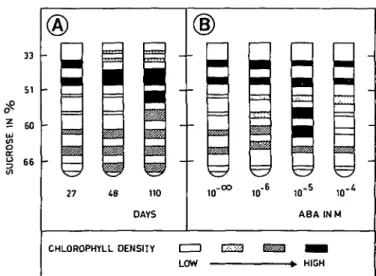
Seven bands were observed in the sucrose gradient (Fig. 1A); they can be classified into three groups, according to their density p:
—Group I : light chloroplasts, density 1.141-1.213 (zones 33 to 42% sucrose) —Group II : medium chloroplasts, density 1.257-1.280 (zones 51 to 57%
sucrose)
—Group III : heavy chloroplasts, density 1.304-1.322 (zones 60 and 63 to 66% sucrose).
In young tissues (27 days), there were more light chloroplasts (group I). Those of group II were almost nonexistent, while those of group III were of few importance. When the tissues became older (48 days), the number of chloroplasts in groups I and III increased. When the tissues were old (110 days), chloroplasts from groups I and III were at about the same level as in the 48 day-old tissues; in contrast, chloroplasts of group II became very abundant. There were a great many middle and high density chloroplasts.
The distribution patterns of chloroplasts, obtained from tissues treated with ABA, were changed in the following way (Fig. IB):
—with 10~6 M ABA, the chlorophyll (Fig. 2) and protein (Fig. 3) content of group I
33 51 £ 60 ul (/I o 5 66 27
z
m
ma I0"6 ID"5 10' ABA INM CHLOROPHYLL DENSITY LOW - > . HIGHFig. 1. Changes in chloroplast distribution patterns (separation in sucrose gradient). (A) With increasing age
of the tissues (in days) cultivated in vitro. (B) Effect of ABA (at several concentrations). Age of cultures: 34 days.
2 0
-51 57 60 SUCROSE IN o /o
Fig. 2. Chlorophyll content (in fig ml zone) of the seven isolated chloroplast sucrose zones. Chloroplasts were
prepared from carrot tissues cultivated on Murashige and Skoog medium in the presence of ABA at several concentrations. Age of cultures: 34 days.
chloroplasts increased and became higher than that of group III. Group II chloroplasts were still very few.
—10~5 M ABA, the chlorophyll and protein levels of group I and group II chloro-plasts was clearly enhanced. As can be seen in Fig. 2, chlorophyll concentration (in /Ag/ml) rose from 5.81 to 13.00 in the 42% zone. In the 57% zone (a very thin band almost undistinguishable in the control), the chlorophyll content rose from 1.44 to 13.49/ig/ml. The 51% zone was stimulated as well as the 60%
2000 - ,
51 57 60 SUCROSE IN °/o
Fig. 3. Protein content (in fig ml zone) of the seven isolated chloroplast sucrose zones. Chloroplasts were
pre-pared from carrot tissues cultivated on Murashige and Skoog medium in the presence of ABA at several concentrations. See Fig. 2.
one, in which chlorophyll content was increased from 5.78/xg/ml to 13.78 /*g/ml. In contrast the 63% and 66% zones "degenerated". The protein content (Fig. 3) was found to change in a similar way.
—with 10~4 M ABA, stimulation of the light chloroplasts of group I was reduced, compared to the effect of 10~5 M ABA while group III zones—60, 63 and 66% sucrose—were destroyed. The chlorophyll (Fig. 2) and protein (Fig. 3) content for these zones was found to be decreased. Such results were expected, since tissues showed necrosis in the presence of ABA at a concentration of 10~4 M.
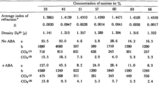
Next, changes in the chloroplast Ru-DP carboxylases were tested (Tables 1 and 2). As more significant results were obtained with 10~5M ABA, a large number of tissue explants were cultivated (fresh weight=300 g) in Erlenmeyer flasks normal MS medium with or without ABA at 10~5 M. The Results are reported in Table 1, as nanomoles of CO2 incorporated per mg of Chi, per ml and per hr, versus sucrose density. As can be seen, ABA has an inhibitory effect on all the zones except on the high density group of chloroplasts.
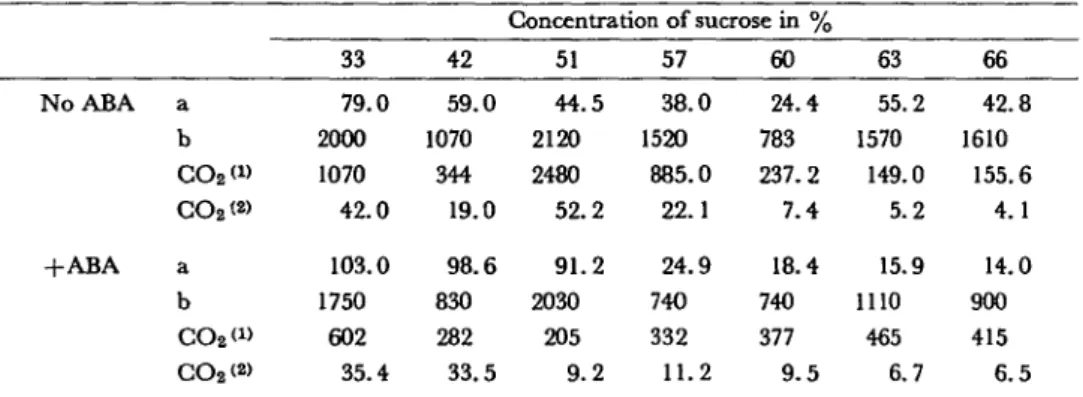
Such an inhibition of Ru-DP carboxylase activity by ABA was abo obtained for the 110 day-old tissues, pretreated 17 hr in a phosphate buffer pH 6.1 (Table 2) containing 10~5 M ABA.
When comparing Ru-DP carboxylase activity of untreated 48 day-old and 110 day-old tissues, it may be noticed that CO2 fixation characterised mainly the chloro-plasts of 33, 42, 51, 57% zones. As a matter of fact, in old tissues, the larger number of heavy chloroplasts (57, 60, 63 and 66% sucrose), less active in CO2 fixation, leads the tissues towards necrosis.
Table I CO2 incorporated in organic compounds expressed in HM for two parameters {per mg ofCHL and per mg of NP) in zones of isolated chloroplasts from carrot tissues (cultivated 48 days on the Murashige andSkoog medium, with or without l-Kh5 M ABA)
Average index of refraction " ± Density D4 2° (/>) No ABA a b CO2<» CO2<2> + A B A a b CO2<» CO2<2> 33 1.3865 0.0030 1.141 35.5 1890 716 13.5 127.0 4380 475 13.8 42 1.4139 0.0047 1.213 92.0 4060 815 18.5 43.5 1240 268 9.3 Concentration of sucrose in % 51 1.4310 0.0028 1.257 4.6 507 821 7.5 8.2 622 311 4.1 57 1.4390 0.0014 1.280 2.8 599 626 2.9 24.0 1290 281 5.2 60 1.4471 0.0041 1.304 28.6 1710 243 4 . 0 28.4 1840 243 3.7 63 1.4526 0.0036 1.316 14.2 1290 301 3 . 3 11.0 1590 449 5.3 66 1.4559 0.0017 1.322 16.5 1290 257 3.3 8.3 1240 356 2.4 a: /*gof CHL/ml b : / i g o f N P / m l
CO2 («: CO2 per mg of CHL/ml/hr CO2 <2>: CO2 per mg of NP/ml/hr
" Evaluation for 9 experiments
Relative standard error in the determination of chlorophylls and protein nitrogen respectively: 6% and 0.25%.
Table 2 CO% incorporated in organic compounds expressed in mi for two parameters {per mg ofCHL and per tng
of NP) in zones of isolated chloroplastsfrom carrot tissues (cultivated 110 days on the Murashige and Skoog medium) and pretreated in a phosphate buffer, pH 6.1, with or without l-lCh5 M ABA for 17 hr
No ABA +ABA a b CO2 <» CO2(» a b CO2W CO2<2> 33 79.0 2000 1070 42.0 103.0 1750 602 35.4 42 59.0 1070 344 19.0 98.6 830 282 33.5 Concentration of sucrose in % 51 44.5 2120 2480 52.2 91.2 2030 205 9.2 57 38.0 1520 885.0 22.1 24.9 740 332 11.2 60 24.4 783 237.2 7.4 18.4 740 377 9 . 5 63 55.2 1570 149.0 5.2 15.9 1110 465 6.7 66 42.8 1610 155.6 4.1 14.0 900 415 6.5
For a, b, COz*1' and CO2;2>, see Table 1.
The chloroplast pattern of ABA-treated tissues is similar to that of old tissues; groups I and III remain. After treatment with ABA, two important populations of chloroplasts appear in the 51 and 57% sucrose zones, in an identical pattern as for 110 day-old tissues. ABA accelerates explant aging and produces the typical chloroplast pattern of old tissues. With 10~4 M, "senescence" is set, almost all groups of chloroplasts are touched. Those of group III are destroyed, and the tissues become necrotic.
Conclusion
To explain the present data, two complementary hypotheses must be briefly presented. ABA may first have a direct action on chloroplast differentiation—as previously reported (7)—in carrot tissues cultivated in vitro, causing thereby an acceleration of tissue maturation. But ABA could also control the metabolism—as already discussed (72)—of the carrot tissues cultured in vitro, inducing some aging processes which could very well explain a rapid increase in the chloroplast content, usually higher in senescent tissues.
On the other hand, whichever type of treatment is applied to the tissues (pre-treatment or continuous application of ABA), it always results in an inhibition of the Ru-DP carboxylase of chloroplasts isolated from the carrot tissues cultivated in vitro and a strong decrease in the 14CC>2 fixation as well.
We express our thanks to Dr. Rose-Marie Hofer, from our Institute, for her helpful participation in the present work.
References
( / ) Amon, D. I.: Copper enzymes in isolated chloroplasts in Beta vulgaris. Plant Physiol. 24: 1—15 (1949).
(2) Bourdu, R., Y. Mathieu, M. Miginiac-Maslow, R. Remy and A. Moyse: Structure granaire,
reduction du NADP et photophosphorylation des chloroplastes isoles de feuilles d'orge. Planta 80: 191-210 (1968).
( 3) Bruinsma, J.: The quantitative analysis of chlorophylls a and b in plant extracts. Photochem.
Photobiol. 2: 241-249 (1963).
(4) Gautheret, R. J.: La culture des tissus vig'etaux. Masson Ed., Paris, 1959.
( 5) Hanson, A. D. and J. Edelman: Photosynthesis by carrot tissue cultures. Planta 102: 11-25 (1972).
( 6) Leech, R. M.: The isolation of structurally intact chloroplasts. Biochitn. Biophys. Ada 79: 637-639 (1964).
( 7) Milborrow, B. V.: The chemistry and physiology of abscisic acid. Ann. Rev. Plant Physiol. 25:259-307(1974).
( 8) Pilet, P. E. and M. Fragata: Prot£ines et gradients auxines-oxydasiques dans les racines de carotte cultivees in vitro. Rev. gin. Bot. 70: 572-587 (1963).
(9) Pilet, P. E.: Auxines et polarity morphologique des tissus de Carotte cultives in vitro, ibid.
70: 572-587 (1963).
(10) Pilet, P. E. and M. Fragata: A propos de l'extraction et de l'absorption du tryptophane dans les
racines de Carotte cultivees in vitro. C. R. Acad. Sci. 261: 513-516 (1965).
(//) Pilet, P. E. and J. Cl. Roland: Effects of abscisic acid on the growdi and ultrastructure of tissues cultivated in vitro. Cytobiologie 4: 41-61 (1971).
(12) Pilet, P. E.: ABA effects on growth in relation to auxin, RNA and ultrastructure. In Hormo-nal Regulation in Plant Growth and Development. Edited by H. Kaldewey and Y. Varder. p. 297-315.
Verlag Chemie, Weinheim, 1972.
(13) Roland, J. Cl. and P. E. Pilet: Modifications ultrastructurales des parois de cellules de Ronce
sous l'effect de l'acide abscissique. C. R. Acad. Sci. 272: 72-75 (1971).
(14) Shumway, L. K., T. E. Weier and C. R. Stocking: Crystalline structures in Viciafaba
chloro-plasts. Planta 76: 182-189 (1967).
(15) Spencer, D. and S. G. Wildman: Observations on the structure of grana-containing chloroplasts and a proposed model of chloroplast structure. Aust. J. Biol. Sci. 15: 599-610 (1962).
(16) Spencer, D. and H. Unt: Biochemical and structural correlations in isolated spinach
chloro-plasts under Uotonic and hypotonic conditions, ibid. 18: 197-210 (1965).
(17) Wildman, S. G., K. Chen, J. C. Gray, S. D. Kung, P. Kwanyuen and K. Sakano: Evolution
of ferredoxin and fraction I protein in the genus Nicotiana. In Genetics and Biogenesis of
Chloroplasts and Mitochondria. Edited by P. S. Perlman, C. W. Birky and T. J. Byers. State