Communication
An enzyme-based screening system for the rapid
assessment of protein N-glycosylation ef
ficiency
in yeast
Alexander D Frey
1,2and Markus Aebi
32
Department of Biotechnology and Chemical Technology, Aalto University, Aalto FI-00076, Finland, and
3Institute of
Microbiology, ETH Zürich, Zürich CH-8093, Switzerland
1To whom correspondence should be addressed: Tel: +358-50-411-6506; Fax: +358-9-462-373; e-mail: alexander.frey@aalto.fi
Received 7 April 2014; Revised 19 November 2014; Accepted 2 December 2014
Abstract
N-Glycosylation ef
ficiency is a key parameter when studying components of the protein
N-glycosylation pathway, but was recently also recognized as an important factor in the production
of glycosylated proteins. We have developed a novel assay to quantify N-glycosylation ef
ficiency
of proteins. This assay is based on the secreted activity of yeast acid phosphatase, the proper folding
and hence secretion of which is strongly dependent on its N-glycosylation status. The results show
that the reporter yields a quantitative measure for protein N-glycosylation in yeast, which is in good
agreement with classically used assay based on protein migration patterns on SDS
–PAGE. However,
the assay is less laborious and is adaptable to high-throughput screening approaches as exempli
fied.
Key words: lipid-linked oligosaccharide, oligosaccharyltransferase, protein N-glycosylation, reporter assay, yeast acid phosphatase
Introduction
Protein N-glycosylation is a fundamental process, which is taking place in the endoplasmic reticulum (ER). Once correctly folded, glyco-proteins move to the Golgi complex where the N-linked glycans are subjected to further modifications (Kornfeld and Kornfeld 1985). The biosynthesis of the branched oligosaccharide, Glc3Man9GlcNAc2,
is initiated at the outer face of the ER membrane by the addition of N-acetylglucosamine (GlcNAc) phosphate from UDP-GlcNAc to do-lichyl phosphate (Dol-P), forming GlcNAc-PP-Dol (Barne et al. 1984). Subsequently, a second GlcNAc residue andfive mannoses are added to the lipid-linked oligosaccharide (LLO) from the activated sugar nu-cleotides. After these transfer reactions the heptasaccharide, Man
5-GlcNAc2-PP-Dol is translocated from the cytoplasmic to the
lumenal side of the ER membrane (Helenius et al. 2002). Once in the ER, four additional mannoses are added before the oligosacchar-ide is capped by the addition of three glucoses (Helenius and Aebi 2004;Aebi 2013). The completed LLO, Glc3Man9GlcNAc2-PP-Dol,
is the optimal substrate for the oligosaccharyltransferase (OST) com-plex that transfers the oligosaccharide to selected asparagine residues of nascent proteins. While nine peptides have been shown to be part of the OST complex in yeast, the structure and function of individual
subunits remain unclear for the most subunits (Knauer and Lehle 1999;Kelleher and Gilmore 2006;Aebi 2013).
Efficiency of protein N-glycosylation is determined by various fac-tors, including the peptide sequence of and around the acceptor site and the availability and structure of the LLO (Jones et al. 2005). Fur-thermore, the composition of the OST complex can have a profound influence on the selection of the acceptor site (Schulz and Aebi 2009). N-Glycosylation efficiency can be measured at the single protein as well as proteome level. In Saccharomyces cerevisiae, the vacuolar pro-tein carboxypeptidase Y (CPY) is typically analyzed to determine gly-cosylation status (Silberstein et al. 1995;Burda et al. 1996). Bearing four N-glycosylation sites, hypoglycosylation of CPY results in a lad-der pattern on membranes when probed with a specific antibody and glycosylation efficiency can be qualitatively scored. Other ER-localized glycoproteins such as Ost1p or Wbp1p have been used for the same purpose (Karaoglu et al. 1995).
Alternatively, N-glycosylation efficiency can be monitored using proteomics tools and several workflows have been described to analyze site occupancy from single-protein level to proteome scale (Hülsmeier et al. 2007;Schulz and Aebi 2009). Qualitative and quan-titative methods exist, which are based on the analysis of intact Advance Access Publication Date: 8 December 2014
Communication
glycopeptides or the comparison of the abundance of glycosylated and non-glycosylated peptide pairs.
More recently, it has been recognized that N-glycosylation ef fi-ciency is also an important quality aspect in the production of thera-peutic proteins and N-glycosylation efficiency of proteins is often negatively affected in heterologous production hosts (Choi et al. 2012;Piirainen et al. 2014).
In this work, we describe a novel marker for the determination of N-glycosylation efficiency. The assay is based on the secreted activ-ity of the highly N-glycosylated yeast protein acid phosphatase (AP). The secreted phosphatase activity is a rapidly accessible marker for N-glycosylation efficiency.
Results
Yeast AP as a reporter of N-glycosylation ef
ficiency
It has previously been shown that secretion of the repressible yeast AP, encoded by PHO5 gene, is strongly dependent on its N-glycosylation state. AP possesses 12 N-glycosylation sites distributed over the whole polypeptide. Efficient folding into its functional conformation and hence secretion is strongly dependent on its N-glycosylation status as deletion of N-glycosylation sites led to accumulation of AP in the ER. It has been shown before that the secreted amount of AP protein was proportional to the secreted activity, indicating that only function-al AP is secreted (Riederer and Hinnen 1991). AP expression is tightly controlled by a network of regulatory proteins and is only induced by phosphate starvation (Oshima et al. 1996).
We have expressed His-tagged AP from constitutive promoters of different strengths. Expression of AP in wild-type yeast resulted in a hypermannosylated protein, which after deglycosylation collapsed into a single-protein band (data not shown). The wild-type yeast strain SS328 transformed with the corresponding expression and control vectors was grown in liquid media and samples for AP measurements were withdrawn during the exponential phase. A linear increase in the absorbance at 405 nm (A405) was observed in samples harvested
from the expression but not from the control cultures. Moreover, the assay readout was proportional to the amount of added super-natant (Figure1). The absence of background activity in control cul-tures indicated that no other phosphatase activity is expressed from either the endogenous repressible (PHO5, PHO11 and PHO12) or constitutive (PHO13) AP encoding genes (Figure1).
Effects of mutations in OST complex components
on AP activity
Yeast OST complex is composed of eight subunits,five of which (Stt3p, Ost1p, Ost2p, Swp1p and Wbp1p) are essential for viability. We selected strains with deletions of OST3 and OST5 genes generat-ing a mild hypoglycosylation phenotype and a strain harborgenerat-ing the temperature sensitive mutant allele wbp1-2, having a more severe hy-poglycosylation phenotype (Karaoglu et al. 1995;Zufferey et al. 1995;
Reiss et al. 1997).
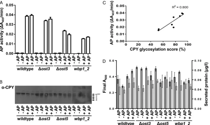
The strains were transformed with the plasmid encoding the re-porter protein or the control plasmid. Samples for AP assay were col-lected during the exponential phase. In addition, samples for the analysis of CPY glycosylation pattern were collected. AP activity was detected in all cultures harboring the reporter construct and was highest in the wild-type strain. AP activity in the cultures origin-ating fromΔost3 and Δost5 strains was only slightly reduced relative to wild-type culture, corresponding to the mild hypoglycosylation phenotype as reported previously (Karaoglu et al. 1995;Reiss et al.
1997). In contrast, a clearly reduced AP activity was observed in the strain carrying the wbp1-2 allele (Figure2A). The glycosylation pat-tern of CPY in the different strains matched the AP activity measure-ments. Whereas full glycosylation of CPY was observed in wild-type cells, increased degrees of hypoglycosylation were observed inΔost3, Δost5 and wbp1-2 strains (Figure2B). In order to compare the results of AP activity and CPY glycosylation, we quantified the relative abun-dance of bands and calculated a CPY glycosylation score. A strong positive correlation (R2= 0.800) of AP activity and CPY glycosylation
was observed (Figure2C). Expression of AP in the different strains did neither significantly affect the total amount of secreted protein nor growth relative to control cultures harboring the plasmid only indicat-ing that the expression of the reporter does not affect strainfitness nor lead to a burden of the secretory pathway (Figure2D). No significant differences were observed between the corresponding cultures (one-way ANOVA).
Effects of gene deletions in the LLO-biosynthesis
on AP activity
Next we addressed the question if the reporter would also faithfully report the expected hypoglycosylation phenotype when providing truncated LLO as substrate to the OST complex. For this experiment, we selected a wild-type control strain, aΔalg9 strain, a Δalg3 strain and a Δalg3 Δalg9 double mutant strain. These strains produce LLOs comprising Man6GlcNAc2and Man5GlcNAc2and
glycosyla-tion efficiency is affected to similar degrees, however, without having any deleterious effects on growth. As expected AP activity in cultures
Fig. 1. Supernatant of wild-type yeast strain transformed with AP encoding (+AP) and control plasmids (−AP) was analyzed for the secretion of AP activity. AP activity was measured by following the turnover of pNPP into pNP at an absorbance of 405 nm (A405). No AP activity was detected in samples from control cultures, revealing the absence of any interfering secreted phosphatase activity in the culture medium. (B) A dilution series of culture supernatant of the yeast strain expressing AP yielded a proportional increase in AP activity.
from wild-type cells was highest. Enzyme activity in cultures originat-ing fromΔalg3 strain was lower, corresponding with previously pub-lished data on glycosylation efficiency in Δalg3 cells. Secreted AP activity was reduced to similar extents inΔalg3 and Δalg9 cells. The combination of both mutations did not have a cumulative effect on the amount of secreted AP activity (Figure3). The glycosylation pat-tern of CPY (data not shown) followed the patpat-tern as previously pub-lished (Burda et al. 1996).
AP activity as readout for the identi
fication of novel POTs
In order to evaluate the applicability of the readout in a screening for-mat, an experiment was designed which aimed at the identification of novel protozoan oligosaccharyltransferases (POT) using a workflow compatible with automation. In contrast to other eukaryotes kineto-plastids, such as Leishmania spp., do not possess an OST complex, ra-ther have a single protein, which is functionally and structurally related to the catalytic subunit of the eukaryotic OST complex. POTs have been shown to functionally replace S. cerevisiae OST com-plex (Nasab et al. 2008;Hese et al. 2009).
A strain having an stt3-7 allele was employed, which encodes a temperature sensitive mutant protein leading to hypoglycosylation above the restrictive temperature. Only by providing a functional POT, the hypoglycosylation phenotype can be cured. POTs isolated from L. brasiliensis, L. infantum and L. major were expressed from low- and high-copy number plasmids in the stt3-7 strain co-expressing the reporter construct (Nasab et al. 2008;Parsaie Nasab et al. 2013).
Strains were inoculated into individual wells of 96-deep-well plates and grown at permissive temperature until reaching saturation. Twenty-five microliters of each culture was transferred into fresh media. The assay plate was grown for 16 h at the restrictive tempera-ture and samples were harvested. N-Glycosylation was restored to different levels, expression of two of the paralogs isolated from L. brasiliensis, termed LbSTT3_1 and LbSTT3_3 led to the highest
Fig. 2. Defects of OST complex reduces secretion of AP. Wild type,Δost3, Δost5 and wbp1-2 yeast strains transformed with either control (−AP) or AP-expression (+AP) plasmid. Samples for AP measurement and immunoblotting were collected in the exponential phase. N-Glycosylation efficiency was measured using AP assay (A) and glycosylation of CPY (B). (C) Correlation of CPY glycosylation score and AP activity. CPY glycosylation pattern was analyzed and quantified using Image Lab software and the data were used to generate CPY glycosylation score. (D) Final A600(black bars) and total amount of secreted protein (gray bars) in culture supernatants. No significant differences in final A600and in the amount of total secreted protein were detected between corresponding control and reporter cultures.
Fig. 3. Defects in LLO synthesis reduces secretion of AP. Wild-type,Δalg3, Δalg9 and Δalg3Δalg9 yeast strains transformed with either control (−AP) or AP-expression (+AP) plasmid. N-Glycosylation efficiency was quantitatively measured using AP assay.
AP activity, both when expressed from high- and low-copy number plasmids. Interestingly, one of the three paralogs (LbSTT3_2 and LiSTT3_3) was not able to restore N-glycosylation in the stt3-7 yeast strain. Interestingly, AP activity in cultures expressing POTs from low-copy number plasmids was higher than when expressed from high-copy number plasmids (Figure4A). In order to compare the experimental data to the CPY glycosylation pattern, cell extracts from a single replicate culture were resolved on SDS–PAGE gels, and the glycosylation pattern of CPY was analyzed using immunoblot ana-lysis. Expression of the proteins yielding highest AP activity (LbSTT3_1 and LbSTT3_3) did not results in any observable hypogly-cosylation phenotype, in contrast LbSTT3_2 and LiSTT3_3, the ex-pression of which did not result in any detectable AP activity, did result in a strong hypoglycosylation of CPY, similar to the levels as
observed in the negative control (Figure4B). The correlation coef fi-cient of AP activity and CPY glycosylation score was 0.678. However, R2was higher when only taking into account the expression of POTs
from CEN/ARS plasmids.
Discussion
Yeast cells have successfully been used to study the N-glycosylation pathway and its individual components, which substantially con-tributed to our current knowledge on N-glycosylation (Helenius and Aebi 2004; Aebi 2013). Furthermore, the capability to modulate N-glycosylation pattern has sparked interest in the use of yeast for the production of therapeutic glycoproteins but also raised concerns regarding glycosylation efficiency in these hosts (Jones et al. 2005;
Choi et al. 2012;Piirainen et al. 2014).
In this work, we have established a system to monitor N-glycosylation efficiency using AP activity. In our tests, the readout responded to various disturbances in the N-glycosylation pathway, namely defects in the OST complex and defects in the LLO synthesis. Moreover, we have shown that the assay can be adapted to multi-well format and is compatible with automation, making this assay an at-tractive tool to study a wide variety of processes having an impact on glycosylation efficiency in yeast. The required time and efforts for sample processing are strongly reduced, enabling the parallel pro-cessing and analysis of a multiple number of samples compared with alternative existing methods.
The data presented here show that N-glycosylation efficiency de-termined using AP activity correlate well with the results obtained using the traditional marker CPY. In order to compare results of AP assay and CPY glycosylation, we have calculated a CPY glycosylation score, which would be 100% if only mature CPY is detected and 0% if only non-glycosylated CPY is expressed. A 50% reduction in the score would imply that only two of the four sites are occupied.
Based on our results, it is obvious that the AP assay is not suitable in conditions where a strong hypoglycosylation is expected. It has been shown previously that AP activity remained at 100% when deleting two of the sites but decreased to 30 and 0% of the initial activity when four or six glycosylation sites, respectively, have been removed (Riederer and Hinnen 1991). However, the AP assays reliably reports N-glycosylation efficiencies in a range of experimental conditions, where a mild-to-medium hypoglycosylation phenotype is observed. Independent of the method used, one must bear in mind that both as-says are based on the analysis of a single protein.
Besides the assay based on AP activity described here, a glycosyla-tion readout based on ER-retained GPF has been published (Losfeld et al. 2012). For this assay, the GFP molecule has been engineered to harbor a single N-glycosylation site. N-Glycosylation of the protein impaired thefluorescence signal. The assay was successfully used to study glycosylation defects in multiple mammalian cell lines.
In order to improve or control N-glycosylation efficiency, the as-says described so far might be very useful to screen for components or conditions, which lead to improved efficiency of the protein N-glycosylation.
Materials and methods
Strains and plasmids
All strains used on this work have been described before. The strains were SS328 (MATα ade2-101 his3Δ200 lys2-801 ura3-52), YG543 (MATa ade2-101 leu2 ura3-52 his3Δ200 lys2-801 stt3-7) (Spirig Fig. 4. Expression of POTs isolated from different Leishmania spp. can recover
secretion of AP in a yeast stt3-7 mutant expressing AP. Low- (Cen/Ars) and high-copy number (2 µ) plasmids were used for the expression of POT. Cells were grown in 96-deep-well plates in quadruplicates at the restrictive temperature. N-Glycosylation efficiency was (A) measured using AP assay and (B) determined using glycosylation of CPY. Results of AP assay are reported as average and standard deviation. Cells from a single replicate were used to analyze N-glycosylation efficiency using CPY protein as readout. (C) Correlation of CPY glycosylation score and AP activity. CPY glycosylation pattern was analyzed and quantified using Image Lab software and the data were used to generate CPY glycosylation score.
et al. 1997), YG0183 (ade2-101 his3Δ200 lys2-801 ura3-52 Δost3:: HIS3), YG0358 (ade2-101 his3Δ200 lys2-801 ura3-52 Δost5::HIS3) (Reiss et al. 1997), YG0411 (MATα ade2-101 his3Δ200 lys2-801 ura3-52), YG0412 (MATα ade2-101 his3Δ200 lys2-801 ura3-52 Δalg3::HIS3), YG0414 (MATα ade2-101 his3Δ200 lys2-801 ura3-52Δalg3::HIS3 Δalg9::KanMX4), YG0413 (MATα ade2-101 his3Δ200 lys2-801 ura3-52 Δalg9::KanMX4) (Burda et al. 1996) and YG0150 (MATα ade2-101 his3Δ200 tyr1 ura3 wbp1-2) (Zufferey et al. 1995). Plasmids for the expression of POTs have been described before (Parsaie Nasab et al. 2013).
DNA manipulation and yeast transformations
Escherichia coli TOP10 (Invitrogen) cells were used as cloning host. The PHO5 gene encoding AP was isolated from genomic yeast DNA using PCR. Oligonucleotides were designed to encompass a 5′ SpeI (forward oligonucleotide 5′-AAACTAGTATGTTTAAATCTG TTGTTTATTC-3′) and a 3′ XhoI restriction site. A C-terminal Hexa-His tag was included (reverse oligonucleotide 5′-AACTCGAGTT AATGGTGATGGTGATGGTGTTGTCTCAATAGACTGGCG-3′. The PCR product was inserted into TOPO pCR-blunt vector (Invitro-gen). Sequence verified PHO5 gene was transferred into SpeI XhoI sites of yeast shuttle vectors pRS426 and pRS425 under control of GPD and TEF promoter. The lithium acetate method was used for transformation of yeast cells (Gietz and Schiestl 2007).
Growth of yeast strains
All experiments were performed with 20 mL media in 100 mL shake flasks. Standard yeast synthetic drop-out media lacking the appropri-ate amino acids was used for all experiments. Cells were grown at 30°C except for temperature sensitive strains. YG0150 was grown at 24°C. YG543 was grown at 24°C and the temperature was shifted to 34°C to induce hypoglycosylation phenotype. The effect of POTs on N-glycosylation efficiency was studied in 96-deep-well plates using 1 mL of media. Experimental cultures were inoculated with 25 µL of saturated precultures and plates were grown for 16 h at 34°C and 300 rpm. Samples for AP assay, protein determination and cells for immunoblot analysis were collected in the exponential growth phase.
Total protein and AP assay
Total protein in culture supernatant was quantified using Bradford protein reagent according to suppliers’ instructions. AP activity was measured from cleared culture broth using an endpoint method. Ten microliters of supernatants were dispensed in triplicates into four-replicate 96-well-assay plates. Prior to the assay, the plates were prewarmed for 5 min at 30°C. The assay was started by addition of 100 µL of 20 mM para-nitrophenyl-phosphate ( pNPP) (Sigma-Aldrich, Buchs, Switzerland) in sodium acetate buffer, pH 4.2, at timed intervals. Reactions were terminated by addition of 200 µL 2 M Na2CO3. Reactions in replicate plates were stopped after 2, 10
and 20 min. For the 0 min incubation plate, 2 M Na2CO3 was
added before the addition of the substrate. Blanks contained only substrate but no culture supernatant. The absorbance was read at 405 nm using a Spectramax photometer (Molecular Devices, Sunnyvale, CA). AP activity was measured as the increase in absorb-ance at A405over time. p-Nitrophenol (Sigma-Aldrich) was used
as standard in order to calculate enzyme activity as release of p-nitrophenol per time (µmol/min). One micromolar of pNP/min equals 0.007456A405/min.
SDS
–PAGE and immunoblotting
Samples for SDS–PAGE and immunoblotting were prepared and ana-lyzed as described (Parsaie Nasab et al. 2013). The primary antibodies were directed against CPY (Burda et al. 1996) and His Tag (Qiagen, Hombrechtikon, Switzerland). Secondary antibodies were anti-rabbit IgG horseradish peroxidase conjugate and anti-mouse IgG horserad-ish conjugate (Santa Cruz Biotechnology, Santa Cruz, CA). CPY gly-cosylation pattern was analyzed and quantified using Image Lab software and the data were used to calculate a CPY glycosylation score. Relative abundances of bands were multiplied with 4, 3, 2 and 1, for mature CPY, and CPY lacking 1, 2 and 3 N-glycans, respectively. Values were summed, divided by 400 and converted to percentage.
Statistical analysis
All statistical analyses were done with GraphPad Prism 6® software.
Con
flict of interest statement
None declared.
Funding
This work was supported by Aalto University and ETH Zurich.
Abbreviations
AP, acid phosphatase; CPY, carboxypeptidase Y; Dol-P, dolichyl phosphate; ER, endoplasmic reticulum; GlcNAc, N-acetylglucosamine; LLO, lipid-linked oligosaccharide; OST, oligosaccharyltransferase; POT, protozoan oligosacchar-yltransferases
References
Aebi M. 2013. N-Linked protein glycosylation in the ER. Biochim Biophys Acta. 1833:2430–2437.
Barne G, Hansen WJ, Holcomb CL, Rine J. 1984. Asparagine-linked glycosyla-tion in Saccharomyces cerevisiae: Genetic analysis of an early step. Mol Cell Biol. 4:2381–2388.
Burda P, te Heesen S, Brachat A, Wach A, Dusterhoft A, Aebi M. 1996. Stepwise assembly of the lipid-linked oligosaccharide in the endoplasmic reticulum of Saccharomyces cerevisiae: Identification of the ALG9 gene encoding a puta-tive mannosyl transferase. Proc Natl Acad Sci USA. 93:7160–7165. Choi BK, Warburton S, Lin H, Patel R, Boldogh I, Meehl M, D’Anjou D, Pon L,
Stadheim TA, Sethuraman N. 2012. Improvement of N-glycan site occu-pancy of therapeutic glycoproteins produced in Pichia pastoris. Appl Microbiol Biotechnol. 95:671–682.
Gietz RD, Schiestl RH. 2007. Large-scale high-efficiency yeast transformation using the LiAc/SS carrier DNA/PEG method. Nat Protoc. 2:38–41. Helenius A, Aebi M. 2004. Roles of N-linked glycans in the Endoplasmic
Re-ticulum. Annu Rev Biochem. 73:1019–1049.
Helenius J, Ng DT, Marolda CL, Walter P, Valvano MA, Aebi M. 2002. Trans-location of lipid-linked oligosaccharides across the ER membrane requires Rft1 protein. Nature. 415:447–450.
Hese K, Otto C, Routier FH, Lehle L. 2009. The yeast oligosaccharyltransferase complex can be replaced by STT3 from Leishmania major. Glycobiology. 19:160–171.
Hülsmeier AJ, Paesold-Burda P, Hennet T. 2007. N-glycosylation site occu-pancy in serum glycoproteins using multiple reaction monitoring liquid chromatography-mass spectrometry. Mol Cell Proteomics. 6:2132–2138. Jones J, Krag SS, Betenbaugh MJ. 2005. Controlling N-linked glycan site
Karaoglu D, Kelleher DJ, Gilmore R. 1995. Functional characterization of Ost3p. Loss of the 34-kD subunit of the Saccharomyces cerevisiae oligosac-charyltransferase results in biased underglycosylation of acceptor sub-strates. J Cell Biol. 130:567–577.
Kelleher DJ, Gilmore R. 2006. An evolving view of the eukaryotic oligosacchar-yltransferase. Glycobiology. 16:47R–62R.
Knauer R, Lehle L. 1999. The oligosaccharyltransferase complex from Sacchar-omyces cerevisiae. Isolation of the OST6 gene, its synthetic interaction with OST3, and analysis of the native complex. J Biol Chem. 274:17249–17256. Kornfeld R, Kornfeld S. 1985. Assembly of asparagine-linked oligosaccharides.
Annu Rev Biochem. 54:631–664.
Losfeld ME, Soncin F, Ng BG, Singec I, Freeze HH. 2012. A sensitive green fluorescent protein biomarker of N-glycosylation site occupancy. FASEB J. 26:4210–4217.
Nasab FP, Schulz BL, Gamarro F, Parodi AJ, Aebi M. 2008. All in One: Leish-mania major STT3 proteins substitute for the whole oligosaccharyltransfer-ase complex in Saccharomyces cerevisiae. Mol Biol Cell. 19:3758–3768. Oshima Y, Ogawa N, Harashima S. 1996. Regulation of phosphatase synthesis
in Saccharomyces cerevisiae– A review. Gene. 179:171–177.
Parsaie Nasab F, Aebi M, Bernhard B, Frey AD. 2013. A combined system for engineering glycosylation efficiency and glycan structure in Saccharomyces cerevisiae. Appl Environ Microbiol. 79:997–1007.
Piirainen MA, de Ruijter JC, Koskela EV, Frey AD. 2014. Glycoengineering of yeasts from the perspective of glycosylation efficiency. New Biotechnol. doi:10.1016/j.nbt.2014.03.001.
Reiss G, te Heesen S, Gilmore R, Zufferey R, Aebi M. 1997. A specific screen for oligosaccharyltransferase mutations identifies the 9 kDa OST5 protein re-quired for optimal activity in vivo and in vitro. EMBO J. 16:1164–1172. Riederer MA, Hinnen A. 1991. Removal of N-glycosylation sites of the yeast
acid phosphatase severely affects protein folding. J Bacteriol. 173:3539–3546.
Schulz BL, Aebi M. 2009. Analysis of glycosylation site occupancy reveals a role for Ost3p and Ost6p in site-specific N-glycosylation efficiency. Mol Cell Proteomics. 8:357–364.
Silberstein S, Collins PG, Kelleher DJ, Gilmore R. 1995. The essential OST2 gene encodes the 16-kD subunit of the yeast oligosaccharyltransferase, a highly conserved protein expressed in diverse eukaryotic organisms. J Cell Biol. 131:371–383.
Spirig U, Glavas M, Bodmer D, Reiss G, Burda P, Lippuner V, te Heesen S, Aebi M. 1997. The STT3 proteinis a component of the yeast oligosacchar-yltransferase complex. Mol Gen Genet. 256:628–637.
Zufferey R, Knauer R, Burda P, Stagljar I, te Heesen S, Lehle L, Aebi M. 1995. STT3, a highly conserved protein required for yeast oligosaccharyl transfer-ase activity in vivo. EMBO J. 14:4949–4960.