ORIGINAL PAPER
Poor performance of microbiological sampling
in the prediction of recurrent arthroplasty infection
Maximilian Schindler&Panayiotis Christofilopoulos&Blaise Wyssa&Wilson Belaieff&
Christian Garzoni&Louis Bernard&Daniel Lew&Pierre Hoffmeyer&Ilker Uçkay
Received: 27 February 2010 / Revised: 27 March 2010 / Accepted: 27 March 2010 / Published online: 27 April 2010 # Springer-Verlag 2010
Abstract During a two-stage revision for prosthetic joint infections (PJI), joint aspirations, open tissue sampling and serum inflammatory markers are performed before re-implantation to exclude ongoing silent infection. We investigated the performance of these diagnostic proce-dures on the risk of recurrence of PJI among asymptom-atic patients undergoing a two-stage revision. A total of 62 PJI were found in 58 patients. All patients had intra-operative surgical exploration during re-implantation, and 48 of them had intra-operative microbiological swabs. Additionally, 18 joint aspirations and one open biopsy were performed before second-stage reimplantation. Recurrence or persistence of PJI occurred in 12 cases with a mean delay of 218 days after re-implantation, but
only four pre- or intraoperative invasive joint samples had grown a pathogen in cultures. In at least seven recurrent PJIs (58%), patients had a normal C-reactive protein (CRP, <10 mg/l) level before re-implantation. The sensitivity, specificity, positive predictive and nega-tive predicnega-tive values of pre-operanega-tive invasive joint aspiration and CRP for the prediction of PJI recurrence was 0.58, 0.88, 0.5, 0.84 and 0.17, 0.81, 0.13, 0.86, respectively. As a conclusion, pre-operative joint aspira-tion, intraoperative bacterial sampling, surgical explora-tion and serum inflammatory markers are poor predictors of PJI recurrence. The onset of reinfection usually occurs far later than reimplantation.
Introduction
A two-stage revision is an acknowledged procedure for the
treatment of infected arthroplasties (PJI) [1]. Infection
recurrence after re-implantation harbours significant
mor-bidity [2, 3], and every effort should be made to identify
patients at risk of such a devastating complication. Normally, an interval of eight weeks is required between infected implant removal and reimplantation, although there are no uniform recommendations about the duration of this
interval, ranging from two to four weeks [1] to several
months [4, 5]. For patients undergoing an antibiotic-free
window before re-implantation, the interval between two stages may be divided into a six-week course of antibiotic
treatment [3], followed by at least two additional weeks of
an antibiotic-free window [1,3,6]. A minimal delay of at
least two weeks seems to be important, because it has been shown that periprosthetic tissue culture sensitivity is less than 50%, if antimicrobial therapy was discontinued less The authors received no financial support, grants, or royalties and
have no financial interests that could lead to a conflict of interest. All authors state that they have read and approved the manuscript. It has not been published elsewhere nor is it under consideration for publication elsewhere.
M. Schindler
:
P. Christofilopoulos:
B. Wyssa:
W. Belaieff:
P. Hoffmeyer
:
I. Uçkay (*)Orthopaedic Surgery Service, Geneva University Hospitals, 4, Rue Gabrielle Perret-Gentil,
1211 Geneva 14, Switzerland e-mail: [email protected] L. Bernard
Division of Infectious Diseases, Bretionneau Hospital, Tours University Hospitals,
Tours, France
C. Garzoni
:
D. Lew:
I. UçkayService of Infectious Diseases,
Geneva University Hospitals and Medical School, Geneva, Switzerland
than 14 days before sampling [5]. In the absence of clinical signs and the presence of normal serum inflammatory
markers [6], re-implantation is undertaken. Additionally, a
joint aspiration or open biopsy is often performed before
re-implantation [3] to exclude asymptomatic persistent
infection. However, this empirical approach in PJI man-agement is mostly based upon speculation and experts’ recommendations, rather than scientific evidence.
Recently, Müller et al. evaluated the values of serum C-reactive protein (CRP) and pre-operative joint aspiration in
the predilection of persistent infection [7]. The utility of
CRP was equally assessed among 109 patients undergoing second-stage reimplantation for infected knee prostheses
[8]. Both studies found only a poor or moderate
perfor-mance for both methods.
In this study, we investigate PJIs that recurred after a two-stage revision with an emphasis on the utility of pre-operative joint aspiration, serum inflammatory markers, intra-operative sampling, surgical exploration and histology obtained during re-implantation.
Materials and methods Setting
The Geneva University Hospitals is a tertiary centre for Orthopaedic Surgery and Traumatology. The Orthopaedic Service has 132 acute care beds, a dedicated Infectious
Diseases physician [9] and performs more than 500
arthroplasties annually. The Service conducts a hip and knee arthroplasty registry with active post-discharge
sur-veillance [10]. One day of hospitalisation costs roughly US
$1200. A surgical tissue sampling for bacterial testing costs $6,500 (including anaesthesia, occupation of the operating theatre, nursing, medical treatment and analyses). The expenses for CRP sampling and leukocyte counts are about $10 each. The cost for the treatment of a single episode of PJI is about $45,000.
Study design
We performed a single-centre retrospective analysis from January 1996 to June 2009. We included all PJI cases treated by two-stage revision with a minimal follow-up of six months post-reimplantation. Two physicians (M.S. and I.U.) independently collected 61 variables for each PJI. Definition of PJI required pus around the affected arthro-plasty and/or at least three identical pathogens in microbi-ological samples. We collected all CRP samples between explantation and re-implantation with the exception of patients with other causes of serum CRP alterations (i.e. active neoplasm, autoimmune disease, cirrhosis Child B
and C). The CRP follow-up of the individual patients under antibiotic treatment was stratified in weekly periods during the first three weeks. The CRP value at the end of each week post-explantation ± one day, e.g. after seven days, or after 14 days post-explanation, was noted on an EXCEL sheet. In contrast, CRP samples performed within 15 days preceding re-implantation were defined as pre-implantational samples. The normal upper limits for CRP, total leukocyte count, and left shift were given by our accredited laboratory and set for <10 mg/l, <10 G/l, and <5% of non-segmented neutrophils, respectively.
Microbiological assessment procedures were unchanged during the study period. They were based on the Clinical
and Laboratory Standards Institute’s recommendations [11].
For direct microscopic examination Gram and Acridine-staining were used. Histology (obtained during reimplanta-tion without frozen secreimplanta-tion) was considered positive if pathogens were detected or if the sample revealed at least ten neutrophils per high-power field. There was no ethical committee approval necessary.
Statistical methods
We investigated potential clinical risk factors for recurrent PJI. To avoid model overfitting and spurious results, no multivariate analysis was performed. Group comparisons between PJI with and without recurrence were performed by the Wilcoxon-rank sum or the Fisher exact test, as
appropriate. P values ≤0.05 (all two-tailed) were
signifi-cant. STATA™ software (9.0, STATA Corp, College
Station, USA) was used.
Results
General and microbiological results
A total of 62 PJI, treated with a two-stage exchange procedure, were found in 58 patients (mean age 68 years; 29 women). Patients were followed-up for an average of 3.3 years after reimplantation. The PJI involved hip arthroplasties (n= 36, 58%), knee arthroplasties (n=23, 37%), hip hemiarthroplasties (n= 2), and one shoulder arthroplasty. The pathogens were Staphylococcus aureus (n=15, of which four were methicillin-resistant), coagulase-negative staphylococci (n = 18), streptococci (n = 15), Enterococcus faecalis (n = 1), anaerobes (n = 2), Gram-negative aerobic bacteria (n = 7), and culture-Gram-negative PJI (n = 4). In 32 cases (32/62, 52%), patients were immuno-compromised by diabetes mellitus (n = 8), severe alcohol-ism (n = 7), neoplasms (n = 6), steroid medication for autoimmune disease (n = 3), HIV (n = 1), or affected by a
combination of several immunosuppressive diseases listed above (n = 7).
Treatment procedure
The median duration of antibiotic therapy after prosthesis explantation was 44 days (range, 28–105 days). In 54 PJI (54/62, 87%), antibiotics had been stopped after an average of 109 days (range, 2–634 days) before re-implantation; in 41 cases this window lasted more than 14 days. The prolonged antibiotic-free windows were due to organisa-tional difficulties, patient’s comorbidities, and social prob-lems rather than probprob-lems related to infection. In eight cases (8/62, 13%), re-implantation took place during ongoing antibiotic treatment. During the prosthesis-free interval, a gentamicin-loaded spacer was used in ten of 23 knee PJIs (43%). For an additional 11 knee PJIs (48%), a spacer without antibiotic loading was used. For all hip PJIs, explantation without transient spacer was the treatment of choice.
Recurrence of PJI
In 12 cases (12/62, 19%), PJI recurred after reimplanta-tion with a mean and median delay of 218 and 88 days,
respectively (Table1). All PJI were surgical site infections
without evidence for haematogenous origin. All recurren-ces were given postoperative antibiotic treatment for a median duration of 46 (range, 9–93) days. Six recurrences were successfully treated with debridement and arthro-plasty retention. In three cases, patients underwent another two-stage exchange. A second recurrence did not occur for these nine cases. One case was treated with lifelong suppressive antibiotic treatment and two cases were lost to follow-up.
Microbiological sampling before re-implantation
A total of 19 invasive diagnostic procedures were performed in 18 patients before re-implantation (18/62, 29%) including 18 joint aspiration procedures (median 33 days before re-implantation, range 0–205 days) and one open biopsy (126 days before). No iatrogenic complications were observed
as a result of these invasive procedures (Table1).
Intraoperative Gram-staining
A total of 48 PJI episodes (48/62; 77%) underwent intra-operative Gram- and Acridine orange staining during re-implantation (average 3.3 samples). In all but one case, the samples were free of leucocytes and staining failed to reveal pathogens. The only exception was a recurrent PJI
due to S. aureus (last case of Table1). This case was also
the only one revealing clinical pus at the operation site among all study patients (1/62; 1.6%).
Intraoperative cultures
In three cases, the presence of bacteria was interpreted as true pathogens, since they were identical to those respon-sible for the PJI and were subsequently treated with antibiotics. In one case, Corynebacterium sp (one of four samples) was interpreted as contamination and was not treated with antibiotics after reimplantation.
In summary, in 53 of 62 PJI, an invasive microbiological sample before reimplantation was performed. Among these, 48 episodes (48/53, 91%) had an antibiotic-free interval longer than 14 days (13/19 preoperative invasive samples, and 35/48 intraoperative invasive samples).
Performance of various tests
In summary, in seven recurrent PJIs (7/12, 58%), pre-reimplantational aspiration, open biopsy, intra-operative surgical status and intra-operative cultures were negative. For these invasive diagnostic procedures altogether, the sensitivity, specificity, positive predictive and negative predictive values for recurrent PJI were 0.58, 0.88, 0.5,
and 0.84, respectively (Table2).
Histology
A total of 18 re-implantations were accompanied by histology. No histological sample revealed the presence of pathogens but four showed marked inflammation (4/18, 22%). Sensitivity, specificity, positive and negative predic-tive values for recurrent PJI were 0.33, 0.8, 0.25, and 0.86,
respectively (Table2).
Serum inflammatory markers before reimplantation Total leukocyte count was elevated in three cases (mean value 6.2 G/l; median three days before reimplantation). Neutrophil left shift was always within normal range.
Pre-reimplantational serum CRP levels (mean value 10.0 mg/l; range 1–54 mg/l) were sampled in 42 cases (42/62; 68%; median three days before reimplantation). In eight of them, CRP values were above normal limit. Among eight patients without normalisation of CRP levels before reimplantation, one had recurrent infection and seven did not (Fisher exact-test, p=1.0). In seven recurrent PJIs (58%), patients had a normal C-reactive protein (CRP, <10 mg/l) level before re-implantation. Sensitivity, speci-ficity, positive predictive and negative predictive values of elevated CRP levels for recurrent PJI were 0.17, 0.81, 0.13,
T able 1 Overview of 12 recurrent arthroplasty infections after two-stage exchange Arthroplasty Gender , age (years) Pathogen Antibiotics a Antibiotic-free interval b CRP c Aspiration/biopsy d Intraoperative samples (during reimplantation) Recurrence Recurrent pathogen Hip F , 80 E. faecalis 93 days 14 days 4 mg/l Four days before, negative E. faecalis (1/4 samples) 46 days later E. faecalis Knee F , 52 Unknown 35 days 14 days 4 mg/l One day before, negative Negative (0/1 sample) 379 days later unknown Hip M, 70 S. epidermidis 28 days 16 days n.a. n.a. Negative (0/2 samples) 27 days later S. hominis Hip M, 37 P . aeruginosa 84 days 585 days 6 mg/l n.a. Negative (0/3 samples) One day later P . aeruginosa Knee M, 89 Str eptococ. sp 42 days 33 days 3 mg/l n.a. Negative (0/3 samples) 313 days later Str eptococcus sp Knee F , 90 S. aur eus 90 days 14 days n.a. 30 days before, negative Negative (0/1 sample) 629 days later S. aur eus Knee M, 77 S. milleri 92 days 14 days n.a. Four days before, negative Negative (0/3 samples) T en days later S. milleri Hip F , 62 S. mitis 46 days 60 days 23 mg/l n.a. Corynbacterium (1/4 samples) 645 days later S. mitis Hip M, 70 E. faecium 90 days 13 days 5 mg/l n.a. E. faecium (1/3 samples) One day later E. faecium Knee M, 61 S. lugdunensis 42 days 12 days 6 mg/l One day before, negative Negative (0/4 samples) 645 days later S. lugdunensis Hip M, 32 S. aur eus 56 days 73 days n.a. n.a. S. aur eus (1/3 samples) 129 days later S. aur eus Hip M, 33 S. aur eus 42 days 221 days 2 mg/l One day before, positive No immediate reimplantation One day later S. aur eus M male, F female, CRP C-reactive protein All Staphylococcus aur eus infections were methicillin-and rifampin-susceptible a D uration of antibiotic treatment before re-implantation b D elay betwe en end of antibiotic trea tment and reimplantation c Last C-reactive prot ein before re-implanta tion d T ime of joint aspi ration before re-implantation
CRP values throughout the antibiotic treatment
In the interval between explantation and reimplantation, a total of 366 CRP samples were performed in the study population without concomitant causes of serum CRP alteration. After starting antibiotic treatment, CRP values fell 90% within a mean of 30 days and a median of 20 days (range, 6–93 days) and returned to normal within a mean of 38 days and a median of 27 days (range, 9–93 days). The patient populations with and without recurrent PJI after reimplantation did not differ in post-explantational peak values (median 208 mg/l vs. 172 mg/l, Wilcoxon rank sum test, p = 0.65), time until CRP normalisation (median 35 days vs. 19 days, Wilcoxon rank sum test, p=0.20) or delay until 90% decrease of initial CRP values (median 26 days vs. 16 days, Wilcoxon rank sum test, p=0.13). The median CRP levels at the end of each week during the first three weeks post-explantation did not differ between infected vs. uninfected patients (all Wilcoxon-rank sum tests; p>0.20).
Comparisons between PJI episodes with and without recurrences
Neither group differed according to key parameters
(Table3).
Discussion
We report a poor performance of pre-operative invasive microbiological sampling, intraoperative surgical explora-tion, or pre-reimplantational serum inflammatory markers for recurrent infections among asymptomatic patients undergoing a two-stage revision. None of the patients presented signs of persistent infection during the
antibiotic-free window, regardless of duration, while awaiting re-implantation. No clinical parameter showed specific
asso-ciation with recurrence (Table 3). Our recurrence risk of
19% was similar to the 20% published by Hanssen and
Osmon [12], the 21% indicated by Ghanem et al. [8] and to
other reports of two-stage [3, 13] or one-stage exchanges
[14, 15], even if lower recurrence rates have also been
reported [6,16]. Therefore, we would exclude a substantial
therapeutic bias.
The landmark publication of Zimmerli et al. cites that at least three intraoperative tissue specimens should be
sampled for culture [1], which is common practice,
although some experts ideally recommend up to six
specimens [17]. In our study population, the average
number of intraoperative microbiologic samples was 3.3, and only a quarter of all reimplantations revealed less than
three samples (Table 1). Therefore we equally exclude a
major sampling bias.
Recurrent PJI occurred far later than the reimplantation
procedure and was solely detected on the basis of patient’s
complaints. While our average delay of recurrence was
seven months after reimplantation, it was 13 months [2] or
66 months [6] in other reports. We think that the usual
six-week antibiotic course may heal, or at least suppress infection, to such a low level that current standard laboratory procedures might not be able to detect them. Pathogens need time to recover and to provoke recurrent clinical infection. It is likely that the length of the antibiotic-free window would not play a major role, since these pathogens may remain dormant for years in the absence of a new implant, and are awakened only in the presence of a new one; a phenomenon observed in recurrent
osteomyelitis [18].
The low accuracy of intraoperative Gram-staining in PJI
diagnosis has been previously reported [19]. Additionally,
Müller et al. evaluated the values of preoperative cultures in
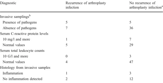
Diagnostic Recurrence of arthroplasty
infection No recurrence of arthroplasty infectiona Invasive samplingsb Presence of pathogens 5 5 Absence of pathogens 7 36
Serum C-reactive protein levels
10 mg/l and more 1 7
Normal values 5 29
Serum total leukocyte counts
10 G/l and more 0 3
Normal values 4 47
Histology from invasive samples
Inflammation 1 3
No inflammation detected 2 12
Table 2 Performances of diagnostic procedures before re-implantation of arthroplasties
a After re-implantation
b Pre-operative needle aspiration
before re-implantation, biopsies and intra-operative microbiologi-cal sampling
the diagnosis of PJI [7]. Interestingly, their sensitivity, specificity, positive and negative predictive values were 0.57, 0.5, 0.78 and 0.29, respectively. While our sensitivity level was similar to theirs (0.58), specificity and negative predictive values were better (0.88 and 0.84, respectively). Of note, the study by Müller et al. investigated a heterogeneous group of 50 patients with suspected PJI, while in our retrospective study, invasive joint aspiration has been performed in proven, formerly severe infections
upon reimplantation [7]. Thus, we initially expected a bias
towards a higher likelihood of detection of persistent infection than reported for first time diagnosis. This was not the case, which was also found by Mont et al., who prospectively assessed infected knee arthroplasties with a two-stage exchange and pre-revisional cultures in one arm
and none in the other. Statistically, the “overall infection
rate” was not significantly different between both groups
(5/35 vs. 3/34 of recurrence) [3].
It is questionable whether normal serum inflammatory markers are to be expected or required prior to
reimplan-tation after long-lasting antibiotic treatment [8]. Ghanem et
al. retrospectively determined the value of erythrocyte sedimentation rate and CRP serum levels before second-stage reimplantation by receiver operating characteristic curves (ROC). They attributed a poor performance to both markers for the predilection of persistent infection. Cut-off
values could not be obtained because of high variance [8].
In contrast, Greidanus et al. prospectively assessed 151 knee arthroplasties and determined a CRP cut-off of 13.5 mg/l for PJI with a sensitivity and specificity of 91%
and 86%, respectively [20]. However, they only assessed
the accuracy for first-time PJI diagnosis and not the accuracy for the detection of persistent infection after treatment, which represents a different clinical situation. In a trial with children with acute osteomyelitis, the peak CRP value was reached on day 2. The decrease was very rapid, Table 3 Comparison between the groups presenting arthroplasty infection recurrence vs. cure after a two-stage revision
All episodes(n=62) No recurrence(n=50) Recurrence(n=12) Odds ratio, 95%
confidence interval
p valuea
Patients
Female sex 25 (50%) 4 (33%) 0.5, 0.1–2.2 0.35
Median age 71 years 66 years 0.39
Chronic immunosuppressionb 27 (54%) 5 (42%) 0.6, 0.1–2.6 0.53
Median ASA-score 2 points 2 points 0.10
Hip arthroplasty compared to knee arthroplasty
29 hips (62%) 7 hips (58%) 0.3, 0.1–2.6 1.0
Infection
Median delay between primary arthroplasty and infection
883 days 623 days 0.63
Median delay between infection and explantation
12.5 days 6.5 days 0.24
Presence of bacteremia 11 (22%) 1 (8%) 0.3, 0.1–2.7 0.43
Infection caused by Staphylococcus aureus 11 (22%) 4 (33%) 1.8, 0.3–8.2 0.46
Infection caused by coagulase-negative staphylococci
17 (34%) 2 (17%) 0.4, 0.1–2.2 0.31
Infection caused by Streptococcus sp 12 (24%) 3 (25%) 1.1, 0.2–5.2 1.00
Infection caused by Gram-negative pathogens
6 (12%) 1 (8%) 0.7, 0.1–6.5 1.00
Before reimplantation
Median duration of antibiotic treatment 43 days 51 days 0.71
Use of spacers 28 (56%) 9 (75%) 2.4, 0.5–15.0 0.33
Use of antibiotic-loaded spacers 14 (28%) 6 (50%) 2.6, 0.6–11.3 0.18
Median duration of antibiotic free window 56 days 25 days 0.53
Median last C-reactive protein value 6.0 mg/l 4.5 mg/l 0.59
Median last total leukocyte count 6.0 G/l 7.5 G/l 0.08
Median percentage of non-segmented neutrophils
1% 1% 0.89
a
Group comparisons were performed with the Wilcoxon rank sum test or the Fisher exact test, as appropriate
with normal values reached within a week (mean 6.9 days)
[21]. Another paediatric study found a normalisation time
of serum CRP levels of only ten days during the treatment
of childhood bone and joint infections [22]. In adults, few
authors have investigated serum CRP levels with the clinical response of surgical site infections after spinal surgery. Although the CRP levels of patients at the four-week-antibiotic treatment time point were lower than in patients that healed well (mean CRP 0.3±0.5 mg/l) as opposed to those with overt persistent infection (continuing drainage, erythema; mean CRP 7.3±3.5 mg/L), CRP values
returned to normal within a few days [23]. In our study,
CRP values tended to decrease more rapidly during antibiotic treatment in patients without recurrent infections than in patients with recurrent PJI. However, these differ-ences were not significant. To our best knowledge, there are currently no prospective trials on serum inflammatory markers in the predilection of persistent infection after antibiotic treatment before second-stage reimplantation. As for the accuracy of peripheral leukocyte counts, previous reports failed to demonstrate a correlation with
osteo-articular infections [21,24].
Our report has several limitations. First, it is a single-centre retrospective study with a small number of cases, limiting the general application of the results. Second, our patients had quasi normal CRP and total leukocyte counts preceding reimplantation. It could be that higher values would predict PJI recurrence. However, in these cases, patients are also likely to be symptomatic. Third, histology and PCR were not sampled in all cases. Only in five PJIs was PCR used. Because of this small number, we excluded PCR from analysis. In the literature, PCR may enhance sensitivity for first-time diagnosis
of PJI [25]. However, its value in the setting of
pre-reimplantational sampling after prolonged antibiotic treatment is not yet established, unlike histology for which a high
accuracy has been attributed in this context [7]. Fourth,
although rare, infection by a new pathogen (instead of mere recurrence) may also occur after a two-stage exchange of PJI. It is debatable whether these new infections should be counted as recurrences as we did in our study. This issue is
only sparsely reported in literature [6]. Fifth, no multivariate
analysis was performed to avoid overfitting and spurious results. Thus, we cannot allow and adjust for potential confounding factors.
As a conclusion, the benefit of pre-reimplantational invasive cultures in the absence of clinical signs is probably too small compared to its expense. Even when negative, these cultures give no guarantee for absence of future
recurrence [3]. Further reports or trials are needed for these
invasive tests. For CRP as the hallmark and the best among currently available clinical serum inflammatory markers
[26], a prospective trial during two-stage exchange is
warranted.
Acknowledgements We are indebted to Christophe Barea and
Mamadou Toure for retrieving data. We thank to all colleagues of the Orthopaedic Service and the Microbiological Laboratory for their help.
References
1. Zimmerli W, Trampuz A, Ochsner PE (2004) Prosthetic-joint
infections. N Engl J Med 351:1645–1654
2. Hanssen AD, Trousdale RT, Osmon DR (1995) Patient outcome with reinfection following reimplantation for the infected total
knee arthroplasty. Clin Orthop Relat Res 321:55–67
3. Mont MA, Waldman BJ, Hungerford DS (2000) Evaluation of preoperative cultures before second-stage reimplantation of a total knee prosthesis complicated by infection. A comparison-group
study. J Bone Joint Surg Am 82:1552–1557
4. Hsieh PH, Shih CH, Chang YH, Lee MS, Shih HN, Yang WE (2004) Two-stage revision hip arthroplasty for infection: compar-ison between the interim use of antibiotic-loaded cement beads and a spacer prosthesis. J Bone Joint Surg Am 86:1989–1997 5. Trampuz A, Piper KE, Jacobson MJ, Hanssen AD, Unni KK,
Osmon DR, Mandrekar JN, Cockerill FR, Steckelberg JM, Greenleaf JF, Patel R (2007) Sonication of removed hip and knee
prostheses for diagnosis of infection. N Engl J Med 16:654–663
6. Kraay MJ, Goldberg VM, Fitzgerald SJ, Salata MJ (2005) Cementless two-staged total hip arthroplasty for deep
peripros-thetic infection. Clin Orthop Relat Res 441:243–249
7. Müller M, Morawietz L, Hasart O, Strube P, Perka C, Tohtz S (2008) Diagnosis of periprosthetic infection following total hip
arthroplasty—evaluation of the diagnostic values of pre- and
intraoperative parameters and the associated strategy to preoper-atively select patients with a high probability of joint infection. J
Orthop Surg Res 21:31–39
8. Ghanem E, Azzam K, Seeley M, Joshi A, Parvizi J (2009) Staged revision for knee arthroplasty infection: what is the role of serologic
tests before reimplantation? Clin Orthop Relat Res 467:1699–1705
9. Uçkay I, Vernaz-Hegi N, Harbarth S, Stern R, Legout L, Vauthey L, Ferry T, Lübbeke A, Assal M, Lew D, Hoffmeyer P, Bernard L (2009) Activity and impact on antibiotic use and costs of a dedicated infectious diseases consultant on a septic orthopaedic unit. J Infect 58:205–212
10. Uçkay I, Lübbeke A, Emonet S, Tovmirzaeva L, Stern R, Ferry T, Assal M, Bernard L, Lew D, Hoffmeyer P (2009) Low incidence of haematogenous seeding to total hip and knee prostheses in
patients with remote infections. J Infect 59:337–345
11. Wayne P (2007) Performance standards for antimicrobial suscep-tibility testing. 17th informational supplement. Clinical and Laboratory Standards Institute. Standard M100-S17
12. Hanssen AD, Osmon DR (2002) Evaluation of a staging system
for infected hip arthroplasty. Clin Orthop Relat Res 403:16–22
13. Nestor BJ, Hanssen AD, Ferrer-Gonzalez R, Fitzgerald RH (1994) The use of porous prostheses in delayed reconstruction of total hip replacements that have failed because of infection. J Bone Joint
Surg Am 762:349–359
14. Jämsen E, Stogiannidis I, Malmivaara A, Pajamäki J, Puolakka T, Konttinen YT (2009) Outcome of prosthesis exchange for infected knee arthroplasty: the effect of treatment approach. Acta Orthop 80:67–77
15. Raut VV, Siney PD, Wroblewski BM (1995) One-stage revision of total hip arthroplasty for deep infection. Long-term followup.
Clin Orthop Relat Res 321:202–207
16. McDonald DJ, Fitzgerald RH Jr, Ilstrup DM (1989) Two-stage reconstruction of a total hip arthroplasty because of infection. J
17. Atkins BL, Athanasou N, Deeks JJ, Crook DW, Simpson H, Peto TE, McLardy-Smith P, Berendt AR (1998) Prospective evaluation of criteria for microbiological diagnosis of prosthetic-joint infection at revision arthroplasty. The OSIRIS Collaborative Study Group. J Clin Microbiol 36:2932–2939
18. Uçkay I, Assal M, Legout L, Rohner P, Stern R, Lew D, Hoffmeyer P, Bernard L (2006) Recurrent osteomyelitis caused by infection with different bacterial strains without obvious source of reinfection. J Clin Microbiol 44:1194–1196
19. Morgan PM, Sharkey P, Ghanem E, Parvizi J, Clohisy JC, Burnett RS, Barrack RL (2009) The value of intraoperative Gram stain in
revision total knee arthroplasty. J Bone Joint Surg Am 91:2124–
2129
20. Greidanus NV, Masri BA, Garbuz DS, Wilson SD, McAlinden MG, Xu M, Duncan CP (2007) Use of erythrocyte sedimentation rate and C-reactive protein level to diagnose infection before revision total knee arthroplasty. A prospective evaluation. J Bone
Joint Surg Am 89:1409–1416
21. Unkila-Kallio L, Kallio MJ, Eskola J, Peltola H (1994) Serum C-reactive protein, erythrocyte sedimentation rate, and white blood
cell count in acute hematogenous osteomyelitis of children.
Pediatrics 93:59–62
22. Pääkkönen M, Kallio MJ, Kallio PE, Peltola H (2010) Sensitivity of erythrocyte sedimentation rate and C-reactive protein in childhood bone and joint infections. Clin Orthop Relat Res 468:861–866
23. Khan MH, Smith PN, Rao N, Donaldson WF (2006) Serum C-reactive protein levels correlate with clinical response in patients treated with antibiotics for wound infections after spinal surgery.
Spine J 6:311–315
24. Duff GP, Lachiewicz PF, Kelley SS (1996) Aspiration of the knee
joint before revision arthroplasty. Clin Orthop Relat Res 31:132–139
25. Vandercam B, Jeumont S, Cornu O, Yombi JC, Lecouvet F, Lefèvre P, Irenge LM, Gala JL (2008) Amplification-based DNA analysis in the diagnosis of prosthetic joint infection. J Mol Diagn
10:537–543
26. Lorrot M, Fitoussi F, Faye A, Mariani P, Job-Deslandre C, Penneçot GF, Bingen E, Bourrillon A (2007) Laboratory studies in
pediatric bone and joint infections. Archives de Pédiatrie 14:86–