Risk factors for secondary dilatation of the aorta after acute
type A aortic dissection
*Franz F. Immer*, Urs Hagen, Pascal A. Berdat, Friedrich S. Eckstein, Thierry P. Carrel
Department of Cardiovascular Surgery, University Hospital, 3010 Berne, SwitzerlandReceived 9 September 2004; received in revised form 24 November 2004; accepted 29 November 2004; Available online 30 December 2004
Abstract
Objectives: Prompt diagnosis of subsequent dilatation of the dissected aorta is crucial to reduce late mortality in these patients. This study focuses on risk factors for dilatation of the aorta after type A aortic dissection (AADA) affecting a normal-sized or slightly dilated aorta. Methods: Overall 531 CT scans were analysed. Patients were included in the study if at least 3 CT scans were available after operative repair. 64 patients (59.8%) out of 107 patients full-field the inclusion criteria. Volumetric analyses of the aorta were performed. Patients were divided in 3 groups: group A included 26 patients (40.6%) without progression of the aortic diameter, group 2, 27 patients (42.2%) with slight progression and group 3, 11 patients (17.2%) with important progression, requiring surgery in 9 patients (81.8%). Risk-factors for progression of the aortic size were analysed and compared between the groups. Results: Patients from group 3 were younger 57.7G13.4 vs. 61.9G11.6 in group 1 (P!0.05) and were more frequent female (45.4 vs. 23.1%; P!0.05). Dissection of the supraaortic branches (100 vs. 80.8%; P!0.05), the presence of preoperative cerebral, visceral or peripheral malperfusion (54.6 vs. 26.9%; P!0.05) and contrast enhancement in the false lumen during the follow-up (72.7 vs. 57.7%; PZ0.07) were additional risk factors for late aortic dilatation in these patients. Conclusions: Acute type A aortic dissection in younger patients, involving the supraaortic branches and/or combined with malperfusion syndrome favour secondary dilatation. A close follow-up is mandatory to prevent acute complications of the diseased downstream aorta following repair of a AADA.
q2004 Elsevier B.V. All rights reserved.
Keywords: Aortic dissection; Follow-up; Risk-factors; Reoperation; Dilatation
1. Introduction
Although results of surgical repair of acute type A aortic dissection (AADA) improved markedly in the last decade
[1,2], late dilatation of the diseased aortic arch and/or the descending aorta remains a threat in these patients[3–6]. The ability to make an accurate prediction of the probability that a dissected aortic arch and/or descending aorta may dilate in the near future, would greatly improve selection of patients requiring close follow-up and late elective surgery. Griepp and colleagues published an important study dealing with the natural history of descending thoracic and thoracoabdominal aneurysms [4–6]. They found that late mortality from rupture was significantly higher in patients presenting with chronic dissections than in patients with non-dissecting aneurysms. Several risk factors for secondary dilatation, such as age, diameter of the aorta, the presence of COPD and a patent false lumen have been described so far [4–7]. In patients suffering from AADA, Bachet
and colleagues described that, in patients with Marfan’s syndrome, elective reoperation must be considered before the occurrence of complications[8].
Since 1994 patients with AADA are followed in our outpatient clinic and CT scans or MRI are performed at regular intervals (6–24 months). All patients undergo clinical examination. The following study focuses on risk factors for secondary dilatation of the aorta after AADA.
2. Patients and methods
Between 01/94 and 12/02, 107 patients (45.8%) out of 234 patients who survived surgery for AADA are followed at our outpatient clinic. Due to the fact, that patients are addressed to our hospital from all over Switzerland, 115 patients have been followed elsewhere by their local cardiologists and/or general practitioners. An earlier anal-ysis of both groups revealed that there are no differences in patients characteristics between the two collectives. Twelve patients have been excluded from the present study, because dilatation of the aorta (O5 cm) was already present at the time of diagnosis of AADA. These patients required reoperation in the early follow-up (less than 6 months after surgery for AADA) and 6 (50%) suffered from
European Journal of Cardio-thoracic Surgery 27 (2005) 654–657
www.elsevier.com/locate/ejcts
1010-7940/$ - see front matter q 2004 Elsevier B.V. All rights reserved. doi:10.1016/j.ejcts.2004.11.031
*
Presented at the joint 18th Annual Meeting of the European Association for Cardio-thoracic Surgery and the 12th Annual Meeting of the European Society of Thoracic Surgeons, Leipzig, Germany, September 12–15, 2004.
*Corresponding author. Tel.: C41 31 632 23 76; fax: C41 31 632 44 43. E-mail address: [email protected] (F.F. Immer).
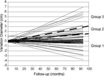
Marfan’s syndrome. Patients were included in the study if at least 3 post-operative CT scans were available and if a De Bakey Type I dissection was present. 64 patients (59.8%) full-fielded the inclusion criterias. Volumetric analyses of the aorta were performed and a linear logistic regression was performed at 5 different levels of the aorta for each patient. Analyses were performed at the aortic arch, in the descending aorta at the level of the pulmonary artery bifurcation, supra- and infradiaphragmal, as well as at the level of infrarenal abdominal aorta. According to the velocity of subsequent dilatation, patients were divided into 3 groups: group 1 included 26 patients (40.6%) without progression of the diameter in the downstream aorta, group 2 included 27 patients (42.2%) with slight progression and group 3, 11 patients (17.2%) with important progression, requiring surgery in 9 patients (81.8%). Logistic regression curve from group 1 was defined as y!0.0167xK0.0167;
group 2yO0.0167xK0.0167 and !0.0333xK0.0333 and group 3 yO0.0333xK0.0333. A slight progression (group 2) was defined as an increasing in size of the aortic arch and/or the descending aorta between 1 and 2 cm over an extrapo-lated time-period of 60 months and an important secondary dilatation (group 3) was defined as expansion of more than 2 cm in a logistic regression model (Fig. 1). Measurement of diameters of the aortic arch and/or descending aorta were performed on-line. Risk-factors were analysed and com-pared between the groups.
2.1. Statistical methods
Data are presented as absolute values or mean valuesG their first standard deviation. A Mann–Whitney U-test and c2 test were used for comparison between groups of continuous and nominal variables, respectively. Linear regression for the relationship between modification of the diameter of the aortic arch and/or descending aorta and time was analyzed. A P-value of less than 0.05 was considered significant.
3. Results
Sixty-four patients full-field the inclusion criteria. Pre-and perioperative characteristics of these patients are summarized in Table 1. Duration of follow-up was similar in all 3 groups, with an average follow-up of 52G14 months. Secondary dilatation, requiring reoperation occurred in 85% of the patients in the first 24 months following initial surgery. In the remaining 2 patients reoperation was performed 37, respectively, 44 months after repair of AADA. Thirty-eight patients (59.4%) showed an increase of the diameter of the aortic arch and/or descending aorta. Out of these 38 patients, 27 (42.2%) showed a slight increase and were assigned to group 2 and 11 (17.2%) showed an important increase and belong to group 3. Subsequent dilatation were mainly found in the supradiaphragmal section. Patients from group 3 had an average age of
Fig. 1. Linear logistic regression curve at the supradiaphragmal section for all patients (nZ64). The variation of the diameter of the descending aorta over time is displayed with a logistic regression curve. The curve consists at least of 3 measurements for each patient (mean 5.4 measurements). The dotted lines represent the repartition in group 1–3, according to the variation of the diameter over time.
Table 1
Patients characteristics from group 1 (nZ26), group 2 (nZ27) and group 3 (nZ11). Results are displayed as absolute values and mean values (G1.SD). * P-value compares group 1 and 3
Group 1 Group 2 Group 3 P-value*
Demographics
No. of patients 26 40.6% 27 42% 11 17%
Mean age (y) 61.9G11.6 60.6G12.1 57.7G13.4 !0.05
Male 20 76.9% 20 74.1% 6 54.6% !0.05 Risk factors History of smoking 9 34.6% 8 29.6% 3 27.3% ns Arterial hypertension 16 61.5% 16 59.3% 4 36.4% !0.05 Marfan syndrome 2 7.8% 1 3.7% 1 9.1% ns Malperfusion syndrome 7 26.9% 16 59.3% 6 54.6% !0.05
Dissection supraaortic branches 21 80.8% 24 88.9% 11 100.0% !0.05
Patent false lumen 15 57.7% 17 63.0% 8 72.7% !0.05
Diameter of the thoracic aorta (cm) 3.4G0.8 3.3G0.6 3.2G0.8
Perioperative data
Ascending aorta replacement 19 73.1% 19 70.1% 7 63.6% ns
Composite graft 7 26.9% 8 29.9% 4 36.4% ns
Including arch replacement 2 7.7% 3 11.1% 2 18.2% ns
Aortic cross clamping (min) 89.2G40.4 77.2G28.3 79.9G26.4 ns
Circulatory arrest (min) 22.8G10.1 24.4G7.6 21.6G5.6 ns
Bicuspid aortic valve 3 11.5% 1 3.7% 0 0% !0.05
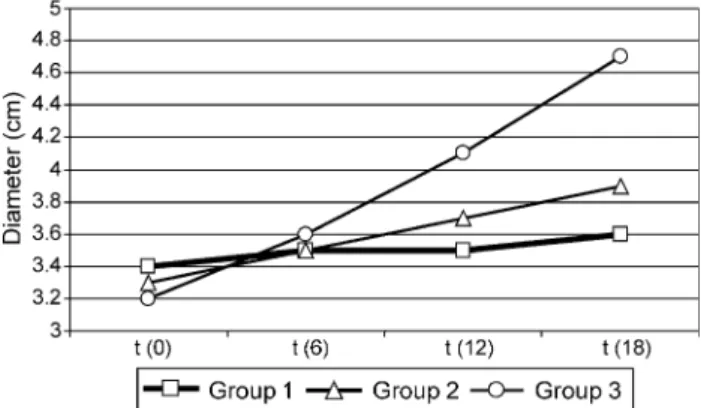
57.7G13.4 years in comparison to an average age of group 1, which was 61.9G11.6 years (P!0.05). 100% of the patients from group 3 showed dissection of the supraaortic branches (arch vessels) in comparison to 80.8% of the patients from group 1 (P!0.05). The initial diameter of the aortic arch and/or the descending aorta after the occurrence of AADA did not influence secondary dilatation. Patients from group 3 showed an average diameter of 3.2G0.8 cm and patients from group 1 3.4G0.7 cm (PZns) in the preoperative or early post-operative CT scan or MRI (before discharge) (Fig. 2). Malperfusion syndrome was much more frequent in group 3, as well as contrast enhancement in the false lumen, in comparison to patients from group 1 (Table 1).
The groups were similar in term of type of initial surgery, without any significant difference between patients receiv-ing a supracoronary replacement of the aorta or a Composite graft, as well as duration of aortic cross clamping and deep hypothermic circulatory arrest (PZns) (Table 1).
As displayed in Table 1, patients with bicuspid aortic valve and AADA, did not show important secondary dilatation of the downstream aorta and were mainly found in group 1.
4. Discussion
Several risk factors which may affect secondary dilatation especially in patients suffering from thoracic aortic aneur-ysms or acute type B aortic dissection have been described
[4–7]. Little is known about risk factors for secondary dilatation of the diseased downstream aorta in patients who underwent surgery for AADA. Bachet and colleagues have shown, that the presence of Marfan’s syndrome favours secondary dilatation after surgery for AADA and that a close follow-up in these patients is mandatory[8]. Aneurysmatic dilatation in these patients is mainly due to abnormal elastic properties of the aorta[9]. As in the present study patients with aneurysmatic dilatation of the aortic arch and/or descending aorta at the time of AADA repair have been excluded, Marfan’s syndrome was not found as an indepen-dent risk factor for subsequent dilatation in mid-term follow-up. However, the incidence of Marfan’s syndrome was 50% in the collective requiring early surgery, with 6 of 12
patients suffering form this type of disease. This confirms the findings reported by Bachet[8].
The typical risk factors described in patients suffering from thoracic aortic aneurysm could not be confirmed in the present study [5–6]. Patients from group 3, who had secondary dilatation of the aortic arch and/or descending aorta were younger than the medium in the collective, history of smoking was not more frequent than in patients from group 1 and the incidence of arterial hypertension— when AADA occurred—was less frequent. However, the extent of AADA, with involvement of the supraaortic branches and/or combined with malperfusion syndrome, seemed to be an important predictor of secondary dilatation in these patients. The high proportion of patients with patent false lumen in group 3, in comparison with group 1 and 2, additionally favours secondary dilatation. Similar findings were recently published, describing that in patients suffering from type B acute aortic dissection patency of the false lumen is the strongest independent risk-factor for dissection related death[7]and secondary dilatation[10].
Extensive destruction of the intimal layer in patients presenting with AADA may reflect an intrinsic tissue damage, due to a genetic defect. It has been shown that biomecha-nical properties of a diseased aorta is influenced by dilatation of the diseased aorta, by the presence of a bicuspid aortic valve (restricted to the ascending aorta) and by genetic defects [9,11,12]. Patients with bicuspid aortic valve disease were mainly found in group 1 and one patient was in group 2, which is consistent with the biomechanical findings described recently[11,12], which are limited to the ascending aorta. Nevertheless a diseased downstream aorta, as it can be found in De Bakey Type I dissections, can secondary dilate, requiring surgery in these patients.
In fact, the diameter of the downstream aorta in patients from group 3 showed no significant difference in comparison to patients from group 1 and 2 at the time of initial diagnosis of AADA. However, the present observations confirm the findings previously described by Griepp and his colleagues, that secondary dilatation is favoured in patients with an enlarged downstream aorta[5,6], which was observed in the follow-up in patients from group 3, where dilatation became more and more important with increasing diameter (Fig. 2). Follow-up was similar in all three groups and secondary dilatation occurred in the majority of our patients in the first 24 months after surgery. A close follow-up in these patients is crucial in this time period, regardless of the presence of the described risk-factors. Once, 24 months after surgery are over, if the patients are at lower risk and if the diameter of the aortic arch and/or the diseased downstream aorta remains stable (Fig. 2) and the false lumen is not patent, prolonged time intervals for the further follow-up of 2–3 years are probably reasonable.
We are aware that group 3 is a very small group, including 11 patients, with the highest risk for secondary dilatation of the aortic arch and/or the diseased downstream aorta. This is the strongest limitation of our study. Based on statistical analysis, the cut off points for the logistic regression curve to separate the 3 groups were chosen to improve the predictive value of the observed risk factors. However, the fact that all patients from group 3 required reoperation but only 2 patients in group 2, may partly counterbalance this
Fig. 2. Maximal diameter of the aortic arch and/or the diseased downstream aorta in patients from groups 1 to 3 at the time of follow-up 6, 12 and 18 months after surgery for AADA.
F.F. Immer et al. / European Journal of Cardio-thoracic Surgery 27 (2005) 654–657 656
limitation, reflecting the importance of the described risk-factors and encouraging our colleagues to perform a follow-up in this collective of patients.
We conclude, that AADA involving the supraaortic branches and combined with malperfusion syndrome, favour secondary dilatation in younger patients. A patent false lumen is per se an independent predictor for secondary dilatation. A close follow-up in all patients, at least up to 24 months after surgery, is mandatory to detect potentially life-threatening complications of the diseased downstream aorta.
References
[1] Strauch JT, Spielvogel D, Lauten A, Lansman SL, McMurtry K, Bodian CA, Griepp RB. Axillary artery cannulation: routine use in ascending aorta and aortic arch replacement. Ann Thorac Surg 2004;78(1):103–8. [2] Barmettler H, Immer FF, Berdat PA, Eckstein FS, Kipfer B, Carrel T.
Rsik-stratification in thoracic aortic surgery: should the EuroSCORE be modified? Eur J Cardiothorac Surg 2004;25(5):691–4.
[3] Carrel T, Beyeler L, Schnyder A, Zurmuhle P, Berdat P, Schmidli J, Eckstein FS. Reoperations and late adverse outcome in Marfan patients following cardiovascular surgery. Eur J Cardiothorac Surg 2004;25(5): 671–5.
[4] Juvonen T, Ergin MA, Galla JD, Lansmann SL, McCullough JN, Nguyen KH, Bodian CA, Ehrlich MP, Spielvogel D, Klein JJ, Griepp RB. Risk factors for rupture of chronic type B dissections. J Thorac Cardiovasc Surg 1999; 1117(4):776–86.
[5] Juvonen T, Ergin MA, Galla JD, Lansman SL, Nguyen KH, McCullough JN, Levy D, de Asla RA, Bodian CA, Griepp RB. Prospective study of the natural history of thoracic aortic aneurysm. Ann Thorac Surg 1997;63(6): 1533–45.
[6] Griepp RB, Ergin MA, Galla JD, Lansmann SL, McCullough JN, Nguyen KH, Klein JJ, Spielvogel D. Natural history of descending thoracic and thoracoabdominal aneurysms. Ann Thorac Surg 1999;67(6):1927–30. [7] Akutsu K, Nejima J, Kiuchi K, Sasaki K, Ochi M, Tanaka K, Takano T.
Effects of the patent false lumen on the long-term outcome of type B acute aortic dissection. Eur J Cardiothorac Surg 2004;26(2):359–66.
[8] Bachet JE, Termignon JL, Dreyfuss G, Goudot B, Martinelli L, Piquois A, Brodaty D, Dubois C, Delentdecker P, Guilmet D. Aortic dissection. Prevalence, cause, adn results of late reoperations. J Thorac Cardiovasc Surg 1994;108:199–206.
[9] Groenink M, de Roos A, Mulder BJ, Verbeeten Jr B, Timmermans J, Zwinderman AH, Spaan JA, van der Waal EE. Biophysical properties of the normal-sized aorta in patients with Marfan syndrome: evaluation with MR flow mapping. Radiology 2001;219(2):535–40.
[10] Suyeoshi E, Sakamoto I, Hayashi K, Yamaguchi T, Imada T. Growth rate of aortic diameter in patients with type B aortic dissection during the chronic phase. Circulation 2004;110(11):256–61.
[11] Nistri S, Sorbo MD, Basso C, Thiene G. Bicuspid aortic valve: abnormal elastic properties. J Heart Valve Dis 2002;11(3):369–73.
[12] Bauer M, Pasic M, Meyer R, Goetze N, Bauer U, Siniawski H, Hetzer R. Morphometric analysis of aortic media in patients with bicuspid and tricuspid aortic valve. Ann Thorac Surg 2002;74(1):58–62.
Appendix A. Conference discussion
Dr C. Hagl (Hannover, Germany): I wonder if you have looked at beta-blockers in this context? Were all patients on beta-blocker therapy during the postoperative period? I think it is an important issue since it has been shown that such a therapy had a significant impact on dilatation of the descending aorta especially during long-term follow-up.
Dr Immer: All the patients discharged from the hospital and followed at the outpatient clinic are on beta-blocking therapies. Radiographic follow-up and flow measurements by MRI showed additionally that also arterial hypertension has to be corrected. If you replace the total aorta, you have to lower blood pressure, which is as important as beta-blockers. But all of our patients are on the beta-blocking agents.
Dr M. Cotrufo (Naples, Italy): So in which case would you suggest an elephant trunk, considering this very high frequency of dilatation of the descending aorta?
Dr Immer: In our institution we try to perform in acute type aortic dissection mainly ascending aortic replacement and a hemiarch. If the aortic arch is dilated or if we know that the descending aorta is already dilated, we try to implant an elephant trunk primarily to avoid secondary clamping at the level of the aortic arch.