603
REVIEW ARTICLE
Composition and characteristics of camel milk
BY ZAKARIA FARAHLaboratory of Dairy Science, Swiss Federal Institute of Technology (ETH) ETH-Zentrum, CH-8092 Zurich, Switzerland
{Received 22 February 1993 and accepted for publication 28 February 1993)
Introduction
Present distribution and milk production Main components
Gross composition
Overall protein composition Casein fractions
Size distribution of casein Whey protein fractions Lipids
Fatty acid composition
CONTENTS PAGE 603 604 605 605 606 607 609 611 612 612 Phospholipids
Physical state and properties of fat globules Lactose
Minerals and vitamins Enzymic coagulation Effects of heat on milk Antibacterial activity Conclusions References INTRODUCTION PAGE 613 614 616 616 617 620 623 623 624
Camels belong to the family Camelidae and thereby to the suborder Tylopoda. The tylopoda themselves belong to the order Artiodactyla or cloven-footed animals. The family of Camelidae contains the genera Camelus (old world camel) and Lama (new world camel). The Camelidae originated in North America where the earliest fossil remains of Camelidae have been found. The genus Camelus migrated from North America in the late Tertiary across the then existing land bridge to Asia and Africa. The llamas on the other hand reached South America in the ice age across the Central American land bridge. Included in the genus Camelus are the one-humped dromedary (Camelus dromedarius) and the two-humped bactrian (Camelus bactrianus). The term dromedary is derived from the Greek 'dromados' (run) and in the strict sense is used for riding camels. The name 'Bactrian' for the two-humped camel refers to the area ' Baktria' in North Afghanistan where this type of camel is thought to have originated. The dromedary is slim, long-legged, short-haired and has its habitat in warm arid and semi-arid areas. The bactrian is stockier, short-legged and has a thicker and longer coat than the dromedary. It mainly occurs in cold and mountainous areas (Simpson, 1945; Zeuner, 1963).
This review is concerned entirely with the one-humped camel; the term ' camel' should therefore be taken to refer to Camelus dromedarius unless specifically stated otherwise.
The majority of the studies conducted on camels concentrate on anatomical features, traditional management and physiological adaptation to desert conditions (Cauvet, 1925; Schmidt-Nielsen, 1964; Bulliet, 1975; Gauthier-Pilters & Dagg, 1981;
604 j 1. Estimated camel Country Africa Table Country Egypt Ethiopia India Kenya Pakistan Somalia Sudan Tunisia Algeria Chad Djibouti Egypt Ethiopia Kenya Libya Mali Mauretania Morocco Niger Nigeria Senegal Somalia Z. FARAH
populations of Africa and the rest of the world in Camel population, thousands 147 26 405 95 960 574 75 198 718 20 350 18 6 5400 Country Sudan Tunisia Upper Volta West Sahara Other regions Afghanistan China India Iraq Mongolia Pakistan Saudi Arabia USSR t From FAO (1979). Camel population, thousands 2904 205 5 86 290 1040 1174 232 615 819 108 230
2. Average milk yields of camels reported from various sourcei Daily yield, kg 3-5-i-5 5-13 7-18 2-12 8-10 3-9 5-10 4 Lactation length, months 9 12-18 15 11-16 12 9-18 10-12 12 Calculated lactation yield of 305 d, 1068-1373 1525-3965 2105-5551 610-3660 2440-3050 915-2745 1525-3050 1220 kg Reference El-Bahay (1962) Knoess (1977) Rao (1974) Field (1979) Knoess (1979) Hartley (1979) El-Amin (1979) Biirgemeister (1974)
Wilson, 1984; Yagil, 1985). Information about camel milk is mostly limited to some data on gross composition. Studies on individual components and their physico-chemical characterization have received very limited attention. The present review attempts to place the scarce available information on camel milk components into a coherent framework. Emphasis is given to comparison with bovine milk. This is natural in as much as bovine milk has been the subject of intensive research over a long period and, although many areas of uncertainty still exist, the general physicochemical properties of all main components of bovine milk are well established.
PRESENT DISTRIBUTION AND MILK PRODUCTION
According to FAO (1979) statistics (Table 1) there are ~ 17 million camels in the world, of which 12 million are found in Africa and 4-9 million in Asia. Of this estimated world population, 15 million are believed to be one-humped camels and 1-9 two-humped. Of the camel population, 60% is concentrated in the four North East African countries Somalia, Sudan, Kenya and Ethiopia; Somalia, with over 5 million, has the largest herd in the world.
Some of the existing data on milk yields of the dromedary are summarized in Table 2. For better comparison, milk yields are calculated for a lactation period of 305 d. Reported milk yields vary from 35 kg for animals under desert conditions up to
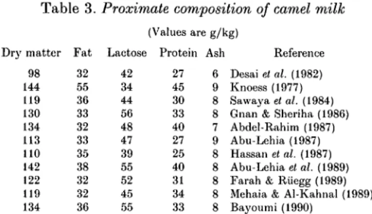
Table 3. Proximate composition of camel milk Dry matter 98 144 119 130 134 113 110 142 122 119 F a t 32 55 36 33 32 33 35 38 32 32 Lactose 42 34 44 56 48 47 39 55 52 45 (Values are g/kg) Protein 27 45 30 33 40 27 25 40 31 34 Ash 6 9 8 8 7 9 8 8 8 8 Reference Desai et al. (1982) Knoess (1977) Sawaya el al. (1984) Gnan & Sheriha (1986) Abdel-Rahim (1987) Abu-Lehia (1987) Hassan et al. (1987) Abu-Lehia et al. (1989) Farah & Riiegg (1989) Mehaia & Al-Kahnal (1989) 134 36 55 33 8 Bayoumi (1990)
It is difficult to estimate the daily milk yield of a camel under pastoral conditions. On the one hand, throughout lactation the calves are still sucking and therefore the actual volumes of milk secreted are higher than the figures presented in Table 2. On the other hand, milking frequency varies among the different pastoral groups. Camels may be milked once a day, among the Murah of Arabia (Cole, 1975), from two to four times among the Somali (Bremaud, 1969; Hartley, 1979) and the Rendille of Kenya (Spencer, 1973) and as many as six or seven times among the Afar of Ethiopia (Knoess, 1977). The Afars may also leave their animals unmilked for a whole day, which may account for sporadic very high estimates. Milk yields will also vary with species, breed, feeding and management conditions, and stage of lactation. The dromedary, like most other species, gives most milk near the beginning of the lactation period. In a study of the camels of Northern Kenya, Field (1979) estimated their daily yield at 21 1 in the second week of lactation falling to 4-8-2-2 1 by the sixteenth week of lactation. The average length of lactation in the camel is 12 months, but it may vary from 9 to 18 months, depending on management and environmental conditions.
The milk yield estimates presented in Table 2 indicate that the camel is potentially a better milker than many African Zebu cows under the same environmental conditions. The daily milk yield of Zebu cows varies between 0-5 and 1-5 kg (Kiwuwa, 1973). Spencer (1973) estimated that in Kenya one Rendille camel produced as much milk as four cows in the same region.
MAIN COMPONENTS
Gross composition
Camel milk is generally opaque-white. It has a sweet and sharp taste, but sometimes can also be salty. The changes in taste are caused by the type of fodder and availability of drinking water. The pH of camel milk ranges from 6-5 to 6-7 with an average value of 6-56, and the density from 1-025 to 1-032 with an average of 1-029. Both values are lower than those of cows' milk (El-Bahay, 1962; Rao et al. 1970; Shalash, 1979; Sawaya et al. 1984; Farah & Bachmann, 1987).
Some recent data on the composition of camel milk are presented in Table 3. The differences among the various sets of data undoubtedly reflect differences in breed and state of lactation of the animals sampled, in sampling procedures and perhaps in analytical procedures as well.
One of the important factors that affects camel milk composition is the water. Yagil & Etzion (1980) examined the effects of restricting drinking water on camel
606 Z. FARAH
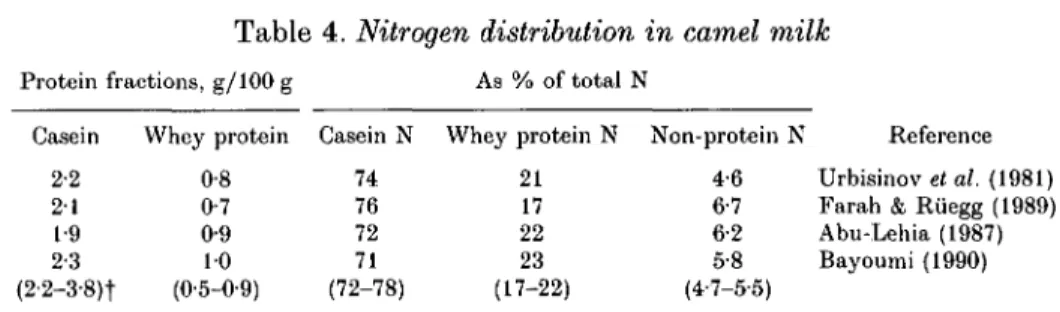
Table 4. Nitrogen distribution in camel milk
Protein fractions, g/100 g As % of total N Casein 2-2 2 1 1-9 2-3 (2-2-3-8)t Whey protein 0-8 0-7 0-9 10 (0-5-0-9) Casein N 74 76 72 71 (72-78) Whey protein N 21 17 22 23 (17-22) Non-protein N 4-6 6-7 6-2 5-8 (4-7-5-5) Reference Urbisinov et al. (1981) Farah & Riiegg (1989) Abu-Lehia (1987) Bayoumi (1990)
•f Values in parentheses are ranges for cows' milk.
milk. While the diet remained unchanged throughout the year, great changes in water content of milk were found. The camels were allowed drinking water ad lib. only during the winter. From spring until the end of summer, the mothers and the calves were allowed to drink only once a week for 1 h. With water freely available the water content of the milk was 86%, but when water was restricted the water content of milk rose to 9 1 % . These changes reflect the reported range and this makes it important when the milk was sampled by the various investigators. This study shows that the lactating camel loses water to the milk in times of drought. This could be a natural adaptation in order to provide necessary fluid to the dehydrated calf.
The colostrum of camels is white and slightly diluted as compared with the colostrum of the cow (Rao et al. 1970; Yagil & Etzion, 1980). Only fragmentary data are available on colostrum composition in camel milk. The most complete data are those reported by Sestucheva (1958) and Abu-Lehia et al. (1989) for Russian and Saudi camels. Sestucheva studied ten Kazakhstan camels. The first colostrum obtained after 3 h post partum contained on average 30-4% total solids, 0-20% fat, 19-4% protein, 7-2% lactose and 3-8% minerals. During the first 2 d of lactation the solids content fell to 18-4%, mainly owing to the decline of total proteins to 3-6% and minerals to 0-1%. The fat content increased to 5-8% whilst the lactose level remained practically unchanged. The composition then remained fairly constant until the tenth day when the experiment ended.
Abu-Lehia et al. (1989) examined the colostrum often Saudi camels (Majaheem breed) during their first season of lactation up to 10 d post partum. At parturition, the contents of total solids, fat, protein, lactose and minerals were 20-5, 0-20, 130, 2-7 and 1-0% respectively. After 3 d total solids decreased to 13-6%, protein to 4-7% and minerals to 0-8%. However, the fat content rose to 1-5% and lactose to 4-4%. Overall protein composition
The N distribution of camel milk from four different regions is presented in Table 4. All authors used the method of Rowland (1938) modified by Aschaffenburg & Drewry (1959) for determining the N fractions.
The average casein and whey protein content in camel milk varies between 2-3 and 1-9% and 0-7 and 1-0% respectively. The values of casein N, whey protein N and non-protein N (NPN), expressed as a percentage of the total milk protein N, lay within the ranges 71-76 %, 17-23 % and 5-8-4-6 % respectively. The results in Table 4 indicate that the protein and N fractions in camel milk are generally similar to those in cows' milk. However, camel milk seems to contain somewhat higher amounts of NPN fractions than cows' milk.
Recent data on amino acid composition of camel milk protein are presented in Table 5. Amino acid content appears generally similar to that of cows' milk protein.
Table 5. Amino acid composition of camel and cows' milk 607 Amino acid Alanine Arginine Aspartio acid Cysteine Cystine Glutamic acid Glycine Histidine Isoleucine Leucine Lysine Methionine Phenylalanine Proline Serine Threonine Tryptophan Tyrosine Valine (Values are Camel (Sawaya et al. 1984) 2-8 3-9 7-6 — 10 23-9 1-7 2-5 5-4 10-4 7 0 2-5 4-6 111 5-8 5-2 1-2 4-5 6 1 g/100 g total protein) Camel
(Mehaia & Al-Kahnal, 1989)
2-7 3 8 6-4 — 0-6 19-5 1-3 2-7 5 0 9-5 7 1 3-6 5-6 111 4-2 4-3 — 4 0 6-9 Cow (Renner, 1991) 3-5 3-7 7-9 — 0-7 21-8 2 1 2-8 6-4 10-4 8-3 2-7 5-2 100 5-6 5 1 1-4 5-3 6-8
Table 6. Caseins of camel milk Casein Molecular mass, Da Reference
a8l 35000 Farah & Farah-Riesen (1985)
<xsl 31000 Larsson-Raznikiewicz & Mohamed (1986)
as2 25000 Larsson-Raznikiewicz & Mohamed (1986) p 35000 Farah & Farah-Riesen (1985)
/? 27000 Larsson-Raznikiewicz & Mohamed (1986)
Casein fractions
According to early nomenclature of cows' milk proteins (Whitney et al. 1976), caseins were defined as those phosphoproteins that precipitate from raw skim milk upon acidification to pH 4-6 at 20 °C. Individual caseins were isolated and defined according to their electrophoretic mobility in alkaline polyacrylamide or starch gels. The latest report on milk protein nomenclature (Eigel et al. 1984) recommends abandoning the use of electrophoresis as a basis for casein classification and identifying caseins according to the homology of their primary structure.
The camel milk caseins reported here and presented in Table 6 are obtained by acid precipitation at pH 4-6 and identified by electrophoresis. At present the primary structure of the individual caseins is not known. Thus the designation a and /? given to camel casein fractions is still uncertain and remains to be confirmed.
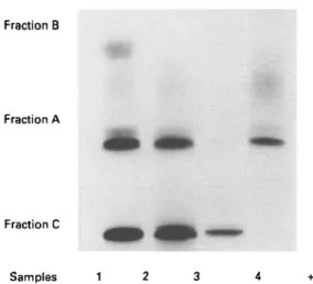
Two casein fractions homologous to bovine a- and /?-casein have been isolated from camel casein (Farah & Farah-Riesen, 1985). The casein fraction was obtained by the usual acid precipitation following the same pattern as that for cows' milk casein. Fig. 1 shows the electrophoretic pattern of camel milk, acid-precipitated camel casein, fraction B, which is isolated according to the /?-casein preparation method of Aschaffenburg (1963), and fraction A, obtained as a-casein by the urea method of Hipp et al. (1952). Fractions A and B can be considered as possibly homologous to bovine a- and /?-casein. Fraction B (^-casein) is clearly separated and
608
Fraction A
Fraction C
Samples 1 2 3 4 +
Fig. 1. PAGE patterns of camel milk and eamel milk casein fractions: 1, fraction A; 2, fraction B; 3, whole camel casein; 4, camel milk (Farah & Farah-Riesen, 1985).
consists of a single strong band. Fraction A (a-casein) shows one strong band and some diffuse slow moving bands. In general a-casein occurs in bovine milk as a mixture of many subfractions such as as l and as2. The fractionation method applied suggests the strong band to be homologous to bovine asl-casein and the diffuse band moving behind to be as2-casein. No protein bands homologous to bovine /c-casein could be clearly detected in the electrophoretic pattern. The molecular masses of the camel casein fractions were estimated by means of SDS-PAGE to be 32 kDa for fraction B (/?-casein) and 35 kDa for fraction A (a-casein). These values are higher than those of bovine caseins, usually reported as 24 kDa for /?-casein, 22 kDa for asl-casein and 25 kDa for as2-casein (Eigel et al. 1984).
Larsson-Raznikiewicz & Mohamed (1986) isolated four casein fractions by ion-exchange chromatography and identified them by PAGE. Amino acid and phosphorus analysis revealed that proteins analogous to the asl-, as2-, /?- and /c-caseins of bovine milk occur in camel milk. For camel milk, asl- and /?-casein
predominated: as2-casein gave diffuse bands. No protein band corresponding to
/c-casein could be detected. This fraction could be isolated only by ion-exchange chromatography and was identified as homologous to bovine /c-casein on the basis of its amino acid composition. Furthermore the asl- and /?-casein of camel milk were
phosphorylated to about the same extent, and as2-casein was more heavily
phosphorylated than the corresponding bovine casein. In Table 7 the amino acid compositions of the different casein fractions isolated by ion-exchange chromato-graphy are given. It shows that there are close similarities in amino acid composition between the camel and bovine casein fractions. The molecular masses
estimated from SDS-PAGE were 27, 31 and 25 kDa for /?-, asl- and as2-casein
respectively. These values differ from those obtained by Farah & Farah-Riesen (1985) for camel a- and /?-caseins, with ^-casein showing the larger deviation.
Pant & Chandra (1980) and Hassan et al. (1987) also reported the occurrence of proteins homologous to cows' milk a- and /?-casein in camel milk, but without sufficient information on the methods used.
Table 7. Amino acid composition of camel and cows' milk casein^
(Values are g/100 g total casein)
/^-Casein /c-Casein <xs] -Casein aa2-Casein
609
Amino acid Alanine Arginine Aspartio acid Cysteinc Glutamic acid Glycine Histidine Isoleueine Leucine Lysine Methionine Phenylalanine Proline Scrine Threonine Tyrosine Tryptophan Valine Camel 2-9 1-9 3-8 0 0 19-5 1-2 1-8 5-7 10-8 5-9 2-9 3-8 18-3 6 1 5 0 2-5 0 0 8-0 Cow 2-4 1-9 4-3 0 0 18-7 2-4 2-4 4-8 10-5 5-3 2-9 4-3 16-7 7-7 4-3 1-9 0-5 9 1 Camel 4-8 2-7 6-2 0-6 17-7 2-2 1-9 6-9 7-2 5-6 1-5 3-6 14-4 6-3 71 3-6 0-7 7 1 Cow 8-3 3 0 7 1 12 160 1-2 1-8 7 1 4-7 5-3 1-2 2-4 11-8 7-7 8-9 5-3 0-6 6-5 Camel 3 0 4-9 9 1 0 0 20-9 2-3 2-3 6-2 8-0 7-3 1-7 2-7 8-4 8 0 4-9 4-6 10 4-8 Cow 4-5 3 0 7-5 0 0 19-6 4-5 2-5 5-5 8-5 8-0 2-5 4 0 8-5 8 0 2-5 5 0 10 5-5 Camel 2-9 1-8 6-5 10 21-8 1-9 2-7 5-3 5 1 10-6 1-6 5 1 5 1 6-7 8-0 5-7 2-2 6 1 Cow 3-9 2-9 8-7 10 19-3 10 1-4 5-3 6-3 11-6 1-9 2-9 4-8 8-2 7-2 5-8 10 6-8 From Larsson-Raznikiewicz & Mohamed (1986).Fig. 2. Freeze-fractured casein micelles in camel milk: cm. casein micelles; sm, submicelles (Farah & Riiegg, 1989).
Size distribution of casein
The casein micelle is the backbone of the colloidal milk system, and determines milk stability. There are no reports that give a detailed picture of the micellar state of casein in milk other than cows' milk. The state of the casein micelle structure in camel milk has seldom been investigated. At present, there are only the following
610 106 105 u § 10" <a . a E 3 2 1O2 10 \ \ <
N
\
\
\
Z.
\
FABA Hi \
I V
I3 x
-#—,
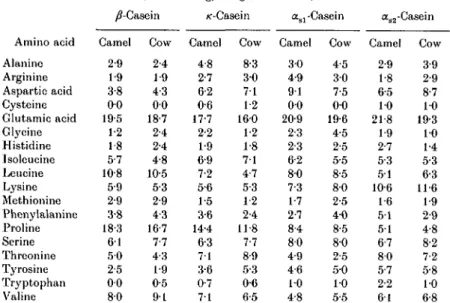
100 200 300 400 Micelle diameter, nm 500 600Fig. 3. Number of particles observed in freeze-fraetured *, camel; x , cows' and O, human milk. The ordinate is logarithmic and gives the number of particles per mm2 fractured areas and per nm classes widths (Farah & Riiegg, 1989).
three reports published on the subject. Gouda et al. (1984) examined the casein micelles of an unspecified number of Egyptian camel milk samples. The milk was solidified with agar and examined in thin sections by electron microscopy. The casein micelles ranged in size from 25 to > 400 nm. The technique did not allow clear identification of smaller micelles. Ali & Robinson (1985) analysed the size distribution of casein micelles in six samples of camel milk using transmission electron microscopy. Milk was collected from individual Bedouin camels reared by nomads from the Sudan. The total number of particles counted was 2448. They found that the majority of the casein micelles had comparatively small diameters of 28-240 nm. The number average diameter of casein micelles was 160 nm. However, this value overestimates the true mean because particles with diameters < 14 nm could not be measured. Parah & Riiegg (1989) studied the casein micellar structure of milk from ten individual camels of Northern Kenya. The size distribution of casein micelles in camel milk was determined by electron microscopy. The milk samples were cryofixed and freeze-fraetured. The total number of particles counted was 6618.
Fig. 2 shows a typical electron micrograph of casein particles in a freeze-fracture sample of camel milk. The mean diameter of the submicelles was on average 15 nm. The average number of particles observed on such freeze-fraetured surfaces is shown graphically in Fig. 3. The distribution is significantly broader than that of cows' and human milk (Riiegg & Blanc, 1982) and shows a greater number of large particles.
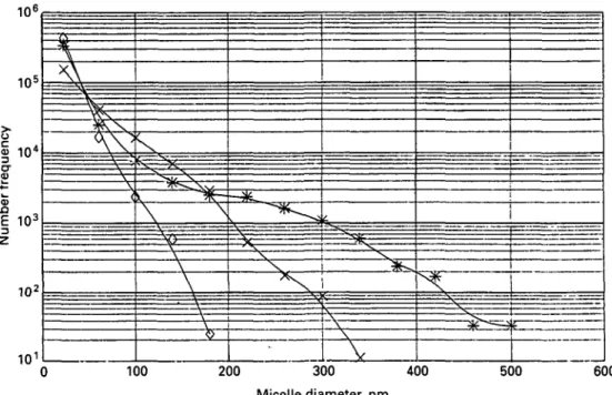
The particles in the lowest size class with diameters < 40 nm comprise ~ 80 % of the observed total number of particles, but represent only 4 - 8 % of the mass or volume of the casein in camel milk. I t is therefore meaningful to consider the weight or volume frequency distribution. Fig. 4 shows the volume frequency of the pooled data for the camel milk samples compared with the distributions found in bovine and
40 r > o c <D 3 22 60 100 140 180 220 260 300 340 380 420 460 500 Micelle diameter, nm 540
Fig. 4. Size distribution of casein micelles in camel milk ( • ) compared with cows' milk (H) and human milk ( • ) (Farah & Riiegg, 1989).
Table 8. Whey proteins of camel milk Whey protein Tmmunoglobulin Serum albumin Serum albumin Serum albumin a-Lactalbumin A a-Lactalbumin B a-Lactalbumin a-Laetalbumin'l' Novel camel wheyt Novel camel wheyf
t Prin Molecular mass, — 66000 — 14000 14000 14000 14600 14000 15000 Da Reference Conti et al. (1985) Conti el al. (1985) Farah (1986) Beg et al. (1987) Conti et al. (1985) Conti et al. (1985) Farah (1986) Beg et al. (1985) Beg et al. (1986) Beg et al. (1987) larv structure determined.
mature human milk (Riiegg & Blanc, 1982). The volume distribution curve of casein micelles in camel milk is broad and shows a maximum between 260 and 300 nm (cows' milk 100-140 nm).
From the few available data on camel casein micelles presented here it can be concluded that camel milk casein differs from cows' milk casein in terms of micellar size distribution. Some possible consequences of this difference in relation to rennet coagulation properties of camel milk will be discussed later.
Whey protein fractions
The term ' whey protein' should be used only in a general sense to describe milk proteins soluble at pH 4-6 and 20 °C (Eigel et al. 1984). Classification of individual whey proteins should be based on the primary sequence of the amino acids in their polypeptide chains, although gel electrophoresis can still be used to characterize and identify the individual whey proteins.
The camel whey proteins reported below and presented in Table 8 were obtained from raw skim milk. The caseins were removed either by acidification to 4-6 or by
612 Z. FARAH
high speed centrifugation. Individual whey proteins have been identified according to their chromatographic and electrophoretic mobilities. Some whey proteins were classified on the basis of the primary sequence of their amino acid chains.
Conti et al. (1985) separated camel whey proteins by gel chromatography on Sephadex G100 and identified some components using electrophoretic methods. The study revealed the presence of immunoglobulins and serum albumin in camel whey proteins. In the study two different a-lactalbumins were isolated and characterized. These two proteins, a-lactalbumin A and B, had similar molecular masses (~ 14 kDa), immunological properties and electrophoretic mobility, but had different isoelectric points, amino acid composition and N-terminal sequence.
Farah (1986) examined whey proteins of camel milk by SDS-PAGE. The study revealed four clearly separated whey proteins. By comparing with standard marker proteins, two protein bands could be shown to be identical to standard bovine serum albumin (molecular mass 66 kDa) and a-lactalbumin (molecular mass 14 kDa). The other whey proteins of molecular masses 23 and 43 kDa could not be identified.
Beg et al. (1985) separated camel whey proteins by gel chromatography on Sephadex G25 and purified them by reversed-phase HPLC. Amino acid and primary structure analysis revealed the presence of whey proteins homologous to bovine a-lactalbumin. The isolated camel a-lactalbumin has a molecular mass of 14-6 kDa and was found to contain 123 amino acid residues, like bovine a-lactalbumin. Mobility on SDS-PAGE was also identical for the two proteins. In other studies Beg et al. (1984, 1986, 1987) isolated and characterized two camel whey proteins that have no homology with known bovine milk whey proteins. One of the novel camel whey proteins with molecular mass 14 kDa is rich in cysteine/half-cystine. Its 117 amino acid residues have 16 half-cystine residues. Such residues usually occur in disulphide links in milk and other extracellular proteins. Thus, the new camel whey protein is likely to have a rigid, highly cross-linked polypeptide chain. This new protein exhibited some structural similarities to bovine /?A2-casein in the N-terminal region. The similarity affected those residues known to be phosphorylated in /?-casein. However, /?-casein lacks cysteine residues. The other new camel whey protein found in these studies has a molecular mass of 15 kDa. It consists of 112 amino acid residues and does not contain cysteine. No obvious structural similarities were noted between the novel milk proteins and other milk proteins characterized.
Lipids
Lipids in milk fat serve nutritionally as an energy source, act as a solvent for the fat-soluble vitamins and supply essential fatty acids. In milks of all species studied to date, triacylglycerols are by far the major lipid class of milk fat, accounting for 97-98 % of the total lipids in most species. The triacylglycerols, which contain a great variety of fatty acids, are accompanied by small amounts of diacylglycerols and monoacylglycerols, cholesterols, free fatty acids and phospholipids.
The fat content in camel milk varies between 2-7 and 3-6%. Most of the data available on camel milk fat are on the fatty acid composition and to a lesser extent on phospholipids and some properties of fat globules.
Fatty acid composition
According to the limited information available, the first data on fatty acid composition in camel milk fat were published by Dhingra (1934), who examined the milk fat of Indian camels using older techniques of fractional distillation. This work was followed by the studies of Glass et al. (1967), who reported on the fatty acid
Table 9. Fatty acid composition of camel and cows' milk fat
(Values are g/100 g total fatty acids)
Camel milk fat Cows' milk fat atty acid 4:0 6:0 8:0 10:0 10:1/1-7 12:0 12:l?i-7 13:0 14:0 14:ln-5 15:0 15:ln-5 16:0 16:ln-7 17:0 17:l>i-8 18:0 18:ln-9 18:2/1-6 18:3n-3 20:0 22:0 2:ln-7 Farah et al. (1989) 0-66 0-37 0-23 0-90 019 079 — — 12-5 11 1-3 0-23 31-5 9-4 0-92 0-60 12-5 191 3-4 1-4 103 — Abu-Lehia (1989) — — 0 1 012 — 0-77 — — 101 1-86 1 62 — 26-6 1040 121 — 12-2 26-3 2-94 1-37 0-57 0-08 057
Gnan & Sheriha (1986) 10 — 0-5 0 1 — 0-5 0 1 0 1 100 1-5 0-5 — 31-5 9 0 0-5 0-5 140 250 3 0 — 0-5 — Abu-Lehia (1989) 3-5 2 1 1-4 21 3 1 — — 104 1-70 2-44 — 26-60 1-70 1-62 — 7-86 290 3-20 110 O i l 0-23
composition of milk fat from 57 species, including camels. Further data on the subject are provided by the studies of Sawaya et al. (1984), Gnan & Sheriha (1986), Hagrass et al. (1987), Abu-Lehia (1989) and Farah et al. (1989). Some representative data on the composition of the main fatty acids in camel milk fat are listed in Table 9. All the data were obtained by gas—liquid chromatography. The list is weighted in favour of the more recent studies and, as far as can be judged, the camels were fed all year round exclusively by grazing with no supplementary feed. Fatty acids of milk from cows living under the same conditions are also given for comparison.
Fatty acid composition is influenced to some degree by environmental and physiological factors such as diet, stage of lactation and genetic differences. Within these limitations, the general pattern of the camel milk fatty acids indicates that short-chain fatty acids, C4-C12, are present in very small amounts in camel milk fat compared with cows' milk fat, but that the concentrations of C14:0, 016:0 and C18:0 are relatively high.
Phospholipids
Phospholipids are a small but important fraction of the lipids of milk and are found mainly in the milk fat globule membrane. At present the work of Morrison (1968a, b) provides the only information available on phospholipids in camel milk. Phospholipids were isolated from the milks of cow, sheep, Indian buffalo, ass, pig, human and camel by quantitative two dimensional thin layer chromatography. The distribution of phospholipids was found to be constant in the milks of all the species studied. However, the study showed that phospholipid fatty acids of camel milk are not entirely characteristic of the ruminant herbivores.
phospho-614 Z. FARAH
lipids; they have only a small proportion of fatty acids with more than two double bonds. Their sphingomyelin contains a high proportion of tricosanoic acid (23:0) but little nervonic acid (24:ln-9).
Camel milk phospholipid fatty acids have high amounts of linoleic acid (18:3?i—3) and long-chain polyunsaturated acids. Its sphingomyelin contains a higher proportion of nervonic acid and lower tricosanoic acid than that of the ruminant herbivores.
Camel milk phosphatidylethanolamine is also unusual in that it contains 15% plasmalogen whereas the largest amount reported in other phospholipids is 4 % plasmalogen in bovine milk phosphatidylcholine (Morrison et al. 1965).
Physical state and properties of fat globules
The bulk of the fat in milk exists in the form of small spherical globules of varying sizes. The surface of these fat globules is coated with a thin layer known as the fat globule membrane, which acts as the emulsifying agent for the fat suspended in milk. Until recently the fat globule membrane of camel milk had not received much attention.
Gouda et al. (1984) reported an electron microscopy study on the size distribution of fat globules of camel milk. Milk from camels was solidified with agar and examined in thin sections under the electron microscope. Fat globules ranged in diameter from 1 to 5 fim with nearly 50% in the 2—3 /an range.
Knoess et al. (1986) analysed camel milk from five different animals for the size of fat globules and found an average globule size between 2-31 and 3-93 /on. He found that the fat globule membrane in camel milk was far thicker than in other types of milk, the term 'thickness' being used in relation to the diameter of the fat globule. In a study on some physical properties of camel milk, Wahba et al. (1988) found that the size of the fat globules in camel milk varied between 2-60 and 3-25 fim with an average of 2-9 fim.
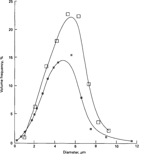
The size distribution of camel milk fat globules was determined by light microscopy (Farah & Riiegg, 1991). A total of 1800 particles was counted. Fig. 5 shows the frequency distribution of fat globules in camel milk compared with the distribution found in cows' milk (Precht et al. 1987). The results show similar globule size distributions in bovine and camel milks. From the light microscopy data a number average diameter of 2-61 fim, a volume to surface mean diameter of 4-40 fim, a weight average diameter of 511 fim and a distribution width of 40-1 % could be calculated.
All the studies presented here indicate that fat globule size distribution of camel milk is similar to that in cows' milk.
The creaming properties of fat globules in camel milk have been recently studied by Farah & Riiegg (1991). The natural creaming of raw and heated camel milk was examined in comparison with cows' milk. The milk samples were heated for 30 min at 55, 60, 62, 68, 70 and 77 °C. The milk samples were left to cream at 4 °C and the cream layer was measured after 5 and 24 h. Compared with cows' milk, camel milk showed a very slow creaming rate at all temperatures. Creaming layers varied from 0-5 to 2 ml at 4 °C.
It is well established that the main factor responsible for rapid formation of the cream layer on cows' milk is a protein adsorbed on cold fat globules, which has the characteristics of a euglobulin. This protein, known as fat agglutinin, promotes clustering of globules (Mulder & Walstra, 1974). To test whether the low creaming capacity of camel milk could be due to a deficiency in agglutinin, creaming capacity
Camel milk
615
25 20 S? 15 a)I
0) E 10 10 12 Diameter, /zmFig. 5. Size distribution of fat globules in rj, camel milk; •, cows' milk (Precht et al. 1987; Farah & Riiegg, 1991).
Table 10. Creaming of camel and cows' milks and of various combinations of their cream and skim milks in 100 ml measuring cylinders at 4 ° C |
Cows' Camel Cows' Camel Cows' Camel Sample milk milk skim + cows' skim + camel skim + camel skim + cows' cream cream cream cream Cream layer after 24 h, ml 12 1 11 1 7 3 Fat in lower 50 ml of cylinders after 24 h, % 0-8 3-2 0-7 3-4 2 0 2-9 t From Farah & Riiegg (1991).
was studied in various combinations of skim milks and creams of raw camel and bovine milks. Results of the cream volume and fat percentage obtained are presented in Table 10. All systems that contained skim camel milk creamed poorly. This could be an indication that camel milk lacks the agglutinating substance required to cluster fat globules. The creaming behaviour of camel milk appears to be similar to
616 Z. FAEAH
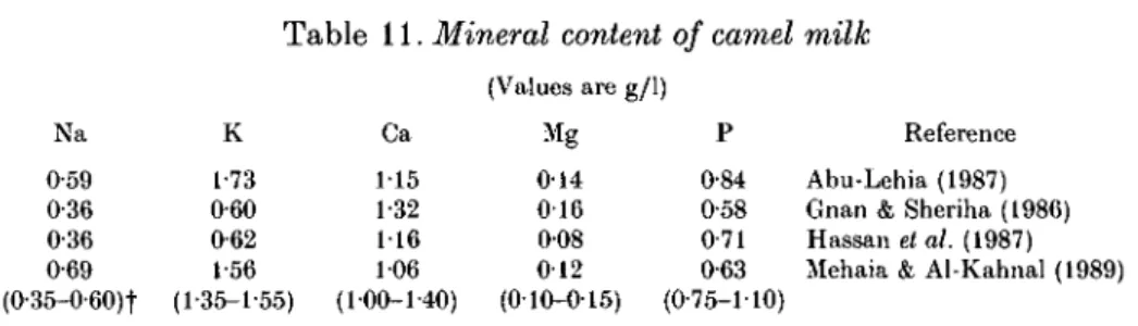
Table 11. Mineral content of camel milk
(Values are g/1) Na 0-59 0-36 0-36 0-69 (0-35-0-60)t K 1-73 0-60 0-62 1-56 (1-35-1-55) Ca 115 1-32 116 106 (100-1-40) Mg 014 0 1 6 008 012 (010-015) P 0-84 0-58 0-71 0-63 (0-75-1-10) Reference Abu-Lehia (1987) Gnan & Sheriha (1986) Hassan et al. (1987) Mehaia & Al-Kahnal (1989)
f Values in parentheses are ranges for cows' milk.
that of buffalo and goat milks, which show poor creaming ability owing to an insufficient quantity of agglutinin (Abo-Elnaga et al. 1966; Parkash & Jenness, 1968).
Lactose
Lactose is the major carbohydrate in the milk of most mammals and it is generally accepted that non-mammalian sources of lactose are very rare.
Nothing is known about the chemistry and properties of lactose in camel milk. The content in camel milk ranges from 3-4 to 5-6%, slightly higher than the lactose content in cows' milk. Hassan et al. (1987) followed the lactose content in camel milk during the lactation period and found minimal variation. Examining the effect of drought on the composition of camel milk, Yagil & Etzion (1980) found that lactose content was low at birth, ~ 2-8%, but within 24 h there was an increase of 36%. There was a further increase up to 5 % as long as drinking water was available. Dehydration led to a decline in the percentage of lactose in the milk until 2-9 % was reached. According to the authors this change in lactose concentration would account for the milk being described as sometimes sweet and other times bitter. Minerals and vitamins
Milk mineral salts are mainly chlorides, phosphates and citrates of Na, Ca and Mg. Although salts comprise < 1 % of the milk, they influence the physical state and stability of milk proteins, particularly the caseinate, which are very dependent on the composition of the salt system. The mineral content of camel milk expressed in ash ranges from 0-6 to 0-8 %. Little is known about the mineral content of camel milk. Table 11 presents results of analysis for the four principal cationic and one principal anionic constituents. These studies appear to be the only ones in which all these constituents were determined in the same sample. Although the salt composition of milk is influenced by factors such as infection of the udder and stage of lactation, the major salt constituents of camel milk seem to be similar to those of cows' milk. The few available data on chloride and citrate content in camel milk (Yagil & Etzion, 1980; Hassan et al. 1987; Farah & Riiegg, 1989) show concentrations similar to those in cows' milk.
Only fragmentary information is available on the vitamin content in camel milk. The data in Table 12 seem to be the only ones in which most vitamins were determined in the same sample. From the results of these studies it appears that camel milk contains less vitamin A and E, thiamin, riboflavin, folic acid and pantothenic acid than cows' milk, while the contents of pyridoxine and vitamin B12 are about the same. The contents of niacin and vitamin C are substantially higher than in cows' milk. In particular the high level of vitamin C in camel milk has been confirmed by several studies (Kon, 1959; Knoess, 1979; Mehaia & Al-Kahnal, 1989;
Vitamin A Thiamin Riboflavin Pyridoxine Vitamin BI S Vitamin E Niacin Folic acid Pantothenio acid Vitamin C 0 1 5 0-33 0-42 0-52 0002 — 4-6 0004 0-88 24 — 0-60 0-80 — — — — — — 23
Table 12. Vitamin content of camel and cows' milk
(Values are mg/kg)
Camel milk Cows' milk
Vitamin Sawaya et al. (1984) Knoess (1977) Farah et al. (1992) Ciba-Geigy (1977) 010 017-0-38 — 0-28-0-90 0-54 1-2-2-0 — 0-40-0-63 — 0002-0007 0-53 0-2-1-0 — 0-5-0-8 — 001-010 — 2-6-4-9 36 3-23
Farah et al. 1992). The availability of a relatively large amount of vitamin C (reported range 25-60 mg/1) in camel milk is of significant nutritional relevance in the arid areas where fruits and vegetables containing vitamin C are scarce.
ENZYMIC COAGULATION
Casein micelles in milk can be coagulated by a number of proteolytic enzymes obtained from animal, plant and microbial sources. The proteolytic enzymes traditionally used in the manufacture of cheese are chymosin (EC 3.4.23.4) and pepsin (EC 3.4.23.1), the former extracted from calf stomach and the latter from adult cow stomach.
According to current knowledge (Dalgleish, 1982; Brown & Ernstrom, 1988) the clotting process occurs in three stages. In the primary stage the /c-casein of the casein micelles is attacked by the proteinase, to yield two fragments of differing properties—a hydrophilic macropeptide which diffuses away from the micelle after /e-casein splitting, and a hydrophobic para-/c-casein which remains on the micelle. The progressive hydrolysis of /e-casein during the primary stage leads to the alteration of the properties of the casein micelles, such that they become capable of aggregation, and this aggregation phase is the secondary stage of the reaction. In the third stage of the reaction, the aggregate of casein micelles forms a firm gel and syneresis occurs.
There is very little information available on the ability of camel milk to undergo enzymic coagulation and the few available data are often contradictory. Some authors have reported that camel milk cannot be coagulated with rennet unless it is mixed with milk of other species such as goats, ewes or buffaloes (Rao et al. 1970; Yagil, 1982). Others reported that camel milk can be coagulated by itself, but a very high dosage of calf rennet is necessary to obtain detectable coagulation (Gast et al. 1969; Chapman, 1985).
The most detailed studies—although still far from complete—available for the moment on the subject are the work of Farah & Bachmann (1987), Ramet (1987), Mehaia et al. (1988) and Mohamed et al. (1989).
Farah & Bachmann (1987) examined the rennet coagulation of ten individual camel milk samples from Northern Kenya using commercial calf rennet powder. The level was adjusted to give a visual coagulation of ~ 5 min in cows' milk. The coagulation properties of camel and bovine milks were measured in a Formagraph according to the procedure of McMahon & Brown (1982). Typical tracings of these
618
,. Coagulation time, R
Fig. 6. Formagraph tracings of duplicate camel (A) and cows' (B) milk samples adjusted to pH 665; coagulation time, R for cow, 330 s; R for camel, 930 s (Farah & Bachmann, 1987).
milks are shown in Fig. 6. As can be seen, with the same amount of rennet, the coagulation time of camel milk was two to three times longer than that of cows' milk. An advantage of the Formagraph is that the process of curd formation can be recorded. This is expressed as the time from the start of gel development until a width of 20 mm is reached. Following this definition, no curd firmness could be measured in camel milk as this width was never reached.
The effects of temperature, pH and CaCl2 on coagulation time were also studied. In camel milk, as in cows' milk, coagulation time was reduced with decreasing pH, increasing temperature and added Ca. This means that the response to the changes in pH, temperature and Ca concentration is the same for camel and cows' milks, but the difference in the coagulation times still remains. The rate of liberation of NPN from casein by the action of rennet was measured by monitoring the increase in 12 % trichloroacetic acid (TCA)-soluble N compounds. In both camel and cows' milks, the amount of NPN released by the action of rennet increased to a maximum at the coagulation point. However, the increase of NPN as a percentage of total casein N was higher in cows' milk (2%) than in camel milk (1-8%).
Ramet (1987) compared the coagulation by calf rennet powder of pooled Saudi camel milk samples and cows' milk. The powder was diluted to obtain an average clotting time of 13 min. The main emphasis of the investigation was to study the effect of adding CaCl2 and Ca(H2PO4)2 on the clotting of the milks. To coagulate camel milk as quickly as cows' milk, four times as much rennet had to be added. A progressive reduction in clotting time occurred as more salt was added, up to a limiting quantity. The reduction was more marked in cows' milk than camel milk. The study showed that adding Ca salt to camel milk prior to rennet substantially reduced the coagulation time. The author recommends that the amount of Ca salt added be limited to 15 g/100 1. This amount of salt reduces the coagulation time by up to 50% of control times without significantly increasing the bitterness of the cheese.
Immobilized chymosin or pepsin is designed to separate the primary from the secondary phase of the coagulation process. For the primary reaction, cooled milk is passed through a column of the immobilized enzyme. The milk effluent from the column with its K-casein converted to para-K-casein is then warmed to start the secondary reaction, allowing coagulation and curd formation (Dalgleish, 1982).
Applying this technique, Mehaia et al. (1988) studied the mechanism of the primary and the secondary phase of enzymic coagulation of camel milk using soluble and immobilized chymosin. When the pH was lowered from 6-6 to 5-6 about a 5-fold reduction in clotting time was obtained by soluble chymosin as compared with a 4-5-fold reduction with immobilized chymosin. The experiments with soluble chymosin suggest that pH adjustment affected both phases of milk clotting, but use of immobilized chymosin indicated that the secondary phase was influenced to a greater extent by acidification. Mehaia & Cheryan (1983) observed similar results with immobilized pepsin and bovine skim milk. By increasing the Ca concentration from 29 to 50 mM a 4-fold reduction in clotting time was observed at pH 6-6 and a 1-3-fold reduction at pH 5-6, with either soluble or immobilized chymosin. It was confirmed that Ca affects only the secondary phase and not the primary phase of the clotting reaction.
Mohamed et al. (1989) studied the coagulation properties of Somali camel milk using chymosin solutions of different concentrations. The coagulation time was two to three times longer than that of cows' milk. With increasing chymosin concentration, the clotting time decreased until it reached a constant value. However, the difference in the coagulation time between camel and cows' milk still remained, and the camel milk coagulum had a lower consistency.
From all these studies it can be concluded that camel milk casein is accessible to chymosin. The action of rennet on camel milk leads to coagulation in the form of floes but no firm coagulum. The reason for this behaviour is still not known, but the following hypotheses can be considered.
Both the dimension and the composition of the casein micelles are of great importance for the coagulation process. Coagulation time varies with micelle size, and reaches an optimum in the medium and small micelles which have higher ^-casein contents than the large micelles (Ribadeau Dumas & Gamier, 1969; Ekstrand et al. 1980). As was pointed out, the size distribution of casein micelles in camel milk is significantly broader than that of cows' milk, with a greater number of larger micelles of 350-500 nm. The poor rennetability could be related to this difference in the size of casein particles, which can affect the availability of /c-casein; it could also be related to the reportedly (Pant & Chandra, 1980; Yagil & Etzion, 1980) low Ca in camel milk, although the distribution of salts between the dissolved and colloidal phases in camel milk is still unknown. The concentration of Ca is crucial for the coagulability of milk casein, and it is well known that the colloidal calcium phosphate in the caseinate micelles is involved in determining whether coagulation occurs when a caseinate system is treated with chymosin (Dalgleish, 1982). Milk coagulation is also affected by pH and ionic strength. Anything that alters the ionic environment surrounding the casein micelles influences coagulation.
Until now, calf rennet has been used for clotting camel milk. However, there are some reports that clotting enzyme from a particular species is more effective and specific with milk from the same species. Rennet extracts from lambs were found to be more effective with ewes' milk than with cows' milk (Herian & Krcal, 1971). Pig chymosin and pig pepsin have shown higher milk clotting activity against porcine milk than against bovine milk (Foltmann et al. 1981). These results suggest an
620 Z. FAR AH
Table 13. Heat denaturation of whey protein of camel and coivs' milkf
Batch no. 1 2 3 Temperature for 30 min, °C Raw 63 80 90 Raw 63 80 90 Raw 63 80 90 t From (WPN Cow 0-88 0-82 0-22 017 0-97 0-90 0-26 018 0-91 0-81 0-26 0-23 Farah ). g/kg Camel 0-77 0-65 0-50 0-41 0-93 0-81 0-63 0-49 100 0-85 0-67 0-49 (1986). of denatured WPN Cow 7 75 81 — 7 73 81 — 7 70 74 Camel 16 35 47 — 13 32 53 — 15 33 51
adaptation between the proteolytic specificities of the gastric proteinases and the structure of the caseins. Accordingly, camel rennet could be more effective with camel milk than bovine calf rennet.
To investigate the action of camel rennet on camel milk, Wangoh et al. (1993) extracted rennet from abomasa of young cow and camel calves. The clotting activity was determined after extraction and activation. Both camel and bovine abomasal extracts were fractionated and the clotting activity of the fractions compared. Camel rennet coagulated camel milk slightly faster than cows' milk, while bovine rennet extract coagulated camel milk less readily than cows' milk. The chymosin fraction of bovine calf rennet showed weak activity with camel milk while the pepsin fraction coagulated it much more readily than cows' milk. The chymosin fraction of camel rennet coagulated cows' and camel milk equally well, whereas the pepsin fraction had higher clotting activity with camel milk. It is concluded that the coagulation of camel milk by bovine calf rennet is primarily due to its pepsin content. The large variations reported in the capacity of bovine rennet to coagulate camel milk can be explained by the differing pepsin content of the rennet used. Camel milk should therefore be coagulated with camel rennet or pepsin.
EFFECTS OF HEAT ON MILK
Heat processing is not used as a means of preserving camel milk. The heat treatments commonly used for cows' milk such as pasteurization and sterilization cause denaturation of the whey proteins. The phenomenon of denaturation has been extensively studied because of its importance in understanding the changes in the properties of milk that occur with heat treatment (Fox, 1982). However, most of these studies are limited to cows' milk owing to its widespread industrial processing and commercial importance. Farah (1986) studied the effect of heat treatment on the whey proteins of camel milk. Heat denaturation of the whey proteins was compared in camel and cows' milk. The milks were heated to 63, 80 and 90 °C for 30 min and whey proteins in raw and heated milk determined using the Aschaffenburg & Drewry (1959) method for determining N fractions. The whey proteins were also examined by PAGE. Table 13 shows the denaturation of the whey proteins expressed as percentage denaturation relative to the control raw milk. The amount of denatured
Origin — "" < a
Immune globulins—
Serum albumin —
a-Lactalbumin —
J3-Lactoglobulin ? ~
Fig. 7. PAGE patterns of whey protein filtrates prepared from camel and cows' milk heated for 30 min at various temperatures. Cows' milk: A, raw; and heated at B, 63 °C; C, 80 °C; D, 90 °C. Camel milk: E, raw; and heated at F, 63 °C; G, 80 °C; H, 90 °C. (Farah, 1986).
whey proteins in the cows' milk was in agreement with reported values (Larsson & Rolleri, 1955; Melachouris & Tuckey, 1966). The lowest time-temperature combination (63 °C, 30 min, which represents the conditions of conventional pasteurization) caused little whey protein denaturation, while stronger heat treatment (80 °C, 30 min, which is more extreme than pasteurization) resulted in 70-81 % denaturation of the whey proteins.
The camel milk whey protein was generally more heat-stable than that in cows' milk. The degree of denaturation of the whey proteins during heating varied in camel milk from 32 to 35% at 80 °C and from 47 to 51 % at 90 °C. The heat stability of camel whey proteins could be confirmed by means of PAGE.
Fig. 7 gives the whey protein gel patterns of raw and heated cows' and camel milks. The electrophoretic pattern obtained for the individual whey proteins in cows' milk agrees with reported findings (Akroyd, 1968). Pasteurization temperature (63 °C) caused no visible change in the whey protein gel pattern. At 80 °C (lane C) immunoglobulins and serum albumin disappeared from the electrophoresis pattern. Portions of/5-laotoglobulins (A and B) and a-lactalbumin remained undenatured at 80 °C, but disappeared after heat treatment at 90 °C (lane D).
The main protein bands of camel milk are designated by the numbers 1,2,3 and 4. The electrophoretic patterns in lanes E, F and G show one faint band in the upper region of the gel (component 1) followed by two sharp bands and one faint band in the lower part of the gel (components 2, 3 and 4). The gel patterns indicate that a pronounced heat effect can be observed only in the 90 °C sample (lane H), where band intensities decrease without totally disappearing.
In order to study the ability of camel milk to withstand higher processing temperatures, the heat coagulation time (HCT) was studied (Farah & Atkins, 1992).
622 Z. FABAH
6-2 6-3 6-4 6-5 6 6 6-7 6-8 6-9 7 0 7-1 7-2
Fig. 8. Heat coagulation time-pH curves for camel milk at Q. 100 °C; A, 120 °C and O, 130 °C; and for cows' milk at 130 °C ( x ) (Farah & Atkins, 1992).
The heat stability of milk can be defined in terms of the time required to induce coagulation at a given temperature. For bovine milk, the most widely used temperature for heat coagulation is 130 or 140 °C. Preliminary experiments showed that in camel milk the HCT at 140 °C was too short (< 1 min) for the assay. HCT was therefore determined at 100, 120 and 130 °C according to the method of Davies & White (1966). Milk was adjusted to various pH in the range 6-5—7-1.
Fig. 8 shows the HCT-pH curves for pooled camel and cows' milks. The shape of the HCT-pH curve for camel milk at low temperature was different from those at high temperatures. The milks heated at 130 and 120 °C were very unstable at all pH values and coagulated in 2-3 min. At 100 °C the heat coagulation time initially increased, then remained constant between pH 6-4 and 6-7 and increased pro-gressively with increasing pH.
The HCT-pH curve in cows' milk is in agreement with reported findings (Rose,
1963; Fox, 1982). It shows a maximum around pH 6-7 and minimum near pH 6-8.
The heat stability increases above 6-9. This type of curve, with pronounced stability maximum and minimum, is called type A. Most cows' milk exhibits type A behaviour, but there are some milks (type B) that give a curve with no maximum or minimum (Fox, 1982).
Considerable interspecies differences in the shape of HCT-pH curves have been reported. The heat stability of individual porcine milks is very low and shows a progressive increase with increasing pH (Hoynes & Fox, 1975). Ovine and caprine
milks show a marked maximum at pH 7-0 in their HCT-pH curves and are very
unstable at all higher pH values (Fox & Hoynes, 1976). Milk from some mares has a HCT-pH curve of a shape similar to that of ovine and caprine milks, while others show a pH response similar to that of porcine milk (Fox & Hoynes, 1976).
The reason for the different heat stabilities in different species is still not well established. However, the importance of/?-lactoglobulin and /c-casein interaction in determining the shape of the HCT-pH curve was confirmed by Tessier & Rose (1964), who showed that the minimum in the HCT-pH curve of a type A milk is eliminated
on enrichment with /c-casein. Conversely a type B milk is converted to a type A milk by removing some of the micellar /c-casein. Tessier & Rose (1964) concluded that the heat stability pattern of milk is controlled by the proportions of surface /c-casein and soluble /Mactoglobulin present.
No protein homologous to /e-casein has yet been clearly detected in camel milk. It is possible that camel casein contains so very little /c-casein that it escaped detection or was obscured by other casein fractions. On the other hand, there is still no evidence for the presence of /?-lactoglobulin in camel milk (Beg et al. 1984, 1987). The lack or deficiency of these two proteins in camel milk may be a cause of its poor stability at high temperatures, but this remains to be confirmed.
ANTIBACTERIAL ACTIVITY
Camel milk is mainly consumed in the raw state by nomads. It may therefore be of interest to determine the activity of natural antimicrobial proteins in camel milk. Moreover there are reports that camel milk could have medicinal properties, which suggests that it contains antimicrobial components.
The ability of camel milk to inhibit growth of pathogenic bacteria and its relation to whey lysozyme have been studied by Barbour et al. (1984). Twenty of 200 samples collected from individual camels inhibited growth of one or more of six pathogenic test organisms. The milk samples with inhibitory properties scored zero in the California mastitis test. The lysozyme content of the 20 samples showing growth inhibition was 648 /<g/100 ml, significantly higher than the average in the 38 samples (62-6 [ig/100 ml) that had no inhibition effect. The reported average lysozyme content of human milk is 40000 fig/100 ml and for cows' milk 120/^g/lOOml (Chandan et al. 1968). Lysozyme is a milk protein that has bactericidal effect as it is capable of degrading the bacterial cell wall and enhancing the activity of the immune antibodies. Its reported high level in camel milk is very significant and needs further elucidation.
El Sayed et al. (1992) extracted lysozyme, lactoferrin, lactoperoxidase, immuno-globulin G and immunoimmuno-globulin A from camel milk. The activity of these protective proteins was assayed against Lactococcus lactis subsp. cremoris, Escherichia coli, Staphylococcus aureus, Salmonella typhimurium and rotavirus. The antibacterial activity spectrum of camel milk lysozyme was similar to that of egg white lysozyme and different from the lysozyme of bovine milk. Bovine and camel milk lactoferrin antibacterial activity spectra were similar. Camel milk lactoperoxidase was bacteriostatic against the positive strains and bactericidal against Gram-negative cultures. The immunoglobulins had little effect against the bacteria but high titres of antibodies against rotavirus were found in camel milk.
CONCLUSIONS
In spite of its economic and ecological advantages the virtues of the camel are almost unknown outside the communities where it is used and until now it has received less attention than other domestic animals.
Research on camel milk was until the early 1970s limited to studies on general composition and milk yields. Since the early 1980s interest in studies on physicochemical properties of camel milk as well as technological problems associated with its utilization has been growing. However, such studies are still fragmentary and by no means systematic. Much of the work so far has been carried out by
624 Z. FAR AH
individuals with little institutional support. Thus the research has tended to remain isolated with little impact on dairy camel production. The wide dispersal of pastoralists in the arid areas certainly made it difficult to develop a proper camel dairy research. The aim of the present review has been to bring together as much information as possible on the properties of camel milk.
REFERENCES
ABDEL-RAHIM, A. G. 1987 World Review of Animal Production 23(1) 9-12
ABO-ELNAGA, I. G., EL-SADEK, G. M. & EL-SOKARY, A. M. 1966 Milchioissenschaft 21 210-215
ABU-LEHIA, I. H. 1987 Milchwissenschaft 42 368-371 ABU-LEHIA, I. H. 1989 Food Chemistry 34 261-272
ABU-LEHIA, I. H., AL-MOHIZEA, I. S. & EL-BEHERY, M. 1989 Australian Journal of Dairy Technology 44 34-36 AKROYD, P. 1968 In Chromatographic and Electrophoretic Techniques, vol. 2, Zone Electrophoresis, 2nd edn,
pp. 399-413 (Ed. I. Smith). London: William Heinemann Medical Books ALI, M. Z. & ROBINSON, R. K. 1985 Journal of Dairy Research 52 303-307 ASCHAFFENBUKO, R. 1963 Journal of Dairy Research 30 259-260
ASCHAFFENBURO, R. & DREWRY, J . 1959 15th International Dairy Congress, London 3 1631-1637
BARBOUR, E. K., NABBUT, N. H., FRERICHS, W. M. & AL-NAKHLI, H. M. 1984 Journal of Food Protection 47 838-840
BAYOUMI, S. 1990 Kieler Milchwirtschaftliche Forschungsberichte 42 3-8
BEG, 0 . U., VON BAHR-LINDSTROM, H., ZAIDI, Z. H. & JORNVALL, H. 1984 Bioscience Reports 4 1065-1070 BEG, O. U., VON BAHR-LINDSTROM, H., ZAIDI, Z. H. & JORNVALL, H. 1985 European Journal of Biochemistry
147 233-239
BEG, O. U., VON BAHR-LINDSTROM, H., ZAIDI, Z. H. & JORNVALL, H. 1986 European Journal of Biochemistry 159 195-201
BEG, O. U., VON BAHR-LINDSTROM, H., ZAIDI, Z. H. & JORNVALL, H. 1987 FEBS Letters 216 270-274 BREMAUD, O. 1969 [Notes on Camel Production in the Northern Districts of the Republic of Kenya.] 105pp.
Maisons-Alfort: Institut de l'Elevage et des Medecins Veterinaires des Pays Tropicaux
BROWN, R. J . & ERNSTROM, C. A. 1988 In Fundamentals of Dairy Chemistry, 3rd edn, pp. 609-633 (Ed. N. P. Wong), New York: Van Nostrand Reinhold
BULLIET, R. W. 1975 The Camel and the Wheel. Cambridge, MA: Harvard University Press
BURGEMEISTER, R. 1974 [Problems of Dromedary Behaviour and Husbandry in South Tunisia.] 95pp. Dissertation, Institut fur Tropisohe Veterinarmedizin, Giessen
CAUVET, G. 1925 Les Chameaux. Paris: Bailliere
CHANDAN, R. C , PARRY, R. M. & SHAHANI, K. M. 1968 Journal of Dairy Science 51 606-607 CHAPMAN, M. J . 1985 World Animal Review no. 55 14-19
CIBA-GEIGY 1977 Wissenschaftliche Tabellen, p. 211. Basel: Ciba-Geigy AG
COLE, D. P. 1975 Nomads of the Nomads: the Almurrah Bedouin of the Empty Quarter. Chicago, I L : Aldine Publishing Corporation
CONTI, A., GODOVAC-ZIMMERMANN, J., NAPOLITANO, L. & LIBERATORI, J . 1985 Milchwissenschaft 40 673-675
DALGLBISH, D. G. 1982 In Developments inDairy Chemistry—1 Proteins, pp. 157-187 (Ed. P. F . Fox). London: Applied Science Publishers
DAVIES, D. T. & WHITE, J . C. D. 1966 Journal of Dairy Research 33 67-81
DESAI, H. K., PATEL, J . N., PANDYA, A. J., UPADHYAY, K. G. & VYAS, S. H. 1982 Gujarat Agricultural University Research Journal 7 131-132
DHINORA, D. R. 1934 Biochemical Journal 28 73-78
E I G E L , W. N., BUTLER, J . E., ERNSTROM, C. A., FARRELL, H . M., HARWALKAR, V. R., J E N N E S S , R. &
WHITNEY, R. M C L . 1984 Journal of Dairy Science 67 1599-1631
EKSTRAND, B., LARSSON-RAZNIKIEWICZ, M. & PERLMANN, C. 1980 Biochimica et Biophysica Ada 630 361-366 EL-AMIN, F . M. 1979 IFS (International Foundation for Science) Provisional Reports no. 6 35-54
EL-BAHAY, G. M. 1962 Veterinary Medical Journal 8(9) 7-18
EL-SAYED, I., EL-AGAMY, S. I., RUPPANNER, R., ISMAIL, A., CHAMPAGNE, C. P. & ASSAF, R. 1992 Journal of Dairy Research 59 169-175
FAO 1979 Statistical Yearbook. Rome: FAO FARAH, Z. 1986 Milchwissenschaft 41 763-765
FARAH, Z. & ATKINS, D. 1992 Journal of Dairy Research 59 229-231 FARAH, Z. & BACHMANN, M. R. 1987 Milchwissenschaft 42 689-692 FARAH, Z. & FARAH-RIESEN, M. 1985 Milchwissenschaft 40 669-671
FARAH, Z., RETTENMAYER, R. & ATKINS, D. 1992 International Journal of Vitamin and Nutrition Research 62 30-33
FARAH, Z. & RUEGG, M. W. 1989 Food Microstructure 8 211-216 FARAH, Z. & RUEGG, M. 1991 Journal of Dairy Science 74 2901-2904
FIELD, C. R. 1979 IFS (International Foundation of Science) Provisional Report no. 6 215-240
FOLTMANN, B., J E N S E N , A. L., LONBLAD, P., SMIDT, E. & AXELSEN, N. H. 1981 Comparative Biochemistry and. Physiology 68B 9-13
Fox, P. F . 1982 In Developments in Dairy Chemistry—1 Proteins, pp. 189-228 (Ed. P. F. Fox). London: Applied Science Publishers
Fox, P. P. & HOYNES, M. C. T. 1976 Journal of Dairy Research 43 433-442
GAST, M., MAUBOIS, J . L. & ADDA, J . 1969 Le Lail et Les Produits Laitiers en Ahaggar. Paris: Centre de Recherehes en Anthropologie Prehistorique et Ethnologie
GAUTHIER-PILTERS, H. & DAGO, A. I. 1981 The Camel. Its Evolution, Ecology, Behavior and Relationship to
Man. Chicago, I L : University of Chicago Press
GLASS, R. L., TROOLIN, H. A. & JENNESS, R. 1967 Comparative Biochemistry and Physiology 22 415—425 GNAN, S. 0 . & SHERIHA, A. M. 1986 Australian Journal of Dairy Technology 41 33-35
GOUDA, A., EL-ZAYAT, A. & EL-SHABRAWY, S. A. 1984 Annals of Agricultural Science 29 755-762
HAORASS, A. E., HASSAN, A. A., SORYAL, K. A., MERVAT, A. S. & EL-SHABRAWY, S. A. 1987 Egyptian Journal of Food Science 15 15-25
HARTLEY, B. J . 1979 1FS (International Foundation for Science) Provisional Reports no. 6 109-123
HASSAN, A. A., HAGRASS, A. E., SORYAL, K. A. & EL-SHABRAWY, S. A. 1987 Egyptian Journal of Food Science 15 1-14
HERIAN, K. & KRCAL, Z. 1971 Prumysl Potravin 22 137-139
H I P P , N. J., GROVES, M. L., CUSTER, J . H. & MCMEEKIN, T. L. 1952 Journal of Dairy Science 35 272-281 HOYNES, M. C. T. & Fox, P. F . 1975 Journal of Dairy Research 42 43-56
KIWUWA, G. H. 1973 East African Agricultural & Forestry Journal 39 110-119 KNOESS, K. H. 1977 World Animal Review No. 22 39-44
KNOESS, K. H. 1979 IFS (International Foundation for Science) Provisional Reports no. 6 109-123 KNOESS, K. H., MAKHUDUM, A. J., RAFIQ, M. & HAFEEZ, M. 1986 World Animal Review no. 57 11-21 KON, S. K. 1959 Milk and Milk Products in Human Nutrition. Rome: FAO (FAO Nutrition Studies no. 17,
p. 6)
LARSON, B. L. & ROLLERI, G. D. 1955 Journal of Dairy Science 38 351-360
LARSSON-RAZNIKIEWICZ, M. & MOHAMED, M. A. 1986 Swedish Journal of Agricultural Research 16 13-18 MCMAHON, D. J . & BROWN, R. J . 1982 Journal of Dairy Science 65 1639-1642
MEHAIA, M. A., ABOU E L - K H E I R , A. M. & HABLAS, M. A. 1988 Milchwissenschaft 43 438-441
MEHAIA, M. A. & AL-KAHNAL, M. A. 1989 Nutrition Reports International 39 351-357 MEHAIA, M. A. & CHERYAN, M. 1983 Milchwissenschaft 38 137-140
MELACHOURIS, N. P. & TUCKEY, S. L. 1966 Journal of Dairy Science 49 1154-1156
MOHAMED, M. A., MURSAL, A. I. & LARSSON-RAZNIKIEWICZ, M. 1989 Milchwissenschaft 44 278-280
MORRISON, W. R. 1968a Lipids 3 107-110 MORRISON, VV. R. 19686 Lipids 3 101-103
MORRISON, VV. R., JACK, E. L. & SMITH, L. M. 1965 J'ournal of the American Oil Chemists' Society 42 1142-1147 MULDER, H. & WALSTRA, P . 1974 The Milk Fat Globule: emulsion science as applied to milk products and
comparable foods, p. 147. Famham Royal: Commonwealth Agricultural Bureaux PANT, R. & CHANDRA, P. 1980 Milchwissenschaft 35 91-93
PARKASH, S. & JENNESS, R. 1968 Dairy Science Abstracts 30 67-87
PRECHT, D., P E T E R S , K. H. & PETERSEN, J . 1987 Milchwissenschaft 42 776-781
RAMET, J . P. 1987 World Animal Review no. 61 11-16
RAO, C. K. 1974 Scheme for the Improvement of Indian Camels: to what extent camels are milked and what the
approximate yield is. New Delhi: Mimeo Item no. 12
RAO, M. B., GUPTA, R. C. & DASTUR, N. N. 1970 Indian Journal of Dairy Science 23 71-78 RENNER, E. 1991 Dictionary of Milk and Dairying, p. 12. Miinchen: Volkswirtschaftliche Verlag
RIBADEAU DUMAS, B. & GARNIER, J . 1969 Comptes Rendus Hebdomadaires des Seances de I'Acade'mie des
Sciences D Sciences Naturelles 268 2504-2506
ROSE, D. 1963 Dairy Science Abstracts 25 45-52 ROWLAND, S. J . 1938 Journal of Dairy Research 9 30-41 RUEGG, M. & BLANC, B. 1982 Food Microstructure 1 25-47
SAWAYA, W. N., KHALIL, J . K., AL-SHALHAT, A. & AL-MOHAMMAD, H. 1984 Journal of Food Science 39 744-747
SCHMIDT-NIELSEN, K. 1964 Desert Animals: physiological problems of heat and water. Oxford: University Press SESTUCHEVA, V. 1958 Molochnaya Promyshlennost' 19 33-39
SHALASH, M. R. 1979 IFS (International Foundation for Science) Provisional Reports no. 6 285-290
SIMPSON, G. G. 1945 The Principles of Classification and a Classification of Mammals. New York: American Museum of Natural History (Bulletin 85 1-350)
SPENCER, P. 1973 Nomads in Alliance: symbiosis and growth among the Rendille and Sambulu of Kenya, 230 pp. London: Oxford University Press
TESSIER, H. & ROSE, D. 1964 Journal of Dairy Science 47 1047-1051
URBISINOV, Z H . K., SERVETNIK-CHALAYA, G. K. & IZATULLAEV, E. A. 1981 Voprosy Pitaniya no. 6 41-42 (Dairy Science Abstracts 45 693)
626 Z. FARAH
WANGOH, J., FARAH, Z. & PUHAN, Z. 1993 Milchwissenschaft 48 322-325
WHITNEY, R. M C L . , BRUNNER, J . R., E B N E R , K. E., FARRELL, H. M., JOSEPHSON, R. V., MORR, C. V.
SWAISGOOD, H. E. 1976 Journal of Dairy Science 59 795-815 WILSON, R. T. 1984 The Camel. Harlow: Longman Group
YAGIL, R. 1982 Camels and Camel Milk. Rome: FAO (FAO Animal Production and Health Paper no. 26) YAGIL, R. 1985 The Desert Camel. Comparative Physiological Adaptation. Basel: Karger
YAGIL, R. & ETZION, Z. 1980 Journal of Dairy Research 47 159-166 ZEUNER, F. E. 1963 A History of Domesticated Animals. London: Hutohinson