Tumor Necrosis Factor-
a
Contributes to Apoptosis in Hippocampal Neurons
during Experimental Group B Streptococcal Meningitis
Inja Bogdan,* Stephen L. Leib,* Marcelle Bergeron, Infectious Diseases Division, San Francisco General Hospital, Department of Neurology, San Francisco VA Medical Center, and Lucian Chow, and Martin G. Ta¨uber
Departments of Medicine and Neurology, University of California San Francisco, San Francisco, California To evaluate the role of tumor necrosis factor-a (TNF-a) in neuronal injury in experimental group
B streptococcal meningitis, infected neonatal rats were treated with a monoclonal antibody against TNF-a (20 mg/kg intraperitoneally) or saline given at the time of infection. Histopathology after 24 h showed necrosis in the cortex and apoptosis in the hippocampal dentate gyrus. Treated animals had significantly less hippocampal injury than did controls (P õ .001) but had similar cortical injury and cerebrospinal fluid (CSF) inflammation. The antibody was then administered directly intracisternally (170mg) to test whether higher CSF concentrations reduced inflammation or cortical injury. Again, hippocampal apoptosis was significantly reduced (Põ .01), while cortical injury and inflammation were not. Thus, TNF-a played a critical role in neuronal apoptosis in the hippocampus, while it was not essential for the development of inflammation and cortical injury in this model.
Bacterial meningitis continues to be an important clinical CSF levels of a are elevated [7, 8]. High levels of TNF-a TNF-are of diTNF-agnostic vTNF-alue for bTNF-acteriTNF-al meningitis TNF-and correlTNF-ate problem, and morbidity and mortality remain unacceptably
high. The prognosis is particularly poor in neonates, as reflected positively with morbidity and mortality [7, 9, 10]. Furthermore, TNF-a may be involved in causing brain damage in bacterial by mortality rates of 20% – 40% and severe long-term
neuro-logic sequelae in as many as one-third of the survivors [1, 2]. meningitis. Work in experimental models of meningitis showed that TNF-a induced enhanced blood-brain barrier permeability Long-term morbidity caused by meningitis includes hearing
loss, hydrocephalus, and sequelae associated with parenchymal and brain edema [11, 12]. Whether TNF-a contributes directly to the development of neuronal injury is not known. In the damage, including learning disabilities, memory loss, cerebral
palsy, and seizure disorders. The mechanisms that lead to neu- present study, we examined the role of TNF-a on the develop-ment of neuronal injury in the infant rat model of GBS meningi-ronal injury in meningitis are under active investigation. We
have established an infant rat model of neonatal meningitis due tis, by use of a monoclonal antibody against TNF-a (TNF MAb). Treatment with antibodies against TNF-a has pre-to group B streppre-tococci (GBS) that is well-suited for examining
factors that contribute to neuronal injury [3]. Two forms of viously been shown to reduce blood-brain barrier permeability in hematogenous Haemophilus influenzae type b meningitis neuronal injury can be distinguished in the model, cortical
neuronal necrosis and apoptosis of neurons in the dentate gyrus and to enhance survival in gram-negative sepsis and in GBS infection [13 – 15].
of the hippocampus [4].
The importance of tumor necrosis factor – a (TNF-a) in bac-terial meningitis has been well-established. Cell wall
compo-Materials and Methods nents of meningeal pathogens induce the release of TNF-a
into the cerebrospinal fluid (CSF), and the cytokine plays an
Infecting organism. A GBS type III isolate, one of the most important role in the subsequent generation of subarachnoid
common types causing neonatal meningitis (gift of C. Rubens, space inflammation [5, 6]. In patients with bacterial meningitis,
University of Washington, Seattle), was used as described [3]. The organism was grown on blood agar plates, cultured overnight in 10 mL of Todd-Hewitt broth, diluted in fresh medium, and grown for 3 h to logarithmic phase. The suspension was centrifuged for Received 13 November 1996; revised 4 April 1997.
10 min at 5000 g and resuspended in normal saline to the desired Presented in part: 8th European Congress of Clinical Microbiology and
Infectious Diseases, Lausanne, Switzerland, 25 – 28 May 1997. density, and the absorbance was measured. An average inoculum This study was approved by the Committee on Animal Research at the size of 104
cfu was used to infect the rats.
University of California, San Francisco, and followed National Institutes of Model of meningitis. Nursing Sprague-Dawley rat pups with Health guidelines for the performance of animal experiments.
their dams were purchased and infected on postnatal day 11 by Financial support: NIH (NS-32553, NS-34028), Swiss National Science
direct intracisternal injection of 10 mL of a suspension of the Foundation (to S.L.L.), Bayer Corp.
Reprints or correspondence (present address): Dr. Martin Ta¨uber, Institute infecting organism with a 32-gauge needle. Control animals re-for Medical Microbiology, University of Berne, Friedbu¨hlstrasse 51, 3010 ceived an identical injection of sterile saline. Treatment with mu-Berne, Switzerland.
rine TNF MAb (Bayer, Berkeley, CA) or saline was started at the * Present affiliations: University Hospital, Basel, Switzerland.
time of infection. In a first set of experiments, animals randomly
The Journal of Infectious Diseases 1997; 176:693 – 7
received an intraperitoneal injection of 20 mg/kg TNF MAb (nÅ
q 1997 by The University of Chicago. All rights reserved.
of experiments, 170 mg (40 mL) of the TNF MAb (nÅ 19) or system for cortical injury had previously been validated in a ran-dom set of animals (nÅ 8) by computer-assisted quantitation of an equal amount of saline (n Å 17) was directly administered
intracisternally 30 min after infection. Pups were returned to their injury performed by an independent investigator. The correlation between the two scoring systems was excellent (rÅ .98, P õ mothers, and 18 h later, they were weighed and assessed clinically
for their ability to ambulate and right themselves. The presence .001). Cortical injury in the present study was scored by a single investigator (M.G.T.) blinded to all experimental data.
of spontaneously occurring seizures was documented during the
following 2 h. A sample of CSF (10 – 20 mL) was obtained by Injured neurons in the dentate granule cell layer of the hippo-campus were defined as cells showing markedly shrunken, con-puncture of the cisterna magna. Bacterial titers in CSF were
deter-mined by plating 10-fold dilutions on blood agar plates and incu- densed nuclei [4, 18]. Brain sections containing all four blades of the dentate gyrus were scored. Each blade was scored separately bating the plates overnight at 377C in room air. CSF lactate and
glucose concentrations were determined by use of a two-channel as follows: 0Å no injury; 1 Å £25% of neurons abnormal; 2 Å 25% – 75% of neurons abnormal; 3Å ú75% of neurons abnormal. autoanalyzer (YSI model 2300 G/L; Yellow Springs Instrument,
Yellow Springs, OH) [16]. Scores for all four blades were added and averaged for all sections. Injury in the hippocampus was scored independently by two inves-Animals were then treated with a single dose of ceftriaxone, 100
mg/kg subcutaneously. Twenty-four hours after infection, animals tigators (M.G.T. and M.B.) blinded to the clinical, microbiologic, and treatment data of the animals, and the two scores were aver-were sacrificed by intraperitoneal injection of pentobarbital (200
mg/kg). If an animal had to be sacrificed prematurely for ethical aged for data analysis and presentation. The two scores of the two investigators correlated well (rÅ .71; P õ .001).
reasons or died spontaneously before 24 h, a randomly chosen
littermate from the comparison group was sacrificed to assure simi- Statistics. Weight loss and bacterial titers are presented as mean{ SD, and differences between groups were analyzed by lar survival times in the experimental groups (intraperitoneal
ex-periments: 23.5{ 1.4 h in active group vs. 22.5 { 1.9 h in controls Student’s t tests. Scores between groups were compared by the Mann-Whitney rank sum test. Seizure incidence and spontaneous [not significant]; intracisternal experiments: 22.4{ 2.0 h in both
groups). Animals that died unobserved were excluded from the death rates were compared by x2 . analysis.
Brain preparation. Immediately after sacrifice, brains were
re-moved and postfixed overnight in 4% paraformaldehyde in PBS Results (pH 7.4) at 47C and then placed in 30% phosphate-buffered
su-Systemic Administration of TNF MAb crose. Brains were cut at 40 – 60 mm thickness on a vibratome,
and sections were mounted on gelatinized glass slides for staining.
Clinical disease. By 18 h after infection, all animals had After dehydration, sections were stained by Nissl’s method with
meningitis, as evidenced by lethargy to obtundation, signifi-cresyl violet, slides were rehydrated, and coverslips were fixed
cantly altered CSF concentrations of lactate and glucose, and with Permount.
End points. Animals were weighed at 0 and 18 h after infection positive CSF bacterial titers (table 1). Treatment with TNF to assess weight loss. The degree of inflammation in the subarach- MAb had no significant effect on CSF bacterial titers and CSF noid space was scored by a predetermined scheme: 0 Å no in- concentrations of lactate and glucose (table 1). As a result of flammation; 1Å occasional inflammatory cells; 2 Å inflammatory infection, all animals lost weight, but the weight loss was more cells forming an infiltrate not involving the entire depth of the pronounced in untreated animals than in animals treated with subarachnoid space; 3Å inflammatory infiltrate involving the en- TNF MAb (3.0{ 0.5 g vs. 2.5 { 0.5 g; P õ .005). Treatment tire subarachnoid space. Neuronal injury was scored for the cortex
with TNF MAb also significantly reduced the incidence of and the hippocampus. Cortical neuronal injury was defined as areas
spontaneous death (24% in treated animals vs. 68% in controls; of decreased density of neurons or frank cortical necrosis and was
Põ .02). quantitated as previously described [4, 17]. Briefly, 12 cortical
Histopathology. Histopathologically, the disease in this sections spanning the middle half of the brain were scored in each
model was characterized by an intense granulocytic inflamma-animal for the presence or absence of neuronal injury, and the
average injury score was used for statistical analysis. The scoring tion in the subarachnoid and ventricular space and by two
Table 1. Cerebrospinal fluid parameters in infant rats with experimental group B meningitis.
Infected (at 18 h)
Parameter Uninfected Controls TNF MAb ip TNF MAb ic
Bacterial titers
(log10cfu/mL) NA 9.6{ 1.1 (36) 9.1{ 1.4 (21) 9.5{ 1.4 (19)
Lactate (mEq/L) 1.5{ 0.9* (16) 6.8{ 1.9 (8) 7.0{ 2.0 (7) 7.0{ 2.0 (10) Glucose (mg/dL) 36.9{ 12.4* (16) 4.9{ 6.6 (8) 5.1{ 4.1 (7) 1.2{ 2.5 (10) NOTE. Data are mean{ SD (n). TNF MAb, monoclonal antibody to tumor necrosis factor-a; ip, intraperitoneally; ic, intracisternally. NA, not applicable.
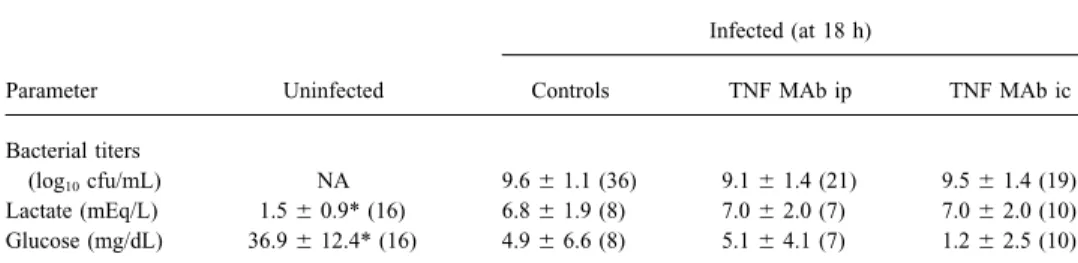
Figure 1. Pattern of neuronal in-jury in infant rats with group B strep-tococcal meningitis. In hippocampus (A), neuronal injury shows charac-teristics of apoptosis [4, 18], with dense, shrunken nuclei (arrowhead) in dentate gyrus (cresyl violet, 11000). In cortex (B), injury is of ischemic origin, with wedge-shaped focal areas of reduced neuronal den-sity (arrowheads) (cresyl violet, 1200).
forms of neuronal injury, cortical necrosis and apoptotic injury in spontaneous death rates did not reach statistical significance in this set of experiments (31% vs. 53%; PÅ .31).
of the dentate gyrus in the hippocampus (figure 1) [3, 4, 17].
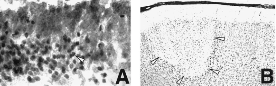
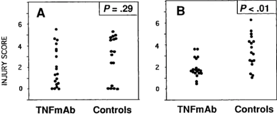
Treatment with TNF MAb had no measurable effect on the Histopathology. Similar to results with systemic administra-tion of antibody, inflammaadministra-tion in the subarachnoid space showed histopathologic scoring of inflammation in the subarachnoid
space, since all animals showed maximal (grade 3) inflamma- no significant difference between treated and untreated animals, and there was no significant effect of the antibody on cortical tion. Similarly, there was no significant reduction in cortical
injury (median [range] for treated animals: 2.4 [0 – 4.4]; con- injury (median [range] in treated animals: 1.3 [0 – 5.6]; controls: 3.5 [0 – 5.0]; PÅ .29) (figure 3A). The effect of TNF MAb on trols: 2.9 [0 – 5.8]; PÅ .30) (figure 2A). In contrast, there was
a significant beneficial effect of TNF MAb treatment on the hippocampal apoptotic injury was confirmed with intracisternal administration (median [range] for treated animals: 1.7 [0.5 – extent of apoptosis in the dentate gyrus of the hippocampus
(median [range] for treated animals: 2.7 [1.2 – 4.6]; controls: 3.5]; controls: 3.2 [1 – 6.2]; Põ .01) (figure 3B). 4.1 [2.3 – 5.8]; Põ .001) (figure 2B). Duration of survival after
induction of infection had no detectable influence on the score
Discussion of both types of injury. Uninfected animals treated with TNF
MAb showed none of the above-described changes.
The present study in a rat model of neonatal meningitis caused by GBS indicates that TNF-a plays a critical role in causing injury in neurons of the dentate gyrus of the
hippocam-Intracisternal Administration of TNF MAb
pus. Previous studies in the same model and in a rabbit model of pneumococcal meningitis have established that this form of Given the lack of a significant effect of systemically
adminis-tered antibody on CSF inflammation and cortical injury, TNF neuronal injury shows the characteristic features of apoptosis [4, 18]. Taken together, these data thus provide a strong link MAb (170 mg) was injected in a second experiment directly
into the cisterna magna to reach higher CSF concentrations. between the action of TNF-a and the induction of apoptosis in a selected neuronal population in vivo during bacterial
men-Clinical disease. As with the systemic administration of
antibody, intracisternal administration did not result in signifi- ingitis.
It has been recognized, largely on the basis of in vitro studies, cant differences in CSF bacterial titers or CSF concentrations
of lactate or glucose (table 1). Also in agreement with the that TNF-a can induce apoptosis in a number of cell types [19, 20]. Some studies have addressed the role of TNF-a in causing systemic experiments, there was a significant difference in
weight loss between intracisternally treated animals and con- cellular injury in the central nervous system. The cytokine can be toxic to cultured human fetal cortical neurons at physiologi-trols (2.3{ 0.4 g vs. 3.0 { 0.4 g; P õ .0001), but the difference
Figure 2. Injury scores in cortex (A) and hippocampus (B) of infant rats with group B streptococcal meningitis treated systemically with monoclonal antibody (MAb) against TNF-a or saline (controls). 20 mg/kg TNF MAb or equal volume of saline was administered intraperitoneally at time of infection. Statistics by Mann-Whitney rank sum test.
Figure 3. Injury scores in cortex (A) and hippocampus (B) of infant rats with group B streptococcal meningitis treated intracisternally with monoclonal antibody (MAb) against TNF-aor saline (controls). TNF MAb (170mg) or equal volume of saline was administered intra-cisternally 30 min after initiating infection. Sta-tistics by Mann-Whitney rank sum test.
cally relevant concentrations, and a recent study using a human infant rat model of meningitis [17]. Thus, TNF-a could exert its effect by modulating excitatory amino acid – induced injury. neuroblastoma cell line differentiated to a neuronal phenotype
with retinoid acid showed that TNF-a induced apoptosis [21, Interestingly, excitatory amino acid receptor stimulation also leads to induction of reactive oxygen intermediates as important 22]. There is also good evidence for a toxic effect of TNF-a
in oligodendrocytes, the cell population responsible for myelin mediators of cellular injury [28]. Thus, regardless of whether TNF-a induces apoptosis in the hippocampus by a direct action production in the central nervous system [23]. However, not
all studies have found that TNF-a is involved in the develop- on neurons or through modulation of the excitatory amino acid pathway, scavenging of oxygen radicals can be expected to be ment of neurotoxicity. In primary cultures of rat neurons,
pre-treatment with TNF-a promoted intracellular calcium homeo- highly protective, as was the case in our study with PBN [4]. TNF-a did not appear to play an essential role in mediating stasis and protected neurons from damage caused by excitatory
amino acids or hypoglycemia [24]. Similarly, a study in mice subarachnoid space inflammation or cortical neuronal injury in the present study. These findings were surprising in light lacking TNF-a receptors found that ischemic and excitotoxic
brain injury were increased compared with that in genetically of previous studies in similar models of bacterial meningitis, in which TNF-a appeared to play an important role in generat-normal control animals, indicating that TNF-a may have
pro-tective properties in this model of acute brain injury [25]. Thus, ing CSF inflammation and in which antibodies to TNF-a were effective in reducing CSF inflammation [5, 11]. Our results the role of TNF-a in acute neuronal injury is complex and
likely influenced by specifics of the experimental system stud- are likely not a reflection of a general lack of biologic activity of the TNF MAb used in the study, since the antibody showed ied. In our rat model of neonatal meningitis, TNF-a appeared
to be predominantly harmful, since several experimental end significant beneficial effects on hippocampal injury as well as systemic parameters, seizure activity, and mortality. Fur-points, including neuronal apoptosis in the dentate gyrus of
the hippocampus, were beneficially affected by treatment with thermore, experiments in which the TNF MAb was adminis-tered intrathecally indicated that its limited effect was not TNF-a antibody.
The exact mechanisms by which TNF-a induced hippocam- likely the result of inadequate CSF concentrations. In these studies,Ç20% of the systemic dose of the antibody was in-pal apoptosis remain to be defined. A wealth of studies have
documented that the cytotoxic effects of TNF-a are related to jected directly into the CSF, resulting in estimated CSF con-centrations 10- to 100-fold higher than after systemic adminis-the production of oxidative radicals [26]. This pathway appears
to be particularly critical in TNF-a – induced apoptosis, includ- tration. An important difference between the present study and previous studies that may explain the discrepancy is the ing in neuronal cells [22]. We have previously found in the
infant rat model of meningitis that scavenging of reactive oxy- time points of examination. Previous studies focused on early time points up to 6 h after infection, while animals were gen intermediates with the spin trapping agent
a-phenyl-tert-butyl nitrone (PBN) was highly protective against apoptotic examined 18 – 24 h after infection in the present study. It is conceivable that TNF-a is critical early in infection, while injury in the hippocampus, suggesting that oxygen
intermedi-ates play an important role in this form of injury in vivo [4]. other inflammatory mediators may compensate for TNF-a in later stages of the disease.
Other mechanisms, however, could account for the effect of
TNF-a. Cytokines may affect excitotoxic neuronal injury The differential effect of TNF MAb on hippocampal and cortical injury underlines the possibility that different mecha-caused by glutamate and other excitatory amino acids, either
by increasing their release from glial cells or by modulating nisms may be involved in these two forms of injury. Previous studies indicated that cortical injury is caused primarily by excitatory amino acid receptors [21]. There is increasing
evi-dence that excitatory amino acids can induce apoptosis in neu- ischemia, which in turn is likely to be a result of the effect of subarachnoid space inflammation on the cerebral vasculature rons under certain circumstances [27], and we have shown that
etiologies: high levels of TNFa indicate bacterial meningitis. J Infect the failure of TNF MAb to affect CSF inflammation resulted
Dis1993; 167:882 – 9. in a corresponding lack of protection against ischemic cortical
10. Dulkerian SJ, Kilpatrick L, Costarino AT, et al. Cytokine elevation in infants injury. Hippocampal injury, on the other hand, does not appear with bacterial and aseptic meningitis. J Pediatr1995;126:872–6. to be of ischemic origin, since the ischemia-sensitive structures 11. Saukkonen K, Sande S, Cioffe C, et al. The role of cytokines in the
generation of inflammation and tissue damage in experimental gram-of the hippocampus (CA1/CA3) are not affected in the
meningi-positive meningitis. J Exp Med1990; 171:439 – 48. tis model and since the dentate gyrus is not considered to
12. Quagliarello VJ, Wispelwey B, Long WJ, Scheld WM. Recombinant hu-be particularly ischemia-sensitive. It appears more likely that
man interleukin-1 induces meningitis and blood-brain barrier injury in soluble mediators, such as TNF-a or excitatory amino acids, the rat. J Clin Invest1991; 87:1360 – 6.
have direct effects on the neurons in the dentate gyrus, possibly 13. Kim KS, Wass CA, Cross AS, Opal SM. Modulation of blood-brain barrier permeability by tumor necrosis factor and antibody to tumor necrosis by diffusing from the ventricular space, which is in close
prox-factor in the rat. Lymphokine Cytokine Res1992; 11:293 – 8. imity to the dentate gyrus and shows uniform inflammation in
14. Silva AT, Baystone KF, Cohen J. Prophylactic and therapeutic effect of the model [17].
a monoclonal antibody to tumor necrosis factor-a in experimental gram-The clinical significance of the hippocampal injury observed negative shock. J Infect Dis1990; 162:421 – 7.
in our model, and thus of the selective beneficial effect of 15. Teti G, Mancuso G, Tomasello F. Cytokine appearance and effect of anti – tumor necrosis factor alpha antibodies in a neonatal rat model of group TNF MAb on this form of neuronal injury, needs to be further
B streptococcal infection. Infect Immun1993; 61:227 – 35. explored. Neonatal meningitis often results in mental
retarda-16. Ta¨uber MG, Sande E, Fournier MA, Tureen JH, Sande MA. Fluid adminis-tion and learning disabilities [2], and the selective injury to the tration, brain edema, and cerebrospinal fluid lactate and glucose concen-hippocampus in our model is intriguing, since this structure trations in experimental Escherichia coli meningitis. J Infect Dis1993; plays a critical role in learning and memory functions. It is 168:473 – 6.
17. Leib SL, Kim SY, Ferriero DM, Ta¨uber MG. Neuroprotective effect of unknown whether similar changes can be observed in humans
excitatory amino acid antagonist kynurenic acid in experimental bacte-after meningitis, but similar findings in pneumococcal
meningi-rial meningitis. J Infect Dis1996; 173:166 – 71.
tis in rabbits indicate that hippocampal injury is not unique to 18. Zysk G, Bru¨ck W, Gerber J, Bru¨ck Y, Prange HW, Nau R. Anti-inflamma-our infant rat model [18]. Independent of these questions, Anti-inflamma-our tory treatment influences neuronal apoptotic cell death in the dentate gyrus in experimental pneumococcal meningitis. J Exp Neurol Neuropa-study significantly expands the understanding of the molecular
thol1996; 55:722 – 8. mechanisms of brain injury in meningitis by documenting a
19. Higuchi M, Singh S, Jaffrezou JP, Aggarwal BB. Acidic sphingomyeli-critical role of TNF-a in causing apoptotic cell death in a
nase-generated ceramide is needed but not sufficient for TNF-induced selected subpopulation of neurons. apoptosis and nuclear factor-kB activation. J Immunol1996; 157:297 –
304.
20. Malorni W, Rainaldi G, Tritarelli E, et al. Tumor necrosis factor-a is a powerful apoptotic inducer in lymphoid leukemic cells expressing the References
P-170 glycoprotein. Int J Cancer1996; 67:238 – 47.
1. Anderson SG, Giulbert GL. Neonatal gram negative meningitis: a 10-year 21. Gelbart HAK, Dzenko K, DiLoreto D, del Cerro K, del Cerro M, Epstein review, with reference to outcome and relapse of infection. J Paediatr LG. Neurotoxic effects of tumor necrosis factor in primary human neu-Child Health1990; 1990:212 – 6. ronal cultures are mediated by activation of the glutamate AMPA recep-2. Edwards MS, Rench MA, Haffar AAM, Murphy MA, Desmond MM, tor subtype: implications for AIDS neuropathogenesis. Dev Neurosci
Baker CJ. Long-term sequelae of group B streptococcal meningitis in 1993; 15:417 – 22.
infants. J Pediatr1985; 106:717 – 22. 22. Talley AK, Dewhurst S, Perry SW, et al. Tumor necrosis factor-a – induced 3. Kim YS, Sheldon RA, Elliot BR, Liu Q, Ferriero DM, Ta¨uber MG. Brain apoptosis in human neuronal cells: protection by the antioxidant N-damage in neonatal meningitis caused by group B streptococci in rats. acetylcysteine and the genes bcl-2 and crmA. Mol Cell Biol1995; 15: J Neuropathol Exp Neurol1995; 54:531 – 9. 2359 – 66.
4. Leib SL, Kim YS, Chow LL, Sheldon RA, Ta¨uber MG. Reactive oxygen 23. D’Souza S, Alinauska K, McCrea E, Goodyer C, Antel JP. Differential intermediates contribute to necrotic and apoptotic neuronal injury in an susceptibility of human CNS-derived cell populations to TNF-dependent infant rat model of bacterial meningitis due to group B streptococci. J and -independent immune-mediated injury. J Neurosci1995; 15:7293 –
Clin Invest1996; 98:2632 – 9. 300.
5. Mustafa MM, Ramilo O, Olsen KD, et al. Tumor necrosis factor a in 24. Cheng B, Christakos S, Mattson M. Tumor necrosis factors protect neurons mediating experimental Haemophilus influenzae type b meningitis. J against metabolic-excitotoxic insults and promote maintenance of cal-Clin Invest1989; 84:1253 – 9. cium homeostasis. Neuron1994; 12:139 – 53.
6. Ling EWY, Noya FJD, Ricard G, Beharry K, Mills EL, Aranda JV. Biochemi- 25. Bruce AJ, Boling W, Kindy MS, et al. Altered neuronal and microglial cal mediators of meningeal inflammatory response to group B streptococcus response to excitotoxic and ischemic brain injury in mice lacking TNF in the newborn piglet model. Pediatr Res1995;38:981–7. receptors. Nature Med1996; 2:788 – 94.
7. Mustafa MM, Lebel MH, Ramilo O, et al. Correlation of interleukin-1b 26. Larrick JW, Wright SC. Cytotoxic mechanism of tumor necrosis factor-and cachectin concentrations in cerebrospinal fluid factor-and outcome from a. FASEB J 1990; 4:3215 – 23.
bacterial meningitis. J Pediatr1989; 115:208 – 13. 27. Bonfoco E, Krainic D, Ankarcrona M, Nicotera P, Lipton ST. Apoptosis 8. Arditi M, Manogue KR, Caplan M, Yogev R. Cerebrospinal fluid and necrosis: two distinct events induced, respectively, by mild and cachectin/tumor necrosis factor-a and platelet-activating factor concen- intense insults with N-methyl-D-aspartate or nitric oxide/superoxide in trations and severity of bacterial meningitis in children. J Infect Dis cortical cell cultures. Proc Natl Acad Sci USA1995; 92:7162 – 6. 1990; 162:139 – 47. 28. Reynolds IJ, Hastings TG. Glutamate induces the production of reactive 9. Glimaker M, Kragsbjerg P, Forsgren M, Olcen P. Tumor necrosis factor-a oxygen species in cultured forebrain neurons following NMDA receptor
activation. J Neurosci1995; 15:3318 – 27. (TNFa) in cerebrospinal fluid from patients with meningitis of different