CDG - AN UPDATE
Oligosaccharyltransferase: the central enzyme
of N-linked protein glycosylation
Elisabeth Mohorko&Rudi Glockshuber&Markus Aebi
Received: 17 December 2010 / Revised: 1 April 2011 / Accepted: 7 April 2011 / Published online: 26 May 2011 # SSIEM and Springer 2011
Abtract N-linked glycosylation is one of the most abun-dant modifications of proteins in eukaryotic organisms. In the central reaction of the pathway, oligosaccharyltransfer-ase (OST), a multimeric complex located at the membrane of the endoplasmic reticulum, transfers a preassembled oligosaccharide to selected asparagine residues within the consensus sequence asparagine-X-serine/threonine. Due to the high substrate specificity of OST, alterations in the biosynthesis of the oligosaccharide substrate result in the hypoglycosylation of many different proteins and a multi-tude of symptoms observed in the family of congenital disorders of glycosylation (CDG) type I. This review covers our knowledge of human OST and describes enzyme composition. The Stt3 subunit of OST harbors the catalytic center of the enzyme, but the function of the other, highly conserved, subunits are less well defined. Some compo-nents seem to be involved in the recognition and utilization of glycosylation sites in specific glycoproteins. Indeed, mutations in the subunit paralogs N33/Tusc3 and IAP do not yield the pleiotropic phenotypes typical for CDG type I but specifically result in nonsyndromic mental retardation, suggesting that the oxidoreductase activity of these subunits is required for glycosylation of a subset of proteins essential for brain development.
Introduction
N-linked glycosylation in the endoplasmic reticulum (ER) N-linked glycosylation is the most frequent protein modi-fication of membrane and secretory proteins in eukaryotes, but it also exists in archaea and bacteria. This essential and highly conserved process in the endoplasmic reticulum (ER) of all eukaryotic cells is characterized by the transfer of a preassembled, uniform oligosaccharide (Glc3Man9Glc NAc2in most eukaryotes) from the isoprenoid lipid carrier dolichol pyrophosphate to the side-chain amide group nitrogen of an asparagine residue contained in a N-X-S(T) sequon of the polypeptide substrate, where X can be any amino acid except proline. The glycosylation reaction is catalyzed by oligosaccharyltransferase (OST), a hetero-oligomeric membrane protein complex in animals, plants, and fungi. In bacteria, archaea, and protozoa, OST is a monomer corresponding to the catalytic subunit of the eukaryotic OST and catalyzes the transfer of a highly defined, lipid-linked oligosaccharide (LLO) donor substrate to a multitude of peptide acceptor sequences located in different substrate proteins. Mutations affecting the biosyn-thesis of the activated Glc3Man9GlcNAc2 oligosaccharide substrate or the biogenesis of OSTs generally have a systemic effect in eukaryotes and alter glycosylation of many different glycoproteins. As a matter of fact, it is the substrate specificity of OST that translates defects in the biosynthesis of the oligosaccharide substrate into a general-ized and multisystemic deficiency observed for the different forms of human congenital disorders of glycosylation type I (Freeze and Aebi2005).
N-linked glycans serve a multitude of functions that can be divided into two main groups correlating with the processing and maturation pathway: they affect the folding
Communicated by: Dirk Lefeber Competing interest: None declared. E. Mohorko
:
R. Glockshuber Department of Biology,Institute of Molecular Biology and Biophysics, ETH Zürich, Schafmatt 20, CH 8093 Zürich, Switzerland
M. Aebi (*)
Department of Biology, Institute of Microbiology, ETH Zürich, Wolfgang Pauli Str. 10, CH 8093 Zürich, Switzerland e-mail: aebi@micro.biol.ethz.ch
and the sorting of proteins in the ER and, after glycan remodeling in the Golgi compartment, confer multiple functions that mediate interactions of the cell or the organism with its environment. In the ER, N-linked glycans primarily assist in proper folding of the newly synthesized polypeptide (Helenius 1994; Paulson 1989). The hydro-philic nature of the covalently attached glycan alters the biophysical properties of the polypeptide, can affect the conformation (Wormald and Dwek1999) of the unfolded polypeptide chain, and influences protein folding: N-linked glycans can direct folding chaperones in the ER and protein folding catalysts such as the ER thiol oxidoreductase ERp57 to specific domains of glycoprotein substrates (Frickel et al. 2002; Parodi 2000; Zapun et al. 1999). In addition, trimming of the N-linked glycans generates defined oligosaccharide structures that are interpreted by the quality control process machinery in the ER, and these signals direct improperly folded polypeptides or non-assembled subunits of protein complexes to the export from the ER back to the cytoplasm and degradation by the proteasome (Aebi et al.2010; Helenius and Aebi2004). Oligosaccharyltransferase reaction in the ER
Figure 1 shows the overall glycosylation reaction in eukaryotes catalyzed by OST, i.e., transfer of the dolichyl-pyrophosphate-linked oligosaccharide to the asparagine of an asparagine-X-serine or threonine (N-X-S/T) sequon in the acceptor polypeptide, where X can be any amino acid residue except proline. Though fully assembled tetradeca-saccharide Glc3Man9GlcNAc2is the preferred substrate of eukaryotic OST, many in vitro studies using OST from dog pancreas or yeast have shown that truncated, lipid-linked oligosaccharides such as GlcNAc2 or GlcNAc2Man also serve as OST substrates, though their binding affinities are low (Bause et al. 1995; Imperiali and Shannon 1991; Karaoglu et al.2001; Sharma et al.1981; Tai and Imperiali 2001). The minimal glycan substrate of OST that still leads to an N-glycosidic linkage is the dolichol-linked monosac-charide Dol-PP-GlcNAc (Tai and Imperiali2001). Interest-ingly, the acetamido group of the reducing-end GlcNAc is an essential substrate property, whereas the presence of the second GlcNAc enhances binding affinity of the substrate toward OST but does not affect the glycan transfer rate. It has therefore been proposed that the acetamido group of GlcNAc and the catalytic center of OST might form a hydrogen bond network required for catalysis (Tai and Imperiali 2001). Alternatively, the acetamido group of GlcNAc might act as a neighboring group in order to stabilize an oxonium intermediate that is generated by cleavage of the phosphate-ester linkage (Wacker et al. 2006). Although the exact reaction mechanism of OST still needs to be elucidated at the atomic level, the mechanism
most likely involves a nucleophilic attack of the side-chain amide nitrogen of the asparagine located in the selected N-X-S/T sequon on the C1 atom of GlcNAc (Bause and Legler1981). A mechanism to increase the intrinsically low nucleophilicity of the amide is currently described by two contradictory mechanistic models (Bause et al. 1997). In the first model, the acidity of the amide is increased by hydrogen bond contacts between the side-chain carbonyl group of asparagine and theα-NH and β-OH groups of the hydroxyl amino acid in the glycosylation sequon (Imperiali et al.1992). The second model favors“inverted” hydrogen-bond interactions between the amide NH2protons as donors and the β-OH group oxygen of the hydroxyl amino acid (Bause et al. 1995; Bause and Legler 1981; Bause et al. 1997). Irrespective of the precise reaction mechanism, both models predict that the N-X-S/T sequon has to adopt a specific conformation that is not compatible with regular secondary structure elements of folded proteins. It is important to note that this does not exclude the possibility that N-linked glycans are covalently linked to asparagine residues located in α−helical segments or β-strand ele-ments of folded glycoproteins. It rather implies that glycosylation in vivo has to occur at the level of the unfolded glycoprotein substrate, i.e., before its folding to the native tertiary structure.
Donor and acceptor substrates of the oligosaccharyltransferase
Though not directly involved in the catalytic transfer, the different units of the OST substrates define substrate affinity. The lipid-linked donor substrate Glc3Man9GlcNAc2 is rec-ognized by OST with high specificity, primarily determined by theα-1,2-linked glucose residue at the nonreducing end of Glc3Man9GlcNAc2. Importantly, addition of this hexose unit represents the final step in a linear assembly pathway of the branched oligosaccharide, ensuring the transfer of completely assembled oligosaccharide to proteins (Burda and Aebi 1999). Absence of this terminal glucose residue causes impaired glycan recognition and correlates with a reduced N-glycosylation site occupancy in glycoprotein substrates. The result is an overall hypoglycosylation of the N-glycoproteome (Hulsmeier et al. 2007). In vitro studies confirmed the lowered binding affinities of incompletely assembled glycans to OST (Karaoglu et al.2001).
In contrast to the lipid-linked oligosaccharide donor, the polypeptide acceptor substrates are less clearly defined. N-X-T sequons are more frequently used than N-X-S sequons, and in rare cases, the glycosylation of asparagines in N-X-C sequons has also been observed (Miletich and Broze1990; Titani et al. 1986; Zielinska et al. 2010). Recently, N-glycosylation was also detected on N-X-V and NG motifs. (Zielinska et al. 2010). Importantly, the existence of the
latter N-glycosylation sites argues against a direct involve-ment of theβ-hydroxyl group of the N-X-S/T motif in the catalytic process. The large-scale proteomics study of the mouse N-glycoproteome allows detailed categorization of glycoproteins and revealed that glycosylation sites are preferentially located in surface-exposed loop and turn regions. Almost half of the glycoproteins detected in mice contain only one modified glycosylation sequon and 22% are doubly glycosylated. The rest of the N-glycoproteins contain three to ten glycan structures, and only 2% of the entire pool of glycoproteins possesses more than ten glycans (Zielinska et al.2010). Glycoprotein expression is highly tissue-specific, as most glycoproteins were found in brain tissue and the majority of glycoproteins discovered in brain tissue were not detected in any other tissues. On the contrary, the minimal figure of glycoproteins was measured in heart tissue, and only few glycoproteins were exclusively present in heart tissue (Zielinska et al.2010).
OST associates with numerous newly synthesized polypeptide chains, but no plausible mechanistic model that defines the site occupancy has been proposed to date. It has been estimated that 65–75% of the potential glycosylation sequons present in secretory proteins are modified (Ben-Dor et al.2004; Petrescu et al.2004; Schulz and Aebi 2009). However, individual glycoproteins may display site occupancy of 98–100% for all glycosylation sites (Hulsmeier et al.2007).
Human oligosaccharyltransferase complex
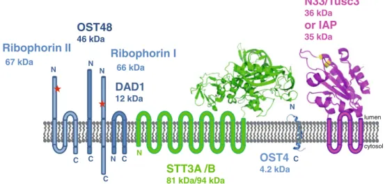
The human OST complex is composed of seven different polypeptide chains, the sequences of which are highly
conserved among mammalian species (Fig.2). Subunits of human OST share a minimum of 90% pairwise sequence identity with the respective homolog in mouse, bovine, canine, rat, and golden hamster OST. Isolation and proteomic characterization of the canine OST complex revealed the presence of two additional, most likely loosely associated, proteins, DC2 and KCP2, that have not yet been verified in the human OST complex (Shibatani et al.2005). However, no functional role of these subunits in the glycosylation process has been reported.
The heptameric human OST complex (Fig. 2) contains the subunits ribophorin I (OST1p), ribophorin II (Swp1p), OST48 (Wbp1p), OST4 (OST4), Stt3-A/Stt3-B (Stt3p), N33/Tusc3 and IAP (OST3p and OST6p), and DAD1 (OST2p). All of these subunits have homologous proteins in yeast OST (shown in brackets), and a cryo-EM structure at 12Å resolution has been reported for this enzyme (Li et al. 2008). Five of the subunit proteins, namely, OST1p, Swp1p, Wbp1p, Stt3p, and OST2p, are essential for yeast viability. Based on our current knowledge, we assume that there is a functional equivalence of the homologous subunits between the yeast and the human OST complex, allowing for a description of human subunit function based on experiments performed in yeast. In the following, we describe the different subunits of the mammalian, specifi-cally the human, OST in more detail.
Stt3
Stt3 is the largest and most conserved polypeptide of the OST complex (Spirig et al. 1997). Stt3 proteins are the catalytic subunits of eukaryotic OST complexes (Burda and Aebi 1999; Kelleher et al.2003; Nilsson et al. 2003; Yan
Fig.1 Oligosaccharyltransferase (OST) reaction catalyzed by the OST complex. Note that the α-glycosidicphosphodiester ester bond connecting the C1 atom of the first GlcNAc of
Glc3Man9GlcNAc2with the
py-rophosphate attached to dolichol (dol) is converted to a β-N-glycosidic bond connecting the C1 atom of the first GlcNAc with the side chain nitrogen atom of asparagines that lie within a glycosylation sequon [Asn-X-Ser(Thr)] of the poly-peptide substrate. The reaction products are the glycosylated polypeptide substrate, dolichol diphosphate, and a proton (Dempski and Imperiali2002)
and Lennarz 2002), and protozoan and bacterial N-linked glycosylation is carried out by a single membrane protein homologous to Stt3p (Castro et al.2006; Hese et al. 2009; Izquierdo et al. 2009; Nasab et al. 2008; Wacker et al. 2002). All Stt3 proteins share the typical signature of a hydrophobic, N-terminal, membrane-embedded part with 11 or 13 predicted transmembrane helices and a soluble C-terminal domain that is located in the lumen of the ER in eukaryotes or in the periplasm of Gram-negative bacteria (Kelleher and Gilmore2006). Mammalian genomes encode two Stt3 isoforms, and either Stt3-A or Stt3-B is incorpo-rated into the mammalian complex (Kelleher et al.2003).
The human variants Stt-3A and Stt3-B share 59% amino acid sequence identity. The larger polypeptide, Stt3-B, consists of 826 residues whereof the first 558 are predicted to form the transmembrane domain with 11 or 13 transmembrane helices. The slightly smaller isoform Stt3-A contains 705 residues and the same predicted membrane topology. Its transmembrane domain has 11 or 13 predicted transmembrane helices, followed by an approximately 230-residue C-terminal domain residing in the ER lumen. Both Stt3 variants are coexpressed in various tissues but differ in expression levels. Stt3-A expression was suggested to be more tissue specific, as high expression levels were observed in muscle, liver, heart, placenta and pancreas
and very low expression in brain, lung, and kidney. On the contrary, Stt3-B expression patterns were more even in all tissues tested (Kelleher et al.2003).
Canine OST isoforms harboring the different Stt3 proteins differ in catalytic activity and substrate selectivity. OST complexes with the Stt3-B isoform are more active; they reach 8- to 12-fold higher Vmax values for glycopep-tide formation than complexes containingStt3-A (Kelleher et al. 2003). The increased catalytic activity for Stt3-B complexes coincides with a reduced selectivity with respect to the oligosaccharide donor substrate. OST complexes with Stt3-B accepted the dolichol-pyrophosphate-activated Glc3Man9GlcNAc2 and Man9GlcNAc2 substrates with roughly the same specificity, whereas OST complexes with Stt3-A were more selective, as reflected by increased Km values forMan9GlcNAc2 relative to Glc3Man9Glc NAc2(Kelleher et al.2003). Similar results were obtained for the human OST complexes isolated from HeLa cells: OST complexes containing Stt3-B were also found to be more active than OST complexes harboring Stt3-A (Ruiz-Canada et al.2009).
Stt3-A and Stt3-B also differ in their acceptor substrate selectivity (Wilson and High2007).The glycosylation state of different membrane proteins was analyzed in Stt3-A- or Stt3-B-deficient HeLa cells using specific small interfering
Ribophorin I
66 kDaN33/Tusc3
36 kDaor IAP
35 kDaDAD1
12 kDaOST48
46 kDaRibophorin II
67 kDaOST4
4.2 kDaSTT3A /B
81 kDa/94 kDa N C N C C N C N N C lumen cytosol NFig. 2 Human oligosaccharyltransferase (OST) complex consists of equimolar amounts of seven different subunits. There are two isoforms of the catalytic subunit (Stt3A and Stt3B) and two isoforms of the subunit with a redox-active, thioredoxin-like domain oriented toward the endoplasmic reticulum (ER) lumen (N33/Tusc3 and IAP), where only one of the respective subunits can be incorporated into the OST complex. Consequently, there are four different variants of the human OST complex, bearing the subunit pairs Stt3A/(N33/Tusc3), Stt3B/ (N33/Tusc3), Stt3A/IAP, or Stt3B/IAP. The scheme depicts the membrane topology of the individual subunits [amino (N) and carboxyl (C) termini] and the number of predicted transmembrane helices for each subunit. The only parts of the OST complex for which three-dimensional structure information is available to date are
represented by a ribbon. Specifically, these are: (i) the crystal structure of the homologue of the luminal domain of Stt3 from Pyrococcus furiosus, (Igura et al. 2008), (ii) the X-ray structure of the homologueof the thioredoxin-like domain of N33/Tusc3 from yeast (OST6) (Schulz et al. 2009) containing the active-site motif CXXC (depicted in yellow), and (iii) the NMR structure of the yeast homologue of the smallest subunit OST4 (Zubkov et al. 2004). In the human OST complex, the ER luminal parts of the subunits ribophorin I, ribophorin II, Stt3A, and Stt3B are also glycosylation substrates, with one glycosylation site each in ribophorin I and ribophorin II, three in Stt3A, and two in Stt3B (Chen et al.2009). The molecular masses (in kDa) of the mature, unglycoslyated subunits are indicated
RNA (siRNA)-mediated knockdown experiments. Stt3-B depletion led to hypoglycosylation of certain membrane proteins. In contrast, a similar approach targeting Stt3-A resulted in complete loss of both isoforms and abrogation of glycosylation (Wilson and High2007). Divergent results for the specific depletion of the Stt3 isoforms were observed in a more recent study (Ruiz-Canada et al. 2009). Here, siRNA depletion of Stt3-A resulted in elevated Stt3-B levels, and conversely, the loss of Stt3-B caused slightly increased levels of Stt3-A. Both regimes decreased ribophorin I levels. Endogenous glycoprotein prosaposin (pSAP) displayed severe hypoglycosylation in the absence of Stt3-A, but Stt3-B depletion had no effect on the glycosylation state of this protein. On the other hand, procathepsin C (pCatC) glycosylation was hardly affected by Stt3-A deficiency, but the loss of Stt3-B resulted in hypoglycosylation of pCatC. The differences between OSTs harboring Stt3-A or Stt3-B were described more specifically when looking at individual glycosylation sites in defined substrates: Stt3-B was found to act preferentially on sites glycosylated posttranslationally, whereas Stt3-A catalyzed cotranslational glycosylation. Such a sequential action of the OST complexes with Stt3-A and Stt3-B was demonstrated for the glycosylation process of two different glycoproteins (Ruiz-Canada et al. 2009). Therefore, the different OST forms (containing either Stt3-A or Stt3-B) differ with respect to substrate specificity as well as in the temporal order in the processing of polypeptides entering the lumen of the ER.
Ribophorin I
Ribophorin I and II and OST48 were the first identified subunits of the mammalian OST complex, which was purified from canine pancreas (Kelleher et al.1992). Initial data suggested a catalytic glycosyltransferase activity for ribophorin I, but subsequent studies clearly assigned this function to the Stt3 subunits (Nilsson et al.2003; Yan and Lennarz 2002). Ribophorin I is highly conserved among various species, and human ribophorin shares >90% sequence identity with homologous proteins in vertebrates. It is a 68-kDa type-I transmembrane protein with a single, predicted transmembrane helix; a large, luminal domain; and a smaller, C-terminal cytosolic domain. Ribophorins are highly abundant ER proteins, exclusively present in the rough ER in about equal amounts relative to membrane-bound ribosomes (Kreibich et al.1978c; Marcantonio et al. 1984). Direct interactions between ribophorins and ribo-somes were detected in cross-linking experiments, and therefore, the cytoplasmically oriented parts of ribophorins were considered to provide binding sites for translating ribosomes (Kreibich et al. 1978a; Kreibich et al. 1978b). Indeed, antibodies against the C-terminal cytosolic domain
of ribophorin I prevent ribosome targeting to the ER membrane by blocking ribosome association to the trans-location channel, and they inhibit protein transtrans-location (Yu et al. 1990). Ribophorin I and ribophorin II were also detected in ribosome fractions isolated from native ER membranes (Gorlich et al. 1992; Menetret et al. 2000; Wang and Dobberstein 1999). In summary, these data provide solid experimental evidence that at least a subset of OST complexes in the ER are associated with the trans-locon (Shibatani et al. 2005), enabling cotranslocational glycosylation of entering polypeptides.
Ribophorin I is not essential for glycosylation per se but, rather, affects utilization of certain glycosylation sites on defined polypeptides (Wilson and High 2007). Single-spanning membrane proteins required ribophorin I expres-sion for efficient glycosylation, but glycosylation of secretory proteins (α1-antitrypsin, transferrin, and tyrosi-nase) and multispanning membrane proteins was not affected by ribophorin I depletion (Wilson et al.2008). A chaperone-like activity of ribophorin I was proposed in the case of the substrate mu-opioid receptor (MOR), a member of the G-protein-coupled receptor family. Direct interac-tions between MOR and ribophorin I were shown by coimmunoprecipitation when His-tagged MOR was puri-fied from neuroblastoma cells. MOR receptor trafficking and surface expression is regulated via glycosylation of the MOR receptor. Ribophorin I overexpression rescued surface expression of intracellular retained MOR variants, but N-linked glycosylation-deficient variants were not rescued by ribophorin I overexpression (Ge et al.2009). Ribophorin II
Ribophorin II is highly conserved in mammals; the human ribophorin II shares >90% sequence identity with ribo-phorin II from mice, rat, and pig. Primary structure analysis suggests a large N-terminal luminal domain followed by a hydrophobic transmembrane domain with three predicted α-helices and a small C-terminal segment located in the cytoplasm (Crimaudo et al. 1987). ER localization of ribophorin II is mediated by independent ER retention signals located in the transmembrane segment and cytosolic part. A 15 amino acid residue luminal region strengthens ER localization, but no defined ER retention signals were found in the ER luminal part (Fu et al.2000).
Ribophorin II expression is elevated in drug-resistant human breast cancer cells (Honma et al. 2008). Down-regulation of ribophorin II restores docetaxel-induced apoptosis in drug-resistant human breast cancer cells, and in vivo delivery of ribophorin II siRNA reduced tumor growth in drug-resistant tumor types. Here, ribophorin II-mediated drug resistance is regulated via P-glycoprotein glycosylation. The general reduction of N-linked
glycosyl-ation by ribophorin II downregulglycosyl-ation also leads to hypoglycoylation of P-glycoprotein and results in functional inactivity. P-glycoprotein is a key player in conferring drug resistance, and stable membrane-bound P-glycoprotein requires complete glycosylation (Honma et al.2008). OST 48
OST48, the third initially identified OST subunit, was copurified with active canine OST (Kelleher et al. 1992). Canine OST48 and yeast Wbp1, an essential subunit of the yeast OST complex, share 25% sequence identity (Silberstein et al. 1992). OST48 is, however, highly conserved in mammals. The human OST48 subunit shares >90% sequence identity with other mammalian OST48 proteins.
Human OST48 consists of 456 residues; the first 42 are suggested to include a signal sequence, followed by a large, lumen-oriented domain. One single transmembrane span constitutes OST48 as type I membrane protein with a very small C-terminal cytosolic segment comprising only nine residues. The C-terminal cytosolic domain of canine OST48 was shown to interact with DAD1, another component of OST, and interactions between the luminal domain of OST48 and ribophorin I were detected by yeast two-hybrid screening and in vitro binding assays. Interest-ingly, OST48 seems to provide a link between ribophorin I and ribophorin II in the active OST complex, as the N-terminal luminal domain of OST48 interacts with the luminal domain of ribophorin I and ribophorin II, but a direct interaction between ribophorin I and ribophorin II was not observed (Fu et al.1997).
Pig OST48 contains a double lysine motif at the very C-terminus, which was suggested to confer ER residency (Hardt et al.2000; Hardt et al.2001). Replacing the lysine motif by leucine residues elicits cell-surface expression of the OST48 mutant protein. However, cell-surface localiza-tion was abolished by ribophorin I overexpression, indicat-ing a direct interaction between OST48 and ribophorin I. The yeast homolog Wbp1p contains a similar dilysine motif that functions as an ER retrieval signal (Gaynor et al. 1994). The yeast Wbp1p was proposed to be responsible for binding of the lipid-linked oligosaccharide donor substrate (Pathak et al. 1995), but further experimental evidence to support this notion is missing.
DAD1
DAD1 (defender against apoptotic cell death), was initially discovered in the search for genes involved in apoptosis. The apoptotic phenotype of a temperature-sensitive cell line at nonpermissive temperature was rescued by human DAD1, and apoptosis was induced by degradation of the mutated DAD1 protein at nonpermissive temperatures
(Nakashima et al. 1993). DAD1 is the most conserved OST subunit: canine, bovine, and human DAD1 share 99% sequence identity; and human, mice, rat, and hamster DAD1 proteins are identical. Human DAD1 is a small, 113-residue polypeptide with two predicted transmembrane span helices in the center of the protein and both termini located in the cytosol.
Analysis of the canine OST complex revealed a tight association of DAD1 with the active OST complex (Kelleher and Gilmore 1997). Moreover, specific interac-tions between the amino terminal domain of DAD1 and OST48 were reported (Fu et al.1997). The instability of the temperature-sensitive DAD1 mutant at restrictive tempera-ture causes a time-dependent degradation of the other OST subunits ribophorin I, ribophorin II, and OST48, disrupting the entire OST complex (Sanjay et al.1998). Consequently, the loss of OST activity caused by DAD1 instability results in severe hypoglycosylation that might induce apoptosis (Makishima et al. 1997; Sanjay et al. 1998).
Interestingly, OST activity was retained in dad1 null (−/−) mice blastocytes (Nishii et al. 1999) but hypoglycosylation of specific glycoproteins was detected (Hong et al. 2000). This observation is in agreement with other data suggesting that N-linked glycosylation is not required in very early embryonic stages but becomes essential at later develop-mental stages (Surani1979). DAD1 might provide structural and functional integrity for the OST complex, and a deficiency causes a time-dependent defect on OST activity (Sanjay et al. 1998). Heterozygous dad1 (+/−) mice are viable and display no severe abnormalities but rather mild forms of thymic hypoplasia and soft tissue syndactyly (Brewster et al.2000; Hong et al.2000; Nishii et al.1999). N33/Tusc3 and IAP
N33/Tusc3 and IAP are two paralogous, mutually exclusive subunits of the mammalian OST complex. Human N33/ Tusc3 and IAP share 70% sequence identity with similar predicted membrane topology, consisting of an N-terminal luminal domain including approximately 170 residues, followed by a transmembrane domain with four transmem-brane segments. The full-length human N33/Tusc3 poly-peptide contains 348 residues, and IAP consists of 335 residues. Both proteins contain an N-terminal signal sequence, which is very likely to be cleaved off after translocation. Human N33/Tusc3 exists in two splicing variants, differing from each other only in the last five residues. Splicing variant 1, N33-1 cosedimented with the active OST complex, but N33-2 and IAP dissociated from the OST complex during membrane solubilization and are most likely weakly associ-ated with the active complex (Kelleher et al.2003).
N33/Tusc3 was initially described as tumor suppressor candidate (MacGrogan et al.1996) based on the location of its
gene on the short arm of chromosome 8. This region is frequently lost in various tumor cells. Normal expression levels of N33/Tusc3 were observed in various tissues, including heart, brain, lung, liver, placenta, pancreas, prostate, testis, and colon (Kelleher et al. 2003; MacGrogan et al. 1996), but expression was downregulated in many colorectal, lung, liver, breast, and pancreatic cancer cell lines (Bashyam et al.2005; Cooke et al.2008; MacGrogan et al.1996).
Based on sequence similarity and secondary structure prediction, N33/Tusc3 and IAP are homologous to the yeast OST subunits OST3p and OST6p, respectively. Recent structural and biochemical studies on the luminal domain of the yeast OST6p showed a thioredoxin-like fold for the luminal domain and revealed oxidoreductase activity for this domain (Schulz et al.2009). Based on sequence similarity, N33/Tusc3 and IAP are proposed to have oxidoreductase activity as well (Fetrow et al. 2001). Though this is the functionally best characterized subunit of eukaryotic OST complexes, there are conflicting results regarding the role of these proteins in vivo. Zhou and coworkers suggest that N33/ Tusc3 and IAP might be involved in magnesium homeostasis (Zhou and Clapham2009). Most interestingly, deficiency of human N33/Tusc3 or IAP results in isolated cognitive defects (Garshasbi et al.2008; Molinari et al.2008) (see below). OST4
The subunit OST4 is a 37-residue peptide with a single transmembrane helix. Its association with the mammalian OST complex was proposed based on sequence similarity to the yeast OST4p subunit of OST, and OST4 has also been identified as a component of the purified canine OST complex (Kelleher and Gilmore2006).
Human OST4 contains a very short luminal segment with one transmembrane span followed by a cytoplasmic part consisting of approximately ten residues. Besides the high conservation among mammalian species, reflected in 100% sequence identity between human, rat, and mice OST4, very little is known about the function of mammalian OST4. In the yeast OST complex, OST4p regulates the incorporation of the two functionally equivalent, but mutually exclusive, subunits OST3p and OST6p (Spirig et al. 2005). Single-point mutations in the transmembrane segment of OST4p disrupt the interaction between OST4p, OST3p, and Stt3p, causing severe growth defects in yeast (Kim et al.2000; Kim et al. 2003).
Human diseases associated with OST
The high substrate specificity of the OST complex with respect to the structure of the lipid-linked oligosaccharide is responsible for the very often severe phenotypes of many of
the congenital disorders of glycosylation (CDG) type I. However, only few cases where OST activity is affected directly are known so far. It can be assumed that most mutations affecting the central components of OST, such as the Stt3 proteins, can have lethal affects in a homozygous situation. It is therefore not surprising that the known cases affect the mutually exclusive paralogs N33/Tusc3 and IAP. Based on genetic evidence, homozygous loss of function mutations in N33/Tusc3 or IAP result in nonsyndromic mental retardation (Garshasbi et al. 2008; Molinari et al. 2008). Interestingly, this specific symptom of the disease is in sharp contrast to the pleiotropic affects induced by mutations altering LLO substrate biosynthesis, and the CDG-typical hypoglycosylation of the marker protein transferrin is not observed in these patients. More generally, serum N-glycome, as analyzed by mass spec-trometry, showed neither quantitative nor qualitative differences in these patients. These observations are in line with the functional analysis of the homologous OST subunits in yeast that point to their role in efficient glycosylation of defined polypeptide substrates by alter-ing the kinetics of their oxidative foldalter-ing (Schulz et al. 2009). Within the framework of this hypothesis, N33/ Tusc3 and IAP might ensure defined glycosylation of a subset of proteins that are required for normal brain development.
A polymorphism in the ribophorin II locus has recently been detected in a screen for mutations occurring in 27 CDG-patients without clear diagnosis at a molecular level. The G374D missense mutation lies in the second half of the N-terminal luminal domain of ribophorin II. Interestingly, the affected patient presents a fairly mild clinical phenotype, but a clear genotype–phenotype correlation for this mutation could not be established (Vleugels et al. 2009).
Outlook
OST is the key player in transferring the glycan onto the selected glycosylation sequons present in many different substrates. The N-linked glycans perform a multitude of functions at a cellular and organismal level. Alterations in the biosynthesis of the unique lipid-linked oligosac-charide substrate of OST therefore affect the glycosyla-tion efficiency and funcglycosyla-tionality of many different proteins and results in a highly diverse set of symptoms that characterize CDG-type I patients. In contrast, the recent identification of deficiencies in OST activity that result in highly specific clinical pictures suggests that mutations affecting the selection of a subset of polypep-tide substrates by OST can yield a more defined and less general phenotype. However, it is difficult to predict the
clinical pictures that might emerge from OST complexes containing a certain mutant subunits. Our knowledge about the function of these subunits in the N-glycosylation process is limited and does not allow predictions for diagnosis. In addition, our tools to analyze the N-linked protein glycosylation pathway are limited. We have to be aware that the output of the N-glycosylation process is a large number of different N-glycoproteins, and the exact description of this output requires quantitation of the site occupancy at every potential N-glycosylation site. This is a typical problem addressed by analytical approaches at a systemic level, and a first step toward the quantitative description of the N-glycoproteome in eukaryotes has recently been published (Zielinska et al. 2010). It is evident that the quantitative description of the N-glycoproteome as a diagnostic tool will lead to identification of mutations that affect polypeptide substrate selection of OST directly. Alternatively, whole-genome sequencing of patient DNA will reveal OST mutations, and the subsequent characterization of the N-glycoproteome will provide additional information to define the molecular function of the different OST components. Eventually, this understanding of OST-mediated catalysis will open the way for the development of drugs that affect the properties of the enzyme. Altering lipid-linked substrate specificity might reduce many of the severe symptoms observed in patients with the different types of CDG-I. To reach this long-term goal, we have to understand the function of a unique enzyme, OST, which modulates the activity of a large number of different proteins and which is of central importance for the generation of the eukaryotic glycome.
Acknowledgments This work was supported by the ETH Zürich and by grants from the Swiss National Science Foundations to RG and MA.
References
Aebi M, Bernasconi R, Clerc S, Molinari M (2010) N-glycan structures: recognition and processing in the ER. Trends Biochem Sci 35:74–82
Bashyam MD, Bair R, Kim YH, Wang P, Hernandez-Boussard T, Karikari CA, Tibshirani R, Maitra A, Pollack JR (2005) Array-based comparative genomic hybridization identifies localized DNA amplifications and homozygous deletions in pancreatic cancer. Neoplasia 7:556–562
Bause E, Legler G (1981) The role of the hydroxy amino acid in the triplet sequence Asn-Xaa-Thr(Ser) for the N-glycosylation step during glycoprotein biosynthesis. Biochem J 195:639– 644
Bause E, Breuer W, Peters S (1995) Investigation of the active site of oligosaccharyltransferase from pig liver using synthetic tripep-tides as tools. Biochem J312(Pt 3):979–985
Bause E, Wesemann M, Bartoschek A, Breuer W (1997) Epoxyethyl-glycyl peptides as inhibitors of oligosaccharyltransferase: double-labelling of the active site. Biochem J 322(Pt 1):95–102 Ben-Dor S, Esterman N, Rubin E, Sharon N (2004) Biases and
complex patterns in the residues flanking protein N-glycosylation sites. Glycobiology 14:95–101
Brewster JL, Martin SL, Toms J, Goss D, Wang K, Zachrone K, Davis A, Carlson G, Hood L, Coffin JD (2000) Deletion of Dad1 in mice induces an apoptosis-associated embryonic death. Genesis 26:271–278
Burda P, Aebi M (1999) The dolichol pathway of N-linked glycosylation. Biochim Biophys Acta 1426:239–257
Castro O, Movsichoff F, Parodi AJ (2006) Preferential transfer of the complete glycan is determined by the oligosaccharyltransferase complex and not by the catalytic subunit. Proc Natl Acad Sci USA 103:14756–14760
Chen R, Jiang X, Sun D, Han G, Wang F, Ye M, Wang L, Zou H (2009) Glycoproteomics analysis of human liver tissue by combination of multiple enzyme digestion and hydrazide chemistry. J Proteome Res 8:651–661
Cooke SL, Pole JC, Chin SF, Ellis IO, Caldas C, Edwards PA (2008) High-resolution array CGH clarifies events occurring on 8p in carcinogenesis. BMC Cancer 8:288
Crimaudo C, Hortsch M, Gausepohl H, Meyer DI (1987) Human ribophorins I and II: the primary structure and membrane topology of two highly conserved rough endoplasmic reticulum-specific glycoproteins. EMBO J 6:75–82
Dempski RE Jr, Imperiali B (2002) Oligosaccharyl transferase: gatekeeper to the secretory pathway. Curr Opin Chem Biol 6:844–850
Fetrow JS, Siew N, Di Gennaro JA, Martinez-Yamout M, Dyson HJ, Skolnick J (2001) Genomic-scale comparison of sequence- and structure-based methods of function prediction: does structure provide additional insight? Protein Sci 10:1005–1014
Freeze HH, Aebi M (2005) Altered glycan structures: the molecular basis of congenital disorders of glycosylation. Curr Opin Struct Biol 15:490–498
Frickel EM, Riek R, Jelesarov I, Helenius A, Wuthrich K, Ellgaard L (2002) TROSY-NMR reveals interaction between ERp57 and the tip of the calreticulin P-domain. Proc Natl Acad Sci USA 99:1954–1959
Fu J, Ren M, Kreibich G (1997) Interactions among subunits of the oligosaccharyltransferase complex. J Biol Chem 272:29687– 29692
Fu J, Pirozzi G, Sanjay A, Levy R, Chen Y, De Lemos-Chiarandini C, Sabatini D, Kreibich G (2000) Localization of ribophorin II to the endoplasmic reticulum involves both its transmembrane and cytoplasmic domains. Eur J Cell Biol 79:219–228
Garshasbi M, Hadavi V, Habibi H, Kahrizi K, Kariminejad R, Behjati F, Tzschach A, Najmabadi H, Ropers HH, Kuss AW (2008) A defect in the TUSC3 gene is associated with autosomal recessive mental retardation. Am J Hum Genet 82:1158–1164
Gaynor EC, Te Heesen S, Graham TR, Aebi M, Emr SD (1994) Signal-mediated retrieval of a membrane protein from the Golgi to the ER in yeast. J Cell Biol 127:653–665
Ge X, Loh HH, Law PY (2009) mu-Opioid receptor cell surface expression is regulated by its direct interaction with Ribophorin I. Mol Pharmacol 75:1307–1316
Gorlich D, Prehn S, Hartmann E, Kalies KU, Rapoport TA (1992) A mammalian homolog of SEC61p and SECYp is associated with ribosomes and nascent polypeptides during translocation. Cell 71:489–503
Hardt B, Aparicio R, Bause E (2000) The oligosaccharyltransferase complex from pig liver: cDNA cloning, expression and func-tional characterisation. Glycoconj J 17:767–779
Hardt B, Aparicio R, Breuer W, Bause E (2001) Analysis of structural signals conferring localisation of pig OST48 to the endoplasmic reticulum. Biol Chem 382:1039–1047
Helenius A (1994) How N-linked oligosaccharides affect glycoprotein folding in the endoplasmic reticulum. Mol Biol Cell 5:253–265 Helenius A, Aebi M (2004) Roles of N-linked glycans in the
endoplasmic reticulum. Annu Rev Biochem 73:1019–1049 Hese K, Otto C, Routier FH, Lehle L (2009) The yeast
oligosacchar-yltransferase complex can be replaced by STT3 from Leishmania major. Glycobiology 19:160–171
Hong NA, Flannery M, Hsieh SN, Cado D, Pedersen R, Winoto A (2000) Mice lacking Dad1, the defender against apoptotic death-1, express abnormal N-linked glycoproteins and undergo in-creased embryonic apoptosis. Dev Biol 220:76–84
Honma K, Iwao-Koizumi K, Takeshita F, Yamamoto Y, Yoshida T, Nishio K, Nagahara S, Kato K, Ochiya T (2008) RPN2 gene confers docetaxel resistance in breast cancer. Nat Med 14:939– 948
Hulsmeier AJ, Paesold-Burda P, Hennet T (2007) N-glycosylation site occupancy in serum glycoproteins using multiple reaction monitoring liquid chromatography-mass spectrometry. Mol Cell Proteomics 6:2132–2138
Igura M, Maita N, Kamishikiryo J, Yamada M, Obita T, Maenaka K, Kohda D (2008) Structure-guided identification of a new catalytic motif of oligosaccharyltransferase. EMBO J 27:234–243 Imperiali B, Shannon KL (1991) Differences between Asn-Xaa-Thr-containing peptides: a comparison of solution conformation and substrate behavior with oligosaccharyltransferase. Biochemistry 30:4374–4380
Imperiali B, Shannon KL, Rickert KW (1992) Role of peptide conformation in Asparagine-Linked glycosylation. J Am Chem Soc 114:7942–7944
Izquierdo L, Schulz BL, Rodrigues JA, Guther MLS, Procter JB, Barton GJ, Aebi M, Ferguson MAJ (2009) Distinct donor and acceptor specificities of Trypanosoma brucei oligosaccharyltrans-ferases. EMBO J 28:2650–2661
Karaoglu D, Kelleher DJ, Gilmore R (2001) Allosteric regulation provides a molecular mechanism for preferential utilization of the fully assembled dolichol-linked oligosaccharide by the yeast oligosaccharyltransferase. Biochemistry 40:12193–12206 Kelleher DJ, Gilmore R (1997) DAD1, the defender against apoptotic
cell death, is a subunit of the mammalian oligosaccharyltransfer-ase. Proc Natl Acad Sci USA 94:4994–4999
Kelleher DJ, Gilmore R (2006) An evolving view of the eukaryotic oligosaccharyltransferase. Glycobiology 16:47R–62R
Kelleher DJ, Kreibich G, Gilmore R (1992) Oligosaccharyltransferase activity is associated with a protein complex composed of ribophorins I and II and a 48 kd protein. Cell 69:55–65 Kelleher DJ, Karaoglu D, Mandon EC, Gilmore R (2003)
Oligosac-charyltransferase isoforms that contain different catalytic STT3 subunits have distinct enzymatic properties. Mol Cell 12:101– 111
Kim H, Park H, Montalvo L, Lennarz WJ (2000) Studies on the role of the hydrophobic domain of OST4p in interactions with other subunits of yeast oligosaccharyl transferase. Proc Natl Acad Sci USA 97:1516–1520
Kim H, Yan Q, Von Heijne G, Caputo GA, Lennarz WJ (2003) Determination of the membrane topology of Ost4p and its subunit interactions in the oligosaccharyltransferase complex in Saccharomyces cerevisiae. Proc Natl Acad Sci USA 100:7460– 7464
Kreibich G, Czako-Graham M, Grebenau R, Mok W, Rodriguez-Boulan E, Sabatini DD (1978a) Characterization of the ribosomal binding site in rat liver rough microsomes: ribophorins I and II, two integral membrane proteins related to ribosome binding. J Supramol Struct 8:279–302
Kreibich G, Freienstein CM, Pereyra BN, Ulrich BL, Sabatini DD (1978b) Proteins of rough microsomal membranes related to ribosome binding. II. Cross-linking of bound ribosomes to specific membrane proteins exposed at the binding sites. J Cell Biol 77:488–506
Kreibich G, Ulrich BL, Sabatini DD (1978c) Proteins of rough microsomal membranes related to ribosome binding. I. Identifi-cation of ribophorins I and II, membrane proteins characteristics of rough microsomes. J Cell Biol 77:464–487
Li H, Chavan M, Schindelin H, Lennarz WJ, Li HL (2008) Structure of the oligosaccharyl transferase complex at 12 angstrom resolution. Structure 16:432–440
MacGrogan D, Levy A, Bova GS, Isaacs WB, Bookstein R (1996) Structure and methylation-associated silencing of a gene within a homozygously deleted region of human chromosome band 8p22. Genomics 35:55–65
Makishima T, Nakashima T, Nagata-Kuno K, Fukushima K, Iida H, Sakaguchi M, Ikehara Y, Komiyama S, Nishimoto T (1997) The highly conserved DAD1 protein involved in apoptosis is required for N-linked glycosylation. Genes Cells 2:129–141
Marcantonio EE, Amar-Costesec A, Kreibich G (1984) Segregation of the polypeptide translocation apparatus to regions of the endoplasmic reticulum containing ribophorins and ribosomes. II. Rat liver microsomal subfractions contain equimolar amounts of ribophorins and ribosomes. J Cell Biol 99:2254–2259 Menetret JF, Neuhof A, Morgan DG, Plath K, Radermacher M,
Rapoport TA, Akey CW (2000) The structure of ribosome-channel complexes engaged in protein translocation. Mol Cell 6:1219–1232
Miletich JP, Broze GJ Jr (1990) Beta protein C is not glycosylated at asparagine 329. The rate of translation may influence the frequency of usage at asparagine-X-cysteine sites. J Biol Chem 265:11397–11404
Molinari F, Foulquier F, Tarpey PS, Morelle W, Boissel S, Teague J, Edkins S, Futreal PA, Stratton MR, Turner G, Matthijs G, Gecz J, Munnich A, Colleaux L (2008) Oligosaccharyltransferase-subunit mutations in nonsyndromic mental retardation. Am J Hum Genet 82:1150–1157
Nakashima T, Sekiguchi T, Kuraoka A, Fukushima K, Shibata Y, Komiyama S, Nishimoto T (1993) Molecular cloning of a human cDNA encoding a novel protein, DAD1, whose defect causes apoptotic cell death in hamster BHK21 cells. Mol Cell Biol 13:6367–6374
Nasab FP, Schulz BL, Gamarro F, Parodi AJ, Aebi M (2008) All in one: leishmania major STT3 proteins substitute for the whole oligosaccharyltransferase complex in Saccharomyces cerevisiae. Mol Biol Cell 19:3758–3768
Nilsson I, Kelleher DJ, Miao Y, Shao Y, Kreibich G, Gilmore R, Von Heijne G, Johnson AE (2003) Photocross-linking of nascent chains to the STT3 subunit of the oligosaccharyltransferase complex. J Cell Biol 161:715–725
Nishii K, Tsuzuki T, Kumai M, Takeda N, Koga H, Aizawa S, Nishimoto T, Shibata Y (1999) Abnormalities of developmental cell death in Dad1-deficient mice. Genes Cells 4:243–252 Parodi AJ (2000) Role of N-oligosaccharide endoplasmic reticulum
processing reactions in glycoprotein folding and degradation. Biochem J 348(Pt 1):1–13
Pathak R, Hendrickson TL, Imperiali B (1995) Sulfhydryl modification of the yeast Wbp1p inhibits oligosaccharyl transferase activity. Biochemistry 34:4179–4185
Paulson JC (1989) Glycoproteins: what are the sugar chains for? Trends Biochem Sci 14:272–276
Petrescu AJ, Milac AL, Petrescu SM, Dwek RA, Wormald MR (2004) Statistical analysis of the protein environment of N-glycosylation sites: implications for occupancy, structure, and folding. Glyco-biology 14:103–114
Ruiz-Canada C, Kelleher DJ, Gilmore R (2009) Cotranslational and posttranslational N-glycosylation of polypeptides by distinct mammalian OST isoforms. Cell 136:272–283
Sanjay A, Fu J, Kreibich G (1998) DAD1 is required for the function and the structural integrity of the oligosaccharyltransferase complex. J Biol Chem 273:26094–26099
Schulz BL, Aebi M (2009) Analysis of glycosylation site occupancy reveals a role for OST3p and OST6p in site-specific N-glycosylation efficiency. Mol Cell Proteomics 8:357–364 Schulz BL, Stirnimann CU, Grimshaw JP, Brozzo MS, Fritsch F,
Mohorko E, Capitani G, Glockshuber R, Grutter MG, Aebi M (2009) Oxidoreductase activity of oligosaccharyltransferase sub-units Ost3p and Ost6p defines site-specific glycosylation effi-ciency. Proc Natl Acad Sci USA 106:11061–11066
Sharma CB, Lehle L, Tanner W (1981) N-Glycosylation of yeast proteins. Characterization of the solubilized oligosaccharyl transferase. Eur J Biochem 116:101–108
Shibatani T, David LL, McCormack AL, Frueh K, Skach WR (2005) Proteomic analysis of mammalian oligosaccharyltransferase reveals multiple subcomplexes that contain Sec61, TRAP, and two potential new subunits. Biochemistry 44:5982–5992 Silberstein S, Kelleher DJ, Gilmore R (1992) The 48-kDa subunit of the
mammalian oligosaccharyltransferase complex is homologous to the essential yeast protein WBP1. J Biol Chem 267:23658–23663 Spirig U, Glavas M, Bodmer D, Reiss G, Burda P, Lippuner V, Te Heesen
S, Aebi M (1997) The STT3 protein is a component of the yeast oligosaccharyltransferase complex. Mol Gen Genet 256:628–637 Spirig U, Bodmer D, Wacker M, Burda P, Aebi M (2005) The 3.4-kDa
OstOST4 protein is required for the assembly of two distinct oligosaccharyltransferase complexes in yeast. Glycobiology 15:1396–1406
Surani MA (1979) Glycoprotein synthesis and inhibition of glycosyl-ation by tunicamycin in preimplantglycosyl-ation mouse embryos: compaction and trophoblast adhesion. Cell 18:217–227 Tai VW, Imperiali B (2001) Substrate specificity of the glycosyl donor
for oligosaccharyl transferase. J Org Chem 66:6217–6228 Titani K, Kumar S, Takio K, Ericsson LH, Wade RD, Ashida K,
Walsh KA, Chopek MW, Sadler JE, Fujikawa K (1986) Amino acid sequence of human von Willebrand factor. Biochemistry 25:3171–3184
Vleugels W, Schollen E, Foulquier F, Matthijs G (2009) Screening for OST deficiencies in unsolved CDG-I patients. Biochem Biophys Res Commun 390:769–774
Wacker M, Linton D, Hitchen PG, Nita-Lazar M, Haslam SM, North SJ, Panico M, Morris HR, Dell A, Wren BW, Aebi M (2002) N-linked glycosylation in Campylobacter jejuni and its functional transfer into E. coli. Science 298:1790–1793
Wacker M, Feldman MF, Callewaert N, Kowarik M, Clarke BR, Pohl NL, Hernandez M, Vines ED, Valvano MA, Whitfield C, Aebi M (2006) Substrate specificity of bacterial oligosaccharyltransferase suggests a common transfer mechanism for the bacterial and eukaryotic systems. Proc Natl Acad Sci USA 103:7088–7093 Wang L, Dobberstein B (1999) Oligomeric complexes involved in
translocation of proteins across the membrane of the endoplasmic reticulum. FEBS Lett 457:316–322
Wilson CM, High S (2007) Ribophorin I acts as a substrate-specific facilitator of N-glycosylation. J Cell Sci 120:648–657
Wilson CM, Roebuck Q, High S (2008) Ribophorin I regulates substrate delivery to the oligosaccharyltransferase core. Proc Natl Acad Sci USA 105:9534–9539
Wormald MR, Dwek RA (1999) Glycoproteins: glycan presentation and protein-fold stability. Structure 7:R155–R160
Yan Q, Lennarz WJ (2002) Studies on the function of oligosaccharyl transferase subunits. Stt3p is directly involved in the glycosyla-tion process. J Biol Chem 277:47692–47700
Yu YH, Sabatini DD, Kreibich G (1990) Antiribophorin antibodies inhibit the targeting to the ER membrane of ribosomes containing nascent secretory polypeptides. J Cell Biol 111:1335–1342 Zapun A, Jakob CA, Thomas DY, Bergeron JJ (1999) Protein folding
in a specialized compartment: the endoplasmic reticulum. Structure 7:R173–R182
Zhou H, Clapham DE (2009) Mammalian MagT1 and TUSC3 are required for cellular magnesium uptake and vertebrate embryonic development. Proc Natl Acad Sci USA 106:15750–15755 Zielinska DF, Gnad F, Wisniewski JR, Mann M (2010) Precision
mapping of an in vivo N-glycoproteome reveals rigid topological and sequence constraints. Cell 141:897–907
Zubkov S, Lennarz WJ, Mohanty S (2004) Structural basis for the function of a minimembrane protein subunit of yeast oligosac-charyltransferase. Proc Natl Acad Sci USA 101:3821–3826