Abstract Pneumocephalus is commonly associated with
head and facial trauma, ear infection or surgical
interven-tions. We describe the rare case of a spontaneous
pneu-mocephalus arising from lateral mastoid air cells. A
48-year-old man presented with a 10-day history of sudden,
repetitive, ‘hammering-like’ acoustic sensations in his left
ear that were followed by word-finding difficulties and
loss of vision in the right visual field. Imaging revealed a
large, left temporal pneumatocele associated with a small
acute intracerebral hemorrhage. Left temporal and
sub-temporal craniotomy and decompression were performed.
Further exploration confirmed a dural and osseous defect
in the anterolateral surface of the mastoid that was
con-secutively closed watertight. Although extremely rare, a
spontaneous pneumocephalus with mastoidal origin should
be considered as a possible diagnosis in patients with
sug-gestive acoustic phenomena and other non-specific
neuro-logical symptoms.
Keywords Spontaneus pneumocephalus · Temporal
bone
Introduction
Pneumocephalus after head and facial trauma,
neurosurgi-cal or otologic surgineurosurgi-cal procedures [1, 2], otitis media [3]
or tumors of the scull base [4, 5] is commonly seen in
otorhinolaryngiology and neurosurgery. In contrast, cases
of spontaneous pneumocephalus originating from the
temporal bone have been reported only rarely in the
liter-ature [6, 7, 8, 9, 10, 11, 12]. Most often, they develop on
the basis of a congenital abnormal pneumatised bone
sur-rounding the middle ear that is in communication with the
intracranial compartment.
Here, we report the case of a patient with a large,
spon-taneous, left temporal pneumatocele arising from a
nor-mally pneumatized mastoid, which initially manifested with
abnormal acoustic sensations, followed by motor aphasia
and visual field disturbances. Craniotomy with surgical
decompression and obliteration of the fistulous
communi-cation ameliorated the symptoms.
Case report
A 48-year-old, otherwise healthy, right-handed man was admitted to the hospital with a history of sudden, short, ‘hammering-like’ noise in his left ear that was also noticed by his wife from outside. This sensation was followed minutes later by an acute onset of word-finding difficulties, loss of vision in the right visual field and mild, left-sided headache. Until 10 days prior to this event, the pa-tient’s medical history was unremarkable. In particular, there was no history of head trauma, surgery or ear infection. He reported having experienced similar hearing sensations in his left ear sev-eral times during the previous 10 days and that a single episode of word-finding difficulties, lasting for minutes, had occurred a week before. At that time an antiplatelet therapy with aspirin was started because a transient ischemic attack was presumed. Neurological examination revealed motor aphasia and a vertical, beam-like deficit in his right visual field. The other neurological functions were normal. An otological examination at the Department of Otorhinolaryngology showed normal findings. Routine laboratory parameters were normal. Cranial computed tomography (CT) ob-tained on admission revealed a large, space-occupying lesion in the white matter of the left temporal lobe of approximately 4×4×
5 cm. The lesion had attenuation values of air (Hounsfield units: –1,000) and a small fluid level in its posterior part. Superior to the lesion, a small intracerebral hemorrhage was present. The lesion led to a significant compression of the left lateral ventricle and of the adjacent sulci and gyri; midline shift with signs of uncal herni-ation and brainstem rotherni-ation were present. A high-resolution CT us-ing a bone algorithm demonstrated a close relation of the pneuma-tocele with air cells in the anterolateral part of the mastoid (Fig. 1a and b). The temporal bone was fluid free and showed no signs of a
Niklaus Krayenbühl · Hatem Alkadhi ·
Hans-Heinrich Jung · Yasuhiro Yonekawa
Spontaneous otogenic intracerebral pneumocephalus:
case report and review of the literature
Eur Arch Otorhinolaryngol (2005) 262 : 135–138 DOI 10.1007/s00405-004-0754-8
Received: 30 July 2003 / Accepted: 12 January 2004 / Published online: 5 March 2004
N E U R O - O TO L O G Y
N. Krayenbühl · H.-H. Jung
Department of Neurology, University Hospital Zurich, 8091 Zurich, Switzerland
H. Alkadhi (✉)
Institute of Neuroradiology, University Hospital, 8091 Zurich, Switzerland
Tel.: +41-1-2558958, Fax: +41-1-2554443, e-mail: hatem.alkadhi@usz.ch
Y. Yonekawa
Department of Neurosurgery, University Hospital, 8091 Zurich, Switzerland
fracture, acute or chronic inflammatory changes. Although no quan-titative measurements were performed, the temporal bones showed a normal and symmetric ventilation on both sides. Similarly, mag-netic resonance imaging (MRI) showed the large intracerebral pneumatocele in the white matter of the left inferior and middle temporal gyrus with minimal perilesional edema. In particular, no pathological enhancement indicative of an underlying tumor was present (Fig. 1c and d). A left temporal osteoplastic and subtempo-ral osteoclastic craniotomy was performed to decompress the brain and to seal the presumed communication. After opening the dura, the cortex was pressed out so that the pneumatocele first had to be punctured before further exploration. Air escaped under high pres-sure, and the cavity collapsed. On the posterior basal part of the in-ferior temporal gyrus, the cortex was adherent to the dura over ap-proxiamtely 1 cm2and had to be carefully detached by bipolar co-agulation. Some of the tissue removed during this procedure was sent for histological examination. Careful inspection revealed mul-tiple tiny holes in the dura and the underlying bone. These defects were localized lateral and anterior to the arcuate eminence. The bone defects were closed watertight with bone wax, a temporalis muscle fascia flap and fibrin glue. An intraoperative Valsalva ma-neuver demonstrated air-tight closure of the defects. Pathological examination showed gliosis of the neuronal tissue; no signs of ma-lignancy were found. After the operation, the patient’s symptoms gradually improved, and the further postoperative course was
un-eventful. The patient was discharged home 5 days after the opera-tion with normal visual fields and minimal speech problems, which were treated with logopedia. The follow-up examination 1 month after demission revealed normal speech and a normal neurological status.
Discussion
The first description of a pneumocephalus was made in
1884 by Chiari for a patient who died of ethmoiditis [13].
In 1913, Luckett was able to demonstrate intracranial air
accumulation in a patient with a scull fracture with the help
of X-rays [14]. The common causes of pneumocephalus
are heterogeneous and comprise head and facial trauma,
ear infections, tumors of the scull base or surgical
inter-ventions [1, 2, 3, 4, 5, 15]. In contrast, reports of
sponta-neous otogenic pneumocephalus are very rare. The first
description of a spontaneous cranial aerocele was made in
1954 by Jelsma [16]. To date, only ten other cases have
been reported in the literature (see Table 1).
136
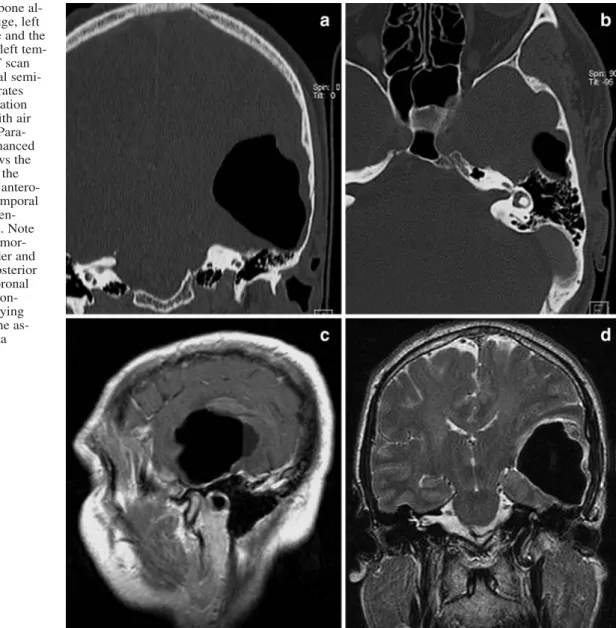
Fig. 1 a Coronal CT (bone al-gorithm) reveals the huge, left temporal pneumatocele and the normally pneumatized left tem-poral bone. b Axial CT scan at the level of the lateral semi-circular canal demonstrates the possible communication of the pneumatocele with air cells in the mastoid. c Para-sagittal gadolinium-enhanced T1-weighted MRI shows the broad-based contact of the pneumatocele with the antero-lateral surface of the temporal bone. No pathological en-hancement was present. Note the small associated hemor-rhage at the upper border and the fluid level in the posterior part of the lesion. d Coronal T2-weighted MRI demon-strates the space-occupying effect of the cyst and the as-sociated minimal edema
Two pathogenetic factors have been suggested to be
important for the development of a spontaneous
pneumo-cephalus. First, a defect in the temporal bone must be
pre-sumed, allowing a communication of air from the mastoid
cells to the intracranial compartment. Second, a pressure
difference is needed between the temporal bone and the
intracranial space to allow the air to enter the cranium.
A “ball valve” mechanism might explain the intracranial
air accumulation: increased air pressure caused by the
Valsalva manoeuvre or ambient pressure changes force air
through the fistula into the intracranial cavity. By the
con-secutive elevation of the intracranial pressure, the brain
and dura are forced over the fistula, and the air becomes
trapped.
There is a wide variation among individuals in number,
size and distribution of the air cells in the temporal bone
[17]. The middle ear pressure is presumed to play an
im-portant role in the extension of the pneumatization.
Con-stantly increased pressure because of Eustachian tube
dys-function or the habit of performing Valsalva manoeuvres
may lead to hyperpneumatization [18]. In addition,
de-fects in the petrous bone might be common in the general
population. In his series of 94 autopsy specimens, Ahren
found that 21% had one and 6% more than five clear
de-fects in the temporal bone [19]. In 6 of the 11 reported
cases of spontaneous otogenic pneumocephalus, a distinct
event causing pressure changes in the middle ear, such as
repetitive Valsalva manoeuvres [9, 11], nose blowing [8,
10], altitude change [15] or scuba diving [7], could be
de-scribed. In the five other cases, no suggestive episodes of
elevated middle ear pressure were documented. It is
pos-tulated as a possible explanation that also an abnormally
low or negative intracranial pressure may lead to air
entrap-ment [5], as is known from some cases after shunt
place-ment [20, 21, 22]. In our patient, the underlying
mecha-nism for the development of the pneumocephalus remains
unknown. Although no quantitative measurements were
performed with the CT data set in our patient, both
tem-poral bones appeared normal and symmetrically
pneuma-tized. Abrupt pressure changes in the middle ear eventually
could be postulated; however, there was no clear
anam-nestic evidence.
The clinical presentations vary among the reported cases,
depending on the location and mass effect of the
pneuma-tocele (see Table 1). Four patients showed epidural, three
subdural and four intraventricular, subarachnoid or
intrac-erebral air collection. Our patient heard sudden,
“ham-mering like” noises prior to the onset of motor aphasia
and visual field disturbances. Only two cases manifesting
with acoustic phenomena have been reported in literature.
In one case, the characteristics of the sensation was not
further described [5]; in the other, the pneumocephalus
manifested with a pulsatile tinnitus 4 years after surgery
for an arteriovenous malformation [23]. In the patient in
this study, a single episode of word-finding difficulties
1 week before presentation led the referring physician to
presume a transient ischemic attack and to initiate an
an-tiplatelet therapy with aspirin. It is most probable that the
intracerebral pneumocephalus was already present at that
time and that an initial cranial CT would have had led to a
correct diagnosis, thereby leading to an early and correct
treatment.
Treatment of spontaneous otologic pneumocephalus is
usually managed surgically in an attempt to relieve
in-tracranial pressure and to prevent the brain from infection.
Most of the cases were treated by closing the existing
fis-tula using muscle fascia flap, cartilage and bone wax to
seal the communication from the extracranial to the
in-tracranial compartment [8, 9, 10, 24]. An alternative
sur-gical option is to perform an otogenic procedure with
puncture of the intracranial air and closure of the mastoid
air cells from the temporal bone. An additional
therapeu-tic approach could be the reduction of the pressure in the
middle ear by Eustachian tube closure [5].
137 Table 1 Review of the literature of spontaneous pneumocephalus
Author year Sex Age Location Symptoms Mechanism Treatment
Jelsma 1954 [16] M 39 Subdural occipital right Hemiplegia, syncope Spontaneous Needle aspiration Markham 1967 [15] F 64 Subdural, intra-parenchymal,
parietal left
Headache, aphasia Pressure change Dural repair
Madeira 1977 [8] M 57 Epidural, occipital left Homonymous
hemianopsia
Pressure change Dural repair
Goldmann 1986 [7] M 26 Subdural, frontoparietal left Headache Pressure change Conservative
Stavas 1986 [10] M 64 Intracerebral, temporal left Expressive aphasia Spontaneous Dural repair, mastoidectomy
Spar 1994 [12] F 27 Subarachnoid,
intraventricular
Headache, vomiting, CSF otorrhea
Spontaneous Dural repair
Maier 1996 [9] M 24 Epidural, parietooccipital
right
Headache, visual scotoma
Valsalva Mastoidectomy
Dowd 1998 [6] F 78 Intraventricular, intracerebral temporal right
Hemiparesis, aphasia, ear noise
Spontaneous Dural repair, eusta-chian tube closure
Park 1998 [24] M 49 Epidural, temporo-parietal
right
Headache Spontaneous Dural repair
Vallejo 1999 [11] M 20 Epidural, temporo-occipital right
In conclusion, a spontaneous pneumocephalus
originat-ing from the temporal bone, although extremely rare,
should be considered in patients reporting extraordinary
acoustic phenomena and other, non-specific neurological
symptoms. The diagnosis is even likely in cases of a
nor-mally pneumatized temporal bone and without signs of
ear infection or tumor growth.
References
1. Clevens RA, Marentette LJ, Esclamado RM, Wolf GT, Ross DA (1999) Incidence and management of tension pneumo-cephalus after anterior craniofacial resection: case reports and review of the literature. Otolaryngol Head Neck Surg 120:579– 583
2. Di Lorenzo N, Caruso R, Floris R, Guerrisi V, Bozzao L, Fortuna A (1986) Pneumocephalus and tension pneumocephalus after posterior fossa surgery in the sitting position: a prospective study. Acta Neurochir (Vienna) 83:112–115
3. Andrews JC, Canalis RF (1986) Otogenic pneumocephalus. Laryngoscope 96:521–528
4. Brunori A, Bruni P, Delitala A, Greco R, Chiappetta F (1995) Frontoethmoidal osteoma complicated by intracranial muco-cele and hypertensive pneumocephalus: case report. Neuro-surgery 36:1237–1238
5. Rappaport JM, Attia EL (1994) Pneumocephalus in frontal si-nus osteoma: a case report. J Otolaryngol 23:430–436 6. Dowd GC, Molony TB, Voorhies RM (1998) Spontaneous
oto-genic pneumocephalus: Case report and review of the litera-ture. J Neurosurg 89:1036–1039
7. Goldmann RW (1986) Pneumocephalus as a consequence of barotraumas. JAMA 255:3154–3156
8. Madeira JT, Summers GW (1977) Epidural mastoid pneumato-cele. Radiology 122:727–728
9. Maier W, Fradis M, Schermet R (1996) Spontaneus otogenic pneumocephalus. Ann Otol Rhinol Laryngol 105:300–302 10. Stavas J, McGeachie RE, Turner DA, Nelson MJ (1987)
Symp-tomatic intracranial pneumatocele from mastoid sinus of spon-taneous origin: case report. J Neurosurg 67:773–775
11. Vallejo LA, Gil-Carcedo LM, Borras JM, De Campos JM (1999) Spontaneous pneumocephalus of otogenic origin. Oto-laryngol Head Neck Surg 121:662–665
12. Spar JA (1994) Spontaneous CFS communication to the mid-dle ear and external auditory canal: a case report. Acta Radiol 35:506–508
13. Chiari H (1884) Über einen Fall von Luftansammlung in den Ventrikeln des menschlichen Gehirns. Zschr F Heilk 5:383–390 14. Luckett WH (1913) Air in the ventricles of the brain following a fracture of the scull. Report of a case. Surg Gynecol Obstet 17:237–240
15. Markham JW (1967) The clinical features of pneumocephalus based upon a survey of 284 cases with report of 11 additional cases. Acta Neurochir (Vienna) 16:1–78
16. Jelsma F, Moore DF (1954) Cranial aerocele. Am J Surg 87: 437–451
17. Dietzel K (1989) Untersuchungen zur “normalen” Pneumatisa-tion des Os temporale. HNO 37:39–47
18. Nyrop M, Bjerre PK, Christensen J, Jorgensen KE (1999) Ex-tensive and symptomatic cranial pneumatization: caused by frequent performance of Valsalva’s manoeuvre? J Laryngol Otol 113:480–482
19. Ahren C, Thulin CA (1965) Lethal intracranial complication following inflation in the external auditory canal in treatment of serous otitis media and due to defect in the petrous bone. Acta Otolaryngol 60:407–421
20. Kanner A, Nageris B, Chaimoff M, Rappaport Z (2000) Spon-taneous pneumocephalus in the posterior fossa in a patient with a ventriculoperitoneal shunt: case report. Neurosurgery 46: 1002–1004
21. Mylonas C (1991) Delayed pneumocephalus in patients with CSF shunts. Br J Neurosurg 5:67–72
22. Villarejo F, Carceller F, Alvarez C, Bencosme J, Perez Diaz C, Goldman L, Pascual A (1998) Pneumocephalus after shunting for hydrocephalus. Child Nerv Syst 14:333–337
23. Saitoh Y, Takeda N, Yagi R, Oshima K, Kubo T, Yoshimine T (2000) Pneumocephalus causing pulsatile tinnitus. Case illus-tration. J Neurosurg 92:505
24. Park P, Chandler WF, Telian SA, Doran S (1998) Spontaneous chronic epidural pneumocephalus resulting from hyperpneu-matization of the cranium causing mass effect: case report. Neurosurgery 42:1384–1386