Cell Tissue Res (2006) 325: 303–313 DOI 10.1007/s00441-005-0119-7
R E G U L A R A RT I C L E
Romano Silvio Kasper . Natallia Shved . Akiyoshi Takahashi . Manfred Reinecke . Elisabeth Eppler
A systematic immunohistochemical survey of the distribution
patterns of GH, prolactin, somatolactin, β–TSH, β–FSH,
β–LH, ACTH, and α–MSH in the adenohypophysis
of
Oreochromis niloticus, the Nile tilapia
Received: 24 June 2005 / Accepted: 3 November 2005 / Published online: 22 March 2006 # Springer-Verlag 2006
Abstract Fish pituitary plays a central role in the control of growth, development, reproduction and adaptation to the environment. Several types of hormone–secreting adeno-hypophyseal cells have been characterised and localised in diverse teleost species. The results suggest a similar distri-bution pattern among the species investigated. However, most studies deal with a single hormone or hormone family. Thus, we studied adjacent sections of the pituitary of Oreochromis niloticus, the tilapia, by conventional staining and immunohistochemistry with specific antisera directed against growth hormone (GH), prolactin (PRL), somatolactin (SL), thyrotropin (β–TSH), follicle–stimulating hormone (β–FSH), luteinising hormone (β–LH), adrenocorticotropic hormone (ACTH) and melanocyte–stimulating hormone (α–MSH). The pituitary was characterised by a close in-terdigitating neighbourhood of neurohypophysis (PN) and adenohypophysis. PRL–immunoreactive and ACTH–im-munoreactive cells were detected in the rostral pars distalis. GH–immunoreactive cells were present in the proximal pars distalis (PPD). A small region of the PPD containedβ–TSH–immunoreactive cells, and β–LH–im-munoreactive cells covered approximately the remaining parts. Centrally, β–FSH–immunoreactive cells were
de-tected in the vicinity of the GH–containing cells. Some of these cells also displayed β–LH immunoreactivity. The pars intermedia was characterised by branches of the PN surrounded by SL–containing and α–MSH–immunoreac-tive cells.The ACTH andα–MSH antisera were observed to cross-react with the respective antigens. This cross– reactivity was abolished by pre–absorption. We present a complete map of the distinct localisation sites for the classical pituitary hormones, thereby providing a solid basis for future research on teleost pituitary.
Keywords Pituitary . Hormones . Localisation . Immunohistochemistry . Mapping . Teleost
Introduction
As in mammals, fish pituitary has been described to play a central regulatory role in the control of growth, develop-ment, reproduction and adaptation to environmental challenges, such as changes in salinity, temperature and stress. Bony fish represent the largest non-mammalian vertebrate group and provide valuable models for basic research on vertebrate physiology. In addition, numerous bony fish species, such as salmonids, seabream and tilapia, have high commercial value and are used in aquaculture, the fastest growing area of food production. Therefore, the scientific results on the regulatory mechanisms involved in growth, development and reproduction that have emerged are of major impact not only in basic science, but also in the aquaculture industry (for reviews, see Reinecke and Collet
1998; Reinecke et al.2005). The susceptibility of fish kept in pond culture or other farming conditions to environ-mental challenges, including pollutants, is an important research field that is increasingly under investigation.
In teleosts, the pituitary is characterised by a close interdigitating neighbourhood between the neurohypophy-sis (pars nervosa, PN) and the adenohypophyneurohypophy-sis. The latter consists of three main parts: the rostral pars distalis (RPD), the proximal pars distalis (PPD), and the pars intermedia This work was supported by the SNF (NRP 50, project 4050–
66580) and by the Hartmann Müller-Stiftung für Medizinische Forschung an der Universität Zürich (grant no. 972).
R. S. Kasper . N. Shved . M. Reinecke . E. Eppler (*)
Division of Neuroendocrinology, Institute of Anatomy, University of Zürich, Winterthurerstrasse 190, Zürich CH–8057, Switzerland e-mail: [email protected] Tel.: +41-44-6355333 Fax: +41-44-6355702 A. Takahashi
Laboratory of Molecular Endocrinology,
School of Fisheries Sciences, Kitasato University, Ofunato, Iwate, Japan
(PI). Unlike mammals, teleost fish lack a hypothalamo– hypophyseal portal system for the transport of neurohor-monal regulators. Instead, a direct axonal transport exists between hypothalamic neurons and pituitary endocrine cells via the hypophyseal stalk and the PN (Weltzien et al.
2004).
The hormone–producing cells of the adenohypophysis have been investigated for several decades by means of standard staining procedures and immunocytochemical and molecular biological methods (for a review, see Agulleiro et al. 2006). With these methods, hormone–secreting adenohypophyseal cells have been characterised and lo-calised in diverse teleost species. Whereas the hormone– producing cell types are arranged in a mosaic pattern in the pituitary of adult tetrapods, teleost pituitary preserves the embryonic compartmental organisation, i.e. each specific hormone–producing cell type is located in a particular compartment. Thus, teleost fish represent interesting mod-els for the study of pituitary ontogeny and phylogeny (for a review, see Weltzien et al.2004).
The potential impact of environmental pollutants on growth hormone (GH) and adrenocorticotropic hormone (ACTH) peptide expression has been pointed out in tilapia (Oreochromis niloticus) indicating the need for further studies (Mousa and Mousa1999a). However, few system-atic data exist on teleost pituitary as the central regulatory organ of numerous physiological processes. Most studies deal with single hormones or hormone families (e.g. Amemiya et al. 1999; Amano et al. 2005; Dores et al.
1996; García Ayala et al.2003; Huang and Specker1994; Joss et al.1990; Kagawa et al.1998; Kawauchi et al.1986,
1989; Mousa and Mousa1999a,b; Nagahama et al.1981; Naito et al.1983; Nishioka et al.1993; Nozaki et al.1990; Olivereau and Rand-Weaver 1994; Parhar et al. 2003; Power 1992; Rand-Weaver et al.1991; Saga et al.1993; Shimizu et al.2003; Siegmund et al. 1987; Weltzien et al.
2003a). A few more extensive studies have been performed (e.g. García-Hernández et al. 1996; Grandi and Chicca
2004; Laiz-Carrión et al.2003; Naito et al.1993; Parhar et al.1998,2002; Sánchez Cala et al.2003; Segura-Noguera et al.2000; Weltzien et al.2003b; Yan and Thomas1991) but no thorough investigation has localised all pituitary hormones by immunohistochemistry on consecutive sections. In detail, during the ontogeny of chum salmon (Oncorhynchus keta), cells expressing ACTH, prolactin (PRL), GH,α–melanocyte–stimulating hormone (α–MSH) and β–thyroid–stimulating hormone (β–TSH) have been localised by using immunocytochemistry (Naito et al.
1993). In the protogynous teleost wrasse Thalassoma duperrey, all adenohypophyseal hormones were investi-gated but cross-reactivities betweenβ–TSH and the gonad-otropin GTH II (β–LH) antisera had to be considered (Parhar et al. 1998); however, the study mainly character-ised endocrine cells at the ultrastructural level and only selected hormones were presented by light microscopy. In an ontogenic study on sturgeon, Acipenser naccarii, the appearance and distribution of all adenohypophyseal hor-mones including β–endorphin were monitored but, as the main focus was laid on early development, the demand for
investigations of the hormone expression pattern in adults was stressed (Grandi and Chicca2004).
An early study of adult Atlantic croaker (Micropogonias undulatus), spotted sea trout (Cynoscion nebulosus) and red drum (Sciaenops ocellatus) investigated the cells express-ing GH, PRL, ACTH,β–TSH and the gonadotropins but the distinct localisation of the gonadotropins remained unclear (Yan and Thomas1991). An immunocytochemical study in Mediterranean yellowtail (Seriola dumerilii) characterised the same hormonal cell types, plus SL and α–MSH. Again, the gonadotropins could not be defin-itively localised (García-Hernández et al.1996). Similarly, in young white seabream (Diplodus sargus), the data for the gonadotropins also remained unclear (Segura-Noguera et al. 2000). In a more recent study in adult male Atlantic halibut (Hippoglossus hippoglossus), a map was created for the distinct localisations of all hormone cell types, whereby β–FSH was localised by in situ hybridisation (Weltzien et al.2003b). However, a clear interpretation of the potential co–localisation of the gonadotropins was not possible. In a study on gonadotropin–releasing hormone receptors re-cently performed in tilapia, the gonadotropins and cells of the GH/PRL family were also investigated by using in situ hybridisation and immunohistochemistry; whereasβ–LH– containing cells were displayed at the gene and peptide expression level, β–FSH–expressing cells were again detected exclusively at the mRNA level (Parhar et al.
2002). The contradictory results led to the explanation that the antisera against salmon β–FSH might recognise an epitope of tilapiaβ–LH (Parhar et al.2003), a proposal that gives new support to the importance of a definite localisa-tion of the gonadotropins, especially with respect to the differences in sexual regulation between salmonids and non–salmonids.
Thus, to our knowledge, no study has as yet succeeded in demonstrating all the hormone-producing cells in the entire pituitary in adjacent or, at least similarly oriented, sections by using the same technique consistently throughout the investigation. However, only this approach can create a basis for investigations of diverse hormone–producing cells under various experimental settings. Furthermore, no complete mapping has been performed in tilapia, a species widely used in aquaculture. Another problem that has not been sufficiently addressed to date is the dual existence and localisation of the gonadotropins, i.e. GTH I and GTH II, more recently shown to be β–FSH and β–LH in higher teleosts (Rosenfeld et al. 2001; Quérat et al. 1990). The obtained contradictory results (see above) have been attributed not only to species differences, but also to antisera–dependent differences (e.g. García-Hernández et al.1996; Segura-Noguera et al. 2000; Parhar et al.2003) and need to be clarified.
In order to provide a basis for future experiments, we wanted to elaborate a defined morphological knowledge of teleost pituitary and thus explored the pituitary of the Nile tilapia (O. niloticus) as a representative model species. Thus, the distribution pattern of GH, PRL and SL, of the POMC family members ACTH and α–MSH, and of β–TSH and the gonadotropins β–FSH and β–LH was
analysed on consecutive Paraplast and semi-thin sections. Special emphasis was laid on elucidating the localisation sites of the gonadotropinsβ–FSH and β–LH. Based on the results obtained, we finally obtained a complete map of the distinct localisation sites of all pituitary hormones.
Materials and methods
Fish culture
Twelve individuals of Oreochromis niloticus (125 days old) kindly provided by Prof. N. Maclean (School of Biological Sciences, University of Southampton, South-ampton, UK), and Dr. J.-F. Baroiller (CIRAD, Montpellier, France) were used. The fish were kept in fresh-water tanks at 27±1°C under a 12 h/12 h light/dark cycle and fed to satiation.
Tissue sampling and preparation
Fish were anaesthetised with 2–phenoxy–ethanol (Sigma, St. Louis, Mo, USA) added to the water and killed. Pituitaries were immediately excised and, for conventional and immunofluoresence microscopy, attachment to the brain was conserved to simplify later orientation. Tissue samples were fixed by immersion in acetic-acid-free Bouin’s solution for 4 h at room temperature. Specimens were dehydrated in an ascending series of ethanol and routinely embedded in Paraplast Plus at 58°C. Sections were cut at 4 μm, mounted on Super Frost Plus slides (Menzel–Gläser, Germany) and dried overnight at 42°C. After being dewaxed in xylol, they were rehydrated in a descending series of ethanol (100%, 96%, 70%).
For semi-thin sections, pituitaries were fixed by immer-sion in a solution containing 2.5% paraformaldehyde, 0.1%
Table 2 HE and PAS staining (PI pars intermedia, PN pars nervosa, PPD proximal pars distalis, RPD rostral pars distalis)
Hormone Region HE stain PAS stain
ACTH RPD Basophilic Negative
β–FSH PPD Basophilic Positive
GH PPD Strongly eosinophilic Negative
β–LH PPD Basophilic Positive
α–MSH PI Basophilic Negative
PRL RPD Strongly eosinophilic Negative
SL PI Eosinophilic Negative
β–TSH PPD Basophilic Positive
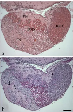
Fig. 1 Histology of tilapia pituitary. Consecutive sagittal Paraplast sections stained with (a) HE and (b) PAS (see Table2). a, b The adenohypophysis is divided into three main parts, the rostral (RPD) and proximal (PPD) pars distalis and the pars intermedia (PI). Sections display the neural connection of the hypothalamus to the pituitary (a, arrows) and the interdigitating branches of the PN reaching the PI (b, arrowheads). b Regions containing cells expressing ACTH (c) and α–MSH (d) investigated by the pre– absorption experiment (see Fig.4). Bar 150μm
Table 1 Primary antisera used for immunofluorescence on Paraplast sections (all antisera raised in rabbit)
Antiserum against Specificity Dilutions Source Reference
ACTH (1–23) Rat 1:2,000 Peptide Inst., Osaka (Japan), cat. no. Y352 –
FSH,β–subunit Chum salmon 1:400 Dr. H. Kawauchi, Kitasato University (Japan), lot 8510 Nozaki et al.1990 FSH,β–subunit Human 1:100 Biogenesis, Poole (England), cat. no. 4560–8055 Grandi and Chicca2004 GH Chum salmon 1:8,000 Dr. H. Kawauchi, Kitasato University (Japan), lot 8502 Kawauchi et al.1986 LH,β–subunit Chum salmon 1:4,000 Dr. H. Kawauchi, Kitasato University (Japan), lot 8506 Nozaki et al.1990 LH,β–subunit Human 1:200 DAKO A/S, Glastrup, Denmark, cat. no. N1543 Siegmund et al.1987 MSH,α–subunit Human 1:1,500 Biogenesis, Poole (England), cat. no. 6045–0400 Amano et al.2005 PRL Chum salmon 1:12,000 Dr. H. Kawauchi, Kitasato University (Japan), lot 8206 Naito et al.1983 SL Chum salmon 1:4,000 Dr. H. Kawauchi, Kitasato University (Japan), lot 8906 Rand-Weaver et al.1991 TSH,β–subunit Human 1:300 Biogenesis, Poole (UK), cat. no. 8926–0004 Grandi and Chicca2004
glutaraldehyde and 0.01% picric acid for 4 h. Thereafter, specimens were dehydrated in an ascending series of ethanol and routinely embedded in LR White (Poly-sciences, Warrington, USA). Serial semi-thin sections (1μm) were cut on an Ultracut E (Reichert-Jung, Zürich, Switzerland) and placed on glass slides.
Haematoxylin–eosin and periodic–acid–Schiff stain For haematoxylin–eosin (HE) staining, slides were im-mersed in Mayer`s haematoxylin for 10 min, briefly treated
with HCl solution, rinsed in tap water for 10 min and subsequently immersed in eosin for another 10 min. For periodic–acid–Schiff (PAS) staining, slides were treated with periodic acid for 5 min and rinsed in tap water. Treatment with Schiff’s reagent for 15 min was followed by exposure of the sections to sodium bisulfite (0.52%) for 10 min. After a rinse in tap water, slides were immersed in Mayer’s haematoxylin. After another wash step, differen-tiation in HCl ethanol was performed followed by a rinse in flowing water. Slides were dehydrated in 100% ethanol and xylol (all reagents obtained from Fluka, Switzerland) and covered with Eukitt (Merck, Switzerland).
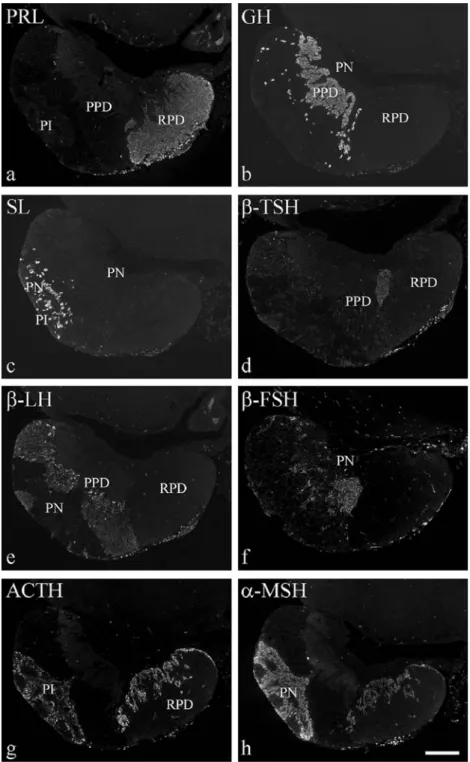
Fig. 2 Localisation of hor-mones in tilapia pituitary by the use of immunofluorescence. Eight consecutive sagittal Para-plast sections adjacent to Fig.1 were stained with specific anti-sera raised in rabbit (see Table1) and visualised with an FITC-coupled anti–rabbit IgG (PI pars intermedia, PN pars nervosa, PPD proximal pars distalis, RPD rostral pars distalis). a–c Anti-sera directed against chum salmon (a) PRL, (b) GH, (c) SL. d Antibody directed against human TSHβ–subunit. e Anti-chum-salmonβ–LH an-tiserum. f Anti-humanβ–FSH antiserum. g, h Antibodies di-rected against (g) rat ACTH (amino acids 1–23) and (h) human MSHα–subunit. Bar 200μm
Immunocytochemistry
To allow unmasking of the antigen for immunofluorescent stain, microwaving (350 W, 5 min) in 0.01 M citrate buffer (pH 6) was performed. Unspecific binding was reduced by treatment with phosphate-buffered saline (PBS, pH 7.4) containing 2% bovine serum albumin (BSA) for 30 min at room temperature. Thereafter, sections were incubated overnight at 4°C with the diverse antisera specific for the various pituitary hormones (diluted in PBS/2% BSA; for dilutions see Table 1). After repeated rinses in PBS/2% BSA, the antisera were detected by incubation in the dark for 30 min at room temperature with a fluorescein– isothiocyanate (FITC)–conjugated goat anti–rabbit IgG (Bioscience Products, Emmenbrücke, Switzerland) diluted 1:100 in PBS. Specificity of the antisera (Table1) had been previously shown by other groups. For instance, pre– absorptions of the antisera against chum salmon GH, SL and PRL with their respective antigens had been shown to abolish immunoreactivity (Mousa and Mousa 1999b; Ominato and Nozaki 2002). In contrast, for the anti-chum-salmon gonadotropin antisera, the reported cross– reactivities (see Introduction) could not be abolished by pre–absorption (Nozaki et al.1990), whereas no immuno-staining was observed after pre–incubation of the anti-human β–FSH and β–TSH antisera with an excess of the appropriate antigens (Grandi and Chicca2004). Thus, in the present study, specificities were further examined by omitting the primary antiserum and by the choice of different antisera (see Table1). In addition, the specificities of the antisera directed against rat ACTH and human α–MSH were tested by pre–incubation for 24 h with 40 or 400μg recombinant human α–MSH (Biogenesis, UK) and ACTH (fragment 1–24, Sigma), respectively, per millilitre of diluted antiserum.
The semi–thin sections were treated with 10 mM gelatin in 10 ml PBS (pH 7.4) containing 0.2 g BSA for 30 min to reduce non–specific binding. After repeated washes in PBS, consecutive semi–thin sections were processed for immunohistochemistry. The first antisera directed against human and chum salmonβ–LH and β–FSH, respectively (see Table1; all diluted 1:30 in gelatin–PBS–BSA) were incubated in a humid chamber for 12 h at 4°C. After repeated washes in PBS, the sections were treated with biotin-coupled goat anti-rabbit IgG (Bio–Science Products; diluted 1:100 in gelatin–PBS) for 30 min at room temperature, followed by incubation with a streptavidin– gold–5–nm complex (Amersham, Little Chalfont, UK; diluted 1:100 in gelatin–PBS) for 1 h at room temperature. After repeated washes in double-distilled water, the sections were treated with the IntenSE M silver enhance-ment kit (Amersham) for 10 min at room temperature and counterstained with methylene blue/azure.
Imaging
Microscopy and photography were performed with a Zeiss Axioscope and Axiovision 3.1. software (Zeiss, Zürich, Switzerland). The fluorochromes were visualised with a fluorescence module for FITC (BP 450–490 nm, FT 510, LP 515–565 nm).
Results
Structure of the pituitary of adult O. niloticus
Consecutive sagittal Paraplast sections of tilapia pituitary attached to the brain were stained with HE (Fig. 1a) and PAS (Fig. 1b). The neurohypophysis (PN) and the
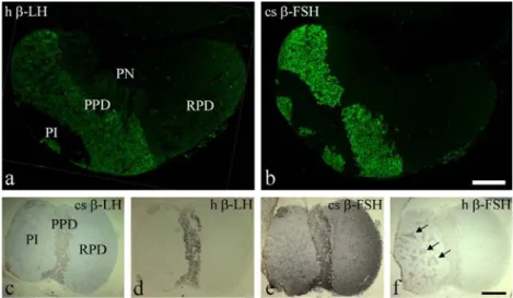
ade-Fig. 3 Localisation of gonadotropins in tilapia pituitary (PI pars intermedia, PN pars nervosa, PPD proximal pars distalis, RPD rostral pars distalis). a, b Consecutive Paraplast sections to those in Figs.1,2 immunostained by rabbit antisera (see Table 1) directed against (a) human (h)β–LH, and (b) chum salmon (cs) β–FSH visualised with an FITC-coupled anti–rabbit IgG. Bar 200 μm. c–f Consecutive
horizontal semi-thin sections of tilapia pituitary immunostained with antisera directed against (c) csβ–LH, (d) h β–LH, (e) cs β–FSH and (f) h β–FSH (see Table 1) visualised with biotin-conjugated anti– rabbit IgG followed by a streptavidin–gold–5-nm complex (arrows branches of the PN within the PI). Bar 300μm
nohypophysis presented an interdigitating neighbourhood. The adenohypophysis was divided into three main parts: the RPD, PPD and PI. Branches of the PN were detected throughout the adenohypophysis, especially in the PI. The hormone–containing cells displayed differential staining behaviour with both techniques (see Table2).
Distribution pattern of tilapia adenohypophyseal hormones analysed by immunofluorescence microscopy
Consecutive sections to Fig. 1were investigated by using antisera (Table1) specific for the classical adenohypophy-seal hormones, i.e. members of the GH/PRL family (GH, PRL, SL), the glycoprotein hormone group constituted by the gonadotropins and thyrotropin (β–FSH, β–LH, β– TSH) and members of the proopio–melanocorticotropin (POMC) family (ACTH,α–MSH).
GH/PRL family
The members of the GH/PRL family were detected in all three parts of the adenohypophysis and showed a strong eosinophilic staining behaviour (Fig. 1a, Table 2). PRL– immunoreactive cells filled the major part of the RPD (Fig. 2a). GH–immunoreactive cells were arranged in regular chains along the branches of the PN within the PPD (Fig.2b). SL–containing cells surrounded the nerve fibres of the PN (Fig.2c).
Glycoprotein hormones
The glycoprotein hormone–containing cells reacted to PAS (Fig.1b, Table2). Near to the border seam between RPD and PPD, a small region contained cells immunoreactive for β–TSH (Fig.2d). Cells immunoreactive to the anti-chum-salmonβ–LH antiserum filled the whole PPD except for the Fig. 4 Specificity test of anti-rat
ACTH antiserum on consecutive Paraplast sections (PI pars inter-media, PN pars nervosa, PPD proximal pars distalis, RPD rostral pars distalis). a–c Immu-nostaining of ACTH in the RPD (compare Fig.1a,b, region c). a With the rat ACTH anti-serum not pre-absorbed. b,c After 24 h pre–incubation withα–MSH peptide at 40 μg/ ml (b) and 400μg/ml (c) anti-serum. d–f Cross-reactive staining ofα–MSH in the PI (compare (Fig.1a,b, region d). d With the ACTH antiserum not pre-absorbed. e, f After 24 h pre– incubation withα–MSH at 40μg/ml (e) and 400 μg/ml (f) antiserum. Bar 50μm. g, h Immunostaining of ACTH in the RPD. g With the anti-rat ACTH antiserum not pre-ab-sorbed. h After 24 h pre –incu-bation with human ACTH 1–24 fragment at 40μg/ml antiserum. Bar 150μm. i Amino acid sequences for tilapia ACTH (1st line), human/rat ACTH (2nd line) andα–MSH (3rd line). Differences are indicated in red, insertions and deletions in blue. Green vertical lines indicate amino acids 1–23, the epitope of the rat ACTH peptide to which the antiserum is directed
GH and β–TSH regions (Fig. 2e). Similar results were obtained with an antiserum directed against humanβ–LH (Fig. 3a). On using the antiserum against chum salmon β–FSH (Fig.3b), an almost identical distribution pattern as with theβ–LH–antisera (Figs.2e,3a) was detected, whereas an antiserum directed against human β–FSH recognised a cell population near to the somatotrophs, mainly in the region of the β–LH–expressing cells (Fig. 2f). Similar results were achieved by immunocytochemistry on hori-zontal semi-thin sections (Fig. 3c–f): anti-chum-salmon (Fig.3c) and anti-human (Fig.3d)β–LH antisera displayed an identical staining pattern in the PPD, and the anti-chum-salmon β–FSH antiserum (Fig. 3e) again recognised the same regions as theβ–LH antisera. In horizontally oriented pituitary sections, even the anti-human β–FSH antiserum stained the same regions, indicating a partial co–localisation of GTH I (β–FSH) and GTH II (β–LH) in the centre of the PPD (Figs.2e,f,3c–f). In addition, distinct staining of the PN branches in the PI was observed with both anti-β–FSH antisera (Fig.3e,f). A similar but markedly weaker staining pattern in the PI was observed forβ–LH (Fig.3c,d). POMC family
The members of the POMC family were only faintly ba-sophilic after HE staining and not stained by PAS (Fig.1, Table2). ACTH-immunoreactive cells formed a fine seam of cuboid to columnar cells in the RPD (Figs.2g,4a) along the region containing the PRL-immunoreactive cells (Fig.2a). In the PI, a large cell population immunoreactive forα–MSH was detected around the PN branches (Figs.2h,4d).
The antisera used for detecting ACTH and α–MSH reacted with both cell types, i.e. with ACTH–containing cells of the RPD and withα–MSH–containing cells of the PI (Fig. 2g,h), although with different staining intensity. Because this reaction pattern might have indicated cross– reactions, we performed preabsorption assays: the antise-rum directed against ACTH was preincubated withα–MSH to saturate potential α–MSH-binding sites. Accordingly, cross-reactivity of the ACTH antiserum to α–MSH cells (Fig.4d) in the PI was strongly reduced by preincubation of the anti–ACTH antiserum with α–MSH at 40 μg/ml (Fig.4e) and completely abolished at 400μg/ml (Fig.4f) antiserum. On the contrary, immunostaining for ACTH– containing cells (Fig.4a,g) in the RPD was not impaired by preabsorption of the antiserum with α–MSH (Fig.4b,c), whereas it was completely abolished (Fig. 4h) after pre– incubation with the human ACTH amino acid fragment 1–24. The anti-rat ACTH antiserum used was thus specific for amino acids 1–23, which are almost identical for human, rat and tilapia ACTH (Fig.4i).
Localisation of the principal hormones in tilapia adenohypophysis
A map was created displaying the localisation of all principal hormones in tilapia adenohypophysis (Fig. 5).
Thus, PRL and ACTH (Figs.2a,g,4a) were localised in the RPD,β–TSH, β–FSH and β–LH (the last two mentioned in partial coexistence) in the PPD (Figs.2d–f,3c–f), and SL
andα–MSH in the PI (Figs.2c,h,4d).
Discussion
An understanding of the central role of the adenohypoph-ysis in teleosts (for a recent review, see Agulleiro et al.
2006) demands profound knowledge of the distribution of the hormone–producing cells. The present study investi-gating all pituitary hormones on consecutive sections in the Nile tilapia (O. niloticus) has revealed a structure and distribution pattern similar to those emerging when combining the results of other groups in various teleosts including tilapia (e.g. Holmes and Ball1974; Leatherland et al. 1974; Joy and Sathyanesan 1980; Siegmund et al.
1987; Yan and Thomas1991).
The hormones GH, PRL and SL belong to the GH–PRL family and are believed to have evolved from a common ancestral gene by duplication and divergence (Ono et al.
1990; Rand-Weaver et al.1991). GH and PRL have been identified in most gnathostome vertebrate classes but SL is thought to be confined to osteichthyes (Nozaki et al.2001). The general occurrence of the GH–PRL family hormones in the three parts of the teleost pituitary as found here has previously been described in the Mediterranean yellowtail and Atlantic halibut (García-Hernández et al. 1996; Weltzien et al. 2003b). In juvenile striped bass (Huang and Specker 1994) and Anguilla anguilla (Grandi et al.
2003), GH immunoreactivity has also been detected in the RPD, and in Mediterranean yellowtail, greater weever fish and juvenile Acipenser naccarii in the PI (García-Hernández et al. 1996; Sánchez Cala et al. 2003; Grandi and Chicca 2004). In young white seabream, GH– Fig. 5 Map of all pituitary hormone localisations. The rostral pars distalis (RPD) is given in red (ACTH region) and yellow (PRL region). The proximal pars distalis (PPD) is coloured in dark blue (GH) and light blue (β–TSH) regions and in striped green symbolising both β–LH (light green) and β–FSH (dark green) areas. The pars intermedia (PI) is coloured as striped violet representing the intermingled α–MSH (pink) and SL (dark violet) cells. Note the pars nervosa (PN) branches (grey) that reach towards the PI
expressing cells have been found in PPD, RPD and PI (Segura-Noguera et al. 2000). In contrast, we have not detected GH–containing cells outside the PPD of the adult tilapia pituitary. Our results are, thus, in agreement with those of the majority of groups (for a review, see Agulleiro et al. 2006). Ominato and Nozaki (2002) have reported a similar localisation in the agnathan sea lamprey in which the likely common ancestral form of GH–like cells is present (Nozaki et al.2001). We assume that the restriction of GH cells to the PPD is characteristic for adult fish and their wide–spread occurrence for developing fish. This hypothesis agrees with recently observed differences in GH gene expression between adult and developing bony fish. In adult four–spine sculpin, Cottus kazika (Inoue et al.
2003) and tilapia (Caelers et al.2005), GH gene expression has been found exclusively in the pituitary. In contrast, in developing rainbow trout, GH mRNA has been detected in the pituitary and other organs, such as brain, gills, liver and ovary (Yang et al.1999) and, in coho salmon, GH has been found not only in the pituitary, but also in the intestine, but exclusively in small fish (Mori and Devlin 1999). The importance of GH during fish development is stressed by the observation that GH in pituitary is expressed even before the separation of the adenohypophysis into PI and PD (Grandi and Chicca2004) and precedes the appearance of primordial germ cells and gonadotropins (Saga et al.
1993).
The defined localisation of cells immunoreactive for PRL in the RPD and for GH around the neurohypophyseal processes in the PPD as found in the present study has been observed for various teleosts (Yan and Thomas 1991; Power1992; García-Hernández et al.1996; Weltzien et al.
2003b; Volckaert et al. 1999; Laiz-Carrión et al. 2003), including tilapia (Nishioka et al. 1993; Nagahama et al.
1981; Mousa and Mousa1999a,b).
In tilapia, SL is localised in epithelial cells arranged around the interdigitating branches of the PN reaching the PI. Such a regular arrangement has also been described in other developing (Villaplana et al.1997; Laiz-Carrión et al.
2003) and adult teleosts (Weltzien et al.2003b). In mature Pacific salmon (Oncorhynchus nerka), pronounced differ-ences in SL expression have been noted during the sexual cycle (Olivereau and Rand-Weaver 1994). However, the sexual cycle of salmonids differs from that of cychlid fish, such as tilapia, which are reproductive throughout the year (Baroiller and Guiguen 2001; Kawauchi et al. 1989; Melamed et al. 1998; Santos et al. 2001; Shimizu et al.
2003; Weltzien et al.2004). Nevertheless, in a comparative analysis of SL-related immunoreactivity in O. niloticus, different staining patterns were observed during sexual maturation (Mousa and Mousa 1999b). The distribution pattern of the SL–immunoreactive cells observed in our study agrees with results in mature tilapia (Mousa and Mousa1999b; Dores et al.1996).
Whereas the distribution of the GH/PRL family has been mostly elucidated among teleosts investigated so far, that of the gonadotropins is still under discussion. In the present study, we obtained a marked co–localisation of β–FSH and β–LH–immunoreactive cells by using the antisera directed
against chum salmon FSH and LH β–subunits. These results are similar to those obtained in Mediterranean yellowtail with the same antisera. However, anti-catfish α,β–GTH antiserum has given different results (García-Hernández et al. 1996). Anti-chum-salmon antisera are widely used for investigating teleost gonadotropins and have been reported to give different results among the diverse teleost species investigated (for a review, see Agulleiro et al. 2006). The discrepancies have been ascribed either to biological variations or to the possibility that heterologous antisera might be inappropriate to investigate the occurrence of two types of gonadotropes (García-Hernández et al.1996; García Ayala et al.2003). Because homologous antisera are often difficult to obtain, different approaches to counter the problem have been chosen, e.g. staining for the commonα–unit followed by staining for β–LH and β–TSH, whereby the remaining cells are presumed to beβ–FSH–containing cells (García Ayala et al. 2003). Human β–TSH antisera have been reported to cross–react well with teleost TSH (for a review, see Agulleiro et al.2006); this has also been found here in tilapia. We have not observed cross–reactivity between antisera to β–TSH and GTH II (β–LH) as described in several studies (García-Hernández et al.1996; Parhar et al.
1998; Segura-Noguera et al.2000) but between the antisera directed against GTH I and GTH II. A moderate cross– reactivity (10% and 12%, respectively) between these antisera has previously been observed (Suzuki et al.1988). Our data obtained in tilapia with anti-chum-salmon antisera nevertheless contrast with those obtained in salmonids against which the antisera have been generated and to which they have been shown to be specific (Kawauchi et al.
1989; Nozaki et al. 1990). No co–localisation of GTH I (β–FSH) and GTH II (β–LH) immunoreactivities has been found in rainbow trout and Atlantic salmon (Nozaki et al. 1990). In our study, the antisera directed against human β–LH and chum salmon β–LH give identical staining patterns. This result is comparable to those obtained with anti-chum-salmon GTH II antiserum and an anti–ovine β–LH antiserum (Mousa and Mousa1999a). These and our data support the postulated good conserva-tion ofβ–LH throughout evolution (Yaron et al.2001) in contrast to GTH I/β–FSH, which is poorly conserved (Kawauchi et al.1989).
Concerning the localisation of β–FSH, the anti-chum-salmon antiserum reacting with the tilapia β–LH region gives results different from those obtained by the use of the anti-humanβ–FSH antiserum that has been used before in sturgeon Acipenser naccarii (Grandi and Chicca 2004). Theβ–FSH localisation sites recognised by the anti-human β–FSH antiserum in the study by Grandi and Chicca (2004) and in the present study correspond well to data obtained so far with homologous antisera, e.g. in greasy grouper Epinephelus coioides (Li et al. 2005) and mum-michog (Shimizu et al. 2003) or with antisera against hormones of closely related species, e.g. rainbow trout Salmo gairdneri and S. salar (anti-coho-salmon antisera; Nozaki et al. 1990) and Bluefin tuna Thynnus thynnus (anti-Thynnus obesus antisera; Kagawa et al. 1998).
Similar results have been achieved even with antisera against hormones of non-closely-related species, e.g. in gilthead seabream (anti-Mediterranean-yellowtail antisera; García Ayala et al.2003) and Uranoscopus japonicus (anti-chum-salmon antisera; Matsuda et al.2005). These studies and our own have revealed a distribution pattern similar to that obtained by in situ hybridisation with species–specific probes. In tilapia, GTH-I–expressing cells are closely associated to the somatotrophs, whereas the GTH-II– containing cells are located peripherally (Melamed et al.
1998; Yaron et al.2001). Thus, we have shown that the use of anti-salmonid β–FSH antisera in non–salmonid fish, such as tilapia, might give misleading results by suggesting a complete co–localisation of β–FSH and β–LH (compare García-Hernández et al. 1996; García Ayala et al. 2003; Parhar et al.2002,2003). Nevertheless, by using immuno-cytochemistry on consecutive semi-thin sections, a partial co–localisation of the smaller number of β–FSH-expres-sing cells with the major population of β–LH-containing cells has been detected in the central PPD. This result is consistent with data obtained by reverse transcription/ polymerase chain reaction (RT–PCR) in seabream in which a larger amount of β–LH mRNA than of β–FSH mRNA has been measured (Elizur et al.2000). Interestingly, both antisera strongly react withβ–FSH and weakly with β–LH in the PI (this might be attributed to PN fibres). This result agrees with those of an RT–PCR study in which high β– FSH gene expression and lower β–LH gene expression were detected in the preoptic area of the brain indicating that β–FSH and β–LH are produced in the brain and subsequently delivered by axoplasmic flow via the neuro-hypophysis to the pituitary (Parhar et al.2003).
The cell population within the PI that we considered as α–MSH-containing cells displayed a pattern similar to that described in the literature. However, the ACTH antiserum also clearly reacted with theα–MSH–containing cells. The cross–reactivity between ACTH and α–MSH antisera with each others’ antigens is a previously described phenome-non and has been attributed to the presence of corticotro-pin–like intermediate peptide (CLIP) after the cleavage of ACTH to produceα–MSH and CLIP (Dores and Lecaude
2005). Thus, α–MSH possesses an amino acid sequence identical to a region of ACTH (e.g. Joss et al.1990; Yan and Thomas1991; Amemiya et al. 1999; Lee et al.1999; Schally et al. 1980; Segura-Noguera et al.2000; Weltzien et al.2003b).
The molecular evolution of the POMC family in vertebrates has recently been further clarified (Takahashi et al.2004; Dores and Lecaude2005). In some studies, the cross–reactivities of the antisera have been used for staining procedures. For instance, in White sturgeon, the cross–reactivity of anti–porcine ACTH with α–MSH has been used to specify the localisation of SL immunoreac-tivity in cells near to theα–MSH cells of the PI (Amemiya et al. 1999). In order to answer the question of cross– reactivity, we have used a preabsorption technique to abolish cross–reactivity. We have thus been able to display α–MSH and ACTH in distinct regions.
Acknowledgements The authors thank Prof. N. Maclean, School of Biological Sciences, University of Southampton, Southampton, UK, and Dr. Jean-François Baroiller and Mr. Frédéric Clota, CIRAD, Montpellier, France for providing fish, and Mrs. Elisabeth Katz and Mrs. Gunthild Krey for skilful technical support.
References
Agulleiro B, García-Hernández MP, García Ayala A (2006) Teleost adenohypophysis: morphofunctional and developmental as-pects. In: Reinecke M, Zaccone G, Kapoor BG (eds) Fish endocrinology. Science Publishers, New Hampshire, pp 290–324
Amemiya Y, Sogabe Y, Nozaki M, Takahashi A, Kawauchi H (1999) Somatolactin in the white sturgeon and African lungfish and its evolutionary significance. Gen Comp Endocrinol 114:181–190
Amano M, Takahashi A, Yamanome T, Oka Y, Amiya N, Kawauchi H, Yamamori K (2005) Immunocytochemical localization and ontogenic development ofα–melanocyte–stimulating hormone (α–MSH) in the brain of a pleuronectiform fish, barfin flounder. Cell Tissue Res 320:127–134
Baroiller JF, Guiguen Y (2001) Endocrine and environmental aspects of sex differentiation in gonochoristic fish. EXS 91:177–201
Caelers A, Maclean N, Hwang G, Eppler E, Reinecke M (2005) Expression of endogenous and exogenous growth hormone (GH) in a GH–transgenic tilapia (Oreochromis niloticus). Transgen Res 14:95–104
Dores RM, Hoffman NE, Chilcutt-Ruth T, Lancha A, Brown C, Marra L, Youson J (1996) A comparative analysis of so-matolactin-related immunoreactivity in the pituitaries of four neopterygian fishes and one chondrostean fish: an immunohis-tochemical study. Gen Comp Endocrinol 102:79–87
Dores RM, Lecaude S (2005) Trends in the evolution of the proopiomelanocortin gene. Gen Comp Endocrinol 142:81–93 Elizur A, Zmora N, Meiri I, Kasuto H, Rosenfeld H, Chaouat S,
Kobayashi M, Zohar Y, Yaron Z (2000) Gonadotropins—from genes to recombinant proteins. In: Norberg B, Kjesbu OS, Taranger GI, Andersson E, Stefansson SO (eds) Proceedings of the 6th International Symposium on the Reproductive Physi-ology of Fish 1999. University of Bergen, Bergen, Norway, pp 462–465
García Ayala A, Villaplana M, García-Hernández MP, Chavez Pozo E, Agulleiro B (2003) FSH–, LH– and TSH–expressing cells during development of Sparus aurata L. (teleostei). An immunocyto-chemical study. Gen Comp Endocrinol 134:72–79
García-Hernández MP, García Ayala A, Elbal MT, Agulleiro B (1996) The adenohypophysis of Mediterranean yellowtail, Seriola dumerilii (Risso, 1810): an immunocytochemical study. Tissue Cell 28:577–585
Grandi G, Chicca M (2004) Early development of the pituitary gland in Acipenser naccarii (Chondrostei, Acipenseriformes): an immunocytochemical study. Anat Embryol 208:311–321 Grandi G, Colombo G, Chicca M (2003) Immunocytochemical
studies on the pituitary gland of Anguilla anguilla L., in relation to early growth stages and diet-induced sex differentiation. Gen Comp Endocrinol 131:66–76
Holmes RL, Ball JN (1974) The pituitary gland—a comparative account. Cambridge University Press, London, New York Huang L, Specker JL (1994) Growth hormone– and prolactin–
producing cells in the pituitary gland of striped bass (Morone saxatilis): immunocytochemical characterization at different life stages. Gen Comp Endocrinol 94:225–236
Inoue K, Iwatani H, Takei Y (2003) Growth hormone and insulin– like growth factor I of a euryhaline fish Cottus kazika: cDNA cloning and expression after seawater acclimation. Gen Comp Endocrinol 131:77–84
Joss JMP, Dores RM, Crim JW, Beshaw M (1990) Immunocyto-chemical location of pituitary cells containing ACTH,α–MSH, and β–endorphin in Acipenser transmontanus, Lepisosteus spatula, and Amia calva. Gen Comp Endocrinol 78:459–468 Joy KP, Sathyanesan AG (1980) Pituitary cytology of the teleost fish
Tilapia mossambica (Peters). Z Mikrosk Anat Forsch Leipzig 94:337–344
Kagawa H, Kawazoe I, Tanaka H, Okuzawa K (1998) Immuno-cytochemical identification of two distinct gonadotropic cells (GTH I and GTH II) in the pituitary of Bluefin tuna, Thunnus thynnus. Gen Comp Endocrinol 110:11–18
Kawauchi H, Moriyama S, Yasuda A, Yamaguchi K, Shirahata K, Kubota J, Hirano T (1986) Isolation and characterization of chum salmon growth hormone. Arch Biochem Biophys 244:542–552
Kawauchi H, Suzuki K, Itoh H, Swanson P, Naito N, Nagahama Y, Nozaki M (1989) The duality of teleost gonadotropins. Fish Physiol Biochem 7:29–38
Laiz-Carrión R, Segura-Noguera MM, Martín del Río MP, Mancera JM (2003) Ontogeny of adenohypophyseal cells in the pituitary of the American shad (Alosa sapidissima). Gen Comp Endocrinol 132:454–464
Leatherland JF, Ball JN, Hyder M (1974) Structure and fine structure of the hypophyseal pars distalis in endigenous African species of the genus tilapia. Cell Tissue Res 149:245–266 Lee J, Danielson P, Sollars C, Alrubaian J, Balm P, Dores RM
(1999) Cloning of a neoteleost (Oreochromis mossambicus) pro–opiomelanocortin (POMC) cDNA reveals a deletion of the γ–melanotropin region and most of the joining peptide region: implications for POMC processing. Peptides 20:1391–1399 Li C-J, Zhou L, Wang Y, Hong Y-H, Gui J-F (2005) Molecular and
expression characterization of three gonadotropin subunits commonα, FSHβ and LHβ in groupers. Mol Cell Endocrinol 233:33–46
Matsuda K, Nagano Y, Uchiyama M, Takahashi A, Kawauchi H (2005) Immunohistochemical observation of pituitary adenylate cyclase–activating polypeptide (PACAP) and adenohypophysi-al hormones in the pituitary of a teleost, Uranoscopus japonicus. Zool Sci 22:71–76
Melamed P, Rosenfeld H, Elizur A, Yaron Z (1998) Endocrine regulation of gonadotropin and growth hormone gene tran-scription in fish. Comp Biochem Physiol C 119:325–338 Mori T, Devlin RH (1999) Transgenic and host growth hormone
expression in pituitary and nonpituitary tissues of normal and growth hormone transgenic salmon. Mol Cell Endocrinol 149:129–139
Mousa MA, Mousa SA (1999a) Immunocytochemical and histo-logical studies on the hypophyseal–gonadal system in the freshwater Nile tilapia, Oreochromis niloticus (L.), during sexual maturation and spawning in different habitats. J Exp Zool 284:343–354
Mousa MA, Mousa SA (1999b) Immunocytochemical study on the localization and distribution of the somatolactin cells in the pituitary gland and the brain of Oreochromis niloticus (Teleostei, Cichlidae). Gen Comp Endocrinol 113:197–211 Nagahama Y, Olivereau M, Farmer SW, Nishioka RS, Bern HA
(1981) Immunocytochemical identification of the prolactin– and growth hormone–secreting cells in the teleost pituitary with antisera to tilapia prolactin and growth hormone. Gen Comp Endocrinol 44:389–395
Naito N, Takahashi A, Nakai Y, Kawauchi H, Hirano T (1983) Immunocytochemical identification of the prolactin–secreting cells in the teleost pituitary with an antiserum to chum salmon prolactin. Gen Comp Endocrinol 50:282–291
Naito N, de Jesus EG, Nakai Y, Hirano T (1993) Ontogeny of pituitary cell–types and the hypothalamo–hypophysial relation-ship during early development of chum salmon, Oncorhynchus keta. Cell Tissue Res 272:429–437
Nishioka RS, de Jesus EGT, Hyodo S (1993) Localization of mRNAs for a pair of prolactins and growth hormone in the tilapia pituitary using in situ hybridization with oligonucleotide probes. Gen Comp Endocrinol 89:72–81
Nozaki M, Naito N, Swanson P, Miyata K, Nakai Y, Oota Y, Suzuki K, Kawauchi H (1990) Salmonid pituitary gonadotrophs. I. Distinct cellular distributions of two gonadotropins, GTH I and GTH II. Gen Comp Endocrinol 77:348–357
Nozaki M, Ominato K, Takahashi A, Kawauchi H, Sower SA (2001) Adenohypophysial cell types in the lamprey pituitary: current state of the art. Comp Biochem Physiol B 129:303–309 Olivereau M, Rand-Weaver M (1994) Immunocytochemical study
of the somatolactin cells in the pituitary of Pacific salmon, Oncorhynchus nerka, and O. keta at some stages of the reproductive cycle. Gen Comp Endocrinol 93:28–35
Ominato K, Nozaki M (2002) Distribution of growth hormone–like cells in the pituitary of adult sea lampreys, Petromyzon marinus. Zool Sci 19:1055–1059
Ono M, Takayama Y, Rand-Weaver M, Sakata S, Yasunaga T, Noso T, Kawauchi H (1990) cDNA cloning of somatolactin, a pituitary protein related to growth hormone and prolactin. Proc Natl Acad Sci USA 87:4330–4334
Parhar IS, Nagahama Y, Grau EG, Ross RM (1998) Immunocyto-chemical and ultrastructural identification of pituitary cell types in the protogynous Thalassoma duperrey during adult sexual ontogeny. Zool Sci 15:263–276
Parhar IS, Soga T, Sakuma Y, Millar RP (2002) Spatio–temporal expression of gonadotropin–releasing hormone receptor sub-types in gonadotropes, somatotropes and lactotropes in the cichlid fish. J Neuroendocrinol 14:657–665
Parhar IS, Soga T, Ogawa S, Sakuma Y (2003) FSH and LH–β subunits in the preoptic nucleus: ontogenic expression in teleost. Gen Comp Endocrinol 132:369–378
Power DM (1992) Immunocytochemical identification of growth hormone, prolactin, and gonadotropin cells in the pituitary of male plaice (Pleuronectes platessa) during gonadal maturation. Gen Comp Endocrinol 85:358–366
Quérat B, Moumni M, Jutisz M, Fontaine YA, Counis R (1990) Molecular cloning and sequence analysis of the cDNA for the putative beta subunit of the type–II gonadotrophin from the European eel. J Mol Endocrinol 4:257–264
Rand-Weaver M, Baker JB, Kawauchi H (1991) Cellular localiza-tion of somatolactin in the pars intermedia of some teleost fishes. Cell Tissue Res 263:207–215
Reinecke M, Collet C (1998) The phylogeny of the insulin–like growth factors. Int Rev Cytol 183:1–94
Reinecke M, Björnsson BT, Dickhoff WW, McCormick SD, Navarro I, Power DM, Gutiérrez J (2005) Growth hormone and insulin–like growth factors in fish: where we are and where to go. Gen Comp Endocrinol 142:20–24
Rosenfeld H, Levavi-Sivan B, Gur G, Melamed P, Meiri I, Yaron Z, Elizur A (2001) Characterization of tilapia FSHbeta gene and analysis of its 5’ flanking region. Comp Biochem Physiol B 129:389–398
Saga T, Oota Y, Nozaki M, Swanson P (1993) Salmonid pituitary gonadotrophs. III. Chronological appearance of GTH I and other adenohypophysial hormones in the pituitary of the developing rainbow trout (Oncorhynchus mykiss irideus). Gen Comp Endocrinol 92:233–241
Sánchez Cala F, Portillo A, Martín del Río MP, Mancera JM (2003) Immunocytochemical characterization of adenohypophyseal cells in the greater weever fish (Trachinus draco). Tissue Cell 35:169–178
Santos EM, Rand-Weaver M, Tyler CR (2001) Follicle–stimulating hormone and its alpha and beta subunits in rainbow trout (Oncorhynchus mykiss): purification, characterization, devel-opment of specific radioimmunoassays, and their seasonal plasma and pituitary concentrations in females. Biol Reprod 65:288–294
Schally AV, Chang RC, Huang WY, Coy DH, Kastin AJ, Redding TW (1980) Isolation, structure, biological characterization, and synthesis of beta–[Tyr9]melanotropin–(9–18) decapeptide from pig hypothalami. Proc Natl Acad Sci USA 77:3947–3951
Segura-Noguera MM, Laiz-Carrión R, Martín del Río MP, Mancera JM (2000) An immunocytochemical study of the pituitary gland of the white seabream (Diplodus sargus). Histochem J 32:733–742
Shimizu A, Tanaka H, Kagawa H (2003) Immunocytochemical applications of specific antisera raised against synthetic frag-ment peptides of mummichog GtH subunits: examining seasonal variations of gonadotrophs (FSH cells and LH cells) in the mummichog and applications to other acanthopterygian fishes. Gen Comp Endocrinol 132:35–45
Siegmund I, Troncoso S, Caorsi CE, González CB (1987) Identi-fication and distribution of the cell types in the pituitary gland of Austromenidia laticlavia (Teleostei, Atherinidae). Gen Comp Endocrinol 67:348–355
Suzuki K, Kanamori A, Nagahama Y, Kawauchi H (1988) Development of salmon GTH I and GTH II radioimmunoas-says. Gen Comp Endocrinol 71:459–467
Takahashi A, Itoh T, Nakanishi A, Amemiya Y, Ida H, Meguro H, Kawauchi H (2004) Molecular cloning of proopiomelanocortin cDNA in the ratfish, a holocephalan. Gen Comp Endocrinol 135:159–165
Villaplana M, García Ayala A, García-Hernández MP, Agulleiro B (1997) Ontogeny of immunoreactive somatolactin cells in the pituitary of gilthead seabream (Sparus aurata L., Teleostei). Anat Embryol 196:227–234
Volckaert FAM, Mugoyo JWM, Lescroart O, Grisez L, Ollevier F (1999) Immunohistochemically detected ontogeny of prolactin and growth hormone cells in the African catfish Clarias gariepinus. Comp Biochem Physiol B 122:423–431
Weltzien FA, Kobayashi T, Andersson E, Norberg B, Andersen Ø (2003a) Molecular characterization and expression of FSHβ, LHβ, and common α–subunit in male Atlantic halibut (Hippoglossus hippoglossus). Gen Comp Endocrinol 131:87–96 Weltzien FA, Norberg B, Helvik JV, Andersen Ø, Swanson P, Andersson E (2003b) Identification and localization of eight distinct hormone–producing cell types in the pituitary of male Atlantic halibut (Hippoglossus hippoglossus L.). Comp Biochem Physiol A 134:315–327
Weltzien FA, Andersson E, Andersen Ø, Shalchian-Tabrizi K, Norberg B (2004) The brain–pituitary–gonad axis in male teleosts, with special emphasis on flatfish (Pleuronectiformes). Review. Comp Biochem Physiol A 137:447–477
Yan HY, Thomas P (1991) Histochemical and immunocytochemical identification of the pituitary cell types in three sciaenid fishes: Atlantic croaker (Micropogonias undulatus), spotted seatrout (Cynoscion nebulosus), and red drum (Sciaenops ocellatus). Gen Comp Endocrinol 84:389–400
Yang BY, Greene M, Chen TT (1999) Early embryonic expression of the growth hormone family protein genes in the developing rainbow trout, Oncorhynchus mykiss. Mol Reprod Dev 53:127–134 Yaron Z, Gur G, Melamed P, Rosenfeld H, Levavi-Sivan B, Elizur A
(2001) Regulation of gonadotropin subunit genes in tilapia. Comp Biochem Physiol B 129:489–502