The C-terminal conserved domain of DNA-PKcs, missing in the SCID mouse, is required for kinase activity
8
0
0
Texte intégral
(2) Nucleic Acids Research, 2000, Vol. 28, No. 7 1507. normally, in contrast to mice defective in the other components (19,25–27). This has raised the possibility that the DNA-PKcsdefective cell lines, and the mouse SCID cell lines in particular, may not represent a null phenotype but retain residual DNA-PKcs function. Since only the extreme C-terminal region is truncated in the SCID mouse leaving the PI 3-K domain intact, it is possible that the residual mutant protein present in SCID cell lines is functional, conferring residual activity not detectable by standard assays. This controversy has been highlighted recently by a study reporting appreciable residual kinase activity in SCID mouse embryo fibroblast (MEFs) cells (28). To address this question, mice knocked out for DNA-PKcs have been generated and, in fact, display phenotypes similar to the SCID mouse (29,30). MEFs derived from DNA-PKcs knock-out mice show a similar signal join-proficient, coding join-deficient phenotype to the mouse SCID cell lines. However, this does not address whether the mouse SCID mutational change results in impaired kinase activity. Since mouse SCID cell lines continue to be utilised as a model DNAPKcs-defective system and since a host of experiments have already been conducted with the mice and the cells, it is important to determine the impact of the SCID mutational change. A structure–function analysis of DNA-PKcs by site-directed mutagenesis has been limited by its large size, although an intact cDNA has recently been constructed and such studies are now emerging (12). In the current work, we have used different approaches to examine the effects of mutations in DNA-PKcs. We demonstrate that: (i) introduction of a mutation in the kinase domain confers a strong growth disadvantage on the cells; and (ii) by examining cultured mouse SCID pre-B lymphocytes that atypically have normal levels of the mutant SCID protein we demonstrate that the SCID mutation results in severely impaired and possibly ablated kinase activity. These results support the suggestion that the kinase activity is required for DSB rejoining and show that the extreme C-terminal region unique to the PI 3-K-related kinases is required for protein kinase activity. MATERIALS AND METHODS YAC site-directed mutagenesis and YAC methodology YAC WIBRy910HO4186, designated YAC155 in this study, harbours the mouse DNA-PKcs gene and was obtained by PCR screening of the Whitehead YAC library as described previously (31). The pop-in/pop-out replacement procedure was as described previously (32,33). Initially a 2.0 kb fragment of DNA-PKcs from YAC155 was amplified by PCR using primers MCS1 (bp 11893 of the DNA-PKcs sequence) (5′-AGC ACC AGC CCT GAG GCC CTT-3′) and HCS5 (5′-GAG CGG AAA GCT CTC AGT GC-3′) cloned into pGEM (Promega) and a C→A mutational change created using the QuickChange™/Site-directed Mutagenesis Kit (Stratagene). This mutational change created an ApoI site, which was verified by restriction digestion and sequencing. The fragment was subcloned into a yeast integrating vector, pRS306, that contained the URA3 gene. This plasmid was transformed into yeast harbouring YAC155 and Ura3 transformants were selected and analysed by PCR and Southern hybridisation. A suitable yeast clone in which the plasmid was integrated by insertional recombination was chosen. Such a clone contained. a direct repeat of the amplified sequence of DNA-PKcs, one mutated and the other containing the wild-type sequence flanking the URA3 gene (a ‘pop-in’ clone). Subsequently, ‘pop-out’ clones were generated by selection on plates containing 0.1% 5-fluoro-orotic acid (5-FOA). The majority of these arise by homologous recombination, with 50% bearing the wild-type sequence and 50% the introduced mutation. ‘Pop-out’ clones were also examined by Southern hybridisation and by PCR followed by restriction digestion with ApoI. The origin of two clones, one containing the mutational change and a second that had a wild-type sequence, was verified by sequencing and these were then utilised for transfer to mammalian cells. The latter clone was used as a control for protoplast fusion to verify that an inactivation mutation in DNA-PKcs was not generated during the ‘pop-in’ stage. Retrofitting of YACs with a dominant selectable marker for mammalian cells and their introduction into cells by protoplast fusion was as described previously (22). Cell culture and survival analysis Cell propagation was as described previously (34). The V3 CHO cell line was derived from AA8-4 cells (35). Cells were maintained as monolayers at 37°C in a 5% CO2 atmosphere in minimal essential medium (Gibco) supplemented with nonessential amino acids, penicillin (100 U/ml), streptomycin (0.1 mg/ml) and 10% foetal calf serum. For the rodent cells, survival following irradiation was as described previously (34). Pre-B bcl2 SCID+ and SCID– cells were derived from bone marrow from SCID and control mice that expressed the bcl2 transgene (36). These cells are non-transformed and retain the capacity to differentiate in the absence of interleukin (36). The cells were grown as described previously in Iscoves medium (Gibco) supplemented with 2% foetal calf serum and containing 10 µM β-mercaptoethanol and 1 µg/20 ml interleukin 7 (IL7; Sigma) on a semi-confluent layer of PA-6 stromal cells irradiated with 20 Gy γ-rays (37). For ‘grow-back’ survival assays, exponentially growing cells were diluted at ~2 × 10⁵ cells/ml into growth medium in flasks containing irradiated stromal feeder cells. Four hours later cells were recounted and irradiated with varying doses of γ-rays. The cells were then counted at daily intervals and diluted with or replaced in fresh medium to maintain the potential for active growth. Care was taken to disrupt clumps of cells whilst limiting contamination of the cell suspension by the stromal feeder cells. These could be distinguished morphologically and were not observed in significant numbers during haemocytometer counting. For the estimation of survival by cloning, cells, with or without irradiation treatment, were diluted at differing concentrations into multiple multiwell plates to which irradiated stromal feeder cells had been added 24 h previously. The plates were scored after 10–14 days and survival estimated from the number of negative wells. Extract preparation, western blotting and kinase assays For V-3 cells, extract preparation and examination was as described previously (22). Whole cell extracts from pre-B cells were prepared using an adapted protocol since they did not lyse by freezing and thawing. In brief, pellets of ~1 × 10⁷ cells were resuspended in 100 µl of extraction buffer (25 mM HEPES pH 7.8, 0.25 mM sucrose, 1 mM EGTA, 5 mM MgSO4, 1 mM DTT and ‘Complete’ protease inhibitors; Boehringer). The.
(3) 1508 Nucleic Acids Research, 2000, Vol. 28, No. 7. resuspended pellets were left on ice for 20 min and then homogenised using a 0.1 ml Jencons homogeniser until lysis was observed by microscopic examination. Then 0.2% Triton X-100 was added and the extracts homogenised with a further 50 strokes. The suspension was centrifuged for 7 min at 4°C and the supernatant recovered. For the experiments involving microfractionation with DNA–cellulose beads, protein extract was mixed with pre-swollen and pre-washed DNA–cellulose beads (5 mg/100 µg protein) and incubated for 30 min at 4°C. After two washes in Z′0.05 buffer (25 mM HEPES pH 7.5, 50 mM KCl, 10 mM MgCl2, 1 mM DTT, 1% NP40 and 20% glycerol), the beads were resuspended in the same buffer and utilised for the kinase assay. The modified DNA-PK assay in which the phosphorylated product is separated by polyacrylamide gel electrophoresis was carried out essentially as previously described (31,38). The DNA-PKcs antibodies utilised were 18-2, a hybridoma antibody raised against purified human DNA-PKcs (39), and DNA-PKcs-3M, which was raised in rabbits against the N-terminal region of mouse DNA-PKcs. Purified DNA-Pkcs, used to verify the origin of the western blotting bands, was obtained from Prof. S. Jackson. Measurement of DNA DSB rejoining using PFGE Exponentially growing cells were radioactively labelled by addition of 0.02 µCi/ml [14C]thymidine (50–60 mCi/mmol; Amersham Life Sciences) for 72 h and then fresh medium added for a 2 h chase period. Cells were then pelleted, washed once in prewarmed phosphate-buffered saline (PBS), resuspended in prewarmed PBS and mixed gently with an equal volume of a 1% solution of low melting point agarose (Gibco BRL) at a final density of 2.5–3.0 × 10⁶ cells/ml. The stromal cells were not recovered by this procedure. The suspension of cells in agarose was dispensed into a mould made from clean, nuclease-free microscope slides clamped around an electrophoresis comb. The mould was placed at 4°C for 30 min to solidify the agarose plugs. The solidified plugs were cut into inserts that contained ~1–2 × 10⁵ cells, corresponding to 0.75–1.25 µg of DNA. These inserts were transferred to 20 vol of complete MEM, incubated on ice for 1 h and irradiated on ice with 20 Gy γ-rays at a dose rate of 12.8 Gy/min. The irradiated cells were either immediately lysed on ice by addition of 3 vol of lysis solution (0.5 M EDTA, 1% N-lauroylsarcosine, 1 mg/ml proteinase K) and incubated at 50°C for 40 h or placed into 7 ml of prewarmed medium in culture dishes and incubated for various times prior to lysis. After lysis, the inserts were washed five times against 100 vol of TE buffer to avoid any remaining proteinase K and detergent contamination. For pulsed-field gel electrophoresis analysis, agarose gels (1.0% Bio-Rad Chromosomal Grade Agarose) were cast in 0.5× TAE buffer (1× TAE = 40 mM Tris–acetate, 1 mM EDTA). The agarose inserts containing sample, control or marker DNA were gently placed into the wells of the gel and sealed with 0.5% low melting point agarose (Gibco BRL). The gel was then subjected to electrophoresis using a Bio-Rad Chef-DR111 with forward and backward pulse times of 30 min. The total electrophoresis time was 48 h at 1.5 V/cm. After electrophoresis, the gel was placed onto DE81 paper (Whatman), dried for 3 h at 50°C and then analyzed on a Phosphorimager (Storm) using the ImageQuant program package.. RESULTS Generation of an inactivating mutation within the DNA-PKcs kinase domain in a YAC In previous work, we utilised a YAC that encompasses the mouse DNA-PKcs gene to complement the DSB repair and V(D)J recombination defect of DNA-PKcs-defective V-3 cells (31). To assess whether the kinase activity of DNA-PKcs is essential for DSB repair, we introduced an inactivating mutation into the kinase domain of DNA-PKcs in yeast bearing this YAC using yeast genetic techniques. We engineered a C→A mutational change that results in substitution of an E for a D at amino acid residue 3941 (D3941E). This is a highly conserved residue within PI 3-K subdomain VII and is involved in ATP binding. Previous studies with the Schizosaccharomyces pombe RAD3 gene, another member of the PI 3-K superfamily, have shown that the equivalent mutation creates a dominantly acting kinase null phenotype (40). The mutation was introduced into the mouse DNA-PKcs YAC using a ‘pop-in/pop-out’ replacement mutagenesis procedure. ‘Pop-in’ YACs bearing a URA3 gene flanked by two copies of a region of DNA-PKcs, one wild-type and one bearing the D3941E mutational change, were selected on plates lacking uracil and their origin verified by PCR, Southern hybridisation and sequence analysis (data not shown). ‘Pop-out’ clones, arising through loss of URA3 and either the wild-type or mutated region of DNA-PKcs, were selected by growth on 5-FOA. Their origin and whether they expressed the wild-type or D3941E mutation were also confirmed by PCR, Southern hybridisation and sequence analysis (data not shown). The mutant and wild-type DNA-PKcs alleles could be distinguished by PCR using primers MCS1 and HCS5 followed by digestion with ApoI. Two YAC clones were chosen for further study, one harbouring the D→E mutational change (designated YAC D3941E) whilst the second (designated wt YAC) retained the wild-type sequence. The control for these experiments, therefore, was not the original complementing YAC but a reciprocal recombination product which served to verify that an inactivating mutation in DNA-PKcs was not generated during the procedure. These two YACs were next retrofitted with a neomycin resistance marker to facilitate selection in mammalian cells and then transferred to DNA-PKcs-defective V-3 cells using a protoplast fusion technique. G418-resistant clones were picked and examined by PCR for the presence of the YAC. Transfer of the wt YAC to V-3 cells resulted in several neoR PCR-positive V-3 clones that were shown to harbour the wt YAC by PCR. Six of these clones were grown to confluence in T25 flasks and then analysed for both DNA-PKcs expression, by western immunoblotting, and for survival to ionising radiation. All the clones proved to be resistant to ionising radiation to a level comparable to the AA8 cell line, showing that the wild-type ‘pop-out’ YAC carries a functionally active DNA-PKcs gene capable of correcting the radiation sensitivity of V-3 cells (Fig. 1). Multiple neoR PCR-positive clones were obtained following fusion with the D3941E YAC although they were always significantly slower growing than the clones expressing the functional gene. These clones were grown to confluence in T25 flasks and analysed in parallel for DNA-PKcs expression and for their survival response to ionising radiation. Of 12 clones examined, only two were.
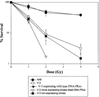
(4) Nucleic Acids Research, 2000, Vol. 28, No. 7 1509. Figure 1. Survival of V-3 and V-3 YAC fusion hybrids following γ-irradiation. Closed circles, AA8; closed inverted triangles, V-3 cells containing YAC 155 and expressing wild-type DNA-PKcs protein; open circles, V-3; closed squares, V-3 clones containing YAC D3941E that did not express DNA-PKcs protein; open inverted triangles, a V-3 clone containing D3941E YAC and expressing DNA-PKcs protein. Survival of V-3, AA8 and a complemented clone represent the means of a minimum of three survival experiments. Five additional complemented clones gave a similar level of resistance. The results from the non-expressing mutant clones represent the means of 10 clones analysed. The expressing mutant clone represents the results of a single experiment carried out on a single clone; similar survival results were obtained with a second expressing mutant clone.. found that expressed DNA-PKcs protein and both showed elevated radiosensitivity compared to parental V-3 cells and produced significantly smaller colonies even without irradiation (Fig. 1). These clones were regrown for further analysis but subsequent extracts failed to express the protein. Despite additional protoplast fusion experiments, no further clones expressing DNA-PKcs protein were obtained. PCR analysis, however, showed that these non-expressing clones retained DNA-PKcs sequences and were resistant to neomycin, the selective marker present on the YAC. Analysis of six clones that failed to express DNA-PKcs showed that their radiosensitivity was similar to V-3 cells. These results provide preliminary evidence that the kinase activity of DNA-PKcs is required for DSB repair, but they were limited since repeat analysis on the expressing clones could not be carried out. The results raise the possibility, however, that there is a strong selective disadvantage for cells expressing the kinase null protein and derivatives that fail to express or degrade the mutant protein rapidly dominate the population. Examination of a pre-B mouse SCID cell line that expresses mutant DNA-PKcs protein SB7 is a pre-B cell line derived from a SCID mouse carrying the anti-apoptotic bcl-2 transgene (36). The expression of bcl2 allows the pre-B cells to grow in culture by preventing. apoptosis (41). It has been shown previously that these pre-B bcl2 SCID– cells fail to undergo V(D)J recombination but continue to express DNA-PKcs protein (37). The kinase activity was also shown to be impaired, although the magnitude of the effect was limited by the sensitivity of the assay. We therefore exploited these cells to investigate whether the mutant SCID DNA-PKcs protein could bind to DNA, could function as a protein kinase and whether the kinase activity was required for DNA-PKcs function in DSB rejoining. As a control we utilised pre-B cells derived from wild-type mice also carrying the bcl2 transgene. These two lines have been designated pre-BSCID+ and pre-BSCID–. These pre-B cells grow in suspension but require co-cultivation with lethally irradiated stromal cells. The stromal cells remain adherent and care was taken to harvest the suspension grown cells without taking the attached stromal cells. Whole cell extracts (WCEs) prepared from pre-BSCID+ and pre-BSCID– cells were first analysed by western immunoblotting using the anti-DNA-PKcs antibody 18-2 (Fig. 2A, lanes 1 and 2). The level of DNA-PKcs present in the pre-BSCID– cell line was reproducibly slightly lower compared with the wild-type line, in line with previous findings (37). To assess the kinase activity, we prepared large-scale WCEs from the two cell lines. Double-stranded DNA–cellulose beads were mixed with these WCEs to allow microfractionation of the DNA-binding proteins following centrifugation and washing. An aliquot of the harvested beads was boiled and the detached proteins were analysed by immunoblotting using the anti-DNA-PKcs 18-2 antibody. Figure 2A (lanes 3 and 4) shows that the relative recovery of DNA-PKcs was similar in SCID– and SCID+ cells, demonstrating that the mutant DNA-PKcs protein can interact efficiently with DNA, consistent with a previous report (15). This served as a control to demonstrate that similar protein levels were utilised in the kinase assays. The microfractionated DNA-bound proteins from the two lines were assayed for DNA-PK activity by measuring their ability to phosphorylate a p53 peptide that is known to be phosphorylated by DNA-PKcs. The phosphorylated proteins were separated by SDS–PAGE electrophoresis. This procedure provides a sensitive assay for DNA-PK activity since, firstly, DNA-PKcs is separated from kinases that lack DNA binding activity and, secondly, the electrophoresis step enables the phosphorylated p53 peptide to be separated from small non-specific proteins or peptides present on the DNA–cellulose beads that may become phosphorylated. The results from a larger and smaller preparation of WCEs are shown in Figure 2 (Fig. 2A, lanes 5 and 6, larger preparation; Fig. 2B, smaller preparation). The gel from the former was over-exposed to enable the detection of any low residual activity in the SCID– cells. Whilst a strong signal representing phosphorylated p53 is seen from the SCID+ cell extracts there is no detectable signal using the SCID– extracts. We estimate that our assay would be capable of detecting a phosphorylated band 100-fold less intense than that present in the SCID+ sample. Thus, if the mutant DNA-PKcs protein has residual activity it must be less than 50-fold the level found in wild-type cells (the level of mutant DNA-PKcs protein recovered from the beads was ~50% of that recovered from SCID+ cells). We conclude that the C-terminal region of DNA-PKcs is required for the protein to function efficiently as a protein kinase but that this activity is dispensible for binding of DNA-PKcs.
(5) 1510 Nucleic Acids Research, 2000, Vol. 28, No. 7. Figure 2. Analysis of DNA-PKcs protein and kinase activity from pre-B bcl2 SCID+ and SCID– cells. (A) Lanes 1 and 2, western blot analysis of WCE (100 µg protein) from a large scale preparation (5 × 10⁷ cells) of SCID+ and SCID– cells using anti-DNA-PKcs antibody 18-2. The origin of the band representing DNA-PKcs was verified using purified DNA-PKcs and since it was absent or severely diminished in extracts from V-3 and mouse SCID fibroblast cells (data not shown). Lanes 3 and 4, western blot analysis of proteins microfractionated from WCE (250 µg) using DNA–cellulose beads. Following microfractionation, the beads were boiled in 2× SDS loading buffer and an aliquot of the detached proteins used for western blot analysis. Lanes 5 and 6, assay of the proteins microfractionated using DNA–cellulose beads for DNA-PK activity. The remainder of the same samples used for the analysis in lanes 3 and 4 were utilised for DNA-PK assays using a p53-derived peptide as a substrate. Following incubation, 2× SDS loading buffer was added, the samples boiled, the beads removed by centrifugation and the samples subjected to PAGE electrophoresis to separate the p53 peptide (lanes 5 and 6). The band representing the phosphorylated p53 peptide is highlighted. Two minor phosphorylation products were also obtained which do not represent the specific p53 phosphorylation event under study. The microfractionation and p53 peptide phosphorylation assay has been utilised extensively by us and others to demonstrate the defect in DNA-PKcs defective cell lines such as V-3 and irs-20, demonstrating that the major kinase phosphorylating p53 in this assay is DNA-PK (see for example 31,38). (B) The results of a similar DNA-PK assay carried out as described in (A) using a smaller number of cells and less extract (10 µg WCE for 1BR3 and 50 µg WCE for pre-B SCID+ and pre-B SCID–; only half the microfractionated proteins were utilised for the kinase assay in this experiment). 1BR3 is a primary human fibroblast cell line used as a control. As reported previously, rodent cells have ~50-fold lower levels of DNA-PK activity compared with human cells. The -peptide lane represents the assay carried out in the absence of p53 peptide. to DNA, which is mediated through interaction with the Ku protein (1,2). Requirement of the kinase activity for DSB repair To determine whether the kinase activity is required for DSB rejoining, we next examined survival following radiation. exposure in the pre-B SCID– and SCID+ cell lines and measured their ability to rejoin DNA DSBs using pulsed-field gel electrophoresis (PFGE). The cells were grown in suspension and in initial experiments were found to have a low cloning efficiency. Therefore, to assess their survival following radiation exposure, we carried out a ‘grow-back’ experiment involving measurement of the growth rate of the cells post-irradiation. The results of a typical experiment for pre-B SCID+ and SCID– cells are shown in Figure 3A and B. The percentage survival can be estimated by back extrapolation once linear growth is established. This procedure has large errors and is unsuitable for quantification of survival at high doses, but nonetheless indicates a clear sensitivity of the pre-B SCID– cell line. Subsequently, we managed to achieve a higher cloning efficiency and therefore also estimated survival using a cloning assay for suspension cells. Figure 3C shows the mean data for three experiments carried out using the ‘grow-back’ protocol and the results of a single cloning experiment are given in the legend. The data clearly show a similar magnitude of γ-ray sensitivity of these pre-B bcl2 SCID– cells compared to the pre-B bcl2 SCID+ cells to that shown by the SCID fibroblast cells that express low levels of DNA-PKcs protein compared to wild-type fibroblasts (42). DSB rejoining was also analysed in the pre-BSCID– and SCID+ cells using PFGE. Cells embedded in plugs were exposed to 20 Gy γ-rays, incubated for varying repair times and subjected to PFGE. The pre-B SCID– cells showed a defect in DSB rejoining relative to pre-B SCID+ cells and of a magnitude comparable to that reported previously for SCID– fibroblasts (Fig. 4; 23,43). These results provide strong evidence that the kinase function of DNA-PKcs is required for DSB repair. DISCUSSION Although DNA-PKcs functions in NHEJ, its precise role remains unclear. Curiously, unlike other components required for NHEJ, DNA-PKcs appears to be essential for rejoining the V(D)J coding junctions and radiation-induced DSBs but is largely dispensible for signal joint formation. An evaluation of its role has been hindered by its large size and by the possibility that the DNA-PKcs-defective model cell lines may not represent null phenotypes. In this study we have taken two approaches to address whether the kinase activity of DNA-PKcs is essential for DSB repair. Our results also address the issue of whether mouse SCID cells have residual kinase activity. In the first approach, we exploited yeast genetics to introduce a kinase-inactivating mutation into a DNA-PKcs gene encoded by a YAC. Although procedures for site-directed mutagenesis of YACs have been described, this approach has not been extensively utilised but is a useful strategy for examining genes with large cDNAs (44). This study revealed the unexpected finding that expression of a ‘kinase-dead’ mutant DNA-PKcs appears to confer a marked growth disadvantage. Two clones initially expressing the protein were significantly slower growing and had elevated radiosensitivity compared to the parent V-3 cells. Furthermore, the protein ceased to be expressed after prolonged growth even though the clones retained the neomycin resistance marker and the region of DNA-PKcs containing the introduced mutational change. Although mutations or sequence loss may have occurred elsewhere in the large DNA-PKcs gene, this did not occur in 6/6 clones expressing the wild-type YAC. It is possible that the.
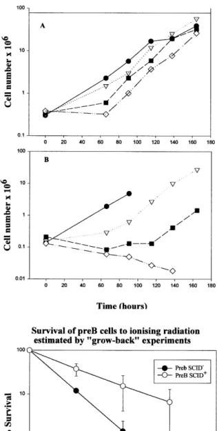
(6) Nucleic Acids Research, 2000, Vol. 28, No. 7 1511. Figure 4. Repair of DNA DSBs in SCID+ and SCID– cells. Cells were exposed to 20 Gy γ-rays and DSB repair measured by PFGE. Open circles, pre-B bcl2 SCID+; closed circles, pre-B bcl2 SCID–. FAR (fraction of [14C] radioactivity released) is the fraction of activity migrating out of the well relative to the total activity loaded (activity in the well and the gel). The results are expressed as a percentage of the value obtained for an irradiated sample that was not allowed time to repair. The background activity in unirradiated samples treated in parallel is subtracted from that present in the irradiated samples.. Figure 3. ‘Grow-back’ survival experiment following exposure to ionising radiation. (A) Pre-B bcl2 SCID+ cells. (B) Pre-B bcl2 SCID– cells. Closed circles, unirradiated cells; inverted triangles, cells irradiated with 1 Gy; closed squares, cells irradiated with 2 Gy; open diamonds, cells irradiated with 3 Gy. (C) Survival of SCID+ and SCID– cells after ionising radiation estimated by ‘grow-back’ experiments. Open circles, pre-B bcl2 SCID+; closed circles, pre-B bcl2 SCID–. The results represent the means of three ‘grow-back’ experiments. No observable recovery of the SCID– cells after 3 Gy was seen in most experiments even after 6 days post-irradiation incubation [see for example (B)]. This point has therefore been omitted from the figure. The survival, however, was clearly lower than that observed with the SCID+ cells. The results of the single cloning survival experiment were 37.6 and 15.3% survival for SCID+ cells and 11.9 and 1.4% survival for SCID– cells following exposure to 1 and 2 Gy, respectively.. gene has become transcriptionally silenced or that the mutant protein was degraded. It is notable that the majority of the available DNA-PKcs-defective cell lines, and indeed most mouse SCID cell lines, have substantially decreased or undetectable expression of the mutant protein (16,17,20–22,31). However, here we only utilised a single mutated YAC clone for protoplast fusion and only analysed the impact of a single mutational change within the kinase domain. Thus we cannot, at present, unequivocally causally relate the impaired growth to the kinase dead phenotype. In contrast to our results, DNAPKcs mutant protein was expressed following the introduction of mutated DNA-PKcs cDNA under the expression of an exogenous promotor (12). It is possible that over-expression of the protein counteracts the degradation or that transcriptional silencing cannot take place with the exogenously controlled plasmid gene. Taken together, our results raise the possibility that expression of a mutant DNA-PKcs protein is detrimental to the cell. Based on the single survival analysis that we were able to carry out with two clones, our results are consistent with the notion that DNA-PKcs kinase activity is required for DNA-PKcs function in NHEJ. To assess the impact of the mouse SCID mutation, we analysed a mouse SCID cell line that, atypically, did express mutant DNA-PKcs protein (37). Despite expressing mutant DNA-PKcs protein, no significant growth impairment is apparent in the pre-B bcl2 SCID– cells. One possibility is that the mutant DNA-PKcs protein renders the cells prone to undergo apoptosis, which is overridden by bcl2 expression in these cells. Pre-B SCID– cells from mice that do not carry the bcl-2 transgene also expressed mutant DNA-PKcs protein but these cells showed higher levels of apoptosis compared to pre-B SCID+ cells (our unpublished observations), consistent.
(7) 1512 Nucleic Acids Research, 2000, Vol. 28, No. 7. with the suggestion that cells expressing mutant DNA-PKcs may have a selective disadvantage. Significantly, DNA-PKcs is cleaved by caspase-3 activation during apoptosis (45). Following a large-scale preparation of mutant SCID protein, we show here that the mutated protein has dramatically decreased ability to function as a protein kinase. We estimate conservatively that the kinase activity of the mutant protein must be at least 50-fold lower than that of the wild-type protein. Since mouse SCID cell lines have 10- to 100-fold lower DNA-PK protein levels compared to wild-type cells, any residual activity in the commonly used cell lines must be less than 1/500th that present in wild-type cells. Our results differ from a previous study reporting that mouse SCID embryo fibroblasts (MEFs) had ~50% of the DNA-PK activity found in wild-type BALB/c MEFs (28). Our results suggest that this cannot be attributed to residual activity of any remaining ‘mutant’ SCID protein. It is possible that reversion of the SCID mutation can occur or that the activity could be assigned to another kinase. Our results also show that the extreme C-terminal region of DNA-PKcs unique to the PI 3-K-related subfamily is required for DNA-PKcs to function as a protein kinase and conversely that the PI 3-K domain present in the lipid kinases is insufficient for protein kinase activity. This was tentatively suggested previously from the study of irs-20 cells, which have a point mutation in the fourth amino acid from the C-terminus, but could not be verified since the complete cDNA had not been sequenced (31). Our findings here also suggest that residual activity is unlikely to provide an explanation for the different phenotype of DNA-PKcs-defective cells compared to Ku-, XRCC4- and DNA ligase IV-defective cells. This conclusion is consistent with studies based on recently generated knock-out DNA-PKcs mice and cell lines, which display a similar phenotype to the mouse SCID cell line (29,30). The pre-B bcl2 SCID– cells show elevated radiosensitivity and impaired DSB repair compared with pre-B bcl SCID+ cells. The magnitude of these defects are similar to that found in SCID cell lines that have low expression of the mutant SCID DNA-PKcs protein (23,42,43). Previous studies have also shown that these pre-B SCID– cells are defective in V(D)J recombination (37). These studies therefore indicate that the kinase activity of DNA-PK is required for the repair of radiationinduced DSBs and the breaks induced during V(D)J recombination. This is consistent with a recent study involving site-directed mutagenesis of a full-length DNA-PKcs cDNA (12). Consistent with a previous report, the mutant SCID protein retains its ability to interact with DNA, probably via interaction with Ku (15). The function of the kinase activity is not known, although it is dispensable for p53 activation and cell cycle arrest (46). The DSB rejoining defect suggests that the kinase activity is directly required for NHEJ and rules out models in which the kinase activity is proposed to function solely to initiate a signal transduction pathway to alert the cell to the presence of a DSB. One possibility is that the kinase activity regulates or activates NHEJ by phosphorylation of the NHEJ component proteins, an appealing model since most of the constituent proteins can be phosphorylated by DNA-PKcs in vitro (8,47). Our findings, however, do not rule out the possibility that the large DNA-PKcs protein has some additional function in the process, such as acting as a scaffold protein, but show that such activity is not sufficient for DSB rejoining.. In conclusion, we have shown that the mouse SCID mutation abolishes the ability of DNA-PKcs to function as a protein kinase (to a level <2% of wild-type). Thus the extreme C-terminal region of DNA-PKcs, which is unique to the PI 3-K-related subfamily of PI 3-Ks, is essential for protein kinase activity. Our results provide supporting evidence that the kinase activity of DNA-PKcs has a direct function in DSB rejoining. ACKNOWLEDGEMENTS We thank Prof. A. R. Lehmann and Dr B. Singleton for invaluable support and advice. We thank Dr G. Smith and Prof. S. P. Jackson for providing purified DNA-PK. Work in the P.A.J. laboratory contributing to this study was funded by European Union grant FI3PCT920007 and grants from the Kay Kendall Leukaemia Fund, the Human Frontier Science Programme and Industry-funded UKCCCR Radiation Research Programme. REFERENCES 1. Dvir,A., Peterson,S.R., Knuth,M.W., Lu,H. and Dynan,W.S. (1992) Proc. Natl Acad. Sci. USA, 89, 11920–11924. 2. Gottlieb,T.M. and Jackson,S.P. (1993) Cell, 72, 131–142. 3. Jeggo,P.A. (1998) In Hall,J.C., Dunlap,J.C., Friedmann,T. and Giannelli,F. (eds), Advances in Genetics. Academic Press, San Diego, Vol. 38, pp. 185–211. 4. Jeggo,P.A. (1997) Mutat. Res., 384, 1–14. 5. Jackson,S.P. (1996) Cancer Surv., 28, 261–279. 6. Schar,P., Herrmann,G., Daly,G. and Lindahl,T. (1997) Genes Dev., 11, 1912–1924. 7. Grawunder,U., Wilm,M., Wu,X., Kulesza,P., Wilson,T.E., Mann,M. and Lieber,M.R. (1997) Nature, 388, 492–495. 8. Critchlow,S.E., Bowater,R.P. and Jackson,S.P. (1997) Curr. Biol., 7, 588–598. 9. Hartley,K.O., Gell,D., Smith,G.C.M., Zhang,H., Divecha,N., Connelly,M.A., Admon,A., Lees-Miller,S.P., Anderson,C.W. and Jackson,S.P. (1995) Cell, 82, 849–856. 10. Hunter,T. (1995) Cell, 83, 1–4. 11. Keith,C.T. and Schreiber,S.L. (1995) Science, 270, 50–51. 12. Kurimasa,A., Kumano,S., Boubnov,N., Story,M.D., Tung,C., Peterson,S.R. and Chen,D.J. (1999) Mol. Cell. Biol., 19, 3877–3884. 13. Blunt,T., Gell,D., Fox,M., Taccioli,G.E., Jackson,S.P., Lehmann,A.R. and Jeggo,P.A. (1996) Proc. Natl Acad. Sci. USA, 93, 10285–10290. 14. Araki,R., Fujimori,A., Hamatani,K., Mita,K., Saito,T., Mori,M., Fukumura,R., Morimyo,M., Muto,M., Itoh,M., Tatsumi,K. and Abe,M. (1997) Proc. Natl Acad. Sci. USA, 94, 2438–2443. 15. Danska,J.S., Holland,D.P., Mariathasan,S., Williams,K.M. and Guidos,C.J. (1996) Mol. Cell. Biol., 16, 5507–5517. 16. Fukumura,R., Araki,R., Fujimori,A., Mori,M., Saito,T., Watanabe,F., Sarashi,M., Itsukaichi,H., Eguchi-Kasai,K., Sato,K., Tatsumi,K. and Abe,M. (1998) J. Biol. Chem., 273, 13058–13064. 17. Errami,A., He,D., Friedl,A., Overkamp,W., Morolli,B., Hendrickson,E., Eckardt-Schupp,F., Oshimura,M., Lohman,P., Jackson,S. and Zdzienicka,M. (1998) Nucleic Acids Res., 26, 3146–3153. 18. Bosma,G.C., Custer,R.P. and Bosma,M.J. (1983) Nature, 301, 527–530. 19. Bosma,M.J. and Carroll,A.M. (1991) Annu. Rev. Immunol., 9, 323–350. 20. Kirchgessner,C.U., Patil,C.K., Evans,J.W., Cuomo,C.A., Fried,L.M., Carter,T., Oettinger,M.A., Brown,J.M., Iliakis,G., Mehta,R. and Jackson,M. (1995) Science, 267, 1178–1183. 21. Peterson,S.R., Kurimasa,A., Oshimura,M., Dynan,W.S., Bradbury,E.M. and Chen,D.J. (1995) Proc. Natl Acad. Sci. USA, 92, 3171–3174. 22. Blunt,T., Finnie,N.J., Taccioli,G.E., Smith,G.C.M., Demengeot,J., Gottlieb,T.M., Mizuta,R., Varghese,A.J., Alt,F.W., Jeggo,P.A. and Jackson,S.P. (1995) Cell, 80, 813–823. 23. Hendrickson,E.A., Qin,X.-Q., Bump,E.A., Schatz,D.G., Oettinger,M. and Weaver,D.T. (1991) Proc. Natl Acad. Sci. USA, 88, 4061–4065. 24. Taccioli,G.E., Cheng,H.-L., Varghese,A.J., Whitmore,G. and Alt,F.W. (1994) J. Biol. Chem., 269, 7439–7442. 25. Custer,R.P., Bosma,G.C. and Bosma,M.J. (1985) Am. J. Pathol., 120, 464–477..
(8) Nucleic Acids Research, 2000, Vol. 28, No. 7 1513. 26. Nussenzweig,A., Chen,C., da Costa Soares,V., Sanchez,M., Sokol,K., Nussenzweig,M.C. and Li,G.C. (1996) Nature, 382, 551–555. 27. Zhu,C.M., Bogue,M.A., Lim,D.S., Hasty,P. and Roth,D.B. (1996) Cell, 86, 379–389. 28. Woo,R.A., McLure,K.G., Lees-Miller,S.P., Rancourt,D.E. and Lee,P.W.K. (1998) Nature, 394, 700–704. 29. Taccioli,G.E., Amatucci,A.G., Beamish,H.J., Gell,D., Torres Arzayus,M.I., Priestley,A., Jackson,S.P., Rothstein,A.M., Jeggo,P.A., Herrera,V.L.M. and Xiang,X.H. (1998) Immunity, 9, 355–366. 30. Gao,Y., Chaudhuri,J., Zhu,C., Davidson,L., Weaver,D.T. and Alt,F.W. (1998) Immunity, 9, 367–376. 31. Priestley,A., Beamish,H.J., Gell,D., Amatucci,A.G., Muhlmann-Diaz,M.C., Singleton,B.K., Smith,G.C.M., Blunt,T., Schalkwyk,L.C., Bedford,J.S., Jackson,S.P., Jeggo,P.A. and Taccioli,G.E. (1998) Nucleic Acids Res., 26, 1965–1973. 32. Duff,K., McGuigan,A., Huxley,C., Schulz,F. and Hardy,J. (1994) Gene Ther., 1, 70–75. 33. Rothstein,R. (1991) In Guthrie,C. and Fink,G.R. (eds), Methods in Enzymology. Guide to Yeast Genetics and Molecular Biology. Academic Press, San Diego, CA, pp. 281–301. 34. Jeggo,P.A., Hafezparast,M., Thompson,A.F., Broughton,B.C., Kaur,G.P., Zdzienicka,M.Z. and Athwal,R.S. (1992) Proc. Natl Acad. Sci. USA, 89, 6423–6427. 35. Whitmore,G.F., Varghese,A.J. and Gulyas,S. (1989) Int. J. Radiat. Biol., 56, 657–665.. 36. Rolink,A., Kudo,A., Karasuyama,H., Kikuchi,Y. and Melchers,F. (1991) EMBO J., 10, 327–336. 37. Grawunder,U., Finnie,N., Jackson,S.P., Riwar,B. and Jessberger,R. (1996) Eur. J. Biochem., 241, 931–940. 38. Finnie,N.J., Gottlieb,T.M., Blunt,T., Jeggo,P.A. and Jackson,S.P. (1995) Proc. Natl Acad. Sci. USA, 92, 320–324. 39. Carter,T., Vancurova,I., Sun,I., Lou,W. and DeLeon,S. (1990) Mol. Cell. Biol., 10, 6460–6471. 40. Bentley,N.J., Holtzman,D.A., Flaggs,G., Keegan,K.S., DeMaggio,A., Ford,J.C., Hoekstra,M. and Carr,A.M. (1996) EMBO J., 15, 6641–6651. 41. Strasser,A., Harris,A.W. and Cory,S. (1991) Cell, 67, 889–899. 42. Fulop,G.M. and Phillips,R.A. (1990) Nature, 374, 479–482. 43. Biedermann,K.A., Sun,J., Giaccia,A.J., Tosto,L.M. and Brown,J.M. (1991) Proc. Natl Acad. Sci. USA, 88, 1394–1397. 44. Tucker,R.M. and Burke,D.T. (1996) Nucleic Acids Res., 24, 3467–3468. 45. Song,Q., Lees Miller,S.P., Kumar,S., Zhang,N., Chan,D.W., Smith,G.C.M., Jackson,S.P., Alnemri,E.S., Litwack,G., Khanna,K.K. and Lavin,M.F. (1996) EMBO J., 15, 3238–3246. 46. Jimenez,G.S., Bryntesson,F., Torres-Arzayus,M.I., Priestley,A., Beeche,M., Saitoll,S., Sakaguchill,K., Appellall,E., Jeggo,P.A., Taccioli,G.E., Wahl,G.M. and Hubank,M. (1999) Nature, 400, 81–83. 47. Leber,R., Wise,T.W., Mizuta,R. and Meek,K. (1998) J. Biol. Chem., 273, 1794–1801..
(9)
Figure

Documents relatifs
