DOh 10.1097/s00268-00t-0043-2
O
WORLD
J o u r n a l o fSURGERY
9 2001 by the Soci&t ~ lntemationate de Chirurgie
Transanal Endoscopic Microsurgical Excision of Rectal Tumors: Indications and
Results
Nicolas D e m a r t i n e s , M.D., 1'3 M a r c u s O. y o n Fl0e, M.D., 2 Felix H. H a r d e r , M.D. t ~Department of Surgery, University Hospital of Basel, Spitalstrasse 21, CH-4031 Basel, Switzerland 2Department of Surgery A, Kantonsspital of Lucerne, Lucerne, Switzerland
3Department of Surgery, University Hospital of Zurich, Raemistrasse 100, CH-8091 Zurich, Switzerland
Abstract. Transanai endoscopic microsurgery (TEM) allows local exci- sion of rectal tumors located 4 to 18 cm above the anal verge. The technique is not yet generally established because of the necessary special instrumentation and tools, the unusual technical aspects of the approach, and the stringent patient selection criteria. The aim of this prospective, descriptive study was to analyze the currently accepted indications for TEM and to evaluate the use of this procedure for treating rectal cancer. Over a 4-year period 50 patients aged 31 to 86 years (mean 64 years) underwent TEM for treatment of rectal tumors located 12 cm above the anal verge (range 4-18 cm). The local complication rate was 4%. Alto- gether, 76% of lesions were benign, and 24% were T1 and T2 tumors. Of 12 cancer cases, 4 required reoperation by total mesorectal resection; the other 8 are currently under follow-up management. Over the follow-up period of 30.6 months (range 11-54 months) the recurrence rate of T1 tumors was 8.3%. TEM is a minimally invasive surgical technique that may benefit a small, specific population of patients with rectal tumors. Compared with conventional transanai resection, TEM provides superior exposure of tumors higher up in the rectum (i.e., up to 18 cm from the anal verge). The greater precision of resection combined with low mor- bidity (I0%, relative to that of anterior resection) and short duration of hospitalization (5.5 days) make this technique a reliable and in some cases more effective surgical approach than laparotomy and low anterior resection.
The introduction of transanal endoscopic microsurgcry (TEM) by Buess et al. in 1983 realized an interesting technical advance in localized surgical treatment of rectal tumors [1]. TEM is a mini- mally invasive technique that allows precise resection of tumors located 2 to t8 cm from the anal verge using an operative micro- scope. Thus laparotomy is no longer the only option for managing such cases. However, the number of patients treated by TEM by Buess et al. during the 15 years since its introduction totaled only 500 [2], so the worldwide experience is limited. Reasons may include the high cost of TEM equipment (up to $80,000), a small candidate pool, the stringent criteria for patient selection and indications for the procedure [3], and a highly demanding surgical technique that requires specialized training [3].
To date, there are only 109 publications on TEM listed by Medline. Most of these published series report the experience of Buess and his current and former staff members, with only a few Correspondence to: N. Demartines, M.D., e-mail: nicolas.demartines@ chi.usz.ch
reports on the results obtained at other medical centers in Europe (Italy, United Kingdom), the United States, and Japan. Thus the reliability of this new method has been demonstrated primarily by its inventor, and it must now be evaluated by the broader surgical community, especially given the current controversy surrounding localized management of rectal cancer [4, 5]. The present study reports the experience of a department of general surgery at a university hospital, with the goals of analyzing the currently ac- cepted indications for TEM and evaluating the use of this proce- dure for treating rectal cancer.
P a t i e n t s , Materials, Methods
Between January 1995 and September 1999, all patients with rectal tumors were examined in our interdisciplinary coloproctol- ogy consultation practice. Following clinical examination, a biopsy was obtained in all patients, and endorectal sonography was per- formed using a 360-degree endoprobe with an inflatable balloon at a frequency of 7 MHz to diagnose possible wall infiltration. This endosonographic technique has approximately 90% diagnostic accuracy. [6]. Simultaneously, we tested the integrity of sphincter function and assessed patient continence using the Kirwan-Parks classification system [7]. Anal manometry was performed only in cases of prolonged postoperative incontinence.
Preoperative assessment to stage and grade the type and sever- ity of tumor resulted in two patient groups: The largest group had benign rectal tumor, and the smallest group had proven rectal cancer. Benign tumor and low risk rectal cancer were considered for TEM resection. Low risk rectal cancer was defined as a well or moderately differentiated (G1, G2) T1 tumor without lym- phangiosis carcinomatosa [8]. All other types of cancer were excluded. Indications for TEM were defined as: (i) a benign tumor of any type > 2 cm in diameter; (2) adenoma recurrence; (3) low risk rectal cancer; and (4) rectal stenosis after fistula or anastomosis.
Tumors located 2 to 18 cm from the anal verge were treated by TEM. Tumors 2 to 4 cm from the a~al verge were considered inappropriate for TEM because this setting prohibited proper introduction of the 40 mm operating microscope. These patients
Demartines et al.: TEM of Rectal Tumors 871
underwent conventional transanal surgical treatment using the Lone Star Retractor (Lone Star Medical Product, Houston, TX, USA) and were excluded from the study.
TEM Technique
The bowel was prepared as for a formal laparotomy by lavage over 4 hours with 3 to 4 liters of polyethylene glycol solution. Antibiotic prophylaxis for gram-negative and anaerobic strains was given at the time of anesthetic induction. Following the technique de- scribed by Buess et al. [1, 9], we used an operative rectoscope of 40 mm diameter and 120 or 200 mm length, with a sixfold mag- nified stereoscopic view. The tip of the rectoscope is beveled downward. The patient's position for surgery therefore depends on the anteroposterior and lateral orientation of the tumor.
To visualize the anatomic relation between tumor and healthy mucosa, CO2 is insufflated to enlarge the intrarectal space and facilitate precise resection. This requires use of a combination suction-insufflation endosurgical unit to ensure constant, high flow of gas and to evacuate the smoke due to coagulation. The operation itself was performed as described originally by Buess et al. [1, 9]. The use of a multifunctional instrument (suction/irriga- tion, coagulation, cutting) (ERBE, Elektromedizin GmbH, Tii- bingen, Germany) eased and sped up the procedure.
To optimize access to the entire tumor, the rectoscope orien- tation must be changed frequently to compensate for the limited operating field and length of the surgical instruments. Finally, the resection specimen is affixed to a preparation plate to allow the pathologists a precise description of the resection margin in 5 ram of healthy tissue
Postoperatively, patients are allowed to sit and walk as soon as they are fully recovered from anesthesia. A liquid diet is main- tained for 24 hours, and patients are discharged on postoperative day 2 or 3. Initial clinical follow-up occurs at 6 weeks postopera- tively and the final clinical examination, including endorectal sonography, at 3 months.
Data Analysis
Patient data were prospectively collected on a personal computer and managed using Microsoft Excel. Descriptive statistics were used, and results are expressed as the absolute value, percent, mean, and range.
Results
Over the 4-year period of study, 50 patients satisfied the indica- tions for TEM and were included in the study. Of these, 19 were female and 31 male, with a median age of 65 years (range 31-86 years). All underwent surgery, for a tumor located a mean 12 cm from the anal verge (range 4-18 cm). Rectal blood loss was the leading symptom in 31 cases (66%). The tumor was found inci- dentally by routine examination in 5 cases (10%). All other pa- tients presented with loss of mucus or nonspecific pain during defecation.
Preoperative endorectal sonography correctly staged the tumor in 45 cases (90%). "Overstaging" occurred in 3 cases (6%) and
"understaging'" in 2 cases (4%).
Of the 51) procedures, 31 were performed in the lateral position (62%). 13 in the "jackknife prone" position (26%), and 6 in the
Table l. TEM resections.
Diagnosis (n = 50) No. % Villous adenoma 35 70 Rectal cancer 12 24 Carcinoid tumor 1 2 Stenosis 1 2 Recurrence (adenoma) 1 2
TEM: transanal endoscopic microsurgical excision.
"lithotomy" position (t2%). Forty-two cases were performed un- der general anesthesia (84%) and 8 under regional anesthesia (16%). The average operating time was 117 minutes (range 45- 240 minutes).
Full-thickness resection (mucosa and serosa until perireetal fat) was performed in 45 cases (90%) and mucosectomy in 5 cases (10%). We decided to convert one case intraoperatively to lapa- rotomy to avoid any oncologic risk due to rectal wall infiltration. This case was not excluded from the TEM data analysis. There were two cases of inadvertent peritoneal entry (2/50, 4%) associ- ated with resection of tumors located 16 and 18 cm from the anal verge, respectively. One case required laparotomy, and the other resolved following a conservative 4-week course of treatment with protective loop sigmoidostomy. In addition, there were five sys- temic complications (5/50, 10%): four minor problems (e.g., urine infection) and one major problem (myocardial infarction). There was no mortality.
Postoperative pain was rare, and the median hospitalization was 5.5 days (range 2-20 days). The patient who sustained a myocar- dial infarction remained hospitalized for 20 days owing to the development of pneumonia. Reoperation was required in 5 of 50 cases (10%), involving laparotomy and total mesorectal resection [10] for four cases of cancer and anterior resection for one case of rectal wall perforation and peritoneal entry followed by abscess.
Assessment of sphincter function revealed one 85-year-old pa- tient who was incontinent (grade III, liquids and solids) preoper- atively and remained so postoperatively. Six patients (12%) who were initially continent developed incontinence (grade II, liquids and gas) during the first 2 weeks after operation. Clinical fol- low-up at 3 months showed that all six patients had fully recov- ered. Manometric studies were not performed in these cases.
Histolo, W
Lesions were benign in 38 cases (76%) and malignant in 12 cases (24%). The histologic diagnosis indicated 32 villous adenomas (64%). 9 T1 (18%) and 3 T2 (6%) rectal tumors, 3 polyps (6%), I carcinoid (2%), 1 recurrent polyp (2%), and 1 rectal stenosis after a high fistula (2%). The results are summarized in Table 1 with the tumor grading and additional treatment in Table 2. A diagnosis of cancer was known in eight cases, suspected in two cases, and surprising in two other cases. Four cases of cancer (two T2 lesions and two T1 tumors) required reoperation by total mesorectal resection (Table 2).
The T1 tumors were resected by TEM with a safe margin of at least 5 mm and are under follow-up management. One 85-year- old patient with a T2 tumor refused additional treatment by laparotomy or radiotherapy and died of concurrent disease 6 months postoperatively.
Table 2. Cancers resected by TENt (n = 12).
Stage Grade Treatment
9 pTI i GI, 6 G2 Follow-up (*~ = 7) I G2 Adjuvant therapy' (n = 1) 1 G3 Total mesorectal resection (n = l) 3 pT2 3 G2 Total mesorectal resection (n = 2)
Folk)w-up (patient in poor condition) (n = 1) Recurrence rate at 30 months was 8.3%.
Over the follow-up period of 30.6 months (range 11-54 months the recurrence rate of T1 tumor was 8.3%; 14 months after TEM resection we detected one case of local recurrence. This patient was consequently treated by laparotomy and total mesorectal resection and is now under follow-up management.
Discussion
As minimally invasive surgical technique, T E M may benefit a small, specific population of patients diagnosed with rectal tu- mors. It offers the promise of localized treatment of these tumors, with relatively few complications and rare mortality. However, experience with TEM has been limited primarily to those who developed the technique and only a few others, making it neces- sary to reexamine functional outcome based on current indica- tions and criteria for patient selection for this procedure.
Our results with T E M indicate that this technique is reproduc- ible after surgical training, and that there is a clear benefit for the patients with minimal complications, avoiding laparotomy with anterior rectum resection. Because they are based on currently published indications and criteria for patient selection, our find- ings suggest that these standards remain adequate for identifying appropriate surgical candidates for TEM. We are not associated with the developers of TEM, so our results provide an indepen- dent perspective on the value of the technique. However, our series is small and nonrandomized, making larger additional pro- spective and randomized studies essential to confirm the true value of TEM for treatment of early rectal cancer.
The TEM technique distinguishes itself from other endoscopic or laparoscopic procedures in various ways. (1) It uses a magnified binocular stereoscopic device. Compared with the view obtained with monocular instruments or video cameras, the view of the operating field obtained with TEM provides a depth of field of extremely good quality. (2) Surgical instruments are inserted and moved in parallel planes unlike taparoscopy, necessitating special- ized training and skill to achieve full tumor exposure. (3) Costly equipment (about $80,000) is required. The cost of TEM equip- ment, combined with the relatively low incidence of the patholo- gies for which this procedure is indicated, have limited worldwide experience with this technique to a few surgical teams.
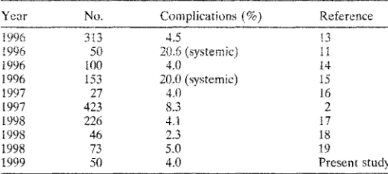
One clear advantage of TEM is an uneventful postoperative course. The postoperative analgesic requirement is slight, gener- ally limited to a few doses of paracetamol [ll]. Moreover, the duration of hospitalization (5.7 days for TEM versus 14.5 days, p < 0.0001) [11] and the complication rate (21% versus 35%) [ l l ] ar e significantly lower than those reported for low anterior resec- tion. Overall, the localized complication rate associated with transanal resection lies between 4.0% and 8.3% of cases [2, 12] and that for systemic complications between 14% and 21% (Table
Table 3. TEM complication rate: review of the relevant literature.
Year No. Complications (%) Reference
t996 3{3 4,5 13 ~996 50 20.6 (systemic) 1I 1996 100 4.0 I4 1996 153 20.0 (systemic) 15 1997 27 4.0 16 1997 423 8.3 2 1998 226 4.1 17 t998 46 2.3 18 1998 73 5.O 19 1999 50 4.0 Present study
3) [11, 15, 20]. The results in our patients are comparable (i.e., a hospital stay of 5.5 days and localized and systemic complication rates of 4% and 10%, respectively, with no mortality). In fact, mortality is rare: Of 109 publications describing the worldwide experience of about 3000 TEM cases (Table 3), only one German study reported a fatality: a unique, lethal complication of retro- peritoneal phlegmon after T E M resection of an adenoma located 7 cm from the anal verge in a 55-year-old patient who died in septic shock after 28 days [21].
Effect on Sphincter Function
It is surprising that prolonged anal dilatation with 4 cm diameter (the operative rectoscope) induces few sphincter function prob- lems. We observed only postoperative transitory grade II incon- tinence in 12% of our patients, with full postoperative recovery after 3 months (excluding one elderly patient with chronic incon- tinence that did not resolve with surgery). Because this duration of incontinence was comparable to that already reported [22-24], we did not conduct manometric studies. However, existing manomet- ric analyses of the effects of anal dilatation indicated a decrease in sphincter tonus ranging from 25% to 37% of preoperative sphinc- ter pressure, with complete recovery to clinical continence within 6 to 16 weeks postoperatively [22-24].
bMications ]'or TEM
The ideal indications for TEM are all types of adenoma located 4 to 18 cm of the anal verge that cannot be treated by colonoscopy. Ideal tumor size ranges in diameter from 20 mm to three-fourths of the lumen circumference. A full-thickness resection is recom- mended to ensure an appropriate margin of safety. In addition, this procedure is technically easier to perform than mucosectomy and decreases the risk of missing a small rectal cancer that may be located inside the villous adenoma. Such "in situ" cancers have been reported in up to 31% of cases [25]. Because the rectal wall is sutured after full-thickness resection, removal of a large seg- ment of the rectum is not a high risk procedure based on the low morbidity reported in the literature (Table 3) and in our own series.
Rectal Cancer. Treatment of rectal cancer by TEM is generally accepted for T1 low risk cancer [2]. The recurrence rate following this application lies between 4% and 8% [2, 18, 19, 26] compared with a local recurrence rate of up to 30% for T1 high risk cancer [27]. Patient selection is therefore crucial to good local and on- cologic results.
Demartines et al.: TEM of Rectal Tumors 873
Other b~dications. The following indications are rather anecdotal but are mentioned for complete information. TEM may be a suitable method for resecting stenoses 5 to t5 cm from the dentate line (e.g., inflammatory stenosis after a high fistula, as in our experience, or colorectal anastomotic stenosis, as described in Japan [28]). Other indications, such as transanal rectopexy origi- nally proposed by Buess, no longer appears in the literature and there are no subsequent reports [29], suggesting that this indica- tion is not to be recommended.
Transcutaneous application of T E M to treat early gastric can- cer was first described in 1997 [30]. However, no follow-up has been reported, making even a preliminary conclusion question- able.
Patient Selection
Careful patient selection is crucial to T E M outcome. Preoperative staging must be precise. Specifically, a specimen should be avail- able for histology, and the tumor should be visualized with the rigid rectoscope to determine its distance from the dentate line and localize it precisely in the quadrant. Endorectal sonography should be performed to assess eventual wall infiltration. Such precise staging is reportedly possible with an accuracy of up to 90% [6, 31]. In the present study, preoperative staging was correct in 90% of cases (n = 45 patients), resulted in an overestimate of tumor severity in 6% (n = 3) and an underestimate in 4% (n = 2). Incorrect estimation is a known phenomenon that depends on the accuracy of endoluminal sonography, which is examiner-depen- dent (learning curve) and described in up to I0% of the cases [6, 31]. Only one procedure had to be converted to laparotomy during the primary operatio n owing to underestimated lesion severity, and it occurred early in our experience with the tech- nique.
Limitations of TEM for Treatment of Cancer
The primary factor limiting the effectiveness of local treatment of early rectal cancer is lymph node invasion. The lymph node metastasis rate of T1 rectal tumors is between 0% and 15.4%, depending on tumor grading [27, 32, 33]. Age less than 45 years is recognized as a significant risk factor for such metastasis [33]. Our population and those in many other studies have a reported age range of 31 to 86 years (median 65 years), suggesting the potential presence of high risk.
Local excision appears to offer a significant advantage. The rate of recurrence for T1 tumors resected by TEM lies between 3.8% and 8.0% at 13 months' follow-up [18, 19, 26] compared with a recurrence rate of 23.0% after conventional transanal surgery [34]. If it is unclear that the T E M resection was radical, it is essential (and recommended) that an anterior resection be sub- sequently performed. This was the decision made in 12% of cases in our present series and in 21.4% of cases in the largest series reported by Buess's group [2]. In a series of 113 patients, Buess et at. found 5 residual tumors among a subset of 39 patients (12.8%) who underwent an anterior resection immediately after TEM. The tymphadenectomy was negative in all cases for T1 tumor and positive in 23.3% of those with T2 and T3 tumors. In the entire series, the failure rate for 81 T1 tumors treated only by TEM was 4.5% (n = 4), and for 22 T1 tumors requiring anterior resection following TEM it was 13.6% (n = 3). The interpretation of these
results is limited because the difference in the failure rates is not significant; moreover, the study was retrospective and may reflect a negative selection [2].
To date, the only published prospective, randomized study comprised 52 patients with T1 tumors treated by TEM or anterior resection [11]. There were no significant differences in group outcome: The 5-year survival was 96%; the local recurrence rate was 4.1% for TEM and 0% for anterior resection; and the me- tastasis rate was 0% for TEM and 4.1% for anterior resection. These results suggest that T E M may offer some advantage relative to anterior resection for T1 rectal cancer, with similar oncologic results [11]. Our results confirm this conclusion. However, our series is small and nonrandomized, making larger additional pro- spective and randomized studies essential to confirm the true value of TEM for treatment of early rectal cancer [26].
TEM and Adjuvant Therapy
Conservative management of rectal cancer with radiotherapy or endocavitary contact radiotherapy has a 30% failure rate [35]. Consequently, the indications for neoadjuvant or adjuvant radio- chemotherapy following local resection of rectal cancer by TEM remain controversial. In fact, local treatment of rectal cancer is limited by the impossibility of removing the potentially positive lymph node, supporting the concept of adjuvant radiotherapy, chemotherapy, or both to achieve local control of the lymph node [36]. The first published report on the combined effect of T E M resection for rectal cancer followed by radiotherapy appears to support such a benefit [18]. Recently published preliminary results suggest its reliability, with a 5-year recurrence-free disease sur- vival of 81% for irradiated patients versus 52% in those treated by local surgery alone [5]. However, except for T1 low grade tumors, there is currently a lack of evidence to recommend the use of TEM for curative treatment of rectal cancer, with or without adjuvant therapy.
TEM and Palliative Therapy
The use of TEM for purely palliative treatment of rectal cancer is not recommended [20]. In our limited experience (two cases), local resection of certain T2 tumors with TEM but without radio- therapy is viabte for compromised patients or those who refuse a laparotomy. However, only a few reports confirm this use of TEM, with a local complication rate of 14% [18, 20, 37]. Palliative resection of a large infiltrative tumor is discouraged by Buess (personal communication), because of a high local complication rate and the difficulty achieving hemostasis. Although local surgi- cal management is possible in such cases, radiochemotherapy should precede palliative surgical treatment and has been shown to facilitate surgical therapy in 83% of these patients [38]. Pre- liminary studies of the use of endoradiotherapy or perioperative radiotherapy in conjunction with local cancer resection of large tumors are under way [38, 39].
Conclusions
Transanal endoscopic microsurgical excision of rectal tumors is not only an additional tool tot transanal resection of low rectal tumors but also a minimally invasive technique for treating minors in the low, middle, and upper rectum. Compared with convert-
tional transanal resection, TEM provides superior tumor expo- sure higher in the rectum (i.e., up to 18 era). The greater precision of resection, low morbidiV (10%, relative to anterior resection), and short duration of hospitalization (5 days) make this technique a reliable and in some cases more effective surgical approach than laparotomy and low anterior resection.
With strict patient selection and precise preoperative staging, the use of transanal endoscopic microsurgery for treatment of low risk T ! carcinoma is possible with a lower complication rate than is seen with radical surgical therapy. Preliminary results suggest no difference in the 5-year survival rate for local and radical surgical therapy. For TEM, tow risk cancer is defined as T I tumors with differentiation G1-G2 without lymphangiosis carei- nomatosa and a resection margin of at least 5 mm in patients older than 45 years. However, this recommendation must be tempered by the lack of controlled studies to provide support. According to the literature and to our own surgical experience to date, all other types of rectal cancer should be treated by total mesorectal excision [10]. The contribution of adjuvant therapy to local treatment of rectal cancer is still under evaluation [4]. R~sum~
La mierochirurgie transanale endoscopique (MTE) permet une excision locale des tumeurs situres entre 4 et 18 cm au-dessus de la tigne anocutanre. La technique n'est pas encore trrs r r p a n d u e en raison du besoin d'une instrumentation et d'outils sprcifiques, une technique un peu sprciale, et une srlection stricte des patients. Le but de cette 6tude prospective, descriptive, a 6t6 d'analyser les indications actuelles de cette technique et d'rvaluer l'utilisation de ce procrd6 pour le traitement du cancer du rectum. En quatre ans, nous avons trait6 50 patients hgrs entre 31 et 86 ans (fige moyen = 64 ans) par MTE pour tumeur rectale situe6 12 cm (en moyenne) de la marge anale (extrrmes 4-18 cm). Le taux de complications locales a 6t6 de 4%. Soixante-six pourcent des 16sions 6taient bdnignes et 24% des tumeurs T1 et T2. Parmi les t2 eas de cancer, quatre ont nrcessit6 une r r o p r r a t i o n pour excision totale du mdsorectum, les huit autres patients sont actuetlement sous surveillance. Pendant la prriode de suivi de 30,6 mois (extrames = 11-54 mois), le taux de rrcidive des tumeurs T1 a 6t6 de 8,3%. La MTE est une technique ehirurgicale mini-invasive qui pourrait 6tre appliqure ~ un sous-groupe de la population atteint de tumeur rectale. Comparre /1 ta rrsection transanale conventionnelle, la M T E permet une meilleure exposition des tumeurs du haut rectum, c'est-~-dire jusqu'/~ 18 cm de la marge anale. Une grande prrcision dans la rrsection, combin6e h une morbidit6 basse (par rapport ~ la rrsection antrrieure) (10%) et une eourte hospitalisation (5,5 jours) rendent cette technique fiable, et dans certains cas, plus efficace que la rrsection antrrieure par laparotomie.
Resumen
La microcirugfa endosc6pica transanat (TEM) permite la reseccidn de tumores rectales localizados de 4 a 18 cm p o t encima dot margen anal. Este procedimiento hasta ahora no se ha generalizado debido a la necesidad de desarrollar instrumentos y herramientas especiales, aspectos trcnicos inusuales de abordaje y criterios estrictos para la selecci6n de los pacientes. El objetivo de este estudio prospectivo y drescriptivo es analizar las indicaciones
actualmente aceptadas para las T E M y evaluar la eficacia de esta tdcnica en et tratamiento dot cfincer de recto. En un periodo de 4 afios, 50 pacientes con edades comprendidas entre 31 y 86 afios (media = 64 afios) fueron tratados mediante el proeedimicnto quirtlrgico TEM, por padecer tumores rectales situados a 12 cm pot encima del margen anal (rango: 4-18 cm). La tasa de complicaciones locales rue del 4%. En el 76% las lesiones fueron benignas y e n el 24% malignas: tumores T1 y T2. De los 12 casos de cfincer, 4 requirieron una reintervencidn con reseccidn total del mesorrecto; los otros 8 est~in actualmente controlados y sometidos a tratamiento. Tras un periodo de seguimiento de 30.6 moses (rango: 11-54 moses) la tasa de recidivas de tumores T1 rue deI 8.3%. La TEM es una trcnica quirdrgica mfnimamente invasiva que puede set may fitil en un ntimero reducido y escogido de pacientes con c~incer de recto. Comparada con la resecci6n transanal convencional, la TEM permite la exposici6n de tumores dot alto recto i.e. hasta 18 em de los mfirgenes del ano. La gran precisidn de la reseccidn, junto con la baja morbilidad (en comparaci6n con la reseccidn anterior) 10% y la corta hospitalizaci6n (5.5 d/as) hace que esta t6cniea sea, fiable y, en algunos casos, m ~ eficaz que la laparotomia con resecci6n anterior baja del recto.
Acknowledgment
We thank Winifred yon Ehrenburg for helpful discussion and editorial advice.
References
1. Buess, G., Hutterer, F., Theiss, J., Brbel, M., Isselhard, W., Pichl- maier, H.: Das System f/ir die transanale endoskopische Rectumop- oration. Chirurg 55:677, 1984
2. Mentges, 13., Buess, G., Effinger, G., Manncke, K., Becker, H.D.: Indications and results of local treatment of rectal cancer. Br. J. Surg. 84:348, 1997
3. Whitlow, C.B., Beck, D.E., Gathright, J.B.: Surgical excision of large rectal villous adenomas. Surg. Oncol. Clin. N. Am. 5:723, 1996 4. Weber, T.K., Petrelli, N,J.: Local excision for rectal cancer: an uncer-
tain future. Oncology (Huntingt.) 12:933, t998
5. Taylor, R.H., Hay, J.H., Larsson, S.N.: Transanal local excision of selected low rectal cancers. Am. J. Surg. 175:360, 1998
6. Massari, M., De Simone, M., Cioffi, U., Rosso, L., Chiarelti, M., Gabrielli, F.: Value and limits of endorectal ultrasonography for preoperative staging of rectal carcinoma. Surg. Laparosc. Endosc. 8:438, 1998
7. Kirwan, W.O., Turnbull, R.B., Jr., Fazio, V.W., Weakley, F.L.: Pull- through operation with delayed anastomosis for rectal cancer. Br. J. Surg. 65:695, 1978
8. Buess, G.F.: Local surgical treatment of rectal cancer. Eur. J. Cancer 3tA:1233, 1995
9. Von Fl~ie, M., Harder, F.: Transanal endoscopic microsurgery (TEM): indications and limitations. Schweiz. Med. Wochenschr. 124:1800, t 994
10. MacFarlane, J., Ryall, R., Heald, R.: Mesorectal excision for rectal cancer. Lancet 341:457, 1993
11. Winde, G., Nottberg. H, Keller, R, Schmid, K.W., Bunte, H.: Surgical cure for early rectal carcinomas (TI): trartsanal endoscopic microsur- gory vs. anterior resection. Dis. Colon Rectum 39:969, 1996 12. Said, S., Stippel, D.: I0 Years experiences with transanal endoscopic
microsurgery: bistopathologic and clinical analysis. Chirurg 67:139, 1996
13. Said, S., Stippel, D.: Transanal endoscopic microsurgery in large, sessile adeaomas of the rectum: a 10-year experience. Surg. Endosc. 9:1106, 1995
Demartines et al.: TEM of Rectal Tumors 875
Scholefield, J.H.: Transanal endoscopic microsurgery--initial experi- ence from three centres in the United Kingdom. Br. J. Surg. 83:207, 1996
15. Smith, L.E., Ko, S.T.. Saclarides, T., Caushaj, P., Orkin, B.A., Khan- duja, K.S.: Transanal endoscopic microsurgery: initial registry results. Dis. Colon Rectum 39:$79, 1996
16. Swanstrom, L.L., Smiley, P., Zelko, J., Cagle, L.: Video endoscopic transanal-rectal tumor excision. Am. J. Surg. 173:383, 1997
17. Morsehel, M., Heintz, A., Bussmann, M., Junginger, T.: Follow-up after transanal endoscopic microsurgery or transanat excision of large benign rectal polyps. Langenbecks Arch. Surg. 383:320, 1998 18. Lezoche, E., Guerrieri, M., Paganini, A.M., Feliciotti, F.: Transanal
endoscopic microsurgical excision of irradiated and nonirradiated rectal cancer: a 5-year experience. Surg. Laparosc.Endosc. 8:249, 1998 19. Saclarides, T.J.: Transanal endoscopic microsurgery: a single sur-
geon's experience. Arch. Surg. 133:595, 1998
20. Turler, A., Schafer, H., Pichlmaier, H.: Role of transanal endoscopic microsurgery in the palliative treatment of rectal cancer. Scan& J. Gastroenterol. 32:58, 1997
21. Klaue, H.J. Bauer, E.: Retroperitoneal phlegmon after transanal en- doscopic microsurgical excision of rectal adenoma. Chirurg 68:84, 1997
22. Hemin~m,vay, D., Flett, M., McKee, R.F., Finlay, I.G.: Sphincter func- tion after transanal endoscopic microsurgical excision of rectal tu- moues. Br.J.Surg. 83:51, 1996
23. Kreis, M.E., Jehle, E.C., Haug, V., Manncke, K., Buess, G.F., Becker, H.D., Starlinger, M.J.: Functional results after transanal endoscopic microsurgery. Dis.Colon Rectum 39:1116, 1996
24. Banerjee, A.K., Jehle, E.C., Kreis, M.E., Schott, U.G., Claussen, C.D., Becket, H.D., Starlinger, M., Buess, G.F.: Prospective study of the proctographie and functional consequences of transanal endoscopic microsurgery. Br. J. Surg. 83:211, 1996
25. Winburn, G.B.: Surgical resection of villous adenomas of the rectum. Am. Surg. 64:1170, 1998
26. Winde, G.: Outcome following transanal endoscopic microsurgery. Dis. Colon Rectum 41:526, 1998
27. Heintz, A., Morschel, M., Junginger, T.: Comparison of results after transanal endoscopic microsurgery and radical resection for Tt carci- noma of the rectum. Surg. Endosc. 12:1145, 1998
28. Kato, K., Saito, T., Matsuda, M, Imai, M., Kasai, S., Mito, M.: Suc-
cessful treatment of a rectal anastomotic stenosis by transanat endo- scopic microsurgery (TEM) using the contact Nd:YAG laser. Surg. Endosc. 11:485, 1997
29. Salm, R, Lampe, H., Bustos, A., Matern, U.: Experience with TEM in Germany. Endosc. Surg. Allied Technol. 2:251, 1994
30. Nakagoe, T.. Sawai, T., Uchikawa, T., Nanashima, A, Yamaguchi, H.. Yasutake, T., Kusano, H., Ayabe, H.: Intragastric endoscopic surgery using the transanal endoscopic microsurgery technique. Br. J. Surg. 84:830, 1997
31. Herzog, U., yon Flue, M., Tondelli, P., Schuppisser, J.P.: How accu- rate is endorectal ultrasound in the preoperative staging of rectal cancer? Dis. Colon Rectum 36:127, 1993
32. Mainprize, K, Mortensen, N., Warren, B.: Early colorectal cancer: recognition, classification and treatment. Br. J. Surg. 85:469, 1998 33. Sizler, PJ., Seow-Cheon, F., Ho, Y.H., Leong, A.P.K.: Lymph node
involvement and tumor depth in rectal cancers: an analysis of 805 patients. Dis. Colon Rectum 40:1472, 1997
34. Warneke, J., Petrelli, N.J., Herrera, L.: Local recurrence after sphinc- ter-saving resection for rectal adenocarcinoma. Am. J. Surg. 158:3, 1989
35. Maingon, P.. Guerif, S., Darsouni, R., Salas, S., Barillot, I., d'Hombres, A., Bone-Lepinoy, M.C., Fraisse, J., Horiot, J.C.: Conser- vative management of rectal adenocarcinoma by radiotherapy. Int. J. Radiat. Oncol. Biol. Phys. 40:1077, 1998
36. Bleday, R., Breen, E., Jessup, J.M., Burgess, A., Sentovich, S.M., Steele, G., Jr.: Prospective evaluation of local excision for small rectal cancers. Dis. Colon Rectum 40:388, 1997
37. Lezoche, E., Guerrieri, M., Paganini, A., Feliciotti, F., Di Pietrantonj, F.: Is transanal endoscopic microsurgery (TEM) a valid treatment for rectal tumors? Surg. Endosc. 10:736, 1996
38. Kim, H.K., Jessup, J.M., Beard, C.J., Bomstein, B., Cady, B., Stone, M.D., Bleday, R., Bothe, A., Jr., Steele, G., Jr., Busse, P.M.: Locally advanced rectal carcinoma: pelvic control and morbidity following preoperative radiation therapy, resection, and intraoperative radiation therapy. Int. J. Radiat. Oncol. Biol. Phys. 38:777, 1997
39. Mendenhall, W.M., Rout, W.R., Vauthey, J.N., Haigh, L.S., Zlotecki, R.A., Copeland, E.M., III: Conservative treatment of rectal adeno- carcinoma with endocavitary irradiation or wide local excision and postoperative irradiation. J. Clin, Oncol. 15:3241, 1997