Neuroradiology (2006) 48: 412–414 DOI 10.1007/s00234-006-0060-9 I N T E RV E N T I O N A L N E U R O R A D I O L O G Y Francesca Siclari Jean H. D. Fasel Philippe Gailloud Received: 19 October 2005 Accepted: 2 December 2005 Published online: 11 March 2006
# Springer-Verlag 2006
Direct emergence of the dorsospinal artery
from the aorta and spinal cord blood supply
Case reports and literature review
Abstract Introduction: Direct emergence of a dorsospinal artery from the aorta is a rare anatomic variant, of which a total of seven cases have been reported. This report offers an additional angiographic ob-servation and reviews the literature. Methods: Two observations of com-mon intercostal trunks documented during spinal angiography are de-scribed. Results: In the first obser-vation, the common intercostal trunk provided complete blood supply to two adjacent vertebral levels (T11 and T12). In other words, the trunk included an intercostal branch and a dorsospinal branch for each level. In the second observation, the common intercostal trunk provided an inter-costal branch for each level (T9 and T10), but only one dorsospinal branch
(T10). An isolated dorsospinal artery (DA) originated separately from the aorta at the T9 level, and provided a significant contribution to the anterior spinal axis. Conclusion: The two reported cases illustrate the concept of “complete” versus “incomplete” common intercostal trunks. In in-stances where an incomplete trunk is documented, a separate DA originat-ing directly from the aorta must be looked for. A review of the literature indicates a tendency for isolated DAs to participate in the blood supply to the spinal cord.
Keywords Spinal angiography . Spinal vascular anatomy . Anterior spinal artery . Spinal cord
vascularization
Introduction
Direct emergence of a dorsospinal artery (DA) from the aorta was initially described by classical anatomists [1,2]. In 1979, Chiras and Merland [2] reported the first three angiographic observations of this rare anatomic variant, of which a total of seven cases have been reported so far [3–5]. We offer one additional angiographic observation and consider the role played by DAs originating from the aorta in relation to the vascularization of the spinal cord. A complementary observation of a common intercostal trunk providing blood supply to the spinal cord is reported as well in order to illustrate the concept of “complete” versus “incomplete” common intercostal trunks.
Case reports
Case 1
A 17-year-old man was investigated for acute paraparesis with a sensory level at T8. Magnetic resonance (MR) imaging only showed a small syringomyelic cavity (max-imum diameter 3 mm) centered at T3, which was not considered to adequately explain the clinical presentation. Spinal angiography was obtained 4 days after initial presentation and documented obliteration of the right L3 segmental artery by a round intraluminal defect suggestive of embolic material. Collateral flow from right L2 was opacifying right L3 distal to the site of occlusion, F. Siclari . P. Gailloud (*)
Division of Interventional Neuroradiology, Department of Radiology and Radiological Sciences, The Johns Hopkins Medical Institutions, Baltimore, MD 21287, USA
e-mail: [email protected] Tel.: +1-410-9558525 Fax: +1-410-6148238 F. Siclari . J. H. D. Fasel Division of Clinical Anatomy, University of Geneva, Geneva, Switzerland
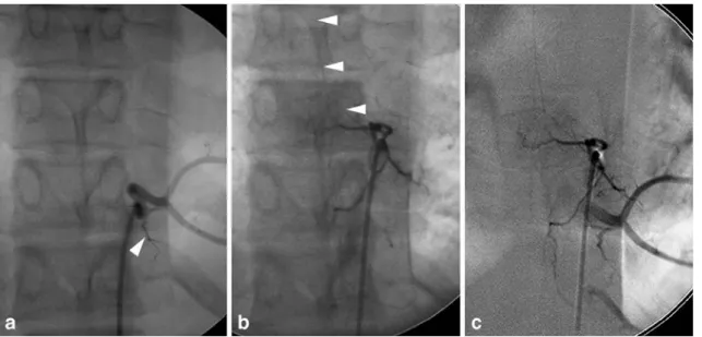
including a major anterior spinal artery contributor. Incidentally, a common trunk supplying both left T9 and T10 was observed (Fig.1a). There was no blush in the T9 left hemivertebra, and the T9 component of the trunk did not have a DA, which was ultimately found at the left T9 level. This direct DA provided a significant anterior spinal branch (Fig.1b,c).
Case 2
A 26-year-old man with a history of cerebral vasculitis was investigated for new onset of rapidly progressive parapa-resis. MR imaging documented an extensive abnormal signal within the spinal cord at the thoracic level. Spinal angiography was unremarkable. Incidental observation was made of a common trunk for left T12 and T11 (Fig. 2). The DA for both levels was opacified by the injection of the trunk, and a hemivertebral blush was observed at both levels.
Discussion
The segmental anatomy of the spine and spinal cord vascularization is relatively simple. A pair of arterial trunks originates from the aorta at most of the thoracic and lumbar vertebral levels. Each of these trunks courses posteriorly along the lateral aspect of the corresponding vertebral body before bifurcating into an anterior branch, the intercostal or
lumbar artery per se and a posterior branch, the DA. The DA itself divides into a dorsal branch that ends within the posterolateral spinal musculature and a smaller radicular branch that courses medially along the corresponding nerve Fig. 1 A 17-year-old man with direct emergence of the T9
dorsospinal artery. a Digital subtraction angiography (DSA), selective left T10 injection, non-subtracted anteroposterior view, showing a common origin for the left T10 and T9 intercostal arteries. Note that a dorsospinal branch is opacified at T10 (arrowhead), but absent at T9. b DSA, selective left T9 injection, non-subtracted anteroposterior view, documenting the direct
emer-gence of the left T9 dorsospinal artery from the thoracic aorta. Note the opacification of an anterior radiculomedullary branch (arrow-heads). c DSA, selective left T9 injection, subtracted anteroposterior view. Contrast reflux into the thoracic aorta provides simultaneous opacification of the T9 dorsospinal artery and of the T9/T10 trunk, clarifying their topographic relationship
Fig. 2 A 26-year-old man with a “complete” form of common intercostal trunk. Selective injection at T12 opacifies both the T12 and T11 intercostal arteries, both of which have a corresponding dorsospinal branch. Note that the dorsospinal artery at T11 (arrow) provides a small anterior radiculomedullary branch (arrowheads)
root to enter the spinal canal through the neural foramen [6]. The functional importance of the radicular branch of the DA lies in its possible contribution to the spinal cord vascularization via small anterior and/or posterior radicular branches supplying the anterior and/or posterior spinal axes, respectively (in which case the radicular artery is classically called a radiculomedullary artery [7]).
The common origin of two (or more) segmental arteries via a single trunk is a variant frequently observed during spinal angiography. In such cases, each segmental vessel must have its own DA in order to be considered complete (Figs.2and3). If a DA is missing, a separate origin of the DA from the aorta must be suspected [2]. The presence or absence of a hemivertebral blush can help distinguish these
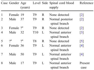
“complete” and “incomplete” forms of common intercostal trunks. At this point, it should be noted that a thromboem-bolic occlusion of a normal DA sparing the corresponding anterior branch could in theory result in a similar angiographic appearance: that is, non-opacification of the DA and absence of the corresponding hemivertebral blush. The seven angiographic observations of direct emer-gence of the DA from the aorta reported to date (including the present case) are summarized in Table1. It is interesting to note that all eight reported instances of direct DA were located at low thoracic levels (T8 to T10), most often at T9 (75% of cases). In four cases, the variant was on the right side and showed either no contribution to the spinal cord vascularization (three cases) or a minor contribution to the posterior spinal axis (one case). On the other hand, all four variants located on the left side were branching off a major anterior spinal branch. Although these numbers remain too small to provide conclusive evidence, we noted a tendency for a left-sided direct DA to carry a major spinal cord feeder. This tendency emphasizes the need for neuroangio-graphers to be familiar with this anatomic variant in order to avoid overlooking a potentially significant anterior or posterior spinal axis contributor during spinal angiography. Fig. 3 Schematic representation of complete and incomplete
common intercostal trunks. a Common trunk of left T9 and T10, “complete type”. Despite its origin from T10, the T9 intercostal trunk normally bifurcates into an anterior intercostal branch and a DA. The injection of the T9/T10 trunk would result in a hemivertebral blush at T9. b Common trunk of left T9 and T10, “incomplete type”. The common trunk for T9 and T10 provides an anterior intercostal branch for T9, but the dorsospinal branch originates separately from the thoracic aorta. The injection of the T9/T10 trunk would not result in a hemivertebral blush at T9
Table 1 Summary of seven published cases of DAs arising from the aorta
Case Gender Age (years)
Level Side Spinal cord blood supply
Reference
1 Female 19 T9 R None detected [2] 2 Male 37 T9 R Normal posterior
spinal branch
[2] 3 Female ?a T9 R None detected [2] 4 Male 32 T10 L Normal anterior
spinal branch
[3] 5 ?a ?a T8 R None detected [5] 6 Female 36 T9 L Normal anterior
spinal branch
[4] 7 Male 50 T9 L Normal anterior
spinal branch
[4] 8 Male 17 T9 L Normal anterior
spinal branch
Present case
a
Information not provided in the original report
References
1. Testut L (1929) Traité d’anatomie humaine, tome 2. Doin, Paris 2. Chiras J, Merland JJ (1979) The
dorsospinal artery. A little known ana-tomical variant. Its importance in spinal angiography. J Neuroradiol 6:93–100 3. Clavier E, Guimaraems L, Chiras J,
Merland JJ, Vasquez J (1987) Isolated dorsospinal artery supplying anterior spinal artery. Neuroradiology 29:213
4. Lefournier V, Bessou P, Gailloud P, et al (1998) Direct emergence of the dorsospinal artery from the aorta supplying the anterior spinal artery: report of two cases. Am J Neuroradiol 19:1961–1962
5. Thron A (1988) Vascular anatomy of the spinal cord. Neuroradiological investigations and clinical syndromes. Springer, Vienna New York
6. Djindjian R, Hurth M, Houdart R (1970) L’angiographie de la moelle epiniere. Masson, Paris
7. Lazorthes G, Gouaze A, Djindjian R (1973) Vascularisation et circulation de la moelle épinière. Masson, Paris 414