HAL Id: hal-01971525
https://hal.umontpellier.fr/hal-01971525
Submitted on 7 Jan 2019
HAL is a multi-disciplinary open access
archive for the deposit and dissemination of
sci-entific research documents, whether they are
pub-lished or not. The documents may come from
teaching and research institutions in France or
abroad, or from public or private research centers.
L’archive ouverte pluridisciplinaire HAL, est
destinée au dépôt et à la diffusion de documents
scientifiques de niveau recherche, publiés ou non,
émanant des établissements d’enseignement et de
recherche français ou étrangers, des laboratoires
publics ou privés.
Prognostic Biomarkers for Oropharyngeal Cancer
Patients
Miren Taberna, Montserrat Torres, María Alejo, Marisa Mena, Sara Tous,
Sandra Marquez, Miquel Pavón, Xavier Leon, Jacinto García, Marta Guix, et
al.
To cite this version:
Miren Taberna, Montserrat Torres, María Alejo, Marisa Mena, Sara Tous, et al.. The Use of
HPV16-E5, EGFR, and pEGFR as Prognostic Biomarkers for Oropharyngeal Cancer Patients. Frontiers in
Oncology, Frontiers, 2018, 8, �10.3389/fonc.2018.00589�. �hal-01971525�
Edited by: Jesper Grau Eriksen, Odense University Hospital, Denmark Reviewed by: Ruveyda Dok, KU Leuven, Belgium Orlando Guntinas-Lichius, Universitätsklinikum Jena, Germany *Correspondence: Miren Taberna mtaberna@iconcologia.net
†These authors have contributed
equally to this work as senior authors
Specialty section: This article was submitted to Head and Neck Cancer, a section of the journal Frontiers in Oncology Received: 29 August 2018 Accepted: 22 November 2018 Published: 11 December 2018 Citation: Taberna M, Torres M, Alejo M, Mena M, Tous S, Marquez S, Pavón MA, León X, García J, Guix M, Hijano R, Bonfill T, Aguilà A, Lozano A, Mesía R, Alemany L and Bravo IG (2018) The Use of HPV16-E5, EGFR, and pEGFR as Prognostic Biomarkers for Oropharyngeal Cancer Patients. Front. Oncol. 8:589. doi: 10.3389/fonc.2018.00589
The Use of HPV16-E5, EGFR, and
pEGFR as Prognostic Biomarkers for
Oropharyngeal Cancer Patients
Miren Taberna1,2,3*, Montserrat Torres4, María Alejo5, Marisa Mena2,6, Sara Tous2,6, Sandra Marquez2, Miquel A. Pavón4, Xavier León7,8, Jacinto García7, Marta Guix9,10, Rafael Hijano11, Teresa Bonfill12, Antón Aguilà13, Alicia Lozano14, Ricard Mesía1,3,15, Laia Alemany2,16†and Ignacio G. Bravo17†
1Department of Medical Oncology, Catalan Institute of Oncology (ICO), IDIBELL, ONCOBELL, L’Hospitalet de Llobregat,
Barcelona, Spain,2Cancer Epidemiology Research Program, Catalan Institute of Oncology (ICO), IDIBELL, L’Hospitalet de
Llobregat, Barcelona, Spain,3Department of Medicine, University of Barcelona, Barcelona, Spain,4Cancer Epidemiology
Research Program, Infections and Cancer Laboratory, Catalan Institute of Oncology-IDIBELL, L’Hospitalet de Llobregat,
Barcelona, Spain,5Department of Pathology, Hospital General de L’Hospitalet, L’Hospitalet de Llobregat, Barcelona, Spain,
6Centro de Investigación Biomédica en Red de Cáncer (CIBERONC), Madrid, Spain,7Otorhinolaryngology Department,
Hospital de Sant Pau, Barcelona, Spain,8Centro de Investigación Biomédica en Red de Bioingeniería, Biomateriales y
Nanomedicina (CIBER-BBN), Madrid, Spain,9Cancer Research Program, IMIM, Hospital del Mar, Barcelona, Spain,
10Department of Medical Oncology, Hospital del Mar, Barcelona, Spain,11Department of Otorhinolaryngology, Hospital del
Mar, Barcelona, Spain,12Department of Medical Oncology, Hospital Universitari Parc Taulí, Sabadell, Barcelona, Spain,
13Department of Otorhinolaryngology, Hospital Universitari Parc Taulí, Sabadell, Barcelona, Spain,14Department of Radiation
Oncology, Catalan Institute of Oncology (ICO), IDIBELL, L’Hospitalet de Llobregat, Barcelona, Spain,15Department of
Medical Oncology, Catalan Institute of Oncology (ICO), Hospital Can Ruti, Badalona, Spain,16Centro de Investigación
Biomédica en Red: Epidemiología y Salud Pública (CIBERESP), Instituto de Salud Carlos III, Madrid, Spain,17French National
Center for Scientific Research (CNRS), Laboratory MIVEGEC (CNRS IRD Uni Montp), Montpellier, France
Background: Anti-epidermal-growth-factor-receptor (EGFR) therapies in combination with radiotherapy are being studied on de-escalation clinical trials for HPV-related oropharyngeal cancer (OPC) patients. The HPV16-E5 oncoprotein increases recycling of activated EGFR to the cell surface, enhancing factor signal transduction. Our aim was to evaluate viral HPV16-E5 oncogene expression as well as EGFR and phosphorylated-EGFR (pEGFR), protein levels as biomarkers for clinical outcome in a retrospective cohort of OPC patients.
Methods: Formalin-fixed-paraffin-embedded OPCs were collected from 1990 to 2013. OPC samples containing HPV-DNA were subject to viral E6∗I mRNA detection
and p16INK4a immunohistochemistry (IHC). HPV16-positive cases were evaluated for HPV16-E5 (RT-PCR) and EGFR/pEGFR (IHC). A stratified and matched random sample of HPV-negative samples was used as control and evaluated for EGFR/pEGFR. Overall survival (OS) and disease free survival (DFS) estimates were assessed for locally advanced OPC patients (stage III, IVa,b 7th edition).
Results: Among 788 OPC patient samples, 53 were double positive for HPV16-DNA/p16INK4a. HPV16-E5 expression was found in 41 of 53 samples (77.4%). EGFR expression was observed in 37.7 vs 70.8% of HPV16-positive vs HPV-negative samples, respectively; (adjusted OR = 0.15) 5% CI = 0.04–0.56]). Expression of pEGFR followed an inverse pattern with 39.6 and 24.9% detection in HPV16-positive and HPV-negative samples; (adjusted OR = 1.58 [95% CI = 0.48–5.17]).
Within HPV16-positive cases, no association between HPV16-E5/EGFR nor pEGFR was observed. With a median follow-up of 39.36 months (min = 0.03 – max = 272.07), the combination of HPV status and EGFR or pEGFR expression were predictors of better OS (p < 0.001, for both) and DFS (p < 0.001 for EGFR and p = 0.003 for pEGFR). Conclusions: HPV16-E5 is highly expressed on HPV16-positive OPCs. Interestingly, HPV16-positive cases expressed significantly more pEGFR while HPV-negative cases expressed more EGFR. The combinations of HPV status and EGFR or pEGFR may be useful biomarkers for evaluating prognosis outcome in OPC patients.
Keywords: head and neck cancer, oropharyngeal cancer, Human Papillomavirus, HPV, EGFR, pEGFR, HPV16, HPV16-E5
INTRODUCTION
Chronic infection by oncogenic Human Papillomaviruses (HPVs) is the principal cause of the increasing incidence rates of oropharyngeal carcinoma (OPC) (1).
Viral transformation concurs with a lower mutational burden, resulting in better prognosis for patients with HPV-related OPCs (2). Given the high morbidity associated with current treatments (3), the scientific community is seeking de-escalation protocols aiming at maintaining the cure rates while reducing toxicity. Therapies targeting the Epidermal Growth Factor Receptor (EGFR) have been proposed as de-escalation strategies in HPV-related OPC patients, as an alternative to cisplatin when given concurrently with radiation therapy, attempting to reduce cisplatin side-effects (4,5). Other de-escalation therapies include reduction of radiation dose given in combination with chemotherapy as primary treatment, reduced dose of radiotherapy with or without cisplatin or reduction of adjuvant chemo-radiotherapy dose following primary treatment with surgery (4,5). EFGR is overexpressed in 90% of head and neck squamous cell carcinomas (HNSCC) (6). Nevertheless, there is limited evidence supporting the role of anti-EGFR therapy in HPV-related cancers, as alteration in EGFR levels in OPCs does not correlate with HPV status (7, 8), and clinical data remain controversial (7). Ligand-dependent EGFR activation leads to auto-phosphorylation, and the activated form pEGFR is the responsible molecular species that triggers the downstream signaling cascade, eventually stimulating cell division (9).
Association with HPV infection, EGFR expression level, and history of tobacco exposure are well-characterized prognosis factors in HNSCC: HPV-related transformation is associated with improved outcome (2); EGFR overexpression is associated with poor prognosis (10); and tobacco may contribute to increased EGFR expression through increased local hypoxia in tumor tissue (6). Along this line, pEGFR has recently been described as a prognosis biomarker related to poorer outcomes in HNSCC (9,11).
The direct effects of the E6 and E7 HPVs oncoproteins at inactivating respectively p53 and retinoblastoma tumor suppressor proteins and thereby disturbing cell cycle regulation are well known as the hallmarks of HPVs-related transformation (5, 12). However, the functions of the third viral oncoprotein, E5, remain poorly understood (13). Expression of HPV16-E5
results in diverse effects, modifying the membrane chemistry and influencing a variety of pathways involved in growth factor-dependent cell proliferation, immune recognition, and altered response to extrinsic and intrinsic pro-apoptotic signals (14). In the present work we have focused on the ability of HPV16-E5 at inhibiting endosomal acidification, thereby promoting recycling of activated pEGFR to the cell surface (15,16). Previous studies have shown that HPV16-E5 encoding transcripts are variably expressed in HPV-related HNSCC, and that E5 gene expression correlates with EGFR overexpression in HPV-related OPC (13). Current data on the differential response of OPCs to anti-EGFR therapies as a function of the HPV status are still inconsistent and importantly, 20% of HPV-related OPSCC patients fail to treatment (2). Here we have analyzed for the first time HPV16-E5 expression, EGFR and pEGFR cellular levels, as well as smoking status in a retrospective cohort of OPC patients. In an era where anti-EGFR therapies are being studied on de-escalation clinical trials we aimed at evaluating the prognostic and predictive values of these biomarkers and their differential connection with clinical outcome.
MATERIALS AND METHODS
Study Population
We carried out a large retrospective cohort study (17), to assess the prognostic and predictive value of HPV-related carcinogenic biomarkers, on consecutively selected OPC cases from four different hospitals from Catalonia (Catalan Institute of Oncology-ICO-Hospital Universitari de Bellvitge; Hospital de Sant Pau, Hospital del Mar and Hospital Parc Taulí) from 1990 to 2013. The current study is nested within this larger cohort and was designed to evaluate the variation in EGFR and pEGFR cellular levels, as well as in HPV16-E5 expression, in the context of clinical outcomes. All HPV16-positive cases [as defined by double positivity for HPV-DNA and p16INK4a,
(n = 53, 6.7%) were eligible to participate. A random set of HPV-negative samples (n = 174) matched for tobacco use were chosen as control group [random sample 1:3 for smokers (<20 cigarettes a day or ≥20 cigarettes a day) and 1:1 for non-smokers]. The 1–3 matching for non-smokers was not possible due to limited sample size in this group.
Cases were confirmed by anatomopathology and metastatic patients were discarded (7th TNM edition). Demographic and
clinical information was extracted from the corresponding clinical reports. Overall survival (OS) and disease free survival (DFS) were assessed up to five years after diagnosis. All methods were carried out in accordance with relevant guidelines and regulations. The protocol was approved by the Institutional Review Broad and ethical committee of each participating hospital (PR323/14).
Laboratory Analysis
Biomarker assessment was centralized at the Catalan Institute of Oncology (Barcelona, Spain) in collaboration with Hospital General de L’Hospitalet (Barcelona, Spain) and the German Cancer Research Center (DKFZ, Heidelberg, Germany), using OPC formalin-fixed paraffin-embedded (FFPE) samples. The detailed methods for immunohistochemistry (IHC), HPV-DNA detection, genotyping, and HPV16-E6∗I mRNA assessment have
been reported elsewhere (18). Briefly, hematoxylin and eosin stained slides were used to confirm presence and estimate the proportion of invasive SCC in the specimen as well as to classify histopathological features. HPV-DNA detection and genotyping was performed using SPF10-DEIA-LiPA25 version 1 system (Labo Biomedical Products, Rijswijk, Netherlands). All DNA positive samples underwent RNA extraction and HPV-E6∗I mRNA detection, as previously reported (19). All HPV16-positive cases were further evaluated for HPV16-E5 mRNA by reverse transcription quantitative PCR (qPCR). The RT-qPCR was designed to generate a 94 base pairs-long amplicon within the HPV16-E5 gene, as previously described (13). BRYT-Green R based RT-qPCR was performed using Light Cycler R 96
Real-Time PCR System (Roche Applied Science, USA) in a final volume of 20 µL reaction using GoTaq R 1-Step RT-qPCR System (Promega, USA), 0.1 µM of each forward and reverse primer and 3 µL of RNA sample. The thermal cycler conditions programmed were 15 min at 37◦C and 10 min at 95◦C, followed by 45 cycles
of 10 s at 95◦C, 10 s at 64◦C and 10 s at 72◦C. Following
amplification, melting curve analysis was performed to assess the nature of the PCR product using a melting program with an increase of 2.2◦C/s from 65 to 97◦C. RNA from cell lines
93-VU-147T (originated from a floor of mouth squamous cell carcinoma, kindly provided by J. Dorsman) and SCC-9 ATCC R CRL-1629TM
(human tongue squamous cell carcinoma) were used as HPV16-positive and HPV-negative controls, respectively. In all samples that tested negative for the presence of HPV16-E5 mRNA, the presence of the target HPV16-E5 DNA was confirmed by PCR using the same amplification primers.
Histopathological Evaluation
We used Roche mtm Laboratories AG IHC (Heidelberg) for
p16INK4adetermination. p16INK4aIHC was considered positive
when the pattern showed a strong and diffuse nuclear and cytoplasmic staining in at least 70% of the cancer tissue (20). A case was consider HPV-related if both HPV-DNA PCR and p16INK4a IHC determination were positive, following the
recommendation from a previous study from our group (17) and from a recent meta-analysis (21). Of note, all samples testing double positive for HPV16-DNA and p16INK4awere also positive for HPV E6∗I mRNA.
All HPV16/p16INK4a-positive samples as well as the stratified
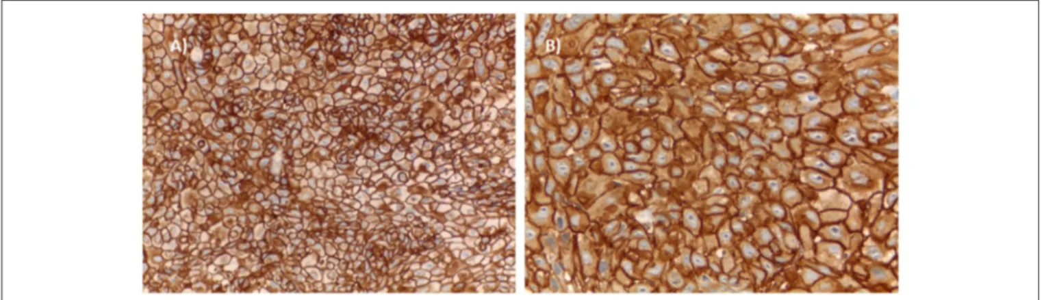
random sample of HPV-negative cases were tested for IHC staining for EGFR [anti-EGFR (3C6), Roche; pre-diluted] and pEGFR [EP774Y, Abcam, (Tyr1068); dilution 1/300]. Membranous and sub-membranous cytoplasmic staining was considered acceptable for either marker, according to manufacturers’ data. EGFR protein and pEGFR expression levels were evaluated semi-quantitatively and classified as follows: Score 0: no staining or membrane staining in <10% of tumor cells; Score 1+: faint membrane staining in >10% of tumor cells; Score 2+: weak or moderate complete membrane staining in >10% of tumor cells and Score 3+: strong, complete membrane staining in >10% of tumor cells. Scores of 0 and 1+ were considered negative for either EGFR or pEGR expression while Scores 2+ and 3+ were considered positive. Representative images for EGFR and pEGFR positive samples are displayed on Figure 1.
Statistical Analyses
Conditional logistic models accounting for matching on tobacco use were used to assess the association between HPV16-positive OPCs and EGFR or pEGFR expression. Crude and
adjusted odds ratios for age, sex, alcohol consumption site, and treatment, and their 95% confidence intervals were estimated. Histological variables were not considered in multivariate analyses, as previously described (22). Differences in baseline characteristics between HPV16-positive and HPV-negative cases were compared using the paired t-test for continuous variables and the Fisher’s exact test for categorical variables. Agreement between EGFR and pEGFR stratified by HPV status was assessed through Kappa index and the distribution of discordant responses from EGFR and pEGFR by using McNemar’s test. Those were also used to assess the agreement/discordant cells between HPV16-E5 and EGFR or pEGFR expression in HPV16-positive OPC cases. For survival analysis only patients with locally advanced disease (stage III, IVa,b 7th edition)
were included. Survival time was calculated from the date of histological diagnosis to either time of death (overall survival, OS) or to time of cancer recurrence (disease-free survival, DFS). The cumulative probability of survival past five years was estimated by Kaplan–Meier analysis. Survival curves were compared by means of the log-rank test. All p-values obtained were corrected for multiple testing by using the Benjamini-Hochberg procedure with alpha = 0.05 significance level. Multivariate Cox’s proportional hazards models were performed under three scenarios to explore whether HPV16-positivity, EGFR and pEGFR expression alone or any combination between them could have a value as prognostic variables, after accounting for other confounding factors (see Table 3 foot note). Hazard ratios were calculated for death and recurrence for OPC patients.
Statistical analyses were performed with STATA SE version 15.0 and R version 3.3.2.
RESULTS
Figure 2 describes the workflow of the OPC targeted cases, samples collected, processed and tested. Among the 1,381 OPC patients targeted, a total of 788 samples yielded a valid DNA result and were finally included in the analysis. HPV-DNA positivity was found in 80 (10.2%) samples and HPV16 was the most frequent genotype, present in 67 cases (83.7% among HPV-DNA positive cases). The number of bona-fide HPV16-positive cases when considering double positivity for HPV16-DNA and p16INK4a IHC was 53 (67.5% among
HPV-DNA positive cases). Hereinafter such double positive cases will be referred to as HPV16-positive cases. A random sample of 174 HPV-negative cases matched for tobacco use was also included in the analysis as a control group. A demographical and clinical descriptive of the OPC patients by HPV status is summarized in Supplementary Table S1. HPV16-positive patients were significantly more likely to be female, non-drinkers,
to have an undifferentiated tumor or to have a primary tonsil tumor. The demographic and clinical characteristics associated with HPV16-E5 expression, EGFR and pEGFR levels, stratified by HPV status, as well as the crude Odds Ratio (OR) measures of association are shown in Supplementary Table S2. The percentage of positive cases expressing also HPV16-E5 was 77.4% (41/53). Since HPV16 integration often concurs with E5 ablation, all samples that tested negative for the presence of HPV16-E5 mRNA (12/53) were further tested for HPV16-E5 DNA, and 58.3% (7/12) were positive. We confirmed that these seven HPV16-E5 mRNA-negative/HPV16-E5 DNA-positive samples were also DNA-positive for HPV-E6∗I mRNA, thus
excluding the possibility of a false negative for mRNA presence by mRNA degradation.
We examined differential EGFR and pEGFR levels between HPV16-positive OPCs and tobacco smoking-matched HPV-negative OPCs and calculated the corresponding ORs (Table 1). EGFR expression levels were significantly lower in HPV16-positive OPCs than in HPV-negative: 37.7% (20/53) vs 70.8% (119/168), respectively (OR = 0.15 [95% Confidence Interval (CI): 0.04–0.56]). There were no differences in pEGFR expression
TABLE 1 | Association of differential EGFR and pEGFR levels between HPV16-positive OPCs and tobacco smoking-matched HPV-negative OPCs.
Biomarker HPV status OR [95% CI]* OR [95%CI]**
HPV negative HPV16 positive No/Total (%) No/Total (%) EGFR EXPRESSION Negative 49/168 (29.2) 33/53 (62.3) Ref. Positive 119/168 (70.8) 20/53 (37.7) 0.15 [0.04–0.56] pEGFR EXPRESSION Negative 127/169 (75.1) 32/53 (60.4) Ref. Positive 42/169 (24.9) 21/53 (39.6) 1.58 [0.48–5.17]
*Adjusted OR by EGFR expression, age, sex, alcohol consumption, and site. Cases matched by tobacco use (see details in Methods section). **Adjusted OR by pEGFR expression, age, sex, alcohol consumption, and site. Cases matched by tobacco use (see details in Methods section).EGFR, Epidermal Growth Factor Receptor; pEGFR, phosphorylated EGFR; HPV, Human Papillomavirus; OPC, Oropharyngeal carcinoma; OR, Odds Ratio; 95%CI, 95% Confidence Interval. HPV16 positive cases are also positive for p16INK4a.
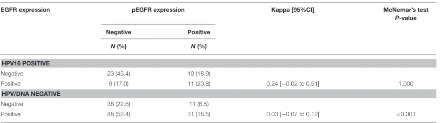
TABLE 2a | Concordance between EGFR and pEGFR expression by HPV status.
EGFR expression pEGFR expression Kappa [95%CI] McNemar’s test
P-value Negative Positive N(%) N(%) HPV16 POSITIVE Negative 23 (43.4) 10 (18.9) Positive 9 (17.0) 11 (20.8) 0.24 [−0.02 to 0.51] 1.000 HPV/DNA NEGATIVE Negative 38 (22.6) 11 (6.5) Positive 88 (52.4) 31 (18.5) 0.03 [−0.07 to 0.12] <0.001
HPV, Human Papillomavirus; EGFR, Epidermal Growth Factor Receptor; pEGFR, phosphorylated EGFR. DNA, Deoxyribonucleic acid; HPV16 positive cases are also positive for p16INK4a.
levels between HPV16-positive and HPV-negative OPCs: 39.6% (21/53) vs 24.9% (42/169), respectively (OR = 1.58 [95%CI: 0.48– 5.17]). Furthermore, as a sensitivity analysis, we reanalyzed the data using unconditional logistic regression obtaining similar results: for EGFR expression levels, OR = 0.22 [95%CI: 0.12– 0.52]; for pEGFR, OR = 2.02 [95%CI: 0.87–4.7]. Table 2a shows the concordance between EGFR and pEGFR expression by HPV status. Among HPV-negative OPC patients we observed an unequal distribution of discordant samples, with an unbalance toward higher proportion of EGFR-positive/pEGFR-negative
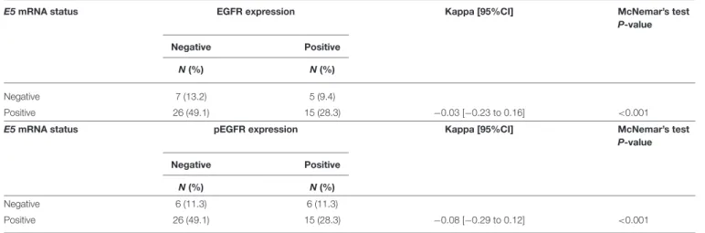
cases, not observed in HPV16-positive cases (McNemar’s test p < 0.001 and 1.0, respectively). Within HPV16-positive cases, no agreement was found between HPV16-E5 expression and EGFR or pEGFR levels (Kappa = −0.03 and Kappa = −0.08, respectively; Table 2b).
With a median follow-up of 39.36 months (min = 0.03 – max = 272.07), survival analyses were performed. We examined the OS and DFS for OPC patients based on Kaplan-Meier curves stratified by different biomarkers. We first evaluated the role of each biomarker alone (Figure 3).
TABLE 2b | Concordance between HPV16-E5 expression and EGFR or pEGFR expression in HPV16 positive OPC cases.
E5mRNA status EGFR expression Kappa [95%CI] McNemar’s test
P-value
Negative Positive
N(%) N(%)
Negative 7 (13.2) 5 (9.4)
Positive 26 (49.1) 15 (28.3) −0.03 [−0.23 to 0.16] <0.001
E5mRNA status pEGFR expression Kappa [95%CI] McNemar’s test
P-value
Negative Positive
N(%) N(%)
Negative 6 (11.3) 6 (11.3)
Positive 26 (49.1) 15 (28.3) −0.08 [−0.29 to 0.12] <0.001
HPV, Human Papillomavirus; EGFR, Epidermal Growth Factor Receptor; pEGFR, phosphorylated EGFR. HPV16 positive cases are also positive for p16INK4a. Kappa index: values <0
as indicating no agreement and 0–0.20 as slight, 0.21–0.40 as fair, 0.41–0.60 as moderate, 0.61–0.80 as substantial, and 0.81–1 as almost perfect agreement.
FIGURE 3 | Overall survival and disease free survival of locally-advance OPC patients based on Kaplan-Meier curves stratified by biomarkers. Legend: Data on 5 years Overall Survival (A–C) and 5 years disease free survival (D–F) according to three different biomarkers: HPV16 (A,D), EGFR (B,E), and pEGFR (C,F). (A,D) Show OS and DFS Kaplan-Meier curve for HPV16-related and HPV negative samples. (B,E) Show OS and DFS Kaplan-Meier curve for EGFR-positive and EGFR-negative samples. (C,F) Show OS and DFS Kaplan-Meier curve for pEGFR-positive and pEGFR-negative samples.
HPV16-positive cases demonstrated better OS (a) Log-rank p < 0.001 and DFS (d) Log-rank p = 0.009. In the same line, EGFR-negative cases demonstrated better OS (b) Log-rank p = 0.033 and DFS (e) Log-Log-rank p = 0.016. We did not find differences related to pEGFR detection alone neither in OS (c) Log-rank p = 0.778 nor in DFS (f) Log-rank p = 0.430.
We further evaluated the differences in OS and DFS as a function of the combination of HPV status and EGFR expression (Figures 4A,C) and of HPV status and pEGFR levels (Figures 4B,D). The analyses showed that stratification after HPV positive/negative and EGFR or pEGFR positive/negative resulted in differences in both OS and DFS (Log-rank p-values indicated in the corresponding figure).
FIGURE 4 | Overall survival and disease free survival of of locally-advance OPC patients stratified by combination of HPV16 status and EGFR or pEGFR. Legend: Data on 5 years Overall Survival (A,B) and 5 years disease free survival (C,D) according to the combination of HPV16 status and EGFR or pEGFR. (A,C) Show OS and DFS Kaplan-Meier curve according to HPV16 status and EGFR expression. (B,D) Show OS and DFS Kaplan-Meier curve according to HPV16 status and pEGFR expression. The H0 assumed that all groups have equal OS and DFS.
FIGURE 5 | Overall survival of locally-advanced HPV16-related patients stratified by HPV16-E5 alone and in combination with EGFR and pEGFR. Legend: Data on 5 years Overall Survival for HPV16-related patients, stratified by HPV16-E5 (A) the combination of HPV16-E5 and EGFR (B) and the combination of HPV16-E5 and pEGFR (C). The H0 assumed that all groups have equal OS.
Among HPV16-positive cases, when analyzing the impact on survival of HPV16-E5 detection, patients with OPC expressing E5 displayed better OS than E5-negative ones, but this difference was not significant (Log-rank p = 0.148; Figure 5A). Finally, evaluation of differences in OS and DFS as a function of the combination of HPV16-E5 and EGFR (Figure 5B) or pEGFR (Figure 5C) expression did not show differences between groups (Log-rank p = 0.104 and Log-rank p = 0.263, respectively).
Hazard ratios (HR) for death and recurrence for OPC patients are presented in Table 3, stratifying by HPV16-positivity, EGFR, and pEGFR expression and their combinations. When all three biomarkers were considered separately and after adjusting for confounding covariates, differences in OS and DFS among OPC patients remained statistically significant for HPV16-positive compared to negative and for EGFR HPV16-positive vs negative regarding adjusted HR for DFS. When combining HPV16 status and EGFR expression, HPV16-positive/EGFR-negative samples demonstrated better OS than any other combination (HR = 0.13 [95%CI: 0.05–0.36]). For HPV16 status and pEGFR expression, all combinations showed better OS compared to patients HPV-negative/pEGFR-positive (p < 0.001).
DISCUSSION
Viral etiology (i.e., HPV-related status) is already well established as an independent predictor of improved OS and treatment responsiveness in OPCs (2,5). Notwithstanding, not all patients with HPV-related OPCs respond equally to treatment nor have the same prognosis. With this study we have aimed at identifying additional biomarkers beyond OPC-relatedness to better classify HPV-related OPC patients, in an era where developing de-escalation clinical trials is a translational research priority.
We have adhered to a strict definition of viral etiology of OPC samples: an OPC was defined as HPV16-positive if it tested positive for the presence of viral HPV16-DNA and also displayed p16INK4a high expression—a cellular surrogate marker for oncoviral activity (17). In our series, HPV16 was the most frequently detected viral genotype, accounting for 83% among HPV-DNA positive OPC samples, matching previous descriptions for the oropharynx (18,23). Among HPV16-positive samples, 77.4% expressed HPV16-E5, in agreement with Um and coworkers, who described variable E5 expression in HPV16-positive OPCs (13).
During chronic infection and malignization, viral DNA can integrate into the cellular DNA (24,25). Usually, this integration event concurs with the loss of particular stretches of the viral genome, often spanning the E2 and the E5 genes. For this reason, in all twelve HPV16-E5 mRNA negative samples we assessed the presence of the corresponding E5 target DNA, as well as the expression of the HPV16-E6∗I mRNA. All twelve
samples were positive for E6 expression, further confirming viral transcriptional activity and thus active viral gene expression. Seven of them (58%) were HPV16-E5 DNA-positive, suggesting that the E5 oncogene was present but its expression had been shut down, either because of upstream disruption of the viral genome or because of epigenetic silencing (26). In five of the
HPV16-E5 mRNA negative samples we could not detect the E5 DNA, suggesting that it had been probably deleted during an integration event.
The E5 oncoprotein enhances the transforming activity of E6 and E7 (13). We therefore aimed at studying whether HPV16-E5 expression could be an additional prognostic biomarker for HPV16-positive OPC patients. In our series, detection of HPV16-E5 mRNA suggested an improved OS, but this trend was not significant, possibly due to the limited size of our cohort (N = 53). Our results thus are concordant with but cannot confirm previous studies reporting an improved DFS, but not OS for patients with cancers displaying high HPV16-E5 expression levels (13).
EGFR is frequently reported to be overexpressed in HNSCC, and such overexpression correlates with poor prognosis: almost 80% of HNSCC studies demonstrate decreased OS and DFS for patients with cancers overexpressing EGFR (27). Mutations in the EGFR gene, such as gene amplification or constitutive activation are common among non HPV-related HNSCCs (8,28,
29). Results from our series at the protein level are consistent with this trend, as HPV-negative samples expressed more often EGFR than HPV16-positive ones (71 vs 38%). Results from the report by Kumar and coworkers. assessing EGFR expression and HPV-relatedness status in OPCs (30) are consistent with those communicated here, but in their case EGFR expression was higher for both groups (93 vs 78%, respectively). This large variation, most notable among HPV-positive cases, can stem from (i) the different evaluation criteria used for assessing EGFR expression (their three-tier vs our two-tier classification scheme), (ii) the HPV-relatedness definition use (more strict in our case), or (iii) may simply be related to sample size, our cohort being almost four times larger size (50 vs 221).
While there is a good agreement in the literature about the higher EGFR expression among HPV-negative OPC cases, this is not the case for pEGFR levels. A previous study has reported higher pEGFR levels among HPV-negative OPCs (31), but our results contradict this view. In our series, the strong increase in EGFR levels among HPV-negative OPCs was accompanied by a sensible albeit non-significant decrease in pEGFR levels, to the extent that HPV16-positive samples displayed higher pEGFR positivity than HPV-negative samples (40 vs 25%, respectively).
Our rationale to seek a connection between HPV16-E5 expression and increased EGFR levels stems from the known functional impact of HPV16-E5 on EGFR-mediated signaling. Extracellular binding of the EGF ligand leads to activation of EGFR through intracellular phosphorylation. The activated form pEGFR is internalized, driven to the endosomes and eventually degraded. Presence of HPV16-E5 results in inability for late endosomal acidification (16), preventing pEGFR degradation, which is instead vehiculated again to the cellular surface in activated state, overall resulting in an increased ligand-triggered EGFR response that is maintained even after the EGF stimulus has disappeared (15). Under this view, we initially hypothesized that higher HPV16-E5 expression would result in increased pEGFR surface recycling, and thus, higher pEGFR positivity. Our results however failed to validate our mechanistic hypothesis, as HPV16-E5 expression was not related to pEGFR expression in
T A B L E 3 | A d ju st e d a n d u n a d ju st e d H a za rd R a tio a n d 9 5 % c o n fid e n c e in te rv a lf o r C o x p ro p o rt io n a lh a za rd m o d e ls p re d ic tin g 5 -y e a rs a ll-c a u se m o rt a lit y a n d 5 -y e a rs re c u rr e n c e in lo c a lly -a d va n c e d o ro p h a ry n g e a l c a n c e r p a tie n ts . A ll c a s e s 5 -y e a rs o v e ra ll s u rv iv a l 5 -y e a rs d is e a s e fr e e s u rv iv a l C ru d e H R [9 5 % C I] P -v a lu e A d ju s te d H R [9 5 % C I] P -v a lu e C ru d e H R [9 5 % C I] P -v a lu e A d ju s te d H R [9 5 % C I] P -v a lu e S C E N A R IO 1 : M A R K E R S C O N S ID E R E D S E P A R A T E LY E G F R e x p re s s io n N e g a tiv e R e f. R e f. R e f. R e f. P o si tiv e 1 .6 9 [1 .0 9 –2 .6 2 ] 0 .0 1 5 1 .3 6 [0 .8 7 –2 .1 4 ] 0 .1 7 4 2 .4 3 [1 .1 9 –4 .9 6 ] 0 .0 0 9 2 .2 5 [1 .0 8 –4 .6 6 ] 0 .0 2 1 p E G F R e x p re s s io n N e g a tiv e R e f. R e f. R e f. R e f. P o si tiv e 1 .0 6 [0 .7 0 –1 .6 1 ] 0 .7 7 9 1 .0 2 [0 .6 6 –1 .5 7 ] 0 .9 2 1 0 .7 7 [0 .3 9 –1 .4 9 ] 0 .4 2 3 0 .9 5 [0 .4 8 –1 .9 1 ] 0 .8 9 3 H P V 1 6 s ta tu s N e g a tiv e R e f. R e f. R e f. R e f. P o si tiv e 0 .2 2 [0 .1 1 –0 .4 3 ] < 0 .0 0 1 0 .2 4 [0 .1 2 –0 .4 8 ] < 0 .0 0 1 0 .2 0 [0 .0 7 –0 .5 9 ] < 0 .0 0 1 0 .1 9 [0 .0 7 –0 .5 7 ] < 0 .0 0 1 S C E N A R IO 2 : C O M B IN E D H P V 1 6 S T A T U S A N D E G F R E X P R E S S IO N H P V 1 6 p o si tiv e a n d E G F R p o si tiv e 0 .3 4 [0 .1 4 –0 .8 2 ] 0 .3 8 [0 .1 5 –0 .9 5 ] 0 .5 3 [0 .1 7 –1 .5 9 ] 0 .4 2 [0 .1 3 –1 .3 1 ] H P V 1 6 p o si tiv e a n d E G F R n e g a tiv e 0 .1 3 [0 .0 5 –0 .3 6 ] 0 .1 7 [0 .0 6 -0 .4 8 ] * * H P V 1 6 n e g a tiv e a n d E G F R p o si tiv e R e f. R e f. R e f. R e f. H P V 1 6 n e g a tiv e a n d E G F R n e g a tiv e 0 .8 4 [0 .5 3 –1 .3 4 ] < 0 .0 0 1 1 .0 7 [0 .6 5 –1 .7 7 ] < 0 .0 0 1 0 .7 1 [0 .3 4 –1 .4 7 ] < 0 .0 0 1 0 .9 5 [0 .4 3 –2 .1 0 ] < 0 .0 0 1 S C E N A R IO 3 : C O M B IN E D h p v 1 6 S T A T U S A N D p E G F R E X P R E S S IO N H P V 1 6 p o si tiv e a n d p E G F R p o si tiv e 0 .1 3 [0 .0 4 -0 .4 2 ] 0 .1 4 [0 .0 4 –0 .4 7 ] * * H P V 1 6 p o si tiv e a n d p E G F R n e g a tiv e 0 .2 1 [0 .0 9 –0 .4 9 ] 0 .2 5 [0 .1 0 –0 .6 0 ] 0 .3 4 [0 .1 0 –1 .0 9 ] 0 .2 6 [0 .0 8 –0 .9 0 ] H P V 1 6 n e g a tiv e a n d p E G F R p o si tiv e R e f. R e f. R e f. R e f. H P V 1 6 n e g a tiv e a n d p E G F R n e g a tiv e 0 .7 7 [0 .5 0 –1 .1 9 ] < 0 .0 0 1 0 .7 9 [0 .5 0 –1 .2 6 ] < 0 .0 0 1 0 .9 5 [0 .4 8 –1 .8 9 ] < 0 .0 0 1 0 .6 9 [0 .3 3 –1 .4 2 ] 0 .0 0 1 P -v a lu e : L o g -l ik e lih o o d ra ti o te s t P -v a lu e . *C o m bi n a ti on s th a t do n o t c o n ta in in di vi du a ls w ith e ve n ts of re c u rr e n c e . C a s e s m a tc h e d by to ba c c o u s e (s e e de ta ils in Me th ods s e c ti on ). In a ll c a s e s , va lu e s in bol d c or re s pon d s h ow s ta ti s ti c a ls ig n ifi c a n t di ff e re n t fr om th e n u ll h ypoth e s is of th e te s t. O v er al l s u rv iv al . V a ri a bl e s in c lu de d in th e a dj u s te d m ode ls : S c e n a ri o 1: a ge , ge n de r, a n d tr e a tm e n t, a n d EG F R e xpr e s s ion or pEG F R e xpr e s s ion or H P V 16 s ta tu s ; S c e n a ri o 2: c om bi n e d H P V 16 s ta tu s a n d EG F R e xpr e s s ion a n d a ge , g e n de r, a n d tr e a tm e n t; S c e n a ri o 3 : c o m bi n e d H P V 16 s ta tu s a n d pEG F R e xpr e s s ion a n d a ge , ge n de r, a n d tr e a tm e n t. D is eas e-fr ee s u rv iv al . V a ri a bl e s in c lu de d in th e a dj u s te d m ode ls : S c e n a ri o 1: a ge , ge n de r, a n d pe ri od of di a gn os is , a n d EG F R e xpr e s s ion or pEG F R e xpr e s s ion or H P V 16 s ta tu s (w ith ou t pe ri od of di a gn os is ); S c e n a ri o 2: c om bi n e d H P V 1 6 s ta tu s a n d EG F R e xpr e s s io n , a g e , g e n de r, pe ri od of di a gn os is a n d s u bs ite ; S c e n a ri o 3: c om bi n e d H P V 16 s ta tu s a n d pEG F R e xpr e s s ion , a ge , ge n de r, a n d pe ri od of di a gn os is .
our series. Nevertheless, our results show that HPV16-positive cancers display lower EGFR levels but higher pEGFR/EGFR ratios than HPV-negative cancers. We are not aware of any viral mechanism to explain this puzzling observation.
High EGFR levels are associated with worse prognosis in HNSCC (32), as well as in a subset of HPV-related OPCs (7). In line with these results and with other studies (30, 33), we confirm in our cohort an association between EGFR expression and worse prognosis. The combination of both biomarkers, HPV-relatedness and EGFR or pEGFR expression, could be useful to better characterize OPC patients. When different variables (i.e., age, tobacco use) were entered into a multivariate Cox proportional hazards model, HPV status and the combination of HPV-relatedness and EGFR or pEGFR expression were predictors of better OS and DFS. This result contradicts however the study by Romanitan and coworkers, where pEGFR was not found to be an optimal prognostic marker for clinical outcome in OPC patients (31). Furthermore, Bossi and coworkers reviewed in a recent article the prognostic and predictive value of EGFR in HNSCC (34), regarding HPV-positive patients they described different trials evaluating the combined effect of HPV status and EGFR expression on prognosis, showing that patients with EGFR-positive/HPV-negative cancer have the poorest outcomes, while the EGFR negative/HPV-positive group showed the best outcome in line with our results. Nevertheless, they suggest that in recent studies including OPC patients, the prognostic role of EGFR expression was related to the association with HPV-negative tumors, and the added value of EGFR analysis seems to be marginal in respect to HPV (34).
Limited preclinical data investigating the combination of radiotherapy and anti-EGFR antibodies are available and clinical data are still controversial for HPV-related patients (35–37). In our series, only four patients with locally advanced HPV16-positive OPC were treated with anti-EGFR therapy concomitant with radiotherapy. So far, no conclusion can thus be drawn about the use of anti-EGFR therapy, HPV-relatedness and HPV16-E5 expression and EGFR and pEGFR levels in the context in our series. Interestingly, two of the phase III clinical trials investigating the combination of anti-EGFR therapy with radiotherapy compared to the standard treatment with chemo-radiotherapy for HPV-positive oropharyngeal cancer patients, the RTOG 1026 and the DE-ESCALaTE study, have been recently presented. Both studies demonstrated superiority for the standard treatment (cisplatin-radiotherapy) for HPV-related OPC patients (38–40).
Our study has a number of limitations. First, the HPV-related attributable fraction among OPCs in Mediterranean countries is lower than in other World regions, such as the US or Northern Europe. Thus, despite having analyzed a large global number of OPC cases, our cohort included a low number of HPV-related OPC patients. This reduced effective sample size limits our power to establish the validity of E5 mRNA expression as a potential biomarker. Second, the analyses for this pilot study were performed on FFPE samples. Although such fixation technique is the most common approach to stabilize tissue prior to anatomopathological analyses, this aggressive chemical treatment may impact the quantity and quality of
the retrieved nucleic acids, potentially rendering lower apparent prevalence values of viral DNA and/or mRNA. Third, pEGFR detection is still not widely implemented in the clinical setting, and the determination here applied was semi-quantitative. All in all, these limitations may have hindered the identification of a putative connection between viral HPV16-E5 expression and the detection of cellular EGFR/pEGFR. On the other hand, the strength of our study is the epidemiology design: we have applied a robust HPV testing method, performed in a single laboratory, to decrease variability; we have only included oropharyngeal samples and excluded other head and neck sub-sites; and we have focused on samples positive only for one single HPV genotype (HPV16).
In summary, our study demonstrates HPV16-E5 to be highly expressed on HPV16-positive OPC samples. HPV-negative samples expressed significantly more EGFR, while HPV16-positive samples expressed more pEGFR. The combinations of HPV-relatedness and EGFR or pEGFR protein levels are useful biomarkers for prognosis outcome in OPC patients. Specifically, OPC patients with HPV16-positive and EGFR negative cancers present better OS prognosis than patients with HPV-negative and EGFR positive cancers. Further studies are warranted to better understand the role of HPV16-E5 in cancer etiology and its relationship with EFGR and pEGFR expression.
AUTHOR CONTRIBUTIONS
MT, MTo, MA, RM, IB, and LA study concept, study design, and interpretation. MT, MM, XL, JG, MG, RH, TB, and AA data acquisition. ST and SM data analysis. MT, MTo, IB, and LA manuscript preparation. All authors reviewed and validated the final version of the manuscript.
FUNDING
This work was supported by grants from Ayudas Merck Serono Investigación_Salud 2000, Instituto de Salud Carlos III-ISCIII (Spanish Government) cofunded by FEDER funds/European Regional Development Fund (ERDF)—a way to build Europe (References: PI1102096, PI1401918, PI1500500, PI1501205, RD12/0036/0056, CIBERESP CB06/02/0073, CIBERONC CB16/12/00401). With the support of the Secretariat for Universities and Research of the Department of Business and Knowledge of the Government of Catalonia. Grants to support the activities of research groups (SGR 2014-2017: 2014SGR756 and 2014SGR1077; 2017-2019: 2017SGR1085). Beca de recerca clínica 2016 de l’Acadèmia de Ciències Mèdiques de Catalunya i Balears, Department of Health of the Generalitat de Catalunya (PERIS-2016-2020, SLT002/16/00404). Asociación Española Contra el Cáncer (personal grant to LA), Rio Hortega-SEOM (ISCIII-Spanish Society of Medical Oncology) (personal grant to MT), and from Sanofi Pasteur MSD and Merck and Co., Inc. None of the funders had any role in the data collection, analysis, interpretation of the results, article writing, and decision to publish.
ACKNOWLEDGMENTS
MT gratefully acknowledges the Rio Hortega-SEOM (ISCIII-Spanish Society of Medical Oncology) grant. The authors want to thank all members of the Study Group: Institut Català d’Oncologia/Hospital Universitari de Bellvitge: Laia Alemany, Oriol Bermejo, Xavier Bosch, Vanesa Camon, Omar Clavero, Ana Esteban, Yolanda Florencia, Montserrat Gomà, Alicia Lozano, Manel Maños, Antonio Marí, Marisa Mena, Ricard Mesía, Julio Nogués, Miquel Ángel Pavón, Beatriz Quirós, Silvia de Sanjosé, Miren Taberna, Montserrat Torres, Sara Tous, Griselda Venturas,
Marleny Vergara, Joan Viñals; Hospital General de L’Hospitalet: María Alejo; Hospital de la Santa Creu i Sant Pau: Jacinto García, Xavier León; Hospital del Mar: Marta Guix, Rafael Hijano, Belén Lloveras; Hospital Universitari Parc Taulí: Antón Aguilà, María Rosa Bella, Carmen Blazquez, Teresa Bonfill.
SUPPLEMENTARY MATERIAL
The Supplementary Material for this article can be found online at: https://www.frontiersin.org/articles/10.3389/fonc. 2018.00589/full#supplementary-material
REFERENCES
1. Ndiaye C, Mena M, Alemany L, Arbyn M, Castellsagué X, Laporte L, et al. HPV DNA, E6/E7 mRNA, and p16INK4adetection in head and neck cancers:
a systematic review and meta-analysis. Lancet Oncol. (2014) 15:1319–31. doi: 10.1016/S1470-2045(14)70471-1
2. Ang KK, Harris J, Wheeler R, Weber R, Rosenthal DI, Nguyen-Tân PF, et al. Human papillomavirus and survival of patients with oropharyngeal cancer. N Engl J Med. (2010) 363:24–35. doi: 10.1056/NEJMoa09 12217
3. Taberna M, Rullan AJ, Hierro C, Navarro V, Vazquez S, Lozano A, et al. Late toxicity after radical treatment for locally advanced head and neck cancer. Oral Oncol. (2015) 51:795–9. doi: 10.1016/j.oraloncology.2015.05.002
4. Masterson L, Moualed D, Masood A, Dwivedi RC, Benson R, Sterling JC, et al. De-escalation treatment protocols for human papillomavirus-associated oropharyngeal squamous cell carcinoma. Eur J Cancer. (2014) 50:2636–48. doi: 10.1016/j.ejca.2014.07.001
5. Taberna M, Marisa M, Pavón MA, Alemany L, Gillison ML, Mesía R. Human papillomavirus related oropharyngeal cancer. Ann Oncol. (2017) 28:2386–98. doi: 10.1093/annonc/mdx304
6. Kalyankrishna S, Grandis JR. Epidermal growth factor receptor biology in head and neck cancer. J Clin Oncol. (2006) 24:2666–72. doi: 10.1200/JCO.2005.04.8306
7. Mirghani H, Amen F, Moreau F, Guigay J, Hartl DM, Lacau St. Guily J. Oropharyngeal cancers: relationship between epidermal growth factor receptor alterations and human papillomavirus status. Eur J Cancer (2014) 50:1100–11. doi: 10.1016/j.ejca.2013.12.018
8. Lawrence MS, Sougnez C, Lichtenstein L, Cibulskis K, Lander E, Gabriel SB, et al. Comprehensive genomic characterization of head and neck squamous cell carcinomas. Nature (2015) 517:576–82. doi: 10.1038/nature14129 9. Aquino G, Pannone G, Santoro A, Liguori G, Franco R, Serpico R, et al.
PEGFR-Tyr 845 expression as prognostic factors in oral squamous cell carcinoma: a tissue-microarray study with clinic-pathological correlations. Cancer Biol Ther. (2012) 13:967–77. doi: 10.4161/cbt.20991
10. Ang K, Berkey B, Tu X, Zhang H, Katz R, Hammond E, et al. Impact of epidermal growth factor receptor expression on survival and pattern of relapse in patients with advanced head and neck carcinoma. Cancer Res. (2002) 62:7350–6.
11. Monteiro L, Ricardo S, Delgado M, Garcez F, do Amaral B, Lopes C. Phosphorylated EGFR at tyrosine 1173 correlates with poor prognosis in oral squamous cell carcinomas. Oral Dis. (2014) 20:178–85. doi: 10.1111/odi.12087 12. zur Hausen H. Papillomaviruses and cancer: from basic studies to clinical
application. Nat Rev Cancer (2002) 2:342–50. doi: 10.1038/nrc798
13. Um HS, Mundi N, Yoo J, Palma DA, Fung K, Macneil D, et al. Variable expression of the forgotten oncogene E5 in HPV-positive oropharyngeal cancer. J Clin Virol. (2014) 61:94–100. doi: 10.1016/j.jcv.2014.06.019 14. DiMaio D, Petti LM. The E5 proteins. Virology (2013) 445:99–114.
doi: 10.1016/j.virol.2013.05.006
15. Leechanachai P, Banks L, Moreau F, Matlashewski G. The E5 gene from human papillomavirus type 16 is an oncogene which enhances growth factor-mediated signal transduction to the nucleus. Oncogene (1992) 7:19–25.
16. Straight SW, Herman B, McCance DJ. The E5 oncoprotein of human papillomavirus type 16 inhibits the acidification of endosomes in human keratinocytes. J Virol. (1995) 69:3185–92.
17. Mena M, Taberna M, Tous S, Marquez S, Clavero O, Quiros B, et al. Double positivity for HPV-DNA/p16ink4ais the biomarker with strongest
diagnostic accuracy and prognostic value for human papillomavirus related oropharyngeal cancer patients. Oral Oncol. (2018) 78:137–144. doi: 10.1016/j.oraloncology.2018.01.010
18. Castellsagué X, Alemany L, Quer M, Halec G, Quirós B, Tous S, et al. HPV involvement in head and neck cancers: comprehensive assessment of biomarkers in 3680 patients. J Natl Cancer Inst. (2016) 108:djv403. doi: 10.1093/jnci/djv403
19. Halec G, Holzinger D, Schmitt M, Flechtenmacher C, Dyckhoff G, Lloveras B, et al. Biological evidence for a causal role of HPV16 in a small fraction of laryngeal squamous cell carcinoma. Br J Cancer. (2013) 109:172–83. doi: 10.1038/bjc.2013.296
20. Grønhøj Larsen C, Gyldenløve M, Jensen DH, Therkildsen MH, Kiss K, Norrild B, et al. Correlation between human papillomavirus and p16 overexpression in oropharyngeal tumours: a systematic review. Br J Cancer (2014) 110:1587–94. doi: 10.1038/bjc.2014.42
21. Prigge ES, Arbyn M, von Knebel Doeberitz M, Reuschenbach M. Diagnostic accuracy of p16INK4a immunohistochemistry in oropharyngeal squamous
cell carcinomas: a systematic review and meta-analysis. Int J Cancer (2017) 140:1186–98. doi: 10.1002/ijc.30516
22. Alemany L, Saunier M, Alvarado-Cabrero I, Quirõs B, Salmeron J, Shin HR, et al. Human papillomavirus DNA prevalence and type distribution in anal carcinomas worldwide. Int J Cancer (2015) 136:98–107. doi: 10.1002/ijc.28963 23. Jordan RC, Lingen MW, Perez-Ordonez B, He X, Pickard R, Koluder M, et al. Validation of methods for oropharyngeal cancer HPV status determination in US cooperative group trials. Am J Surg Pathol. (2012) 36:945–54. doi: 10.1097/PAS.0b013e318253a2d1
24. Vojtechova Z, Sabol I, Salakova M, Turek L, Grega M, Smahelova J, et al. Analysis of the integration of human papillomaviruses in head and neck tumours in relation to patients’ prognosis. Int J Cancer (2016) 138:386–95. doi: 10.1002/ijc.29712
25. Parfenov M, Pedamallu CS, Gehlenborg N, Freeman SS, Danilova L, Bristow CA, et al. Characterization of HPV and host genome interactions in primary head and neck cancers. Proc Natl Acad Sci USA. (2014) 111:15544–9. doi: 10.1073/pnas.1416074111
26. Mesri EA, Feitelson MA, Munger K. Human viral oncogenesis: a cancer hallmarks analysis. Cell Host Microbe (2014) 15:266–82. doi: 10.1016/j.chom.2014.02.011
27. Nicholson R, Gee JM, Harper M. EGFR and cancer prognosis. Eur J Cancer (2001) 37:9–15. doi: 10.1016/S0959-8049(01)00231-3
28. Hayes DN, Van Waes C, Seiwert TY. Genetic landscape of human papillomavirus-associated head and neck cancer and comparison to tobacco-related tumors. J Clin Oncol. (2015) 33:3227–34. doi: 10.1200/JCO.2015.62.1086
29. Keck MK, Zuo Z, Khattri A, Stricker TP, Brown CD, Imanguli M, et al. Integrative analysis of head and neck cancer identifies two biologically distinct HPV and three non-HPV subtypes. Clin Cancer Res. (2015) 21:870–81. doi: 10.1158/1078-0432.CCR-14-2481
30. Kumar B, Cordell KG, Lee JS, Worden FP, Prince ME, Tran HH, et al. EGFR, p16, HPV titer, Bcl-xL and p53, sex, and smoking as indicators of response to therapy and survival in oropharyngeal cancer. J Clin Oncol. (2008) 26:3128–37. doi: 10.1200/JCO.2007.12.7662
31. Romanitan M, Näsman A, Munck-Wikland E, Dalianis T, Ramqvist T. EGFR and phosphorylated EGFR in relation to HPV and clinical outcome in tonsillar cancer. Anticancer Res. (2013) 33:1575–84.
32. Chung CH, Ely K, McGavran L, Varella-Garcia M, Parker J, Parker N, et al. Increased epidermal growth factor receptor gene copy number is associated with poor prognosis in head and neck squamous cell carcinomas. J Clin Oncol. (2006) 24:4170–6. doi: 10.1200/JCO.2006.07.2587
33. Young RJ, Rischin D, Fisher R, McArthur GA, Fox SB, Peters LJ, et al. Relationship between epidermal growth factor receptor status, p16(INK4A), and outcome in head and neck squamous cell carcinoma. Cancer Epidemiol Biomarkers Prev. (2011) 20:1230–7. doi: 10.1158/1055-9965.EPI-10-1262 34. Bossi P, Resteghini C, Paielli N, Licitra L, Pilotti S, Perrone F. Prognostic
and predictive value of EGFR in head and neck squamous cell carcinoma. Oncotarget (2016) 7:74362–79. doi: 10.18632/oncotarget. 11413
35. Fakhry C, Zhang Q, Nguyen-Tan PF, Rosenthal D, El-Naggar A, Garden AS, et al. RTOG Ret 0129/0522 recurrence and HPV status: human papillomavirus and overall survival after progression of oropharyngeal squamous cell carcinoma. J Clin Oncol. (2014) 32:3365–73. doi: 10.1200/JCO.2014. 55.1937
36. Giralt J, Trigo J, Nuyts S, Ozsahin M, Skladowski K, Hatoum G, et al. Panitumumab plus radiotherapy versus chemoradiotherapy in patients with unresected, locally advanced squamous-cell carcinoma of the head and neck (CONCERT-2): a randomised, controlled, open-label phase 2 trial. Lancet Oncol. (2015) 16:221–32. doi: 10.1016/S1470-2045(14)7 1200-8
37. Ou D, Levy A, Blanchard P, Nguyen F, Garberis I, Casiraghi O, et al. Concurrent chemoradiotherapy with cisplatin or cetuximab for locally advanced head and neck squamous cell carcinomas: does human papilloma virus play a role? Oral Oncol. (2016) 59:50–7. doi: 10.1016/j.oraloncology.2016.05.019
38. Mehanna H, Robinson M, Hartley A, Kong A, Foran B, Fulton-Lieuw T, et al. Radiotherapy plus cisplatin or cetuximab in low-risk human papillomavirus-positive oropharyngeal cancer (De-ESCALaTE HPV): an open-label randomised controlled phase 3 trial. Lancet (2018). doi: 10.1016/S0140-6736(18)32752-1. [Epub ahead of print].
39. National Cancer Institute press. Available online at: https://www.cancer.gov/ news-events/press-releases/2018/oropharyngeal-hpv-cisplatin. (Accessed August 14, 2018).
40. Gillison ML, Trotti AM, Harris J, Eisbruch A, Harari PM, Adelstein DJ, et al. Radiotherapy plus cetuximab or cisplatin in human papillomavirus-positive oropharyngeal cancer (NRG Oncology RTOG 1016): a randomised, multicentre, non-inferiority trial. Lancet (2018). doi: 10.1016/S0140-6736(18)32779-X. [Epub ahead of print].
Conflict of Interest Statement:RM has received personal fees and non-financial
support from Merck, and personal fees from AstraZeneca, Merck, Brystol-Myers and MSD. MT has received non-financial support from Merck and Astra Zeneca, and personal fees from Merck, Brystol-Myers and AstraZeneca. Cancer Epidemiology Research Program (LA, MM, SM, ST, MTo, MP) has received sponsorship for grants from Merck and co. None of these funders had any role in the data collection, analysis, interpretation of the results, article writing, and decision to publish.
The remaining authors declare that the research was conducted in the absence of any commercial or financial relationships that could be construed as a potential conflict of interest.
Copyright © 2018 Taberna, Torres, Alejo, Mena, Tous, Marquez, Pavón, León, García, Guix, Hijano, Bonfill, Aguilà, Lozano, Mesía, Alemany and Bravo. This is an open-access article distributed under the terms of the Creative Commons Attribution License (CC BY). The use, distribution or reproduction in other forums is permitted, provided the original author(s) and the copyright owner(s) are credited and that the original publication in this journal is cited, in accordance with accepted academic practice. No use, distribution or reproduction is permitted which does not comply with these terms.