HAL Id: hal-03128448
https://hal.inrae.fr/hal-03128448
Submitted on 2 Feb 2021
HAL is a multi-disciplinary open access
archive for the deposit and dissemination of sci-entific research documents, whether they are pub-lished or not. The documents may come from teaching and research institutions in France or abroad, or from public or private research centers.
L’archive ouverte pluridisciplinaire HAL, est destinée au dépôt et à la diffusion de documents scientifiques de niveau recherche, publiés ou non, émanant des établissements d’enseignement et de recherche français ou étrangers, des laboratoires publics ou privés.
Distributed under a Creative Commons Attribution| 4.0 International License
(Hymenoptera, Chalcididae) in Saudi Arabia:
re-evaluation of its limits and description of three new
species
Muhammad Athar Gul, Ahmed Mostafa Soliman, Neveen Samy Gadallah,
Hathal Mohammed Al Dhafer, Muhammad Gul, Ahmed Soliman, Neveen
Gadallah, Hathal Al Dhafer, Gérard Delvare
To cite this version:
Muhammad Athar Gul, Ahmed Mostafa Soliman, Neveen Samy Gadallah, Hathal Mohammed Al Dhafer, Muhammad Gul, et al.. The genus Phasgonophora Westwood, 1832 (Hymenoptera, Chalci-didae) in Saudi Arabia: re-evaluation of its limits and description of three new species. Journal of Hymenoptera Research, Sofia : Pensoft Publishers; [s.l.] : International Society of Hymenopterists., 2020, 76, pp.1-38. �10.3897/jhr.76.38340�. �hal-03128448�
The genus Phasgonophora Westwood, 1832
(Hymenoptera, Chalcididae) in Saudi Arabia:
re-evaluation of its limits and description
of three new species
Muhammad Athar Gul1,2, Ahmed Mostafa Soliman2,3, Neveen Samy Gadallah4, Hathal Mohammed Al Dhafer2, Gérard Delvare5
1 Department of Environmental and Biological Sciences, University of Eastern Finland, P.O. Box 1627,
F1-70211 Kuopio, Finland 2 Plant Protection Department, College of Food and Agriculture Sciences, King Saud University, P.O. BOX 2460, Riyadh 11451, Saudi Arabia 3 Zoology Department, Faculty of Science (Boys), Al-Azhar University, P.O. Box 11884, Nasr City, Cairo, Egypt 4 Entomology Department, Faculty of Science, Cairo University, Giza, Egypt 5 Centre de Coopération Internationale en Recherche Agronomique pour le Déve-loppement (CIRAD), Montpellier SupAgro, INRA, IRD, Univ. Montpellier, Montpellier, France
Corresponding author: Neveen Samy Gadallah (n_gadallah@hotmail.com)
Academic editor: P. Jansta | Received 17 July 2019 | Accepted 25 February 2020 | Published 27 April 2020
http://zoobank.org/8C7E1DDE-BCFA-47C0-A38D-18458AD9221E
Citation: Gul MA, Soliman AM, Gadallah NS, Al Dhafer HM, Delvare G (2020) The genus Phasgonophora Westwood, 1832 (Hymenoptera, Chalcididae) in Saudi Arabia: re-evaluation of its limits and description of three new species. Journal of Hymenoptera Research 76: 1–38. https://doi.org/10.3897/jhr.76.38340
Abstract
A phylogenetic study based on 25 species of Phasgonophorinae (Hymenoptera: Chalcididae) and 36 characters was carried out for ensuring the generic placement of three new species from Saudi Arabia. As a result of this study, the genera Trigonura Sichel, 1866, Bactrochalcis Kieffer, 1912, Centrochalcis Cameron, 1913, Centrochalcidia Gahan & Fagan, 1923, Chalcidellia Girault, 1924, Urochalcis Nikol’skaya, 1952,
Trigonurella Bouček, 1988, and Muhabbetella Koçak & Kemal, 2008 are synonymized with Phasgonophora
Westwood, 1832. This genus is recorded in Saudi Arabia for the first time, represented here by P. rubens (Klug), and newly described species P. baiocchii Soliman & Gul, sp. nov., P. granulis Delvare, sp. nov., and
P. magnanii Gadallah & Gul, sp. nov. An illustrated key to species of the Arabian Peninsula is provided.
The relevant specimens were mostly reared from buprestid species infesting Acacia sp. and Dodonaea
vis-cosa in Al-Baha, Asir and Riyadh regions, Saudi Arabia.
Keywords
Al-Baha, Asir, Riyadh, Buprestidae, new species, Phasgonophora, Phasgonophorini, phylogeny
http://jhr.pensoft.net
Copyright M. Athar Gul et al. This is an open access article distributed under the terms of the Creative Commons Attribution License (CC BY 4.0), which permits unrestricted use, distribution, and reproduction in any medium, provided the original author and source are credited.
Introduction
Phasgonophorinae were first recognized as discrete group by Steffan (1951) who list-ed their features and classifilist-ed them within Brachymeriinae. They were later classifilist-ed within Chalcidinae (Bouček 1988, 1992; Narendran 1989; Wijesekara 1997a; Delvare 2017), but quite recently were raised to subfamily level (Cruaud et al. 2020) appearing as the sister group of Brachymeriinae s.s. (sensu Wijesekara 1997b, as Phasgonophorini). This rather small subfamily includes 66 described species, but many of them, particularly in Phasgonophora, still await description, especially in the tropics where the subfamily is the most diverse. The subfamily was extensively studied by Steffan (1951, 1956, 1973).
Phasgonophorinae are parasitoids of wood-boring beetles belonging to the families Buprestidae, Curculionidae (including Scolytinae), Cerambycidae and Anthribidae (Steffan 1951, 1973; Burks 1959; Mateu 1972; Bouček 1988, 1992; Narendran 1989; Roscoe 2014; Narendran and van Achterberg 2016).
The subfamily currently includes two tribes (Cruaud et al. 2020), Phasgonophorini and Stypiurini, namely the Phasgonophora and Stypiura groups of Steffan (1951). Phas-gonophorini themselves would comprise a single genus based on the phylogenetic results using the ultra-conserved elements (UCE) (Cruaud et al. 2020). Furthermore, Trigonu-ra appeared polyphyletic, merging on two different bTrigonu-ranches, the first one together with Muhabbetella Koçak & Kemal, 2008 (replacement name for Trigonurella Bouček, 1988 nec Maa, 1963) on one hand, and Phasgonophora Westwood, 1832 on the other hand.
Phasgonophorini have the following features: a hardly sclerotized body, most often with rasp-like sculpture on pronotum and mesonotum; malar sulcus absent; antennal scrobes quite deep and entirely delimited by carinae; antennal toruli on about the level of lower ocular line; both mandibles with three teeth; propodeum often strongly slop-ing; mesopleuron frequently with a ventral shelf that is regularly longer than epicne-mium; procoxa with a deep depression anteriorly, delimited by an oblique carina raised into a flange; metatibia without spur; postmarginal vein short, only surpassing stigmal vein in length; petiole very short, either entirely concealed dorsally or visible here as a ring-like sclerite, but better visible ventrally; first tergite with dorsolateral costulae and syntergum often elongated into a stylus; ovipositor sheaths and valvulae straight.
Phasgonophora with 38 described species is distributed in arid and temperate zones of Asia (21 species), New World (11 species), Africa (2 species) and Australia (4 spe-cies). The genus parasitizes xylophagous beetles of the families Anthribidae, Bupresti-dae, Cerambycidae and Curculionidae (including Scolytinae) (Waterston 1922; Stef-fan 1951; Burks 1959; Mateu 1972; Narendran and van Achterberg 2016).
Until now Phasgonophora was represented by only two species in the Arabian Pen-insula, P. ninae (Nikol’skaya, 1952) and P. rubens (Klug, 1834), reported by Delvare (2017) from the United Arab Emirates (under Trigonura). In the present study, the genus is recorded for the first time from Saudi Arabia (from Al-Baha, Asir and Riyadh regions) with four species, namely P. rubens (Klug), and the newly described P. baiocchii Soliman & Gul, sp. nov., P. granulis Delvare, sp. nov., and P. magnanii Gadallah & Gul, sp. nov., mainly reared from Buprestidae attacking Acacia sp. (Fabaceae) or Dodonaea viscosa (L.) Jacq. (Sapindaceae) dead wood.
A phylogenetic study based on morphology was carried out for exploring the pos-sible congruence between molecular and morphological data and hypothesizing the systematic placement of the species collected in Saudi Arabia.
Material and methods Phylogenetic study
Sampling (Table 1). Outgroups include Brachymeria minuta (Linnaeus, 1761) (Bra-chymeriinae), and within the Phasgonophorinae, three species of Stypiurini, namely Kopinata partirubra Bouček, 1988, and two Stypiura species that were chosen together with 21 species of Phasgonophorini belonging to the genera presently recognized. Phas-gonophora sulcata Westwood, 1835, PhasPhas-gonophora (Trigonura) crassicauda Sichel, 1866, and Chalcis euthyrrhini Dodd, 1921, respectively type species of Phasgonophora West-wood, 1832, Trigonura Sichel, 1866, and Chalcidellia Girault, 1924 are included in the sample, which also comprises the specimens that were used for the phylogenetic infer-ence with Ultra Conserved Elements (UCE) (Cruaud et al. 2020). The material from Saudi Arabia was reared from xylophagous Coleoptera of the family Buprestidae (Chrys-obothris sp.) attacking Acacia sp. and D. viscosa dead wood collected by Daniele Baiocchi and Gianluca Magnani (Roma, Italy) or collected using sweep net on Calotropis procera (Aiton) (Apocynaceae) trees from different wadis in Al Baha, Asir and Riyadh regions.
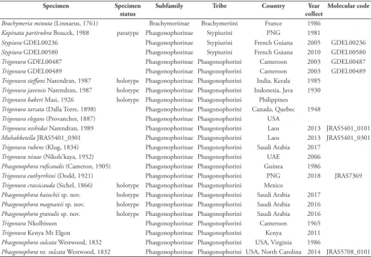
Table 1. Specimens used for the phylogenetic study. Generic names as in the present literature.
Specimen Specimen
status Subfamily Tribe Country collectYear Molecular code Brachymeria minuta (Linnaeus, 1761) Brachymeriinae Brachymeriini France 1986
Kopinata partirubra Boucek, 1988 paratype Phasgonophorinae Stypiurini PNG 1981
Stypiura GDEL00236 Phasgonophorinae Stypiurini French Guiana 2005 GDEL00236
Stypiura GDEL00580 Phasgonophorinae Stypiurini French Guiana 2010 GDEL00580
Trigonura GDEL00487 Phasgonophorinae Phasgonophorini Cameroon 2003 GDEL00487
Trigonura GDEL00489 Phasgonophorinae Phasgonophorini Cameroon 2003 GDEL00489
Trigonura steffani Narendran, 1987 holotype Phasgonophorinae Phasgonophorini India, Kerala 1985
Trigonura javensis Narendran, 1987 holotype Phasgonophorinae Phasgonophorini Indonesia, Java 1930
Trigonura bakeri Masi, 1926 holotype Phasgonophorinae Phasgonophorini Philippines
Trigonura tarsata (Dalla Torre, 1898) Phasgonophorinae Phasgonophorini Canada, Quebec 1948
Trigonura elegans (Provancher, 1887) Phasgonophorinae Phasgonophorini USA
Trigonura nishidai Narendran, 1989 Phasgonophorinae Phasgonophorini Laos 2013 JRAS5401_0101
Muhabbetella JRAS5401_0301 Phasgonophorinae Phasgonophorini Laos 2013 JRAS5401_0301
Trigonura rubens (Klug, 1834) Phasgonophorinae Phasgonophorini Saudi Arabia 2017
Trigonura ninae (Nikols’kaya, 1952) Phasgonophorinae Phasgonophorini UAE 2006
Phasgonophora ruficaudis (Cameron, 1905) Phasgonophorinae Phasgonophorini Guinea 1986
Trigonura euthyrrhini (Dodd, 1921) Phasgonophorinae Phasgonophorini PNG 2018 JRAS7369
Trigonura crassicauda (Sichel, 1866) holotype Phasgonophorinae Phasgonophorini Mexico
Phasgonophora baiochii sp. nov. holotype Phasgonophorinae Phasgonophorini Saudi Arabia 2017
Phasgonophora magnanii sp. nov. holotype Phasgonophorinae Phasgonophorini Saudi Arabia 2016
Phasgonophora granulis sp. nov. holotype Phasgonophorinae Phasgonophorini Saudi Arabia 2016
Trigonura Nkolbisson Phasgonophorinae Phasgonophorini Cameroon 1965
Trigonura Kenya Mt Elgon Phasgonophorinae Phasgonophorini Kenya 2011
Phasgonophora sulcata Westwood, 1832 Phasgonophorinae Phasgonophorini USA, Virginia 1986
Phylogenetic inference. A matrix of 36 characters (Tables 2, 3) was analyzed with maximum parsimony in PAUP* version 4.0a (Swofford 2001). PAUP analysis was first performed with equally weighted and non-additive character states. Eight characters that were initially stated irreversible, as reversals involving a separation of claval seg-ments and gastral tergites following their fusion or the reappearance of the metatibial spur after its loss, are biologically inconceivable. A traditional heuristic search was conducted using 100 random addition sequences (RAS) to obtain an initial tree and “tree bisection and reconnection (TBR)” as branch swapping option. We then used a successive weighting method with the weight assigned to each character proportional to the maximum rescaled consistency index. We also screened the effect of ordering/ non-ordering of characters. Robustness of the topology (equally weighted characters) was assessed by bootstrap procedures (100 replicates).
Examination and imaging
Specimens were examined using a Leica M205 C stereomicroscope. Some specimens were photographed using a digital microscope Keyence VHX-5000. Photographs were digitally optimized (artifacts removal, background standardization) using the photo-shop V-program. The photos made with the aforementioned equipment were used for measurements of the types (holotypes and some paratypes). Further photographs were taken using Canon EOS camera attached to a Leica MZ 125 stereomicroscope. Individual source images were then stacked using HeliconFocus v.6.22 (HeliconSoft Ltd) extended depth of field software. Further image processing was done using the software Adobe photoshop CS5.1 (v.12.1) and Adobe photoshop Lightroom v.5.2 Fi-nal [ChingLiu]. The distribution of Phasgonophora species in Saudi Arabia was plotted (Fig. 17) using DIVA-GIS (v.7.17).
Morphological terminology
Morphological terminology follows Burks (1959) and Delvare (2017); body sculpture terminology follows Harris (1979).
Species identification
We examined the types of 18 species of Phasgonophora sensu lato, thus including those described in Trigonura. The relevant species included all those described from the New World, the west Palaearctic and the Afrotropical regions, and part of those described from the Oriental region. We used keys and descriptions provided by Narendran (1989), Narendran and van Achterberg (2016) for comparison of species described from Saudi Arabia to the rest of the Oriental species.
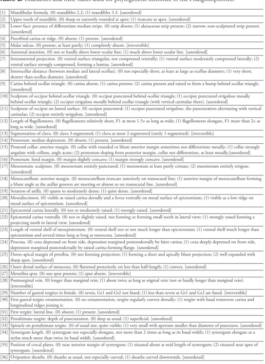
Table 2. Characters and their states used for phylogenetic inference of the Phasgonophorini. [1] Mandibular formula. (0) mandibles 2.2; (1) mandibles 3.3. [unordered]
[2] Upper tooth of mandible. (0) sharp or narrowly rounded at apex; (1) truncate at apex. [unordered]
[3] Lower face: presence of differentiate median stripe. (0) strip absent; (1) alutaceous strip present; (2) narrow, non-sculptured strip present. [unordered]
[4] Preorbital carina or ridge. (0) absent; (1) present. [unordered]
[5] Malar sulcus. (0) present, at least partly; (1) completely absent. [irreversible]
[6] Antennal insertion. (0) not or hardly above lower ocular line; (1) much above lower ocular line. [unordered]
[7] Interantennal projection. (0) ventral surface triangular, not compressed ventrally; (1) ventral surface moderately compressed laterally; (2) ventral surface strongly compressed, forming a lamina. [unordered]
[8] Interocellar distance (between median and lateral ocellus). (0) not especially short, at least as large as ocellus diameter; (1) very short, shorter than ocellus diameter. [unordered]
[9] Carina behind ocellar triangle. (0) carina absent; (1) carina present; (2) carina present and raised to form a bump behind ocellar triangle. [unordered]
[10] Sculpture of occiput behind ocellar triangle. (0) occiput punctured behind ocellar triangle; (1) occiput punctured strigulose mesally behind ocellar triangle; (2) occiput strigulose mesally behind ocellar triangle (with vertical carinulae there). [unordered]
[11] Sculpture of occiput on lateral surface. (0) occiput punctured; (1) occiput punctured strigulose, the puncturation alternating with vertical carinulae; (2) occiput entirely strigulose. [unordered]
[12] Length of flagellomeres. (0) flagellomeres relatively short, F1 at most 1.5× as long as wide; (1) flagellomeres elongate, F1 more than 2× as long as wide. [unordered]
[13] Segmentation of clava. (0) clava 3-segmented; (1) clava at most 2-segmented (rarely 1-segmented). [irreversible] [14] Pronotum: median depression. (0) absent; (1) present. [unordered]
[15] Pronotal collar: anterior margin. (0) collar with rounded or blunt anterior margin sometimes not differentiate mesally; (1) collar strongly angulate with collum, angle acute; (2) pronotum sloping from posterior margin, collar not differentiate, at least mesally.[unordered] [16] Pronotum: hind margin. (0) margin slightly concave; (1) margin strongly concave. [unordered]
[17] Mesonotum: sculpture. (0) mesonotum entirely punctured; (1) mesonotum at least partly cristate; (2) mesonotum entirely strigose. [unordered]
[18] Mesoscutellum: anterior margin. (0) mesoscutellum truncate anteriorly on transscutal line; (1) anterior margin of mesoscutellum forming a blunt angle as the axillar grooves are meeting or almost so on transscutal line. [unordered]
[19] Setation of axilla. (0) sparse to moderately dense; (1) quite dense. [unordered]
[20] Mesodiscrimen. (0) visible as raised carina dorsally and a fovea ventrally on mesal surface of epicnemium; (1) visible as a low ridge on mesal surface of epicnemium. [unordered]
[21] Epicnemial carina laterally. (0) not or moderately raised; (1) strongly raised. [unordered]
[22] Epicnemial carina ventrally. (0) not or slightly raised, not forming or forming small tooth in lateral view; (1) strongly raised forming a projecting tooth in lateral view. [unordered]
[23] Length of ventral shelf of mesepisternum. (0) ventral shelf not or not much longer than epicnemium; (1) ventral shelf much longer than epicnemium and several times long as long as mesocoxa. [unordered]
[24] Procoxa. (0) coxa depressed on front side, depression margined posterodorsally by faint carina; (1) coxa deeply depressed on front side, depression margined posterodorsally by raised carina forming flange. [unordered]
[25] Dorso-apical margin of protibia. (0) not forming projection; (1) forming a short and apically blunt projection; (2) well expanded with sharp apex. [unordered]
[26] Outer dorsal surface of metacoxa. (0) flattened posteriorly, on less than half-length; (1) convex. [unordered] [27] Metatibia spur. (0) one spur present; (1) spur absent. [irreversible]
[28] Postmarginal vein. (0) longer than marginal vein; (1) about twice as long as stigmal vein (not or hardly longer than marginal vein). [irreversible]
[29] Number of gastral tergites in female. (0) seven, Gt1 and Gt2 not fused; (1) less than seven as Gt1 and Gt2 are fused. [irreversible] [30] First gastral tergite ornamentation. (0) no ornamentation, tergite regularly convex dorsally; (1) tergite with basal transverse carina and
longitudinal ridges joining it.
[31] First tergite: lateral line. (0) absent; (1) present. [unordered]
[32] Penultimate tergite: depth of puncturation. (0) deep as usual; (1) superficial. [unordered]
[33] Spiracle on penultimate tergite. (0) of usual size, quite visible; (1) very small with aperture smaller than diameter of punctures. [unordered] [34] Syntergum length. (0) syntergum not especially elongate, not more than 2 times as long as its basal width; (1) syntergum elongate as a
stylus much more than twice its basal width. [unordered]
[35] Position of cercal plates. (0) near anterior margin of syntergum; (1) situated about at mid length of syntergum; (2) situated near apex of syntergum. [unordered]
[36] Ovipositor sheaths. (0) sheaths as usual, not especially curved; (1) sheaths curved downwards. [unordered]
Acronyms for museums and other institutions
Natural History Museum, London, United Kingdom (BMNH); Efflatoun Bey col-lection, Entomology Department, Faculty of Science, Giza, Egypt (EFC); King Saud
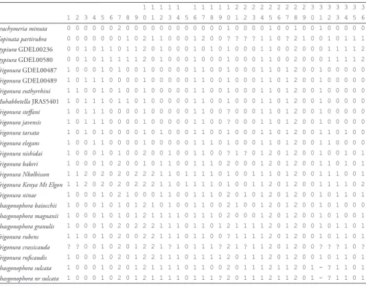
Table 3. Data matrix for the phylogenetic inference of the Phasgonophorini (Chalcididae). 1 1 1 1 1 1 1 1 1 1 2 2 2 2 2 2 2 2 2 3 3 3 3 3 3 3 1 2 3 4 5 6 7 8 9 0 1 2 3 4 5 6 7 8 9 0 1 2 3 4 5 6 7 8 9 0 1 2 3 4 5 6 Brachymeria minuta 0 0 0 0 0 0 2 0 0 0 0 0 0 0 0 0 0 0 0 1 0 0 0 0 1 0 0 1 0 0 1 0 0 0 0 0 Kopinata partirubra 0 0 0 0 0 0 0 1 0 2 1 1 0 0 0 1 2 0 0 ? ? ? ? 1 1 0 ? 2 1 0 0 1 0 1 1 1 Stypiura GDEL00236 0 0 1 0 1 1 0 1 1 2 0 1 0 0 0 1 0 1 0 1 0 0 0 1 1 0 0 2 0 0 0 1 1 1 1 2 Stypiura GDEL00580 0 0 1 0 1 1 1 1 1 2 0 1 0 0 0 1 0 0 0 1 0 0 0 1 1 0 0 2 0 0 0 1 1 1 1 2 Trigonura GDEL00487 1 0 0 0 1 0 1 0 0 1 0 0 0 0 0 1 1 0 0 1 0 0 0 1 1 0 1 2 0 0 1 0 0 0 0 0 Trigonura GDEL00489 1 0 1 1 1 0 0 0 0 1 0 0 0 0 0 1 1 0 0 1 0 0 0 1 1 0 1 2 0 0 1 0 0 0 0 0 Trigonura euthyrrhini 1 1 0 0 1 0 1 0 0 1 0 0 0 0 0 1 1 0 0 1 0 0 0 1 1 0 1 2 0 0 1 0 0 0 0 0 Muhabbetella JRAS5401 1 0 1 1 1 0 1 1 0 1 0 0 0 0 0 1 1 0 0 1 0 0 0 1 1 0 1 2 0 0 1 0 0 0 0 0 Trigonura steffani 1 0 1 1 1 0 0 0 0 1 0 0 0 0 0 1 1 0 0 ? 0 0 0 1 1 0 1 2 0 0 1 0 0 0 0 0 Trigonura javensis 1 0 1 1 1 0 0 0 0 1 0 0 0 0 0 1 1 0 0 ? 0 0 0 1 1 0 1 2 0 0 1 0 0 0 0 0 Trigonura tarsata 1 0 1 0 1 0 0 0 0 1 0 1 0 0 0 1 1 0 0 1 0 0 0 1 1 0 1 2 0 0 1 1 0 1 0 0 Trigonura elegans 1 0 0 1 1 0 0 0 0 1 0 0 0 0 0 1 1 1 0 1 0 0 0 1 1 0 1 2 0 0 1 1 0 0 0 0 Trigonura nishidai 1 0 0 0 1 0 1 0 0 2 0 0 1 0 0 1 1 0 0 ? 1 ? 0 1 2 0 1 2 0 0 1 0 0 1 0 1 Trigonura bakeri 1 0 0 0 1 0 2 0 0 1 0 1 1 0 0 1 1 1 0 2 0 0 0 1 2 0 1 2 0 0 1 1 0 1 0 1 Trigonura Nkolbisson 1 1 2 0 2 0 2 0 2 2 2 1 1 0 1 1 1 1 0 1 0 0 1 1 1 0 1 2 0 0 1 1 1 0 0 1
Trigonura Kenya Mt Elgon 1 1 2 0 2 0 2 0 2 2 2 1 1 0 1 1 1 1 0 1 0 0 1 1 2 0 1 2 0 0 1 1 1 1 0 2 Trigonura ninae 1 0 0 0 1 0 2 1 0 0 0 1 1 0 0 1 1 1 0 2 0 1 0 1 2 0 1 2 0 0 1 0 1 1 0 1 Phasgonophora baiocchii 1 0 0 0 1 0 1 0 1 2 1 0 1 0 0 1 1 0 0 2 1 0 0 1 2 0 1 2 0 0 1 0 1 0 0 0 Phasgonophora magnanii 1 0 0 0 1 0 1 0 1 2 1 1 1 1 0 1 1 1 0 2 0 0 0 1 2 0 1 2 0 0 1 0 1 0 0 1 Phasgonophora granulis 1 0 0 0 1 0 2 0 2 2 2 1 1 1 0 1 1 0 1 2 1 1 1 1 2 0 1 2 0 0 1 0 1 1 0 1 Trigonura rubens 1 1 0 0 1 0 2 0 0 2 2 1 1 1 0 1 1 0 0 ? 1 1 1 1 2 0 1 2 0 0 1 0 1 1 0 1 Trigonura crassicauda ? ? 0 0 1 0 2 0 1 2 2 1 ? 1 0 1 1 1 ? 2 1 ? 1 1 2 0 1 2 0 0 ? ? ? 1 0 ? Trigonura ruficaudis 1 0 0 0 1 0 2 0 1 2 2 1 1 1 0 1 1 1 1 2 0 1 1 1 2 0 1 2 0 0 1 0 1 1 0 1 Phasgonophora sulcata 1 0 0 0 1 0 2 0 1 2 1 1 1 1 0 1 1 0 0 2 0 1 1 1 2 1 1 2 0 1 - ? 1 1 0 1 Phasgonophora nr sulcata 1 0 0 0 1 0 2 0 1 2 1 1 1 1 0 1 1 1 ? 2 0 1 1 1 2 1 1 2 0 1 - ? 1 1 0 1
University for Arthropods, Plant Protection Department, College of Food and Agri-culture Sciences, King Saud University, Riyadh, Saudi Arabia (KSMA); Museum für Naturkunde, Berlin, Germany (MNB); National Museum of Natural History, Smith-sonian Institution, Washington, United States of America (USNM).
Abbreviations
F1−F3 = first to third funicular segments; Gt = gastral tergite; MGV = marginal vein of fore wing; OOL = distance between lateral ocelli and inner eye margin; PMV = postmarginal vein; POL = distance between lateral ocelli; Rs = radial sector; r-m = radio-medial cross vein; SMV = submarginal vein of fore wing; STV = stigmal vein.
Results
Phylogeny of Phasgonophorini
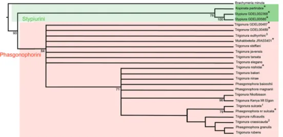
The initial analysis provided 33 equally parsimonious trees with a length of 88 steps, and values of 0.489, 0.831 and 0.406 respectively for the consistency (CI),
reten-Figure 1. Strict consensus tree of the Phasgonophorini achieved from phylogenetic inference using
par-simony. Bootstrap support below nodes. A, B, C denote the supported clades; * denote specimens used for the phylogenetic study using the Ultra Conserved Elements (Cruaud et al. 2020); 1, type species of
Phasgonophora Westwood; 2, type species of Trigonura Sichel; 3, type species of Chalcidellia Girault.
Figure 2. Preferred tree of the Phasgonophorini from phylogenetic inference using parsimony after
suc-cessive weighting. Legend identical with Fig. 1. Black rectangles denote synapomorphies, white rectangles homoplastic derived states and dark triangles putative reversals.
tion (RI), and rescaled consistency (RC) indices. Stypiurini and Phasgonophorini are retrieved monophyletic with moderately strong supports (respectively 78, 64 for the bootstrap values) in the strict consensus tree (CST) (Fig. 1). Regarding Phasgonophorini, the tree shows a basal polytomy, including seven species, identi-fied as Trigonura and one as Muhabbetella when using the traditional classification. The tree then shows a basal clade (Fig. 2, clade B), itself with a polytomy including seven species. Two of the newly described species from Saudi Arabia are retrieved here; all species in clade B would be assigned to Trigonura with the available keys (Bouček 1988; Narendran 1989). Finally, a second, terminal clade (Fig. 2, clade C) especially includes P. sulcata and T. crassicauda – the type species of Phasgonophora and Trigonura – together with P. granulis, one of the new described species from Saudi Arabia. This clade would otherwise comprise most of the species presently identified as Trigonura.
The successive weighing procedure provided three trees with length of 34.232, CI, RI, and RC, respectively of 0.665, 0.933 and 0.658. Ordering versus non-ordering characters do not change the topology. The preferred tree is presented as the Fig. 2. It is entirely congruent, for the appropriate species, with the tree achieved when using the UCE (Cruaud et al. 2020). Here the polytomies observed in the CST are solved, but the corresponding nodes do not have any support as they are sustained by a few derived, mostly homoplastic character states. Again P. granulis appears as sister to T. rubens, while P. magnanii as sister to clade C, a relationship sustained by the presence of a median depression on the pronotum. P. baiocchii merges on a node just basal to P. magnanii, but its position varies when the characters are equally weighted. Thus, it is sometimes sister to P. magnanii, sister to T. nishidai Narendran, 1989 or even merges on a basal node within the clade C, hence sister to the other species of the clade B taken together.
Phasgonophora Westwood, 1832
Phasgonophora Westwood, 1832: 432 (fig. 77). Type species: Phasgonophora sulcata Westwood, 1832, by monotypy.
= Phasganophora [sic] subg. Trigonura Sichel, 1866: 358−376. Type species: Phasgan-ophora (Trigonura) crassicauda Sichel, 1866, by monotypy, syn. nov.
= Trigonura Kirby, 1883: 54, 59–60 (raised to genus level), syn. nov.
= Bactrochalcis Kieffer, 1912: 463. Type species: Bactrochalcis reticulata Kieffer, by monotypy. Synonymized with Trigonura by Steffan, 1951: 147, syn. nov.
= Centrochalcis Cameron, 1913: 92. Type species: Centrochalcis ruficaudis Cameron, by monotypy. Synonymized with Trigonura by Waterston, 1922: 10, syn. nov. = Centrochalcidea Gahan & Fagan, 1923. Replacement name for Centrochalcis
Cam-eron, 1913, nec CamCam-eron, 1905, syn. nov.
= Chalcidellia Girault, 1924: 3. Type species: Chalcis euthyrrhini Dodd, 1921, by origi-nal designation. Synonymized with Trigonura by Bouček, 1988: 63−64, syn. nov.
= Urochalcis Nikol’skaya, 1952: 91. Type species: Urochalcis ninae Nikol’skaya, 1952, by original designation. Synonymized with Trigonura by Nikol’skaya, 1960: 90, syn. nov.
= Trigonurella Bouček, 1988: 64. Type species: Trigonurella elegans Bouček, 1988, by original designation, syn. nov.
= Muhabbetella Koçak & Kemal, 2008: 3. Replacement name for Trigonurella Bouček, 1988 nec Trigonurella Maa, 1963, syn. nov.
The above synonymies are just the taxonomic implications resulting from phylogenetic inference using UCE (Cruaud et al. 2020) and the present study from morphological data. In these two studies, Trigonura appears paraphyletic relative to Phasgonophora and Muhebbetella. In addition, the type species of Phasgonophora and Trigonura are included in supported clade B (values of the bootstrap 100 and 71 respectively for the UCE and the morphology tree) (Figs 1, 2). Thus, Trigonura cannot be sustained anymore and the species belonging to the clade B must be classified in Phasgonophora which may just be considered as a derived form of Trigonura. It would have been pos-sible to classify the species belonging to the clade A in another genus and Chalcidellia Girault is available for that. Bouček (1988) was confronted to the same dilemma and wrote the following: ‘‘For some time I thought that Chalcidellia could be retained as a subgenus of Trigonura, because its type species [Chalcis euthyrrhini Dodd] differs from the typical Trigonura in having a distinct flat and punctured interantennal space and in the female the epipygium [syntergum] is short, with cercal tubercles placed right at the beginning of the sculptured part. The antennae are relatively short and very slightly thickened apically. On the contrary, the type species of Trigonura and many other tropical species have the interantennal space narrow, the female epipygium is prolonged with cercal tubercles removed distinctly from base, and the antennae long and filiform or even tapering apically. However, more recently I found species combin-ing these features in varycombin-ing degrees, which makes me regard Chalcidellia as only a spe-cies group of Trigonura”. The distribution of character states within the morphological matrix just confirm Bouček’s observations (Tables 2, 3). His opinion is shared here and is reinforced by the fact that the clade A is not at all supported and forms a polytomy in the strict consensus tree (Fig. 1).
Taxonomic study of Saudi Arabian species
Key to the species of Phasgonophora Westwood from the Arabian Peninsula (based on females)
1 Gaster shortly acute (Figs 5A, 12A). Syntergum short, about 1.1−1.4× as long as wide when seen from dorsal view (Fig. 5A) ...2 – Gaster lanceolate (Figs 15C, 16A, B). Syntergum evidently longer, 2.5−4.0×
2 Body entirely red (Figs 3A−C, E, 5A). Fore wing with setae white, sparse and short (Fig. 4C). Propodeum without spiracular teeth (Fig. 4A). Pronotal col-lar without median depression, regucol-larly convex (Fig. 4B). Mesoscutellum truncate anteriorly (Fig. 3E). Pedicel about 1.8 × as long as wide (Fig. 3D). Anellus hardly transverse, about 0.8× as long as wide (Fig. 3D) ... ... P. baiocchii Soliman & Gul, sp. nov. – Meso- and metasoma mostly black (Figs 10E, 11A, 12A). Fore wing with dark
setae (Fig. 11B). Propodeum with sharp spiracular teeth (Fig. 10E, F). Prono-tal collar with evident median depression (Fig. 10E). Mesoscutellum bluntly angulate anteriorly (Fig. 10E). Pedicel as long as wide (Fig. 10D). Anellus quite transverse, about 0.45× as long as wide (Fig. 10D) ... ...P. magnanii Gadallah & Gul, sp. nov. 3 Propodeum without spiracular teeth (Fig. 16B). Pronotum sloping from pos-terior margin mesally, uniformly convex (Fig. 16B, C). Mesoscutellum con-vex, and bluntly angulate anteriorly as axillar grooves are joining to each other on transscutal line (Fig. 16B) ... P. ninae (Nikol’skaya) – Propodeum with sharp spiracular teeth (Fig. 7E). Pronotum with collar sepa-rated by evident angulation from collum (Figs 7E, 8A, 14A) ...4 4 Body 7.0–9.6 mm in length. Gena sparsely punctured (Fig. 14A, B). Pronotum
with shallow median depression (Fig. 14D). Pronotal collar and mesonotum clearly cristate (transverse crests) (Fig. 14A), not at all punctured. Setation of axilla not especially dense, not masking integument beneath (Fig. 14D). Pro-podeum strongly sloping posteriorly, almost vertical. Epicnemial carina moder-ately raised ventrally. Fore wing without pigmented tracks of Rs and r-m (Fig. 15B). Gt1 with curved carinulae dorsally, sparsely setose laterally (Fig. 15C) .... ...P. rubens (Klug) – Body 9.5–13.6 mm in length. Gena densely punctured (Figs 7D, 8A). Prono-tal collar with evident median depression (Fig. 7E). PronoProno-tal collar and me-sonotum cristate-punctured, the anterior wall of punctures raised (Fig. 8A). Axillae densely setose, setation masking integument beneath (Fig. 7E). Pro-podeum less strongly sloping than in alternate. Epicnemial carina strongly raised ventrally, forming sharp tooth mesally (Fig. 8A). Fore wing with evi-dent pigmented tracks of Rs and r-m (Fig. 8D); Gt1 with superficial, irregular wrinkles, densely setose laterally (Fig. 9A) ...P. granulis Delvare, sp. nov.
Review of Phasgonophora species from Saudi Arabia
Table 4 represents the absolute measurements of the female holotypes and male paratypes. Selected ratios are quoted in Tables 5−7. They are not repeated in the following descriptions.
Phasgonophora baiocchii Soliman & Gul, sp. nov.
http://zoobank.org/75C97023-EFBA-437E-A031-23A13760231B
Figs 3A−E, 4A−E, 5A−D, 6A−C
Type material. Holotype ♀: Kingdom of Saudi Arabia, Riyadh, Ad Diriyah, Al Uyaynah, Wadi Al Hesiyah (40 NW of Riyadh) [24°55'22.44"N, 46°12'15.13"E, Alt. 790 m], 8.IV.2017, reared from Anthaxia sp. (Buprestidae), e.l. Acacia, leg. D. Baioc-chi [KSMA]; Paratype 1♂, same data as for holotype [KSMA].
Diagnosis. Body mostly red; fore wing hyaline with white setation (Fig. 4C); seta-tion on body and wings sparse and short (Figs 3A−C, E, 4C); flagellomeres
moder-Table 4. Measurements of the types of the described species of Phasgonophora (in µm). Character Phasgonophora
baiocchii holotype ♀ granulisPhasgonophora holotype ♀ magnaniiPhasgonophora holotype ♀ baiocchiPhasgonophora paratype ♂ magnaniiPhasgonophora paratype ♂
head width 1537 1705 1897 1276 1821
head maximal length 784 989 1038 691 1054
head length on median
line 511 608 654 447 717
eye length 532 648 737 455 690
temple length 121 182 64 138 163
frontovertex width 774 926 1026 740 989
distance between lateral
ocelli 263 455 353 289 370
ocular – lateral ocellus
distance 137 74 186 122 168
diameter of lateral
ocellus 158 182 167 122 152
distance between
median and lateral ocelli 95 142 109 102 98
head height 1058 1269 1477 1079 1284
eye height 571 744 781 584 798
distance lower edge antennal torulus – ventral margin of clypeus (ATC)
314 481 500 317 –
distance lower edge antennal torulus – lower edge of median ocellus (ATOM)
538 603 719 455 –
length of malar space 455 513 781 396 –
width of oral fossa 551 679 781 505 –
scape length 570 730 815 444 – pedicel length 127 131 109 98 110 pedicel width 80 108 125 76 106 anellus length 59 59 62 33 37 anellus width 75 98 125 79 102 2nd flagellomere (= F1) length 159 280 308 139 301 2nd flagellomere width 102 127 161 133 163 8th flagellomere (= F7) length 110 172 232 136 272 8th flagellomere width 112 105 151 133 159 clava length 310 292 446 234 472
Table 5. Calculated ratios for the females of Phasgonophora from measurements of Table 4.
Ratio Phasgonophora baiochii
holotype ♀ Phasgonophora granulisholotype ♀ magnaniiPhasgonophora holotype ♀
head width : head maximal length 1.960 1.724 1.827
head width : head length on median line 3.010 2.804 2.902
head width : head height 1.453 1.343 1.285
fronto-vertex width : eye height 1.356 1.245 1.313
ocular – lateral ocellus distance : diameter of lateral ocellus 0.520 0.406 1.115 distance between median and lateral ocelli : diameter of
lateral ocellus 0.600 0.781 0.654
ATC : ATOM 0.583 0.798 0.696
length of malar space : eye height 0.798 0.690 1.000
length of malar space : width of oral fossa 0.826 0.755 1.000
scape length : eye height 1.000 0.981 1.043
pedicel length : pedicel width 1.585 1.218 0.873
anellus length : anellus width 0.789 0.600 0.492
F1 length : F1 width 1.558 2.200 1.914
F7 length : F7 width 0.982 1.638 1.539
mesosoma length : mesosoma (= mesoscutum) width 1.600 1.640 1.612
mesosoma length : mesosoma height 1.538 2.335 2.265
pronotum width : pronotum maximal length 1.707 1.714 1.849 pronotum width : pronotum length on median line 3.559 2.754 2.688
pronotum width : mesoscutum width 1.077 0.977 1.042
mesoscutum length : pronotum length on median line 1.729 1.475 1.375 mesoscutellum length : mesoscutellum width 0.898 0.810 0.978
fore wing length : fore wing width 2.858 2.690 2.263
marginal vein length : costal cell length 0.348 0.269 0.284 marginal vein length : stigmal vein length 3.512 3.016 3.643 marginal vein length : postmarginal vein length 4.800 2.603 4.857
metacoxa length : metacoxa width 2.153 1.747 2.000
metafemur length : metafemur width 1.764 1.774 1.684
syntergum length : mesotibia length 0.276 1.226 0.546
ately long (Fig. 3D); clava 1-segmented in both sexes (Figs 3D, 6B); mesoscutellum moderately convex, truncate anteriorly ((Fig. 4A); propodeal spiracular tooth absent (Figs 4A, 6C); fore wing setation sparse and very short, distributed on both sides with-out line of setae on Rs (Fig. 4C); Gt1 dorsally smooth (Fig. 5A); syntergum 0.276× as long as mesotibia (Fig. 5A, B).
Etymology. This species is dedicated to Daniele Baiocchi, who reared this species from Anthaxia spp. (Buprestidae) infesting Acacia sp. (Fabaceae).
Condition of holotype. Specimen glued on rectangular card, metasoma glued sep-arately. Head and mesosoma partly covered with a thin artifactual layer in bottom of areoles, appearing artificially dull rather than glossy by places; second to fifth terga with sides wide apart from each other, probably resulting from immersion in some medium.
Description of holotype ♀: Body length 5.0 mm. Colour. Body reddish brown; antennal scape and pedicel, anellus and basal half of F1 reddish (Fig. 3D), the rest of flagellum dark brown, almost black (Fig. 3D); mandibular teeth black (Fig. 3B); palpi brown; mesoscutellum apically and metanotum dark (Fig. 3E); wings hyaline (Fig. 4C), SMV testaceous, MGV, STV and PMV dark brown (Fig. 4C); tegula brownish; all legs
Table 6. Calculated ratios for the males of Phasgonophora from measurements of Table 4.
Ratio Phasgonophora baiocchii paratype ♂ Phasgonophora magnanii paratype ♂
head width : head maximal length 1.847 1.727
head width : head length on median line 2.855 2.538
head width : head height 1.183 1.418
fronto–vertex width : eye height 1.267 1.240
ocular – lateral ocellus distance : diameter of lateral ocellus 1.000 1.107 distance between median and lateral ocelli : diameter of
lateral ocellus 0.833 0.643
ATC : ATOM 0.696 –
length of malar space : eye height 0.678 –
length of malar space : width of oral fossa 0.784 –
scape length : eye height 0.759 –
pedicel length : pedicel width 1.286 1.038
anellus length : anellus width 0.414 –
F1 length : F1 width 1.041 1.850
F7 length : F7 width 1.020 1.718
mesosoma length : mesosoma (= mesoscutum) width 1.671 1.781
mesosoma length : mesosoma height 1.521 1.605
pronotum width : pronotum maximal length 3.296 4.064 pronotum width : pronotum length on median line 1.047 1.130
pronotum width : mesoscutum width 1.556 2.574
mesoscutum length : pronotum length on median line 0.878 1.000 mesoscutellum length : mesoscutellum width 2.650 2.892
fore wing length : fore wing width 0.307 0.367
marginal vein length : costal cell length 2.629 3.143 marginal vein length : stigmal vein length 4.182 2.973 marginal vein length : postmarginal vein length 2.154 1.901
metacoxa length : metacoxa width 1.875 1.784
reddish, but tarsi testaceous; metafemur with black teeth on ventral margin (Fig. 4E); metasoma reddish brown (Fig. 5A, B), tip of ovipositor sheaths black (Fig. 5B).
Head (Fig. 3A−C). Slightly wider than maximal width of mesosoma; with sparse, short and thin setae; vertex and frons densely punctured (Fig. 3A, B), lower face and especially gena sparsely punctured, with interspaces as large as punctures on its mesal surface; lower face and frons strongly convex, without preorbital ridges (Fig. 3B); both mandibles 3-toothed, teeth of same length, somewhat blunt at apex (Fig. 3B); clypeus roundly protruding at free margin (Fig. 3B); tentorial pits well visible (Fig. 3B); genal carina strongly raised (Fig. 3C); scrobal cavity completely transversely strigose, reaching median ocellus (Fig. 3B); lateral margins of depression slightly converging dorsally; in-terantennal projection as wide as diameter of antennal torulus, subtriangular, and with punctulate front surface, with sharp carina above it, 0.33× as long as scape (Fig. 3B); occiput vertically strigulose behind ocellar triangle, punctured laterally (Fig. 3A).
Antenna (Fig. 3D). Apex of scape reaching level of median ocellus; pedicel 1.58× as long as wide, without basal bottle neck; anellus hardly transverse, tapering basally; flagellomeres pubescent, bearing numerous, not raised, multiporous plate sensilla in several intricate rows; F1 somewhat tapering basally, 1.59× as long as wide, slightly longer than each of F2 and F3; clava 1-segmented, conical, not much longer than F7 and very narrowly truncate at apex.
Table 7. Comparison between the sexes of P. baiocchii sp. nov. and P. magnanii through ratios calculated
from measurements of Table 4.
Ratio Phasgonophora
baiocchii holotype ♀ baiocchiiPhasgonophora paratype ♂ magnanii Phasgonophora holotype ♀ magnaniiPhasgonophora paratype ♂ head width : head maximal length 1.960 1.847 1.827 1.727 head width : head length on median line 3.010 2.855 2.902 2.538
head width : head height 1.453 1.183 1.285 1.418
fronto–vertex width : eye height 1.356 1.267 1.313 1.240 ocular – lateral ocellus distance : diameter of
lateral ocellus 0.520 1.000 1.115 1.107
distance between median and lateral ocelli :
diameter of lateral ocellus 0.600 0.833 0.654 0.643
ATC : ATOM 0.583 0.696 0.696 –
length of malar space : eye height 0.798 0.678 1.000 – length of malar space : width of oral fossa 0.826 0.784 1.000 –
scape length : eye height 1.000 0.759 1.043 –
pedicel length : pedicel width 1.585 1.286 0.873 1.038 anellus length : anellus width 0.789 0.414 0.492 0.360
F1 length : F1 width 1.558 1.041 1.914 1.850
F7 length : F7 width 0.982 1.020 1.539 1.718
mesosoma length : mesosoma (=
mesoscutum) width 1.600 1.671 1.612 1.781
mesosoma length : mesosoma height 1.538 1.521 2.265 –
pronotum width : pronotum maximal length 1.707 3.296 1.849 1.605 pronotum width : pronotum length on
median line 3.559 1.047 2.688 4.064
pronotum width : mesoscutum width 1.077 1.556 1.042 1.130 mesoscutum length : pronotum length on
median line 1.729 0.878 1.375 2.574
mesoscutellum length : mesoscutellum width 0.898 2.650 0.978 1.000 fore wing length : fore wing width 2.858 0.307 2.263 2.892 marginal vein length : costal cell length 0.348 2.629 0.284 0.367 marginal vein length : stigmal vein length 3.512 4.182 3.643 3.143 marginal vein length : postmarginal vein
length 4.800 2.154 4.857 2.973
metacoxa length : metacoxa width 2.153 1.875 2.000 1.901 metafemur length : metafemur width 1.764 – 1.684 1.784
syntergum length : mesotibia length 0.276 – 0.546 –
Mesosoma (Figs 3E, 4A, B). Slightly convex in lateral view (Fig. 4B), pronotum and mesonotum bearing short thin setae, adpressed on pronotum and suberect on me-sonotum (Fig. 3E); pronotum entirely punctured, its dorsal outline regularly convex, without median depression (Fig. 3E); lateral panel with oblique crenulae ventrally; mesonotum cristate-punctured, the transverse crests moderately raised (Fig. 4B); no-tauli not much impressed (Fig. 3E); tegula bearing three very short setae basally; mes-oscutellum short, convex in lateral view (Fig. 4B), truncate anteriorly as the axillae are widely separated, broadly rounded at apex, with fine longitudinal carinae; postscutel-lum as trapezoidal areola with secondary sculpture (Fig. 3E); propodeum not much sloping, without anterolateral spiracular tooth (Figs 3E, 4A), with irregular costula and poorly delimited median areola; mesepisternum with mesodiscrimen as faint carina dorsally, bifurcate ventrally delimiting a shallow fovea (Fig. 4A); epicnemial carina strongly raised at mid-height, moderately raised ventrally (Fig. 4B); ventral shelf
virtu-Figure 3. A−E Phasgonophora baiocchii Soliman & Gul, sp. nov., female (holotype) A head (dorsal view) B head (frontal view) C head (lateral view) D antenna E mesosoma (dorsal view).
ally smooth; adscrobal area of mesepisternum, entire mesepimeron and metepimeron with dense setiferous punctures, the setae are short and adpressed as on pronotum (Fig. 4B); femoral scrobe of mesopleuron entirely strigose (Fig. 4B).
Wings (Fig. 4C). Fore wing lacking marginal fringe, with microtrichiae on both sides, MGV 0.35× as long as SMV, PMV 0.20× as long as MGV, STV slightly longer than PMV; hind wing with three similar closely set hamuli.
Legs (Fig. 4D, E). Procoxa deeply depressed anteriorly, the depression delimited laterodorsally by strongly raised carina (Fig. 4D). Protibia with thin apicodorsal sock-eted spine (Fig. 4D). Mesotibia without dorsal pegs. Hind leg bearing sparse, thin and suberect setae on ventral side of coxa, femur and tibia (Fig. 4E); metafemur sparsely
Figure 4. A−E Phasgonophora baiocchii Soliman & Gul, sp. nov., female (holotype) A mesosoma (part,
dorsal view) B mesosoma (lateral view) C fore wing D proleg E hind leg.
punctulate on outer side, its ventral margin with a row of 11 regularly distributed equal teeth, basal tooth not prominent, no basal inner tooth (Fig. 4E). All tarsi thin, bearing slender claws.
Metasoma (Fig. 5A, B). Petiole quite transverse in dorsal view, ventral surface virtually smooth. Gaster short, only slightly longer than mesosoma; Gt1 2.6× as wide as long, as long as Gt2 and Gt3 combined, smooth on disc, solely with a row of three thin and short setae on either side (Fig. 5A); Gt2–5 smooth, except for the setiferous punctures in front of their posterior margin, laterally with a complete row of setae and a partial row in front of it (Fig. 5A); penultimate tergite entirely densely and deeply
Figure 5. A−D Phasgonophora baiocchii Soliman & Gul, sp. nov. A, B female (holotype): A metasoma
(dorsal view) B syntergum (lateral view) C, D male (paratype): C head (dorsal view) D head (frontal view). punctured, with three rows of setiferous punctures, spaces between punctures smooth and shiny; spiracle very small, hardly visible at lateral edge of punctured surface as its peritreme is not raised; syntergum very short, only 0.276× as long as mesotibia, its basal part, in front of cercal plates (Fig. 5B); extremely short, median ridge present; tergum coarsely punctured laterally; sternites smooth and bare; tip of hypopygium at about half length of gaster.
Male (Figs 5C, D, 6A−C). Length 4.2 mm; similar to female except for the fol-lowing characters: black parts better expanded especially on occiput, pronotum and mesonotum; scape bright reddish brown (Fig. 6B); head less transverse in dorsal view with anterior outline of frons more convex and temples relatively longer (Fig. 5C), gena mostly smooth, with very sparse punctures (Fig. 6A); frons with faint preorbital ridges, carina above interantennal projection almost reaching dorsal margin of scrobal depression (Fig. 5D); scape fusiform, 3.4 × as long as wide, anellus transverse, strongly tapering basally (Fig. 6B).
Recognition. None of the described Phasgonophora from the Afrotropical region have the short syntergum exhibited by P. baoicchii. Considering the Oriental species, the species would run, using Narendran (1989), either to Trigonura steffani Narendran, or T. javensis Narendran, 1987. The first species (holotype examined) is quite different, especially the deeply impressed notauli and the strongly convex mesoscutellum. In the second species (holotype examined), the lower face has a differentiate median strip similar to that of Muhattebella, and fore wing bears dark setae among other characters.
Figure 6. A−C Phasgonophora baiocchii Soliman & Gul, sp. nov., male (paratype) A head (fronto-lateral
view) B antenna C head and mesosoma (dorsal view).
From this species group, especially in P. euthyrrhinii, the type species of Chalcidiella, P. baiocchii differs from all species examined by the non-segmented clava, the meso-discrimen, not raised as median crest, and the white setae of the fore wing versus clava 3-segmented, mesodiscrimen raised as a carina dorsally and fore wing setation dark.
Distribution. Only known from Saudi Arabia, in Riyadh Region (Fig. 17).
Host. Anthaxia (Haplanthaxia) abdita Bílý, 1982 and A. (H.) kneuckeri ssp. zabran-skyi Bílý, 1995 (Coleoptera, Buprestidae).
Phasgonophora granulis Delvare, sp. nov.
http://zoobank.org/5EB72879-1E9C-4A89-BCD8-FF37534B7172
Figs 7A−E, 8A−D, 9A, B
Type material. Holotype ♀: Kingdom of Saudi Arabia, Al-Baha, Al Mikhwa (Sha-da Al-Ala Natural Reserve) [19°50'51"N, 41°18'06.12"E, Alt. 1358 m], 14.IV.2016,
e.l. Acacia, leg. D. Baiocchi [KSMA]. Paratypes: 7♀, same data as holotype [KSMA]; 2♀, same data as holotype [BMNH]; 2♀, same data as holotype [USNM]; 3♀, same data as holotype [EFC]; 2♀, same data as holotype but differing as for the coordi-nates [19°51'39.96"N, 41°18'15.84"E, Alt. 1248 m] and collection date, 29.III.2017 [KSMA]; 2♀, Kingdom of Saudi Arabia, Asir, Muhayil, Wadi Sabian (28 km SSE of Muhayil) [18°17'54.89"N, 42°07'41.11"E, Alt. 809 m], 05.IV.2017, e.l. Acacia, leg. D. Baiocchi [KSMA].
Diagnosis. Gaster longer than mesosoma and acuminate, with syntergum longer than mesotibia (1.15×) (Fig. 9A, B); gena densely and entirely punctured (Figs 7D, 8A); occiput completely strigulose (Fig. 7A); flagellum filiform, with all flagellomeres much longer than wide, F1 2.5× as long as wide (Fig. 7C); mesosomal dorsum some-what flattened (Fig. 8A); pronotal collum and mesonotum cristate punctured (Fig. 8A); axillae densely setose, setation masking integument posteriorly (Fig. 7E); propodeum with sharp spiracular teeth (Fig. 7E); mesepisternum with epicnemial carina forming sharp tooth mesoventrally (Fig. 8A); fore wing with dense but short setation, and pig-mented track of Rs and r-m (Fig. 8D); Gt1 with weak wrinkles dorsally (Fig. 9A); Gt6 with deep punctures and very small, hardly visible, spiracle; cerci removed from base of syntergum, situated at half of its length (Fig. 9A, B).
Etymology. The name is chosen in reference to the secondary sculpture of the areoles on the head and mesonotum, giving to them a dull, granulose appearance (see Fig. 8B).
Condition of holotype. Specimen glued on rectangular card. Head and mesosoma partly covered with a thin layer on the bottom of areoles; second to fifth tergites with sides wide apart from each other, probably resulting from immersion in some medium.
Description of female holotype. Body 8.4 mm. Colour. Head and mesosoma entirely black (Fig. 7A−E), metasoma brown (Fig. 9A), with syntergum darker laterally (Fig. 9B); tegula brownish (Fig. 8A); fore and mid legs dark brown, but knees, apex of tibiae and tarsi testaceous; hind leg dark brown (Fig. 8C), ventral femoral teeth and ventral side of tibia black (Fig. 8C), tarsus lighter; antenna entirely black (Fig. 7C); wings hyaline, veins dark brown (Fig. 8D).
Head (Figs 7A, B, D, 8A). Hardly wider than mesosoma; with moderately dense setation, the setae long, thin and suberect, regularly distributed according to punc-tures; lower face and frons strongly convex, without preorbital ridges (Fig. 7D); vertex, frons and lower face densely punctured (Fig. 7B, D), gena more coarsely punctured (Fig. 8A); both mandibles 3-teethed (Fig. 7D), lower tooth the largest and somewhat removed from the mid one; clypeus hardly protruding at free margin (Fig. 7D); edge of oral fossa thickened (Fig. 7D); tentorial pits absent (Fig. 7B); scrobal depression en-tirely transversely strigose, reaching median ocellus (Fig. 7B); interantennal projection (Fig. 7B) strongly compressed laterally, narrower than antennal torulus, punctulate on front surface (one row of punctures only), narrowly produced upwards, but without flange above it; vertex with short but distinct carina behind median ocellus (Fig. 7A); POL 6× OOL (Fig. 7A); occiput entirely strigulose, except for a row of punctures be-hind posterior edge of eye (Fig. 7A).
Antenna (Fig. 7C). Scape linear, its apex with level of vertex; pedicel 1.2× as long as wide, with slight basal bottle neck; anellus slightly transverse, 0.8× as long as wide,
Figure 7. A−E Phasgonophora granulis Delvare, sp. nov., female (holotype) A head (dorsal view) B head
(frontal view) C antenna D head (frontolateral view) E mesosoma (dorsal view).
tapering basally; funicular segments pubescent, bearing numerous, not raised multi-porous plate sensilla in several intricate rows; F1 2.2× as long as wide, shorter than F2; F2 as long as F3; F7 1.64× as long as wide; clava 2-segmented (suture nevertheless hardly distinct), narrowly rounded apically.
Mesosoma (Figs 7E, 8A). With setae about twice as long puncture diameter, curved and suberect; setae regularly distributed on punctures, but pronotum in front of prep-ectus, axillae and propodeum laterally around the spiracle, densely setose, the setae ad-pressed there; dorsum of mesosoma somewhat flattened (Fig. 8A), with dorsal outline of pronotal collar and mesonotum straight; punctures with secondary, very fine, sculpture on their bottom (visible only at very high magnification: 800×) (as in Fig. 8B), thus appearing dull; pronotal collum transversely strigose (Fig. 7E); pronotal collar and
mes-Figure 8. A−D Phasgonophora granulis Delvare, sp. nov., female (holotype) A head and mesosoma
Figure 9. A, B Phasgonophora granulis Delvare, sp. nov., female (holotype) A metasoma (dorsal view) B syntergum (lateral view).
onotum uniformly cristate punctured, the anterior wall of punctures forming crests (Fig. 8A); pronotal collar with shallow mesal depression, its sides strongly convex (Fig. 8A); pronotal carina visible laterally, forming a tooth in dorsal view; lateral panel mostly flat,
with longitudinal carinulae dorsally and raised curved carina ventrally; notauli impressed (Fig. 7E); tegula with a tuft of about 10 setae anteriorly (Fig. 8A); mesoscutellum trun-cate anteriorly, rounded apically, its posterior margin raised and surpassing postscutel-lum (Fig. 7E); propodeum strongly sloping anteriorly, more strongly so posteriorly, with sharp spiracular tooth (Fig. 7E) and two irregular costulae; mesepisternum with meso-discrimen as faint carina all over, without ventral fovea; epicnemial carina moderately raised laterally, strongly protruding mesoventrally, appearing as a sharp tooth in lateral view (Fig. 8A); ventral shelf in mesepisternum very weakly sculptured; adscrobal area, mesepimeron, and metepimeron coarsely areolate, the later bearing long setae; femoral depression of mesepisternum with only a few low carinae (Fig. 8A).
Wings (Fig. 8D). Fore wing densely setose but bare on basal cell, basal and cubital folds, marginal cell with a single, incomplete row of setae on the underside; setae gen-erally very short on the disc of the wing, somewhat longer below MGV, PMV and Rs track; setae uniformly short on the underside of wing; MGV 0.27× as long as SMV; PMV 0.38× as long as MGV; STV 0.33× as long as PMV; hind wing with 4 hamuli, the basal one the largest, removed from the followings.
Legs (Fig. 8C). Procoxa with deep depression anteriorly, margined dorsolaterally with carina raised into flange. Protibia with sharp, non-socketed apical spine. Mesotib-ia without dorsal pegs. Metacoxa sparsely punctured ventrally, densely so on outer sur-face of metafemur, with dense and fine setiferous punctures, ventral edge with irregular row of unequal teeth, outer ventral margin with a row of 8−10 teeth, basal tooth not prominent but wider than other teeth; inner basal tooth absent; apical truncation of metatibia forming a curved spine. Tarsi slender.
Metasoma (Fig. 9A, B). Petiole not visible dorsally. Gaster lanceolate, longer than mesosoma; Gt1 with weak wrinkles dorsally, setose laterally, the setae progres-sively longer towards the side (Fig. 9A); Gt2−5 smooth, with posterior rows of setifer-ous punctures, a tuft of sublateral setae longer (Fig. 9A); posterior margin of tergites hardly concave; penultimate tergite smooth anteriorly, with moderately coarse setifer-ous punctures posteriorly; spiracle hardly visible in sublateral position, its aperture much smaller than puncture diameter; syntergum elongate (Fig. 9A, B), 1.23× as long as mesotibia, densely and deeply punctured, with dorsal median ridge (Fig. 9A); cerci removed from base of syntergum, situated at half of its length.
Male. Unknown.
Distribution. Known from Saudi Arabia only in Al-Baha and Asir Regions (Fig. 17).
Host. Anthaxia (Haplanthaxia) abdita Bílý, 1982 and A. (H.) kneuckeri ssp. zabran-skyi Bílý, 1995 (Coleoptera, Buprestidae).
Phasgonophora magnanii Gadallah & Gul, sp. nov.
http://zoobank.org/EFFB564A-B742-47FB-8C59-A9DEDBA2B07C
Figs 10A−F, 11A−D, 12A−D, 13A, B
Type material. Holotype ♀: Kingdom of Saudi Arabia, Asir, Abha (Garf Ray-dah Natural Reserve) [18°12'14.04"N, 42°24'42.84"E, Alt. 2809 m], 16.IV.2016,
e.l. Dodonaea viscosa, reared from Chrysobothris sp. (Buprestidae), leg. G. Magnani [KSMA]; Paratypes: 1♀, same data as holotype but differing as for the collection date, 11−13.IV.2019 and the collector, D. Baiocchi [KSMA]; 1♂, same data as holo-type [KSMA].
Diagnosis. Body mostly black with head predominantly red (Figs 10A−F, 12A); setation of wings dark (Fig. 11B); frons strongly convex (Fig. 10C), and occiput quite concave (Fig. 10A); vertex with transverse mesal carina behind ocellar trian-gle (Fig. 10A); pedicel short with basal bottle neck (Fig. 10D); funicular segments elongate (Fig. 10D); clava 2-segmented (Fig. 10D); pronotum with mesal depression (Fig. 10E), notauli hardly impressed (Fig. 10E); mesoscutellum bluntly angulate ante-riorly (Fig. 10E); propodeum with sharp spiracular teeth (Fig. 10E, F); surface of pro-podeum with long and dense setae lateral to costula (Fig. 10F); mesepisternum with mesodiscrimen as moderately raised carina, without ventral depression (Fig. 11A); epicnemial carina not raised laterally, but raised mesoventrally; forming a tooth in lateral view (Fig. 11A); gaster short with syntergum about half as long as mesotibia (Fig. 12A); vertex of male without transverse carina behind ocellar triangle (Fig. 12B); clava 1-segmented (Fig. 12D).
Etymology. The new species is dedicated to Gianluca Mangani (Roma, Italy) who reared this species from Chrysobothris sp. (Buprestidae) infesting Dodonaea viscosa (L.) Jacq. (Sapindaceae).
Condition of holotype. Specimen glued on rectangular card; head and mesosoma partly covered with a thin artifactual layer on bottom of areoles, appearing artificially dull by places; second to fifth tergites with sides widely separated from each other, probably resulting from immersion in some medium.
Description of holotype ♀: Body length 6.5 mm. Colour. Head mostly red (Fig. 10A−C), ocellar triangle, occiput laterally, gena ventrally, interantennal projec-tion and supraclypeal strip, black (Fig. 10A–C); antenna black (Fig. 10D), scape and pedicel with faint brownish tint; meso- and metasoma black (Figs 10E, 12A), pronotal collar and shoulder (Fig. 10E), mesoscutum laterally and anteromedially (Fig. 10E), mesoscutellum dorso-laterally (Fig. 10E), posterior margin of Gt1, gasterlaterally, tip of syntergum and ovipositor sheaths basally, brownish (Fig. 12A); fore wing slightly infuscate, with track of Rs pigmented, veins dark brown to black (Fig. 11B); tegula glassy yellowish brown (Fig. 10E); fore and mid legs dark brown to black, tarsi brown (Fig. 11C); hind leg black (Fig. 11D), coxa apically, femur ventrally, tibia dorsally brownish, tarsus brown.
Head(Fig. 10A−C). Subequal to maximal width of mesosoma; with moderately dense long thin and suberect setae (Fig. 10A−C), setae longer towards oral fossa; lower face and frons strongly convex, without preorbital ridges (Fig. 10C); both mandibles 3-toothed, lower tooth the largest and somewhat removed from the mid one (Fig. 10B); clypeus protruding at free margin, but projection truncate (Fig. 10B); tentorial pits present, but not well distinct from other punctures (Fig. 10B); lower face and gena densely punctured (Fig. 10B, C), interspaces 0.2× punctures diameter; gena with deep sulcus along genal carina (Fig. 10C); scrobal depression piriform, entirely transversely strigose, reaching median ocellus (Fig. 10A); interantennal projection foveolate, nearly
Figure 10. A−F Phasgonophora magnanii Gadallah & Gul, sp. nov., female (holotype) A head (dorsal
view) B head (frontal view) C head (lateral view) D antenna E mesosoma (dorsal view) F propodeum (posterodorsal view, showing spiracular teeth).
as wide as diameter of antennal torulus, 0.45× as long as scape (Fig. 10B); vertex and frons densely areolate (Fig. 10B), vertex with distinct curved carina behind ocellar triangle (Fig. 10A); occiput with vertical carinulae behind ocellar triangle; punctured-strigose laterally, with oblique crests (Fig. 10A).
Antenna (Fig. 10D). Apex of scape reaching level of median ocellus; anellus strongly transverse; pedicel short, with strong basal bottle neck; flagellomeres pubes-cent, bearing numerous, not raised, multiporous plate sensilla in several intricate rows; F1 1.8× as long as wide, scarcely shorter than F2 or F3 (0.93×); clava 2-segmented, narrowly rounded apically.
Figure 11. A−D Phasgonophora magnanii Gadallah & Gul, sp. nov., female (holotype) A mesosoma
(lateral view) B fore wing C fore leg D hind leg.
Mesosoma (Figs 10E, F, 11A). Pronotum and mesonotum bearing short, adpressed and thin setae (Fig. 10E); pronotum with deep median depression, only angulate later-ally for distinction of collar, which is densely punctured, the anterior walls of which
are raised into crests, especially on either side of the median depression (Fig. 10E); pronotal collum transversely strigose; lateral panel flat, with a single oblique carina (Fig. 11A); dorsal outline of mesonotum straight, mesoscutum and mesocutellum be-ing flattened, crests transverse and hardly raised on mesoscutum, better raised and inter-rupted between each puncture on mesoscutellum (Fig. 11A); notauli hardly impressed posteriorly; tegula with a patch of about 10 setae posteriorly; mesoscutellum rhombic and angulate anteriorly as axillar grooves are joining to each other on transscutal line (Fig. 10E); frenum distinctly sloping; posterior margin of mesoscutellum rounded (Fig. 10E); propodeum moderately sloping, with sharp spiracular teeth and raised but ir-regular costulae (Fig. 10E, F); surface of propodeum with long and dense setae lateral to costulae (Fig. 10E); mesepisternum with mesodiscrimen appearing as moderately raised carina, without ventral depression (Fig. 11A); epicnemial carina not raised later-ally, but raised mesoventrlater-ally, forming a tooth in lateral view (Fig. 11A); ventral shelf of mesepisternum punctured-strigose (Fig. 11A); adscrobal area, mesepimeron, and metepimeron coarsely areolate, bearing long, thin and erect setae (Fig. 11A).
Wings (Fig. 11B). Fore wing densely setose, but basal cell, basal and cubital folds bare; marginal cell with a single row of hairs on the underside; MGV 0.35× as long as SMV, PMV 0.2× as long as MGV, STV 1.3× as long as PMV; hind wing with three hamuli, the basal one larger and somewhat removed from the followings.
Legs (Fig. 11C, D). Procoxa depressed anteriorly, the depression delimited latero-dorsally by a raised carina (Fig. 11C). Protibia with apicodorsal, not socketed spine. Mesotibia without pegs. Metacoxa densely punctured on outer ventral side, with long fine setae along its whole surface (Fig. 11D); metafemur with dense fine setiferous punctures on outer side, ventral margin with a row of 11 teeth, basal tooth not promi-nent but wider than other teeth, no inner basal tooth (Fig. 11D). Apical truncation of metatibia forming a curved spine (Fig. 11D).
Metasoma (Fig. 12A). Petiole not visible from above, entirely concealed within propodeal foramen. Gaster slightly longer than mesosoma; Gt1 1.35× as wide as long, as long as Gt2–5 combined, faintly transversely striolate mesally, broadly setose postero-laterally; Gt2–5 with 1 row of setae in front of the slightly concave posterior margin; pe-nultimate tergite densely and coarsely punctured on the whole dorsal surface; spiracle very small, hardly distinct; syntergum short, 0.55× as long mesotibia, without median ridge, densely coarsely punctured laterally; sternites sparsely finely punctulate; tip of hypopygium 0.60 of gaster length.
Male (Figs 12B−D, 13A, B). Length 5.8 mm. Differs from female mostly through the following characters: interantennal projection better raised and laterally com-pressed (Fig. 12C); gena with dense umbilicate punctures (Fig. 12C); carina behind ocellar triangle vestigial (Figs 12B, 13A); flagellomeres shorter with clava only 1-seg-mented (Fig. 12D); mesosoma more elongate with dorsal outline slightly convex in lateral view; Gt2–5 with 2–3 rows of setiferous punctures posteriorly (Fig. 13B).
Recognition. None of the Afrotropical species described in Trigonura or Phasgo-nophora has the short syntergum exhibited by P. magnanii. In the key of the Oriental species provided by Narendran (1989), it would run to T. samarensis Narendran, 1987.
Figure 12. A−D Phasgonophora magnanii Gadallah & Gul, sp. nov. A female (holotype) metasoma
Figure 13. A, B Phasgonophora magnanii Gadallah & Gul, sp. nov., male (paratype) A head and
meso-soma (dorsal view) B metameso-soma (dorsolateral view).
It differs from this species by the gaster being longer than the mesosoma versus shorter in samarensis; it also lacks the infuscate spot around the stigma, and Gt1 is transversely striolate on the disc versus smooth and shiny in T. samarensis.
Distribution. Only known from Saudi Arabia in Asir Region (Fig. 17).
Phasgonophora rubens (Klug, 1834)
Figs 14A−D, 15A−C
Chalcis rubens Klug, 1834: tab. 37, fig. 7, n. 2. Phasganophora rubens (Klug), Sichel, 1866: 368. Urochalcis maura Nikol‘skaya, 1952: 91–92.
Material examined. Type material. Two conspecific, pinned, ♀ syntypes, labelled “Abissynien /Ambukohl /Ehrbg. L’ [manuscript, black ink, green label] ‘rubens Kl’ [manuscript, black pencil] ‘type’ [red label] ‘GBIF-ChalcISE /ID: Chalc0656’ [MNB].
Other material (all from Saudi Arabia): 1♀, 2♂, Al-Baha, 2 km E of Nawan [19°32'48"N, 41°11'34"E, Alt. 117 m], 31.III.2017, e.l. Acacia, leg. D. Baioc-chi [KSMA]; 1♂, Asir, Abha, N of Khamis Mushait [18°25'25"N, 42°42'05"E, Alt. 1944 m], 17.IV.2016, e.l. Acacia, leg. D. Baiocchi [KSMA]; 2♀, 3♂, Riyadh, Ad Di-riyah, Al Uyaynah, Al Bodah (30 km NW Riyadh) [24°53'33"N, 46°17'39.84"E, Alt. 761 m], 10.IV.2016, e.l. Acacia, leg. D. Baiocchi [KSMA]; 1♂, the same previous data but differing as for collection date (08.IV.2017) [KSMA]; 13♀, 14♂, Riyadh, Ibex Re-serve Protected Area (W of Hutat Bani Tamim) [23°27'26’’N, 46°33'37’’E, Alt. 721 m], 11.IV.2017, e.l. Acacia, leg. D. Baiocchi [KSMA]; 3♀, 2♂, Riyadh, Ibex Reserve Pro-tected Area (W of Hutat Bani Tamim) [23°21'06.62"N, 46°21'35.94"E, Alt. 709 m], 11.IV.2017, e.l. Acacia, leg. D. Baiocchi [KSMA]; 1♀, Riyadh, Rimah, Rawdat Khuraim (100 km NE Riyadh) [25°22'59.06"N, 47°16'42.58"E, Alt. 559 m], 18.II.2012, sweep net (A), Calotropis procera, leg. unknown [KSMA]; 1♂, Riyadh, Rimah, Raw-dat Khuraim (100 km NE Riyadh) [25°25'56.64"N, 47°13'51.96"E, Alt. 572 m], 28.IV.2012, pitfall trap (B), leg. unknown [KSMA]; 1♀, same data but differing as for the trap (Malaise trap (B)) [KSMA]; 9♀, 7♂, Riyadh, Rimah, Rawdat Khuraim (100 km NE Riyadh) [25°23'13’’N, 47°16'45’’E, Alt. 550 m], 09.IV.2016, e.l. Acacia, leg. D. Baiocchi [KSMA]; 2♀, 1♂, Riyadh, Rimah, Rawdat Khuraim (100 km NE Riyadh) [25°22'59.06"N, 47°16'42.58"E, Alt. 559 m], 09.IV.2016, e.l. Acacia, leg. D. Baioc-chi [EFC]; Riyadh, Rimah, Rawdat Khuraim (100 km NE Riyadh) [25°22'59.06"N, 47°16'42.58"E, Alt. 559 m], 09.IV.2017, e.l. Acacia, leg. D. Baiocchi [12♀, 13♂ in KSMA; 1♀, 1♂ in EFC]; 6♀, 8♂, Riyadh, Wadi Al Hesiyah (40 NW of Riyadh) [24°55'22.44"N, 46°12'15.13"E, Alt. 790 m], 08.IV.2017, e.l. Acacia, leg. D. Baioc-chi [KSMA]; 1♀, Riyadh, Wadi Huraymila (86 km NW of Riyadh) [25°04'44.20"N, 46°03'29.80"E, Alt. 798 m], 08.IV.2017, e.l. Acacia, leg. D. Baiocchi [KSMA].
Diagnosis. Female with gena sparsely setose (Fig. 14B); flagellomeres long, F1 2× as long as wide (Fig. 14C); pronotal collar angulate with collum, with shallow median depression (Fig. 14D); mesonotum flattened dorsally, entirely cristate (Fig. 14A); pro-podeum with spiracular teeth (Fig. 15A), sloping posteriorly; fore wing with moderate-ly dense setation, without pigmented track of Rs and r-m (Fig. 15B); metasoma lanceo-late (Figs 14A, 15C); Gt1 with evident curved carinae dorsally, sparsely setose laterally
Figure 14. A−D Phasgonophora rubens (Klug), female A lateral habitus B head (anterolateral view) C antenna D mesosoma (dorsal view).
Figure 15. A−C Phasgonophora rubens (Klug), female A mesoscutellum and propodeum (dorsal view) B fore wing C metasoma (dorsal view).
(Fig. 15C); penultimate tergite densely and deeply punctured (Fig. 15C); syntergum (Fig. 14A) longer than mesotibia (1.25×), sparsely shallowly punctured (punctures dense at base), with median ridge (Fig. 15C). Male. Length 3.1–4.6 mm. Similar to female but antenna stouter; denser pale setae on fore wing; propodeal spiracular teeth slightly shorter; metasomal petiole narrow.
Distribution. General distribution. ALGERIA: mostly northwestern and central Sahara, less common in southern Sahara and Sahel (Mateu, 1972); EGYPT: surrounds
Figure 16. A−C Phasgonophora ninae (Nikol’skaya), female A lateral habitus B dorsal habitus C