Morphologic and molecular diversity of the foraminiferal genus
Globocassidulina
in Admiralty Bay, King George Island
WOJCIECH MAJEWSKI1* and JAN PAWLOWSKI2
1
Institute of Paleobiology, Polish Academy of Sciences, Twarda 51/55, 00-818 Warszawa, Poland 2
Department of Zoology and Animal Biology, University of Geneva, Sciences III, 30 Quai Ernest Ansermet, CH 1211 Gene`ve 4, Switzerland
*wmaj@twarda.pan.pl
Abstract: Four distinctive morphological types can be found among living Globocassidulina in surface sediments of Admiralty Bay (King George Island, South Shetland Islands). The molecular analysis of the SSU and ITS rDNA indicates that they are monospecific and belong to Globocassidulina biora, except for minute forms from deeper than 200 m water depth which probably represent G. subglobosa. The morphological types of G. biora that show doubled or branched apertures, varied test size and shape as well as colour of cytoplasm, represent populations at different stages of ontogenetic development. However, the variability among large G. biora from the same locations is difficult to comprehend. It seems probable that
G. biora is the only recent, large, shallow water Globocassidulina represented throughout the Antarctica,
while G. crassa is typical for the Magellan region.
Received 17 November 2009, accepted 26 January 2010 Key words: Antarctica, benthic foraminifera, Globocassidulina biora, rDNA
Introduction
The genus Globocassidulina belongs to the most common benthic calcareous foraminifera in the western Antarctic Peninsula region inner bay settings (Ishman 1990, Li et al. 2000, Majewski & Anderson 2009) as well as on eastern side of the Peninsula (Milam & Anderson 1981). The species include G. crassa, G. crassa rossensis, (considered by some as an autonomous species G. rossensis), as well as
G. biora, and G. subglobosa. The first three are larger and
more inflated in shape than G. subglobosa. The last species tends to be rather small in test size, compact, globular, and with a single comma-shaped aperture perpendicular to the basal suture of the final chamber. Globocassidulina crassa,
G. rossensis, and G. biora are all closely related and differ
primarily by apertural shape. Globocassidulina crassa has a single aperture that is a part of the basal suture of the ultimate chamber, or parallel to it. Globocassidulina
rossensis shows a bifurcated aperture, with its branch
perpendicular to the basal suture, while G. biora has two apertures parallel to the basal suture of the ultimate chamber (the two apertures while roughly parallel, may join at one end (Quilty 2003)).
There is a considerable degree of inter-gradation between these three types (Finger & Lipps 1981), with significant ontogenetic changes in morphology of particular species (Nomura 1984), and different understandings of their taxonomic and evolutionary relationships. According to Quilty (2003), the three morphologically close foraminifera are subspecies of G. crassa. Fillon (1974) discussed in detail the close relation between G. crassa and G. biora and
suggested that the latter is ancestral to the former, as he has not found G. biora among ‘‘living’’ fauna in the Ross Sea. However, G. biora was recorded from Recent sediments around Antarctica by Finger & Lipps (1981), Violanti (1996), Igarashi et al. (2001), and Majewski (2005). Fillon (1974) also suggested that specimens of G. crassa from the Vestfold Hills described by Crespin (1960) are immature specimens of
G. biora that form a continuous range of ontogenetic stages
between the immature and mature types. On the other hand, Ward & Webb (1986) suggested that some G. biora-like specimens of Fillon (1974, pl. 1, fig. 8) may belong rather to
G. crassa and do not show characteristics of immature
specimens.
As indicated above, it is rather difficult in practice to unequivocally assign specimens to particular Globocassidulina species, which gives rise to important taxonomic discrepancies between reports from the same regions that apparently discussed very similar fauna. Comparison of different studies on modern foraminifera from bays of King George Island provides an excellent example. Li et al. (2000) and Yoo et al. (2006), who analysed Holocene cores from Maxwell Bay, noted the presence of G. biora and G. crassa rossensis. On the other hand, Mayer (2000) who investigated shallow water assemblages, (down to 30 m), from surface sediment of Potter Cove (part of Maxwell Bay) acknowledged the presence of
G. crassa and G. subglobosa. Chang & Yoon (1995) noted G. biora, G. crassa, and G. subglobosa in roughly similar
numbers from Marian Cove (another part of Maxwell Bay) and water depths less than 100 m. All specimens of
Globocassidulina collected by Majewski (2005) from surface
Globocassidulina is an important Antarctic genus of
foraminifera. According to Ishman (1990), G. subglobosa and G. biora both dominate biofacies from western Antarctic Peninsula inlets. Li et al. (2000) noted the association of
G. biora with coarse sediment. They speculated that high
abundance of this foraminifer may imply a high-energy environment and considered G. biora to be the most important species for palaeoenvironmental reconstructions in shallow water inlet settings due to its high abundance and robustness. This foraminifer was also critical for some palaeoenvironmental reconstructions (e.g. Khim et al. 2001, Yoo et al. 2006, Majewski & Anderson 2009). All those facts highlight the need for a better understanding of morphological and molecular variability within the Antarctic Globocassidulina.
Material and methods
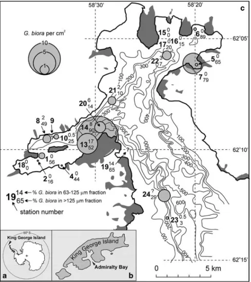
Surface sediment samples for population and morphological study of Globocassidulina were taken at 21 locations in Admiralty Bay from water depths between 8 and 254 m in Table I. List of sampling stations indicating site location, its water
depth, and method of sampling.
Station Water depth GPS location Method
(m) KG1 17.5–25 62810.998'S, 058837.306'W tube-sampler KG2 17.5 62811.216'S, 058837.492'W tube-sampler KG4 17 62810.688'S, 058832.115'W tube-sampler KG5 35 62806.247'S, 058819.777'W tube-sampler KG6 47.5 62804.416'S, 058819.932'W tube-sampler KG7 30 62806.220'S, 058819.676'W tube-sampler KG8 119 62809.650'S, 58834.774'W Van Veen KG9 114 62809.623'S, 58834.510'W Van Veen KG10 107 62809.584'S, 58834.287'W Van Veen KG13 108 62809.461'S, 58829.737'W Van Veen KG14 100 62809.290'S, 58829.439'W Van Veen KG15 16.3 62805.123'S, 058823.471'W diving KG16 29–31.4 62804.760'S, 058821.495'W diving KG17 20.8 62805.319'S, 058823.777'W diving KG18 8.0–12.0 Goulden Cove, ,150 m from ice edge diving KG19 40 South slopes of Napier Rock diving
KG20 249 62809.053'S, 058830.435'W Van Veen
KG21 254 62807.488'S, 058827.545'W Van Veen
KG22 233 62805.610'S, 058822.944'W Van Veen
KG23 223 62813.361'S, 058822.892'W Van Veen
KG24 179 62812.057'S, 058823.666'W Van Veen
Fig. 1. The location of a. King George
Island, b. Admiralty Bay, and
c. sampling stations (KG1 through KG24)
with G. biora abundances shown by circles expressing number of specimens per cm2, and percentages in total Rose Bengal stained assemblage. Stations with the percentages only were sampled by scuba divers. Dark gray indicates inland glacier free areas (Battke 1990). The bathymetric contour lines in metres after Straten (unpublished).
early 2007 (Table I, Fig. 1). They were wet sieved through 63 mm and 125 mm sieves and stained with Rose Bengal (1g l-1) and 70% ethanol diluted in seawater. The stained residues were washed in tap water and dried. In most samples, all ‘‘living’’ (stained) foraminifera were picked from the 63–125 mm and .125 mm fractions. Few faunally-rich samples were divided using a dry microsplitter. The maximum test diameter of all stained Globocassidulina from these samples was measured optically at 63x magnification with ± 16 mm precision. Apertural shapes of selected specimens were analysed under SEM.
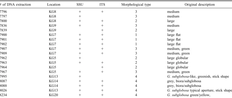
Nineteen specimens of Globocassidulina representing different morphological types were fixed for DNA examination (Table II). They were isolated from the upper 2–3 cm of surface sediments collected at seven locations. As soon as possible after collection, the sediment was washed with cold seawater and wet sieved through 125 mm sieves. The residues were stored in the refrigerator at about 28C, for periods of several days. The foraminifera were sorted under a binocular microscope from the residues kept cool on a tray of ice. Specimens for molecular study were transferred one by one into guanidine or AP1 (DNAEasy, Qiagen) extraction buffers.
A 3' fragment of the SSU rRNA gene was successfully amplified for 17 DNA isolates (specimens). Foraminiferal
specific primer s14F3 and universal eukaryotic primer sB were used for the first amplification. Foraminiferal specific primer s14F1 and primer sB were used for reamplification. PCR conditions and primer sequences are described in Pawlowski (2000).
The ITS region (ITS1 1 5.8S 1 ITS2) was successfully amplified for 18 DNA isolates. The PCR amplification was performed using the foraminiferal-specific primer pair s14F3 and 2TAIC and re-amplified with the universal eukaryotic primer sBr and foraminiferal specific primer 2TAIC. The compatibility of the SSU and ITS sequences was tested by amplifying a longer fragment, using primers s14F1 and For2 situated in the central part of ITS region (Fig. 2). Primers used for amplification of ITS region are described in Pawlowski et al. (2007).
The amplified PCR products were purified using High Pure PCR Purification Kit (Roche Diagnostics). They were ligated in the Topo Cloning vector (Invitrogen), and cloned using ultracompetent cells XL2-Blue MRF’ (Stratagene). Sequencing reactions were prepared using an ABI-PRISM Big Dye Terminator Cycle Sequencing Kit and analysed with an ABI-PRISM 3100 sequencer (Applied Biosystems), all according to the manufacturer’s instructions.
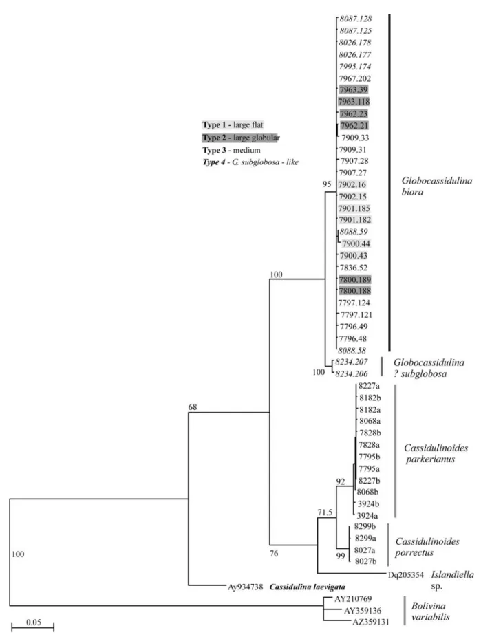
In the SSU analysis, 31 sequences obtained after cloning of 17 PCR products were manually aligned using Seaview Table II. List of DNA samples used in this study.
# of DNA extraction Location SSU ITS Morphological type Original description
7796 KG8 1 1 3 medium 7797 KG8 1 3 medium 7800 KG8 1 1 2 large 7836 KG9 1 1 3 medium 7839 KG9 1 2 large 7900 KG7 1 1 1 large flat 7901 KG7 1 1 1 large flat 7902 KG7 1 1 1 large flat 7907 KG7 1 1 3 medium, green 7909 KG7 1 1 3 medium, green 7962 KG5 1 2 large globular 7963 KG5 1 1 2 large globular 7964 KG5 1 2 large globular 7967 KG5 1 1 3 medium, green
7995 KG13 1 4 G. subglobosa-like, greenish, stick shape
8087 KG14 1 1 4 grey, biora/subglobosa
8088 KG14 1 1 4 grey, biora/subglobosa
8026 KG13 1 1 4 G. subglobosa typical aperture, stick shape
8234 KG20 1 1 4 G. subglobosa green/yellow,
Fig. 2. Schema of rDNA showing the
position of primers used for amplification of SSU and ITS rDNA (dark and bright gray arrows, respectively) and for testing the compatibility of the SSU and ITS sequences (black arrows).
software (Galtier et al. 1996) to 21 sequences of foraminifera from Cassidulinidae family, obtained mostly from the same region. Highly variable regions of the alignment were removed, leaving 1163 sites for phylogenetic analysis. The phylogenetic tree was constructed using maximum likelihood method with GTR 1 GI model, using Treefinder (Jobb et al. 2004). The same programme was used to analyse 34 sequences of the ITS region.
Results
Morphologic diversity of Globocassidulina in Admiralty Bay
Among the stained samples from the recent survey in Admiralty Bay, Globocassidulina was found everywhere except at station KG15 located within Martel Inlet, next to the Keller Peninsula (Table I, Fig. 1). In eight samples, substantial populations (.50 specimens) of stained
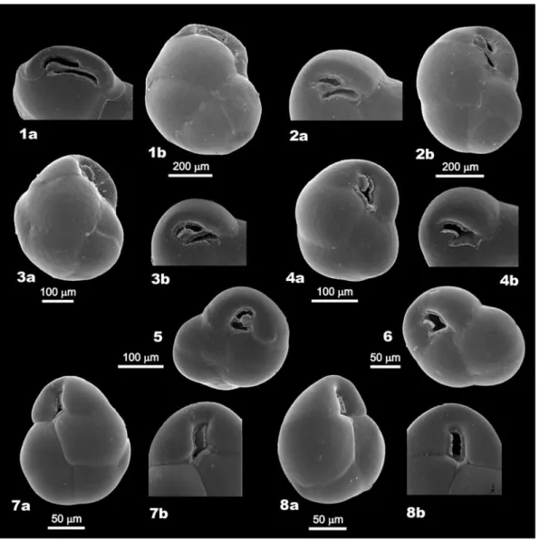
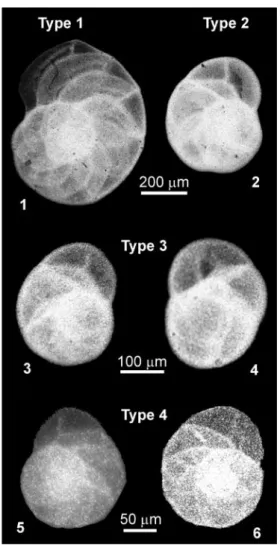
Fig. 3. SEM images of Globocassulina from Admiralty Bay, belonging to the four morphologic types. Type 1 (large flat): 1. KG7;
type 2 (large globular): 2. KG8; type 3 (middle in size): 3. KG7, 4–6. KG8; type 4 (G. subglobosa-like): 7. KG20, 8. KG23.
Table III. Morphological types in the population of Globocassidulina from in Admiralty Bay, based on living specimens from sites KG1 through KG24
(Table I, Fig. 1).
Morphological Colour of Apertural shape Test size Bathymetric
type cytoplasm range (mwd)
1 large flat yellow, pink doubled aperture parallel to the basal suture 500–800 mm 30–179
2 large globular pink to green doubled aperture parallel to the basal suture 500–800 mm 30–179
3 medium in size green doubled or bifurcated aperture 100–600 mm 30–254
4 small, grey, yellow, single aperture, perpendicular or oblique to . 200 mm 223–254
Globocassidulina were encountered. Among truly living
specimens, recognized by coloured cytoplasm rather than by Rose Bengal staining, four morphological types were identified. They include 1) large inflated forms with broad apertural area, doubled aperture and pink to yellow cytoplasm, 2) large forms with more globular outline, doubled aperture and pink to green cytoplasm, 3) medium in size with bifurcated or double aperture and greenish cytoplasm, and 4) small forms with single aperture and greenish or yellowish cytoplasm. The presence of four morphological types among
Globocassidulina in Admiralty Bay was supported by SEM
inspections of stained specimens (Fig. 3). Their characteristic features are summarized in Table III.
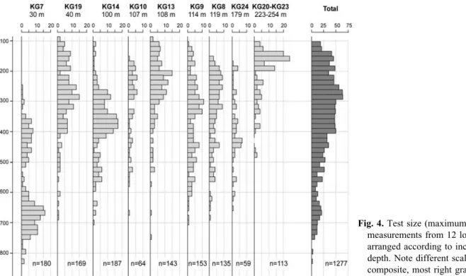
Test size measurements (Fig. 4) on the total of 1277 specimens confirmed the presence of considerable morphological heterogeneity in Globocassidulina population. In the shallowest sample with substantial population of stained specimens (KG7, 30 m depth), 180 specimens show bimodal test size distribution. Most specimens have tests of 600–750 mm, while the rest are smaller than 500 mm. The group of samples from water depths between 40 and 179 m (KG8, KG9, KG10, KG13, KG14, KG19, and KG24) show a more homogenous test size distribution with only a few specimens larger than 600 mm and the overwhelming majority within a broad range between 100 and 600 mm, but rather smaller than 400 mm. In contrast, the deepest water samples from 223 to 254 m (KG20, KG21, KG22, KG23) show the greatest numbers of specimens between 100 and 200 mm, and significantly fewer specimens that are up to 500 mm in test size. To summarize, the presence of at least three morphological types within Globocassidulina
population in Admiralty Bay, is generally confirmed by the test size results.
Genetic diversity of Globocassidulina in Admiralty Bay
To verify whether observed morphological differences correspond to genetic differentiation between the morphological types, we amplified and sequenced a 3' fragment of the SSU and the complete ITS region of the rDNA of specimens representing all morphotypes. The analysis of SSU rDNA (Fig. 5) shows considerable homogeneity between specimens, except for the isolate 8234, which may represent G. subglobosa. All morphologically distinctive types of Globocassidulina from Admiralty Bay merge into one group clearly distinctive from other
Cassidulinidae. Because of the presence of a doubled
aperture in the largest, most characteristic specimens, we consider this group to represent G. biora. Moreover, four isolates suspected of belonging to G. subglobosa (7995, 8026, 8087, 8088) blend within the G. biora cluster, suggesting that they may be in fact immature G. biora.
The test whether the genetic homogeneity of the SSU rDNA sequences is not due to the relatively low mutation rate in this gene (Bowser et al. 2006), we also sequenced more rapidly evolving ITS rDNA region. Phylogenetic analysis of ITS sequences gives a tree similar to the one obtained using SSU rDNA, with no clear differentiation between different morphotypes, except for the DNA isolate 8234, probably representing G. subglobosa (Fig. 6). Within the G. biora group, the distances between sequences obtained from
Fig. 4. Test size (maximum length)
measurements from 12 locations, arranged according to increasing water depth. Note different scale on the composite, most right graph.
different isolates (inter-isolate variation) averaged 0.49%, with the range between 0 and 1.21, while between the two sequences obtained from the isolate 8234 and G. biora the distances were almost an order of magnitude larger and averaged 3.9% with the range between 3.59 and 4.40%. The intra-isolate variation was an average of 0.60%, actually exceeding the inter-isolate variation within the G. biora cluster. To conclude, comparison of the ITS sequences obtained from the four morphological types within G. biora falls within 0.38–0.60% range and reveals no significant differences between intra- and inter-morphological variability
(Table IV), confirming the monospecific character of the
G. biora cluster.
Measurement of the proloculus
For analysing sizes of proloculi, 32 specimens of
Globocassidulina from Admiralty Bay were X-rayed (Fig. 7).
In the case of 28 micrographs, it was possible to measure the sizes of proloculi. In the larger specimens (type 1 and 2) a degree of uncertainty in identification of proloculi existed. However, dissection of 30 specimens from KG7 and KG8 confirmed presence of large proloculi (,120 mm) in majority of specimens. Only three dissected specimens were found to be microspheric. The plot of the proloculi sizes against test diameters (Fig. 8) shows no significant difference in the size of proloculus between types 1 and 2. Their average diameter of ,140 mm exceeds values measured on dissected specimens, due to rather obscured X-ray view of inner whorls in the larger and thicker-walled specimens. Type 3 shows proloculi at
Fig. 6. ITS rDNA tree showing sequences obtained from
different morphotypes of G. biora.
Table IV. Average sequence divergence (in percentage) between
different morphological types of G. biora from Admiralty Bay, based on the analysis of the ITS fragment of the rRNA gene.
Type 1 Type 2 Type 3 Type 4
Type 1 0.38 0.48 0.50 0.44
Type 2 0.48 0.55 0.6
Type 3 0.46 0.52
Type 4 0.34
Fig. 7. X-ray images of four morphological types of
Globocassidulina from Admiralty Bay. 1. Type 1, KG7,
2. Type 2, KG8, 3 & 4. Type 3, KG8, KG14, 5 & 6.
,125 mm in average, which stays in agreement with the values measured on the dissected specimens of types 1 and 2. On contrary, the smallest specimens, represented by type 4, show relatively small proloculi (,80 mm on average or less). All specimens of type 4, that were X-rayed, were from samples 20 and 23 (249 and 223 m) and they may represent
G. subglobosa, similar to the isolate 8234 that was the only
one from below 200 m analysed genetically in this study and which SSU and ITS sequences clearly differed from remaining
Globocassidulina isolates (Figs 5 & 6).
Discussion
What is the reason for morphological diversity of G. biora?
Despite the presence of considerable morphological variability within Globocassidulina in Admiralty Bay (Table III, Fig. 3), the molecular analysis of the SSU and ITS fragments of rRNA gene indicates that except for minute forms from deeper than 200 m probably representing
G. subglobosa, the rest of Globocassidulina from Admiralty
Bay is monospecific and belongs to G. biora (Table IV, Figs 5 & 6). What could be the reason for the significant morphologic variability within G. biora? There appears to be a few possibilities: 1) development of distinctive micro and megalospheric generations, 2) a presence of distinct cohorts that at the time of collection were at different growth stages, and/or 3) it could be due to environmental variability in benthic habitats throughout Admiralty Bay that resulted in development of different morphologies.
The first possibility was tested by analysing inner-test morphology of the four types of G. biora. Although the life cycle is more varied in foraminifera than in any other group of protists (Goldstein 1999) and no specific information on life cycle in Cassidulinidae is available, in general, the
foraminiferal life cycle is characterized by alternation of sexual and asexual generations. As a result, megalospheric gamonts with a large proloculus and relatively small overall test diameter, and microspheric agamonts with tiny proloculus but relatively large overall test may develop (Goldstein 1999). Nomura (1984) reported the presence of megalospheric and microspheric forms in G. biora from raised beaches and modern deposits of the Lu¨tzow–Holm Bay, East Antarctica. He reported the presence of two types of tests having a proloculus of 72 mm in average and of less than 10 mm in diameter.
It appears that among G. biora, morphologic dimorphism, i.e. a distinction between micro and megalospheric generations, is not responsible for the morphological variability observed. However, clear difference in the size of proloculus is observed between G. biora and G. subglobosa (,120 mm vs ,80 mm; Fig. 8). Moreover, megalospheric proloculi of G. biora from Admiralty Bay ( ,120 mm) are on average larger than those reported from Lu¨tzow–Holm by Nomura (1984) (72 mm).
The second possibility of significant morphological changes throughout ontogeny among Globocassidulina was noted by Finger & Lipps (1981). They suggested that smaller specimens of G. crassa rossensis and G. biora are similar to
G. subglobosa, but grade into larger and more compressed
forms with branched or doubled apertures. In the same study, they reported dissection revealing that earlier chambers of adult G. crassa rossensis and G. biora display fully developed apertures without any indications of apertural growth stages. Thus, their final conclusion was not unequivocal. Our data suggest that a large part of morphological variability observed among G. biora in Admiralty Bay may be a result of observing specimens at different growth stages. The presence of single, bifurcated, and doubled apertures among the four morphological types that, according to the molecular analysis all fell into the G. biora cluster, suggests that throughout ontogeny the aperture becomes first bifurcated and then matures into doubled. Similarly, the test shape becomes more inflated in the largest specimens, as observed in type 1 (Fig. 3). All these findings are in agreement with the conclusions of Nomura (1984) who presented analogous morphological variability through ontogenic development of G. biora from the Lu¨tzow–Holm Bay.
On the other hand, the ontogenic changes cannot explain morphological differences between type 1 and type 2, both representing relatively large specimens (Table III, Fig. 3) and falling within a single group on the proloculus vs test size plot (Fig. 8). Dissection of 30 specimens revealed that in both types the megalospheric forms dominate; however, a few microspheric forms are also present. Significant intraspecific morphologic variability is not unknown among benthic foraminifera. Study of Ammonia showed wide morphologic diversity in natural habitats and cultured populations. Up to six different ‘‘morphospecies’’ were found in their natural habitats and in clonal cultures that
Fig. 8. Test versus proloculus diameter among morphologic
types of Rose Bengal stained Globocassidulina in Admiralty Bay. Note that average proloculus diameter measured on dissected specimens from type 1 and 2 was on average ,120 mm. The difference is due to obscured X-ray views of inner whorls in the larger and thicker-walled specimens.
were suggested to be ecotypes of Ammonia beccarii developed due to environmental factors (Schnitker 1974).
A review of ecological parameters that may have impact on benthic foraminiferal tests morphology was presented by Boltovskoy et al. (1991). It appears that the differences in test shape between types 1 and 2 of G. biora may be also a consequence of ecological variability in benthic habitats throughout Admiralty Bay. A problem with this interpretation however, is that the large globular specimens of morphological type 2, that are encountered at several locations (KG5, KG8, KG9, KG13, KG19, KG24), at all locations except KG5 and KG24 blend with the inflated specimens of type 1. Therefore it is difficult to propose any hydrographic (e.g. temperature, salinity, calcium carbonate solubility etc.) or substrate-related factor that could lead to development of two different morphologies in G. biora at the same locations. Among the ecological parameters, reviewed by Boltovskoy et al. (1991), only the nutrition, or more precisely food type could be a candidate for the development of the two different types of large G. biora. There are numerous studies showing morphological and cytological response of benthic foraminifera to heavy metal pollution (e.g. Alve 1991, Le Cadre & Debenay 2006) and it was suggested that the pollutants may penetrate the foraminiferal cell together with food (Yanko et al. 1998). However, so far no data show that different types of food could actually lead to development of different test morphologies in benthic foraminifera. In the case of G. biora from Admiralty Bay, the difference in cytoplasm colouration between types 1 and 2 (Table III) may give slight support for such possibility.
Is there only one large Globocassidulina (G. biora) in the South Shetland Islands and Antarctica?
The first detailed study on Recent benthic foraminiferal distribution in Admiralty Bay in the 2002/03 summer season used only the . 125 mm fraction (Majewski 2005). Specimens of Globocassidulina were encountered at all locations, between 8 and 520 m, except for a single site in Herve´ Cove (8 m), where only a few Quinqueloculina were found. All relatively large specimens of Globocassidulina were reported as G. biora. However, a considerable variability in their apertural shapes was noted. It was also clear that although G. biora inhabits practically the entire Admiralty Bay down to at least 520 m, it dominates shallow water assemblages at less than 200 m (Majewski 2005). However, other studies in South Shetland Islands (Finger & Lipps 1981, Chang & Yoon 1995, Mayer 2000, Li et al. 2000, Gray et al. 2003, Yoo et al. 2006), have reported the presence of more than one representatives/species of
Globocassidulina. Keeping this fact in mind, along with
an investigation of living monothalamous foraminiferal assemblages in Admiralty Bay (Majewski et al. 2007), particular attention was paid also to morphological diversity among living Globocassidulina.
The molecular homogeneity, despite considerable morphological variation suggests that G. crassa reported from modern sediments at King George Island by earlier studies (e.g. Chang & Yoon 1995, Mayer 2000) may be in fact G. biora as might G. crassa rossensis reported from Holocene sections by Li et al. (2000) and Yoo et al. (2006). In studies of recent Antarctic benthic foraminifera, Finger & Lipps (1981) have already suggested a common over-reporting of the true G. crassa type of d’Orbigny (1839). They suggested that many workers have ignored the diagnostic apertural branches that differentiate other taxa from G. crassa, having only a single aperture. Interestingly, despite the comment of Finger & Lipps (1981), Gray et al. (2003) who subsequently studied recent deposits at the same locations, listed G. crassa along with G. biora as present at Deception Island, even though their specimen illustrated as G. crassa seems to have a doubled aperture. They did, however, point out difficulties in classifying
Globocassidulina in cases of poor aperture preservation.
Finger & Lipps (1981) acknowledged the presence of two large Globocassidulina taxa in Deception Island (,150 km south-west from King George Island). They dissected some large specimens of G. biora and what they regarded as G. crassa rossensis and noted that their inner chambers displayed fully developed apertures without any indication of apertural growth stages. Their observation was questioned by Nomura (1984), who observed changes in apertural shape from single, through L-shaped into double, parallel in mature stage of G. biora, in agreement with another suggestion of Finger & Lipps (1981) about potential difficulties in discriminating between immature specimens of G. biora and G. crassa rossensis. In the view of the present findings, it seems to be likely that all specimens reported from Deception Island may in fact belong to G. biora.
It appears that at least some of the morphological types of G. biora distinguished in Admiralty Bay may have broad pan-Antarctic distribution. Crespin (1960) who described
G. biora from Vestfold Hills illustrated large, globular
specimens that could correspond to type 2 from this study. Similarly, large specimens from raised beaches of Lu¨tzow–Holm Bay (Nomura 1983, 1984) and from the Firth of Tay Holocene section (Majewski & Anderson 2009) resemble type 2. On the other hand, in the Ross Sea, Violanti (1996) reported forms that may correspond to type 1 in this study. She noted G. subglobosa, G. crassa, G. rossensis, and
G. biora in a single nearshore sample (IB 3) from near Terra
Nova Station (Ross Sea) and noted a more compressed shape of the largest G. biora as compared with G. crassa. A large and inflated specimen of G. biora was illustrated by Ward & Webb (1986) from materials deposited onshore Ross Island by the ice shelf. It seems to correspond to the type 1 from Admiralty Bay as well. The living specimens of type 1 morphotype were also observed by one of us (JP) at shallow depths in McMurdo Sound.
According to Quilty (2003), the three morphologically close species (i.e. G. crassa, G. rossensis, and G. biora) are in fact subspecies of G. crassa. Their very close phylogenetic relationship is in concordance with the ontogenetic development of the aperture in G. biora (Nomura 1984) and also reflected in the molecular homogeneity among forms with differently developed apertures in Admiralty Bay. The absence of the true G. crassa in Deception Island and possibly at other Antarctic locations, as suggested by Finger & Lipps (1981), may imply that G. crassa, originally reported north of the Polar Front, and G. biora, common in recent Antarctic settings, could represent two distinct species with populations restricted to the Magellan region and the Antarctic, respectively. It seems probable that large, typical specimens of
G. biora are more representative for shallow waters (Bernhard
1987, Li et al. 2000, Majewski 2005) than for greater water depths (Fillon 1974, Osterman & Kellogg 1979, Kellogg & Kellogg 1987), mainly due to control by water mass properties with respect to CaCO3dissolution (e.g. Anderson 1975, Ishman
& Szymcek 2003). This bathymetric constraint could limit
Globocassidulina gene flow between the Antarctic and the
Magellan region. Conclusions
Four distinctive morphological types found among living
Globocassidulina in Admiralty Bay include 1) large inflated
forms with a broad apertural area, doubled aperture and pink to yellow cytoplasm, 2) large forms with a more globular outline, doubled aperture and pink to green cytoplasm, 3) medium forms with a bifurcated or double aperture and greenish cytoplasm, and 4) small forms with a single aperture with greenish or yellowish cytoplasm. The molecular analysis of the SSU and ITS rDNA indicates that they are monospecific and belong to G. biora. Among the smallest forms, only a single isolate, the only one from deeper than 200 m, stands out in the molecular analyses. It may represent the true G. subglobosa. The morphological types of G. biora seem to represent populations at different stages of ontogenetic development. However, the variability among large G. biora collected at the same locations is difficult to interpret. It seems that different types of food may be the most likely factor that could lead to development of different test morphologies in adult G. biora. It also seems probable that G. biora is the only recent, large, shallow water Globocassidulina represented throughout South Shetland Islands and possibly throughout the Antarctica, while G. crassa is typical of the Magellan region north of the Polar Front.
Acknowledgements
This study was supported by a grant of Polish Ministry of Science and Higher Education No N307 115135, a grant of Swiss National Science Foundation 31003A_125372, and
the G. & A. Claraz Donation. We would like to thank Professor Johann Hohenegger (Vienna University) for his kindest help in obtaining X-ray micrographs, Dr Jose´ Fahrni and Jackie Guaird for technical assistance, and Drs Be´atrice Lecroq and Fre´de´ric Sinniger for help in collecting material. We would also like to thank Alessandra Asioli and an anonymous reviewer for useful suggestions for improvement to this manuscript.
References
ALVE, E. 1991. Benthic foraminifera in sediment cores reflecting heavy metal pollution in Sorfjord, western Norway. Journal of Foraminiferal
Research, 21, 1–19.
ANDERSON, J.B. 1975. Ecology and distribution of foraminifera in the
Weddell Sea of Antarctica. Micropaleontology, 21, 69–96.
BATTKE, Z. 1990. Admiralty Bay, King George Island. Map 1: 50 000.
Warsaw: Institute of Ecology, Polish Academy of Sciences.
BERNHARD, J.M. 1987. Foraminiferal biotopes in Explorers Cove,
Antarctica. Journal of Foraminiferal Research, 17, 286–297. BOLTOVSKOY, E., SCOTT, D.B. & MEDIOLI, F.S. 1991. Morphological
variations of benthic foraminiferal tests in response to changes in ecological parameters: a review. Journal of Paleontology, 65, 175–185. BOWSER, S.S., HABURA, A. & PAWLOWSKI, J. 2006. Molecular evolution of foraminifera. In KATZ, L. & BHATTACHARYA, D., eds. Genomics and
evolution of microbial eukaryotes. Oxford: Oxford University Press,
78–93.
CHANG, S.-K. & YOON, H.I. 1995. Foraminiferal assemblages from bottom sediments at Marian Cove, South Shetland Islands, West Antarctica.
Marine Micropaleontology, 26, 223–232.
CRESPIN, I. 1960. Some recent foraminifera from Vestfold Hills, Antarctica.
Reports of the Tahoku University (Geology), Special Volume, Series 2,
4, 19–31.
FILLON, R.H. 1974. Late Cenozoic foraminiferal paleoecology of the Ross
Sea, Antarctica. Micropaleontology, 20, 129–151.
FINGER, L.F. & LIPPS, J.H. 1981. Foraminiferal decimation and
repopulation in an active volcanic caldera, Deception Island, Antarctica. Micropaleonthology, 27, 111–139.
GALTIER, N., GOUY, M. & GAUTIER, C. 1996. SEAVIEW and
PHYLO_WIN, two graphic tools for sequence alignment and molecular phylogeny. Computer Applications in the Biosciences, 12, 543–548.
GOLDSTEIN, S.T. 1999. Foraminifera: a biological overview. In SENGUPTA, B.K. ed., Modern Foraminifera. Dordrecht: Kluwer, 37–56.
GRAY, S.C., STURZ, A., BRUNS, M.D., MARZAN, R.L., DOUGHERTY, D., LAW, H.B., BRACKETT, J.E. & MARCOU, M. 2003. Composition and distribution of sediments and benthic foraminifera in a submerged caldera after 30 years of volcanic quiescence. Deep-Sea Research II, 50, 1727–1751. IGARASHI, A., NUMANAMI, H., TSUCHIYA, Y. & FUKUCHI, M. 2001.
Bathymetric distribution of fossil foraminifera within marine sediment cores from the eastern part of Lu¨tzow–Holm Bay, East Antarctica, and its paleoceanographic implications. Marine Micropaleontology, 42, 125–162.
ISHMAN, S.E. 1990. Quantitative analysis of Antarctic benthic foraminifera:
application to paleoenvironmental interpretations. PhD thesis, The Ohio
State University, Columbus, 266 pp. [Unpublished]
ISHMAN, S.E. & SZYMCEK, P. 2003. Foraminiferal distributions in the former
Larsen-A Ice Shelf and Prince Gustav Channel region, eastern Antarctic Peninsula margin: a baseline for Holocene paleoenvironmental interpretation. Antarctic Research Series, 79, 239–260.
JOBB, G., VON HAESELER, A. & STRIMMER, K. 2004. TREEFINDER: a powerful graphic analysis environment for molecular phylogenetics.
KELLOGG, D.E. & KELLOGG, T.B. 1987. Microfossil distributions in
modern Amundsen Sea sediments. Marine Micropaleontology, 12, 203–222.
KHIM, B.-K., YOON, H.I., KIM, Y. & SHON, I.C. 2001. Late Holocene stable
isotope chronology and meltwater discharge event in Maxwell and Admiralty bays, King George Island, Antarctica. Antarctic Science, 13, 167–173. LE CADRE, V. & DEBENAY, J.-P. 2006. Morphological and cytological
response of Ammonia (foraminifera) to copper contamination: Implication for the use of foraminifera as bioindicators of pollution.
Environmental Pollution, 143, 304–317.
LI, B., YOON, H.I. & PARK, B.K. 2000. Foraminiferal assemblages and CaCO3dissolution since the last deglaciation in the Maxwell Bay, King
George Island, Antarctica. Marine Geology, 169, 239–257.
MAJEWSKI, W. 2005. Benthic foraminiferal communities: distribution and ecology in Admiralty Bay, King George Island, West Antarctica. Polish
Polar Research, 26, 159–214.
MAJEWSKI, W. & ANDERSON, J.B. 2009. Holocene foraminiferal
assemblages from Firth of Tay, Antarctic Peninsula: Paleoclimate implications. Marine Micropaleontology, 73, 135–247.
MAJEWSKI, W., LECROQ, B., SINNIGER, F. & PAW"OWSKI, J. 2007.
Monothalamous foraminifera from Admiralty Bay, King George Island, West Antarctica. Polish Polar Research, 28, 187–210. MAYER, M. 2000. Zur O¨ kologie der Benthos-Foraminiferen der Potter
Cove (King George Island, Antarktis). Berichte zur Polarforschung, 353, 1–126.
MILAM, R.W. & ANDERSON, J.B. 1981. Distribution and ecology of recent benthonic foraminifera of the Ade´lie–George V continental shelf and slope, Antarctica. Marine Micropaleontology, 6, 297–325.
NOMURA, R. 1983. Foraminifera from the raised beach deposits on the east coast of Lutzow–Holm Bay, Antarctica. Memoirs of National Institute of
Polar Research, 28, 219–230.
NOMURA, R. 1984. Cassidulinidae (Foraminiferida) from the Eastern part
of the Lu¨tzow–Holm Bay, Antarctica. Transactions, Proceedings,
Paleontological Society of Japan, 136, 492–501.
OSTERMAN, L.E. & KELLOGG, T.B. 1979. Recent benthic foraminiferal
distributions from the Ross Sea, Antarctica: relation to ecologic and oceanographic conditions. Journal of Foraminiferal Research, 9, 250–269. PAWLOWSKI, J. 2000. Introduction to the molecular systematics of
foraminifera. Micropaleontology, 46 (sup.1), 1–12.
PAWLOWSKI, J., FAHRNI, J., LECROQ, B., LONGET, D., CORNELIUS, N., EXCOFFIER, L., CEDHAGEN, T. & GOODAY, A.J. 2007. Bipolar gene flow in deep-sea benthic foraminifera. Molecular Ecology, 16, 4089–4096. QUILTY, P.G. 2003. Neogene foraminifers and accessories, ODP Leg 188,
Sites 1165, 1166, and 1167, Prydz Bay, Antarctica. ODP, Scientific
Results, 188, 1–41. Available at http://www-odp.tamu.edu/publications/
188_sr/volume/chapters/009.pdf.
SCHNITKER, D. 1974. Ecophenotypic variation in Ammonia beccarii
(Linne´). Journal of Foraminiferal Research, 4, 216–223.
VIOLANTI, D. 1996. Taxonomy and distribution of recent benthic foraminifers
from Terra Nova Bay (Ross Sea, Antarctica), Oceanographic Campaign 1987/1988. Palaeontographia Italica, 83, 25–71.
WARD, B.L. & WEBB, P.N. 1986. Late Quaternary foraminifera from raised
deposits of the Cape Royds–Cape Barne area, Ross Island, Antarctica.
Journal of Foraminiferal Research, 16, 176–200.
YANKO, V., AHMAD, M. & KAMINSKI, M. 1998. Morphological deformities
of benthic foraminiferal tests in response to pollution by heavy metals: implications for pollution monitoring. Journal of Foraminiferal
Research, 28, 177–200.
YOO, K.-C., YOON, H.I., PARK, B.-K. & KIM, Y. 2006. Advance of the outlet glaciers during regional warming as inferred from Late Holocene massive diamicton in the King George Island fjords, the South Shetland Islands, West Antarctica. Quaternary Research, 65, 57–69 (Retracted).