841
CONCISE COMMUNICATIONS
Persistence of Human Herpesvirus 7 in Normal Tissues Detected by Expression
of a Structural Antigen
Werner Kempf, Volker Adams, Prisco Mirandola, Departments of Dermatology and Pathology, University Hospital, Zurich, Switzerland; Heart-Center Leipzig, University of Leipzig, Laura Menotti, Dario Di Luca, Norbert Wey,
Leipzig, Germany; Department of Experimental Pathology, Section on Beatrix Mu¨ller, and Gabriella Campadelli-Fiume
Microbiology and Virology, University of Bologna, Bologna, and Department of Experimental and Diagnostic Medicine, University of Ferrara, Ferrara, Italy Human herpesvirus 7 (HHV-7) infection in histologically normal human tissues was investigated by
immunohistochemical detection of the 85-kDa tegument phosphoprotein (pp85) encoded by the U14 gene. So far, two cell types were recognized as sites of HHV-7 infection in vivo: CD4/T lymphocytes, believed to be the site of latent infection, and epithelial cells of salivary glands, the site of productive infection and viral shedding. Unexpectedly, cells expressing the HHV-7 structural antigen were detect-able in lungs, skin, and mammary glands. Morphologically and phenotypically, they were distinct from lymphocytes. Liver, kidney, and tonsils were positive, although the number of HHV-7 – positive cells was low. Large intestine, spleen, and brain were negative. Different from the current notion of the state of HHV-7 in humans, the results show that a variety of tissues harbor cells at a late stage of infection and suggest that HHV-7 causes a persistent rather than a true latent infection.
Human herpesvirus 7 (HHV-7) was isolated from peripheral glands. The initial observation that HHV-7 can be isolated from blood mononuclear cells (PBMC) and saliva of healthy individ- cultured PBMC following mitogenic or antigenic stimulation uals and of individuals affected by chronic fatigue syndrome led to the idea that HHV-7 establishes a latent infection in [1 – 3]. It was considered an ‘‘orphan virus,’’ that is, without CD4/ T lymphocytes [1]. By contrast, in epithelial cells of association with any known human disease. Discoveries of the salivary glands, the virus establishes a productive infection, past years are changing this perspective, as they led to the which results in virus shedding in saliva [10]. Similarly to the recognition that primary infection results sporadically in exan- in vivo tropism, even the in vitro tropism of HHV-7 appears them subitum or in febrile illnesses without rash [4, 5], that to be very narrow, as CD4/T lymphocytes — either primary during primary infection the virus may have access to the cultures or cell lines — are the only cell type known to support central nervous system and induce neurologic complications productive infection. In investigations focused on the detection [6], that infection or reactivation may take place in bone mar- of herpesviruses in Kaposi’s sarcoma, some of us made the row and renal transplant recipients and complicate engraftment unexpected observation that, in the context of this tissue, of transplanted organs [7, 8], and that viral DNA sequences HHV-7 can also infect cells expressing the CD68 antigen, a are present in lesions and PBMC of individuals affected by marker of monocyte/macrophage lineage [11]. This observation pityriasis rosea [9]. These findings suggest possible associa- raised the possibility that HHV-7 infection in humans might tions between HHV-7 infection and diseases and also raise the not be limited to CD4/T lymphocytes and salivary glands and possibility that HHV-7, similarly to other herpesviruses, may hence that the tissue and cell tropism of the virus may be become a pathogen under conditions of immunosuppression. broader than known so far.
Two cell types have been recognized as targets of HHV-7 While the entire HHV-7 genome sequence was reported re-infection in vivo, namely CD4/ T lymphocytes and salivary
cently [12], knowledge of the proteins specified by HHV-7 and of their biologic properties is scanty, mainly due to difficulties in growing the virus and consequently to few available specific Received 22 December 1997; revised 2 April 1998. reagents. One of our laboratories has characterized a highly Informed consent was obtained from all patients. immunogenic complex of proteins designated phosphoprotein Grant support: At University of Bologna, AIDS Project from Istituto
Superi-85 (ppSuperi-85) [13, 14], which localizes to the outer layers of virion ore di Sanita´, BIOMED2 BMH4 CT95 1016 from UE, Target Project in
Bio-technology, MURST 40% and 60%; at University of Zu¨rich, Gertrud-Rueegg- tegument [15]. pp85 is encoded by the U14 gene [15]. Mono-Stiftung; at University of Ferrara, AIDS Project from Istituto Superiore di clonal antibody (MAb) 5E1 reacts with pp85, does not cross-Sanita´.
react with HHV-6, and hence recognizes in pp85 an HHV-7 – Reprints or correspondence: Dr, G. Campadelli-Fiume, Dipartimento di
Pa-tologia Sperimentale, Sezione di Microbiologia e Virologia, Via San Giacomo specific epitope [13 – 15]. Comparison of the HHV-7 and 12, 40126 Bologna, Italy.
HHV-6 U14 sequences shows regions at the C-terminus of the The Journal of Infectious Diseases 1998; 178:841 – 5 proteins with sufficient divergence to harbor potential
HHV-q 1998 by the Infectious Diseases Society of America. All rights reserved.
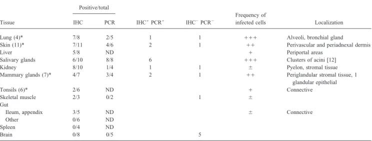
Table 1. Detection of HHV-7 in normal tissues by immunohistochemical staining (IHC) and polymerase chain reaction amplification (PCR). Positive/total
Frequency of
Tissue IHC PCR IHC/PCR/ IHC0PCR0 infected cells Localization
Lung (4)* 7/8 2/5 1 1 /// Alveoli, bronchial gland
Skin (11)* 7/11 4/6 2 1 // Perivascular and periadnexal dermis
Liver 5/8 ND / Periportal areas
Salivary glands 6/10 8/8 6 /// Clusters of acini [12]
Kidney 8/10 1/4 1 1 { Pyelon, stromal tissue
Mammary glands (7)* 4/7 3/4 2 1 // Periglandular stromal tissue, 1
glandular epithelial
Tonsils (6)* 2/6 ND / Connective
Skeletal muscle 2/3 0/2 1 {
Gut
Ileum, appendix 3/5 ND { Connective
Other 0/6 ND
Spleen 0/4 ND
Brain 0/8 0/5 5
NOTE. ND, not done. Frequency of infected cells per positive field, evaluated subjectively, is expressed by increasing symbols.
* Bioptic specimens were obtained from individuals undergoing plastic surgery, tonsil resection or diagnostic lung biopsies (in parentheses, no. of bioptic samples of total). Rest were autoptic samples from patients who died of heart failure, lung embolism, or old age but not of cancer, viral (including human immunodeficiency virus) or bacterial infections, or organ transplantation.
nation. DNA extraction, PCR, and gel electrophoresis were done in
To shed light on the natural history of HHV-7 infection, we
separate laboratories. Successful amplification of a b-globin fragment
investigated if the in vivo tropism of HHV-7 extends to cells
indicated that the samples were adequate for PCR analysis and that
and tissues other than CD4/ T lymphocytes, salivary glands,
no inhibitors were present [17].
and Kaposi’s sarcoma – associated CD68/cells. We screened
HHV-7 – specific sequences were amplified by nested PCR with
histologically normal bioptic or autoptic tissues for
immunohis-two sets of primers. The first set was external primers HV7/HV8
tochemical detection of pp85 [11], since expression of a
struc-and internal primers HV10/HV11, struc-and PCR conditions were as
tural protein is indicative of late-stage infection. detailed elsewhere [18]. About 50% of the specimens analyzed
with this set of primers were also analyzed with a second set of primers specific for pp85. The external primers were H145E,
5*-Materials and Methods
CAAAGCGCTTAAATCAAGTGTC-3*, and H143E, 5*-GAC-ACTTGATTTAAGCGCTTTG-3*; the internal primers were
Antibodies. The MAbs used in these studies were 5E1 specific
H145I, 5*-GCATTGGAATCCAAAGACAACC-3*, and H143I, for HHV-7 pp85 [11, 13–15], MAb 30 to herpes simplex virus
5*-GGTAACTGAAAGGCTGCAAGC-3*. PCR conditions were glycoprotein D [16], a MAb (CD68 KP1, no. M848; Dako, Glostrup,
35 cycles of 957C for 30 s, 587C for 30 s, and 727C for 60 s and Denmark) reacting with the KP1 epitope of CD68 as a specific marker
a final extension at 727C for 10 min. The amplification products of monocyte/macrophage lineage, MAb 0T01 05 Clotimmun to
Fac-were subjected to electrophoresis in a 2% agarose gel and stained tor XIIIa (Behring, Marburg, Germany), MAbs to CD3 (A452; Dako)
with ethidium bromide. Positive and negative controls were in-and CD4 (NCL-CD4-1F6; Novocastra Laboratories, Newcastle upon
cluded routinely. The positive controls consisted of DNA extracted Tyne, UK), and MAb to HHV-6 p100 (Chemicon, El Segundo, CA).
from HHV-7 – infected cord blood mononuclear cells. The negative
Tissue specimens. The bioptic tissue specimens taken from
indi-controls consisted of tubes containing all PCR reagents, but with-viduals undergoing plastic surgery, tonsil resection, or diagnostic lung
out DNA, every 5th or 10th analyzed sample. HHV-6 PCR was biopsies and the autoptic specimens taken at necropsies were routinely
performed as described [11]. fixed in 10% buffered formalin and embedded in paraffin.
Immunohistochemistry (IHC). IHC was performed as detailed
Polymerase chain reaction (PCR) analyses. DNA was extracted
previously [11]. Tissue sections were deparaffinized with xylene from deparaffinized tissues by proteinase K digestion with standard
and briefly digested with pronase (0.1% for 7 min). The endoge-procedures, as detailed previously [11]. Briefly, in order to avoid
nous peroxidase was blocked by incubation with 1% H2O2in
meth-contamination and product carryover, sterile materials were used
anol. The sections were exposed to primary antibodies for 90 min throughout the procedure, and the microtome blade was cleaned
ex-or overnight at room temperature. After several rinses with PBS, tensively with xylene after cutting of each specimen. Blank controls
the sections were reacted with an avidin-biotin-peroxidase coupled consisting of empty tubes containing sterile water were included with
antibody (Vectastain ABC kit; Vector Laboratories, Burlingame, every 10th specimen and simultaneously subjected to DNA extraction
CA). The chromogen was 3-amino 9-ethyl carbazole or neufuchsin, and PCR amplification. In addition, in each set of specimens being
used according to the manufacturer’s protocol. Finally, sections analyzed, unrelated tissue specimens—previously found not to
con-were counter-stained with 1% hematoxylin. Serial sections with a tain any HHV-7 amplifiable sequence—were included among the
maximum thickness of 4 mm were stained with different antibodies. specimens being cut and processed for PCR; their results were
843 JID 1998; 178 (September) Concise Communications
sections. As a routine, each specimen was processed in parallel tively for HHV-7 were reacted with a MAb to glycoprotein
without primary antibody (i.e., with secondary antibody and sub- D of herpes simplex virus, a ubiquitous herpesvirus, and the strates). specimens did not stain (figure 1G). They were also stained
Computerized image analysis. Ektachrome slides of serial sec- with a MAb to HHV-6 p100 (figure 1E). As the specimens tions were scanned using a high-resolution film scanner (Kodak
contained amplifiable HHV-6 DNA sequences, there was a
RFS 3570) and further processed with Photoshop software (Adobe,
positive staining for HHV-6. The cells harboring the HHV-6
Seattle, WA) on a Macintosh Power PC 8100. An area representing
structural protein differed from those harboring HHV-7 pp85,
Ç60% related to the original image size was processed for image
supporting the conclusion that the IHC reactivity was not the
analysis. The images were artificially colored. Images of sections
result of unspecific binding of MAbs. As an additional criterion,
reacted with the anti – HHV-7 MAb were colored with a blue
back-we note that MAbs to cellular markers employed in this study
ground and a red immunoreactive signal. Images of sections
re-acted with anti-CD68 antibody were artificially colored with a blue did not stain the cells reactive to MAb 5E1, strengthening
background and a brown immunoreactive product. The images the conclusion that this latter reactivity was not the result of
were then overlaid in transparent layers, allowing the identification unspecific binding. Of relevance is the finding that coinfection of co-localization of two immunohistochemical signals within the of HHV-6 and -7 in the same cell seems to be specific for same cell in two serial sections. Kaposi’s sarcoma [11], as it was not detected in normal tissues.
Detection of HHV-7 infection by PCR. To confirm the IHC results, a portion of the samples was analyzed by PCR. The
Results
IHC-positive samples included lungs, skin, and mammary and salivary glands; the IHC-negative samples included brain, skin,
Detection of HHV-7 infection by IHC. Of the tissues
exam-ined, some lung, skin, and mammary gland samples were biop- and skeletal muscle. As all samples were formalin-fixed and paraffin-embedded, preservation of DNA was controlled by tic and some were autoptic. The remainder were autoptic
sam-ples. HHV-7 expression was detected by IHC analysis with the PCR amplification of the b-globin gene [17]. Although this represents a good criterion for a eukaryotic gene present in HHV-7 – specific MAb 5E1 to pp85. The results summarized
in table 1 and illustrated in part in figure 1 show that a number two copies per cell, it is not necessarily good when applied to sequences that are present in much lower amounts, as was the of human tissues, in addition to salivary glands, stain positively
for pp85 and therefore harbor HHV-7 – infected cells at a late case for HHV-7 in these samples. It was expected that the partial DNA degradation, inevitably present in paraffin-embed-stage of infection. The tissues that contained the highest relative
number of infected cells were lungs, skin, and mammary glands ded archival samples, might yield a positive b-globin amplifi-cation reaction but result in a negative HHV-7 reaction, even (figure 1). Liver, kidney, tonsils, and appendix vermiformis
contained a very low number of infected cells but could defi- if some viral sequences were indeed present. About 50% of PCR-suitable IHC-positive samples contained amplifiable nitely be scored as positive. Skeletal muscle and ileum
con-tained very few positive cells, while gut (other than ileum HHV-7 sequences (table 1). The rest may have contained amounts of preserved HHV-7 sequences too low to yield detect-and appendix vermiformis), spleen, detect-and brain were constantly
negative. able amplification. The IHC-negative samples were generally
negative by PCR (table 1). To ascertain if the HHV-7 – infected cells belonged to T
lym-phocytes, serial sections were stained with antibodies to CD3 and CD4. Most frequently, the HHV-7 – positive cells did not
Discussion
belong to the T lymphocytes. To determine if the positive cells
in lungs belonged to the monocyte/macrophage lineage, as is In this study, HHV-7 – infected cells, morphologically and immunophenotypically distinct from T lymphocytes, were the case in Kaposi’s sarcoma, and if the positive cells in skin
belonged to dendritic cells, serial sections of the specimens were present in a number of normal tissues. This finding changes our notions on the life of HHV-7 in humans and allows three stained with MAb 5E1 to pp85 and with MAbs to CD68 or to
factor XIIIa (markers of monocyte/macrophages and of dermal major conclusions to be drawn.
First, the natural history of HHV-7 infection may best be dendritic cells, respectively). The images were overlaid by
com-puter analysis. In lung specimens, the cells expressing pp85 were defined as a steady-state low-level reactivation from latency or as a low-level persistence. On the basis that CD4/T lympho-not positive for CD68 staining and vice versa (figure 1A – C).
In skin specimens, the cells expressing pp85 did not stain posi- cytes are the site of latency and that salivary glands are the site of virus production, two scenarios are conceivable. Reacti-tively for factor XIIIa (data not shown). The results rule out that
the infected cells in lungs and skins belonged to CD68/mono- vation from latency would take place in CD4/T lymphocytes; the reactivated virus would then spread to other cells. In low-cyte/macrophage or dendritic lineage, respectively. In lungs, the
HHV-7 – infected cells were epithelial-like and probably were level persistence, the virus produced in salivary glands (or in as yet unidentified sites of replication) would represent the pneumocytes and bronchial gland cells. In all other cases, the
infected cells localized to stromal connective tissue. most likely source of virus that sustains the persistent infection. Second, the range of HHV-7 – susceptible cells in humans is The specificity of the immunohistochemical reaction was
845 JID 1998; 178 (September) Concise Communications
7. Chan PKS, Peiris JSM, Yuen KY, et al. Human herpesvirus 6 and human targets — CD4/ T lymphocytes, salivary glands, and CD68/
herpesvirus 7 infection in bone marrow transplant recipients. J Med cells in the context of Kaposi’s sarcoma — HHV-7 can also
Virol1997; 53:295 – 305.
infect epithelial (lung) and mesenchymal cells. Of interest, cells 8. Osman HK, Peiris JS, Taylor CE, Karlberg JP, Madeley CR. Correlation with the CD68/marker were not infected by HHV-7 in tissues
between the detection of viral DNA by the polymerase chain reaction in peripheral blood leukocytes and serological responses to human her-other than Kaposi’s sarcoma. Therefore, infection of this cell
pesvirus 6, human herpesvirus 7, and cytomegalovirus in renal allograft lineage seems to be restricted to Kaposi’s sarcoma tissue, as
recipients. J Med Virol1997; 53:288 – 94
discussed previously [12].
9. Drago F, Ranieri E, Malaguti F, Losi E, Rebora A. Human herpesvirus 7 Third, the major impact of the present finding is on future in pityriasis rosea. Lancet1997; 349:1367 – 8.
studies aimed to correlate HHV-7 infection or reactivation with 10. Di Luca D, Mirandola P, Ravaioli T, et al. HHV-6 and HHV-7 in salivary glands and shedding in saliva of healthy and HIV positive individuals. specific diseases. For highly seroprevalent viruses that undergo
J Med Virol1995; 45:462 – 8.
latency, seroepidemiologic studies are of little help in
identi-11. Kempf W, Adams V, Wey A, et al. CD68/cells of monocyte/macrophage
fying diseases associated with reactivation, and efforts to define
lineage in the environment of AIDS-associated and classic-sporadic correlations often rest on evidence of viral replication in patho- Kaposi’s sarcoma are singly or doubly infected with human herpesvirus logic specimens. Our data highlight that the mere presence of 7 and 6B. Proc Natl Acad Sci USA1997; 94:7600 – 5.
12. Nicholas J. Determination and analysis of the complete nucleotide se-HHV-7 – infected cells, even of cells expressing a structural
quence of human herpesvirus 7. J Virol1996; 70:5975 – 89.
antigen, does not necessarily imply an etiologic relationship
13. Foa`-Tomasi L, Avitabile E, Ke L, Campadelli-Fiume G. Polyvalent and between HHV-7 and the disease state. monoclonal antibodies identify major immunogenic proteins specific for human herpesvirus 7 (HHV-7) – infected cells. Low cross reactivity with HHV-6. J Gen Virol1994; 75:2719 – 27.
14. Foa`-Tomasi L, Fiorilli MP, Avitabile E, Campadelli-Fiume G.
Identifica-References
tion of a Mr 85,000 phosphoprotein as an immunodominant protein
1. Frenkel N, Schirmer EC, Wyatt LS, et al. Isolation of a new human specific for human herpesvirus 7 – infected cells. J Gen Virol1996; 77: herpesvirus from human CD4 T-cell. Proc Natl Acad Sci USA1990; 511 – 8.
87:748 – 52. 15. Stefan A, Secchiero P, Baechi T, Kempf W, Campadelli-Fiume G. The 2. Berneman ZN, Gallo RC, Ablashi DV, et al. Human herpesvirus 7 (HHV- 85 KDa phosphoprotein of human herpesvirus 7 is encoded by U14 7) strain JI: independent confirmation of HHV-7. J Infect Dis1992; ORF and localizes to a tegument substructure in virion particles. J Virol
166:690 – 1. 1997; 71:5758 – 63, 8084.
3. Black JB, Inoue N, Kite-Powell K, Zaki S, Pellett PE. Frequent isolation 16. Brandimarti R, Huang T, Roizman B, Campadelli-Fiume G. Mapping of of human herpesvirus 7 from saliva. Virus Res1993; 29:91 – 8. herpes simplex virus 1 genes with mutations which overcome host
4. Ueda K, Kusuhara K, Okada K, et al. Primary human herpesvirus 7 infec- restrictions to infection. Proc Natl Acad Sci USA1994; 9:5406 – 10.
tion and exanthema subitum. Pediatr Infect Dis J1994; 13:167 – 8. 17. Bauer H, Ting Y, Greer CE, et al. Genital human papilloma-virus infection 5. Portolani M, Cermelli C, Mirandola P, Di Luca D. Isolation of human in female university students as determined by a PCR-based method.
herpesvirus 7 from an infant with febrile syndrome. J Med Virol1995; JAMA1991; 265:472 – 7.
45:282 – 3. 18. Berneman ZN, Ablashi DV, Li G, et al. Human herpesvirus 7 is a T-6. Torigoe S, Koide W, Yamada M, et al. Human herpesvirus 7 infection lymphotropic virus and is related to, but significant different from, associated with central nervous system manifestations. J Pediatr1996; human herpesvirus 6 and human cytomegalovirus. Proc Natl Acad Sci
USA1992; 89:10552 – 6.
129:301 – 5.
Figure 1. Immunohistochemical detection of HHV-7 pp85 in lung (A – G), mammary gland (H), and skin (I) specimens with monoclonal antibody (MAb) 5E1. A – C show lack of co-localization of HHV-7 – infected cells (A and B, arrows) with cells bearing the CD68 marker (B and C, arrowheads) in lung tissue. Staining of HHV-7 pp85 antigen with MAb 5E1 (A and B, arrows). Staining with an anti-CD68 antibody (C, arrowheads). B, Overlaid serial sections from A and C. D and E show lack of co-localization of HHV-6 – infected cells (E, arrowheads). Corresponding cells are indicated in D and E with arrows and arrowheads, respectively. F and G, Red immunostaining of HHV-7 pp85 in lung specimen (F) and lack of staining of serial section (G) with unrelated antibody: MAb directed to HSV glycoprotein D. H (mammary gland) and I (skin) show cells expressing HHV-7 pp85 (arrows).