Effect of a short period of warm ischemia after cold preservation on
reperfusion injury in lung allotransplantation
1Uz Stammberger
a, Ralph A. Schmid
a,*, Sven Hillinger
a, Thomas Singer
b, Othmar M. Scho¨b
a,
Andreas Zollinger
b, Walter Weder
aaDepartment of Surgery, Uni6ersity Hospital, Ramistrasse100, CH-8091Zu¨rich, Switzerland bDepartment of Anesthesiology, Uni6ersity Hospital, Zu¨rich, Switzerland
Received 29 September 1997; received in revised form 19 January 1998; accepted 10 February 1998
Abstract
Objective: A short period of warm ischemia during lung allograft implantation is inevitable. We studied the effect of 2 h of
warm ischemia before implantation after 18 h of cold preservation on reperfusion edema and pulmonary hemodynamics in a large animal model. Methods: Left lung transplantation was performed in ten weight-matched pigs (25 – 31 kg). Donor lungs were flushed with 1.5 l cold (1°C) LPD solution and preserved for 20 h. In Group I (n = 5) the grafts were preserved for 20 h at 1°C and topically cooled with ice slush during implantation until reperfusion. In Group II (n = 5) lungs were stored at 1°C for 18 h followed by 2 h preservation at room temperature (20°C). Topical cooling was not used during implantation. At 1 h after reperfusion the recipient contralateral right pulmonary artery and bronchus were ligated to assess graft function only. Extravascular lung water index (EVLWI), intrathoracic blood volume (ITBV), mean pulmonary artery pressure (PAP) and cardiac output (CO) were assessed during a 4 h observation period. Quantitative myeloperoxidase (MPO) activity and thiobarbituric acid-reactive substance (TBARS) levels as an indicator for lipid peroxidation were determined in allograft tissue samples taken 5 h after reperfusion. Results: In Group II a tendency to improved pulmonary vascular resistance and cardiac output was noted. Surprisingly, lung edema, assessed by EVLWI, did not increase in animals with warm ischemia. Even a tendency to a reduced EVLWI was noted. However, differences between groups did not reach statistical significance. Gas exchange did not differ statistically significant between groups. Conclusion: Our results indicate that a short period of warm ischemia before reperfusion does not lead to increased pulmonary edema. In animals with a short period of warm ischemia before reperfusion, even a tendency to reduced posttransplant lung reperfusion injury was noted. In this model, topical graft cooling during lung implantation did not improve posttransplant graft function. © 1998 Elsevier Science B.V. All rights reserved.
Keywords: Reperfusion injury; Lung transplantation; Swine; Topical cooling
1. Introduction
Observing the clinical practice of different lung trans-plant groups, a great difference in the use of topical cooling with ice slush during implantation was noted. In
experimental designs, even a cooling jacket [1] was used. And indeed, the importance of rewarming of the organ before reperfusion has not been studied extensively in the field of lung transplantation.
Storage at low temperatures is of course the basic measure in organ preservation. However, reperfusion of the ice cold organ might have detrimental effects. A slow rewarming phase during implantation which allows bet-ter activation of protective endothelial mechanisms at the time of reperfusion might be beneficial, as for example synthesis of prostaglandins and nitric oxide production. * Corresponding author. Tel.: + 41 1 2553147; fax: + 41 1
2554617.
1Presented at the 11th Annual Meeting of The European Associa-tion for Cardio-Thoracic Surgery, Copenhagen, Denmark, 28 Sep-tember – 1 October 1997.
1010-7940/98/$19.00 © 1998 Elsevier Science B.V. All rights reserved. PIIS1010-7940(98)00047-5
In this study topical cooling during implantation was compared to a slow rewarming phase before reperfu-sion in a large animal model of reperfureperfu-sion injury following unilateral lung transplantation.
2. Materials and methods
2.1. Animals and operati6e procedure
Ten weight-matched pairs of outbred pigs served as donors and recipients. Harvest and left lung transplan-tation were performed as previously reported [2].
2.2. Donor procedure
Donor animals were intubated with an endotracheal tube and anesthetized with halothane 1.5% (Synmedic, Zu¨rich, Switzerland). Mechanical ventilation was estab-lished with 100% oxygen at a tidal volume of 550 ml, a rate of 20 breaths/min and 5 cm H2O of positive
end-expiratory pressure (PEEP). An FiO2 of 1.0 was
maintained throughout the whole procedure. After a median sternotomy, the superior and inferior venae cavae, the ascending aorta, the main pulmonary artery (PA) and the trachea were isolated. Animals were hep-arinized (400 U/kg), and a curved metal tipped cannula was inserted through a purse string suture in the main PA just distal to the valve. Before administration of the flush solution, 250 mg prostaglandin E1 (PGE1) (Prostin VR Pediatric; The Upjohn Company, Kalama-zoo, MI) was injected directly into the main PA. Car-diac inflow was occluded by ligation of the superior and inferior venae cavae 20 s after the infusion of PGE1.
The inferior vena cava was cut proximal to the ligation and the tip of the left atrium was excised for decom-pression of the PA flush. The lungs were perfused immediately, at a pressure of 40 cm H2O, with 1.5 l
LPD solution (Perfadex®, Upjohn/Medisan
Pharma-ceuticals AB, Uppsala, Sweden). During the flush the lungs were cooled topically by flooding the chest with cold (4°C) saline solution (0.9%). When the flushing was completed, the trachea was clamped at mid-inspira-tion and the heart – lung block was excised. The har-vested organs were stored in LPD solution (1°C) for 20 h before implantation.
2.3. Recipient procedure
Recipient animals were placed in the right lateral decubitus position after intubation with a single lumen endotracheal tube and ventilated with FiO2 1.0 and
1.5% isoflurane (Abbott, Baar, Switzerland). The left jugular vein and artery were exposed and cannulated for the introduction of a Swan – Ganz catheter and the thermistor-tipped fiberoptic catheter.
A left thoracotomy was performed in the fifth inter-costal space. The hemiazygos vein was ligated and divided, and a left pneumonectomy completed after dissection of the hilum. The right PA, the pulmonary arterial branch to the right upper lobe, and the right intermediate bronchus were encircled with 1-0 silk liga-tures or umbilical tape. This was in preparation to exclude the right lung from perfusion and ventilation following reperfusion to assess allograft function only. The donor left lung was then isolated from the heart – lung block.
With a partial occlusion clamp in place the left atrium was opened between the superior and inferior pulmonary vein and prepared for anastomosis. The left atrial anastomosis was carried out with a running monofilament suture (Prolene 5-0) using an everting mattress technique. The pulmonary artery anastomosis was completed with a monofilament suture (Prolene 5-0) using a continuous over-and-over technique. A Fogarty venous embolectomy catheter was passed across the field as a bronchial blocker after division of the left mainstem bronchus. The bronchial anastomosis was carried out with a running 4-0 monofilament suture (Prolene) using simple non-telescoping technique. One hour after reperfusion the right main PA, the arterial branch to the upper lobe and the right intermediate bronchus were ligated. The right upper lobe was ex-cluded from ventilation by advancing the tracheal tube to the carina. Subsequently, two chest tubes were in-serted into the thoracic cavity and placed on suction. The thoracotomy was closed with umbilical tape and continuous sutures.
2.4. Study groups
In Group I (n = 5) donor lungs were harvested as described above and preserved for 20 h at 1°C. During separation and implantation the lungs were cooled top-ically with ice slush. In Group II (n = 5) the lungs were harvested in identical technique and preserved for 18 h at 1°C. Following cold preservation, the lungs were kept at room temperature (20oC) during separation of
the left lung. Topical cooling with ice slush was not used and the lung graft was exposed to the body temperature of the recipient during implantation.
All animals received humane care in compliance with the European Convention on Animal Care. The proto-col was approved by the local animals study committee.
2.5. Assessment
One hour after reperfusion of the transplanted lung, the right pulmonary arteries and the right main bronchus were ligated to assess allograft function only. During the assessment period anesthesia was main-tained with fluothane 1.5%. Systemic arterial, PA,
cen-tral venous and left atrial pressure were recorded con-tinuously. Arterial and mixed venous blood were col-lected for gas analysis every 60 min.
At the end of the assessment period, 5 h after reperfusion, the animals were sacrificed. Upper lobe allograft samples were submitted to histologic exami-nation and tissue MPO and TBARS assay.
2.6. Extra6ascular lung water
A fiberoptic catheter (System Cold Z-021, Pulsion, Munich, Germany) is advanced via the external carotid artery into the descending aorta. The indica-tor bolus consists of two components. Indocyanine green serves as intravascular marker and ice cold 5% glucose as a thermal intra- and extravascular indica-tor. The bolus is injected via the external jugular artery with a temperature controlled injector. The di-lution curves for dye and temperature are recorded simultaneously in the descending aorta with the ther-mistor tipped fiberoptic catheter. Thoracic intravascu-lar and extravascuintravascu-lar fluid volumes are determined based on the measurement of the mean transit times for thermal and dye indicators and of the decay time volumes calculated from the indicator dilution curves as described previously [3]. The lung water computer (System Cold Z-021, Pulsion) determines the mean transit time for the thermal indicator and for the dye indicator and calculates total thermal volume (ITTV), intrathoracic blood volume (ITBV), and extravascular thermal volume (ETV) [4]. ETV, respectively EVLW are calculated as follows: ETV = ITTV − ITBV. All measurements were made hourly in triplicate. The mean value was used for analysis.
2.7. Myeloperoxidase assay
Lung samples were frozen immediately and stored at − 70°C until assay. Quantitative MPO activity was determined as previously described [5]. Frozen lung tissue (100 mg) was homogenized in 1 ml of 0.5% hexadecyltrimethylammonium bromide (to release myeloperoxidase (MPO) from the primary granules of the PMN), 5 mmol/l EDTA, and 50 mmol/l potas-sium phosphate buffer (pH 6.2) with a tissue grinder. The homogenate was centrifuged at 10000 × g for 15 min at 4°C. The supernatant was assayed for total soluble protein by the method of Pierce Laboratories [6] and for MPO activity. Enzyme activity was mea-sured spectrophotometrically: 10 mg of 5-fold super-natant was combined with 0.6 ml Hanks’ BSA, 0.5 ml of 100 mmol/l potassium phosphate buffer (pH 6.2), 0.1 ml 0.05% H2O2, and 0.1 ml of 1.25 mg/ml
o-dianisidine. Color development was stopped by addition of 1% NaN3 after five, respectively, 20 min
at room temperature. The optical density was mea-sured at 460 nm with a spectrophotometer (Kadas 100, Dr.Lange AG Zu¨rich, Switzerland). Color devel-opment from 5 to 20 min was linear. Enzyme activity is expressed as change in optical density unit per mil-ligram of tissue protein per minute (DOD/mg per min).
2.8. Thiobarbituric acid reacti6e substances
Lung samples were frozen immediately and stored at − 70°C until assay. TBARS were measured ac-cording to the method of Ohkawa et al. [7] in 10% wet weight per volume homogenate. Aliquots (0.2 ml) of this homogenate were added to tubes containing 0.2 ml of 8.1% sodium dodecyl sulfate, 1.5 ml of 20% acetic acid solution adjusted to pH 3.5 with NaOH, and 1.5 ml of 0.8% solution of thiobarbituric acid. The mixture was brought to a volume of 4 ml by the addition of distilled water, heated at 95°C for 60 min, and then cooled with tap water. A volume of 1 ml of distilled water and 5 ml of butanol/pyridine (15:1, v/v) were added (all chemicals by Fluka AG, Switzer-land). The solution was centrifuged at 4000 rpm for 10 min. The absorbance of the upper layer was mea-sured at 532 nm with a spectrophotometer (Kadas 100). The TBARS levels were determined by reference to a standard curve of 1,1,3,3-tetramethoxypropane (Sigma), and the results were expressed as picomoles of malondialdehyde per gram of wet lung.
2.9. Statistical analysis
All values are given as the mean9standard error of the mean (S.E.M). Analysis for repeated measures was performed by ANOVA (Statistica 4.5 StatSoft 1993) and planned comparison was used. Differences were considered significant at the PB0.05 level.
3. Results
3.1. Characteristics of experimental groups
Donor weight was 28.190.9 kg and recipient weight 27.692.3 kg in Group I. In Group II the weights were 28.291.3 and 27.891.3 kg, respec-tively. There was no statistical difference between groups.
Preservation time was 1217.6916.5 min in Group I and 1215.0916.0 min in Group II (difference not significant). Mean warm ischemia in Group II was 148.297.3 min. Body temperatures of recipients did not differ in both groups (35.390.26 in Group I, 35.290.37oC in Group II).
3.2. Gas exchange
PaO2 did not differ at baseline (63.996.8 kPa in Group I and 66.993.1 kPa in Group II). At 1 h after occlusion of the right lung, the PaO2dropped to 50.89 13.1 kPa in Group I, whereas in Group II, a higher PaO2 of 57.797.4 kPa was measured. This difference
was not statistically significant. At 4 h after reperfusion, the difference in PaO2 was even higher (Group I:
48.7912.4 vs. Group II: 61.497.8 kPa), but not statis-tically significant.
3.3. Hemodynamic parameters
Cardiac output was lower at the baseline in Group II (Fig. 1). At 1 h after occlusion of the right lung, CO was 3.0690.10 l/min in Group I and 3.5690.15 l/min in Group II (P = 0.06). This trend to an improved cardiac output in the group with warm ischemia per-sisted during the whole observation period, however, no statistically significant difference was noted.
Mean pulmonary artery pressure was the same in both groups at baseline. At 1 h after occlusion, a slightly lower PAPmeanin Group II compared to Group I was observed (32.491.9 vs. 34.091.8 mmHg, P= 0.62). At the end of the assessment, the mean PAP was 32.093.0 mmHg in Group I and 29.891.3 mmHg in Group II (P = 0.65).
Pulmonary vascular resistance was higher in Group II at baseline (159.0927.3 vs. 121.8916.8 dyne/s per cm− 5, P = 0.45). At 1 h after occlusion of the
con-tralateral right lung PVR was higher in the group with topical cooling (603.0944.0 vs. 532.0948.5 dyne/s per cm− 5, P = 0.41) but 4 h after occlusion PVR was lower
in Group I than in Group II (554.59109 vs. 570.99
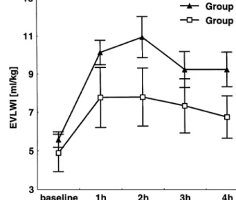
Fig. 2. EVLWI at baseline and 1, 2, 3 and 4 h after occlusion of the right contralateral lung (mean9S.E.M.).
62.1 dyne/s per cm− 5, P = 0.73). No statistical
signifi-cant difference in pulmonary vascular resistence was found during the whole observation period.
3.4. Reperfusion edema
After occlusion of the right lung, one hour after reperfusion, only EVLWI of the graft was measured. EVLWI at this time point was lower in Group II (7.7791.57 ml/kg) compared to Group I (10.1290.64 ml/kg, P = 0.25). This trend to reduced edema (Fig. 2) in the group without topical cooling persisted until four hours after occlusion (6.7491.1 vs. 9.2290.90 ml/kg, P = 0.23). However, differences were not significant.
Intrathoracic blood volume showed no differences between the groups.
3.5. PMN migration
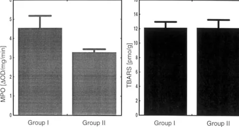
A tendency to a reduced MPO-activity in Group II (3.2590.19 DOD/mg per min), compared to Group I (4.5290.64 DOD/mg per min), was noted at the end of the observation period (PB0.08) Fig. 3.
3.6. Lipid peroxidation
No difference of TBARS in tissue homogenates be-tween groups was seen 5 h after reperfusion: Group I 12.0790.86 pmol/kg compared to Group II 12.079 1.18 pmol/g (Fig. 3).
4. Discussion
Various experiments have been conducted to evaluate the optimal preservation conditions in lung transplanta-Fig. 1. Cardiac output at baseline and 1, 2, 3 and 4 h after occlusion
Fig. 3. MPO-Assay and TBARS in graft tissue at the end of the observation period (mean9S.E.M.). tion; and the mechanisms of ischemia-reperfusion
in-jury have been studied extensively in the field of tho-racic organ transplantation. However, to our knowledge the beneficial effect of topical lung cooling during implantation with subsequent rapid reperfusion of the cold organ on posttransplant lung edema and hemodynamics has not been evaluated.
In the present experiment we compared the effect of topical cooling during implantation to prewarming of the organ for a 2-h period at room temperature prior to reperfusion. Surprisingly we found that prewarming of the lung allograft and avoidance of topical cooling with ice slush did not impair post-transplant graft function. On the contrary, a tendency to improved posttransplant hemodynamics, reduced reperfusion edema, and re-duced PMN migration to the allograft was noted. The beneficial effect on pulmonary vascular resistance was most marked in the early phase of reperfusion.
Without focusing directly on the problem of rewarm-ing a number of previously performed studies by other groups support our finding that low temperature of the organ at the time of reperfusion might enhance the development of reperfusion injury and vascular dys-function. Moriyasu et al. [8] studied the effect of an initial warm (20°C) crystalloid flush before regular reperfusion after a 6-h preservation period in an ex vivo rabbit model. The flush was used to eliminate toxic metabolites from the vascular system. The observed improvement of pulmonary vascular resistance after reperfusion might be as well a consequence of rewarm-ing of the graft before reperfusion. Bhabra et al. demonstrated in a rat lung transplant model that initial low-pressure reperfusion for 10 min reduces posttrans-plant lung edema [9]. Also in this experiment it is possible that the amelioration of reperfusion injury is at least partially due to a rewarming effect before reperfusion.
Van Raemdonk et al. demonstrated that lung flush is the most effective way to change the temperature of the graft [10]. Reperfusion of the organ with blood at body temperature, therefore, induces a rapid increase of the tissue temperature in the graft which might disturb reorganisation of the cytoskeletal components of the endothelial cells which demonstrate abnormal configu-ration already during cold preservation [11]. It was shown that lungs are able to maintain aerobic metabolism during storage at 10oC and that this
tem-perature seems to be the ideal storage temtem-perature for lungs [12,13]. Prewarming activates cell metabolism in a similar way which might have a beneficial effect on posttransplant lung injury.
In addition, Ingemansson et al. demonstrated in a series of experiments that cold preservation severely impairs endothelial dependent and endothelial indepen-dent vascular relaxation after reperfusion [14] and that cold ischemia and reperfusion independently induce vascular dysfunction. Our results indicate that reperfu-sion with subsequent rapid rewarming of the lung allo-graft further increases vascular dysfunction. On the other hand we could not demonstrate that the short period of warm ischemia before reperfusion enhances the production of oxygen free radicals substantially as lipid peroxidation products, assessed by the measure-ment of tissue-TBARS 5 h after reperfusion, were not increased in the group without topical cooling. We also observed a strong tendency towards a reduction of PMN migration to the allograft which correlates in many models with the severity of posttransplant lung injury. This reduction might be caused by reduced neutrophil sequestration in the lung, due to reduced sheer forces by improved hemodynamics, and decreased PMN and endothelial activation.
In addition, reperfusion injury in this new experimen-tal setting was evaluated by the measurement of ex-travascular lung water which is an excellent parameter
to assess minor differences in posttransplant lung edema [4]. Clinical studies demonstrated that EVLWI correlates better to the pathological status of the lung than gas exchange and radiological signs of edema [15,16]. A 20-h preservation time was chosen because following this ischemic injury the recipients still tolerate ligation of the right pulmonary artery which allows to assess isolated graft function. If a longer preservation time is used a rapid decrease of allograft gas exchange is noted and the animals die from right heart failure. In our hands temperature measurement in the allo-graft bronchus was unreliable, therefore mimicking the clinical situation, the preservation temperature in the ice box, the room temperature and the recipient’s body temperature were measured and kept constant in all experiments. Measuring the lung core temperature would have harmed the graft. So we are not able to prove the hypothesis that the 2-h period of warm ischemia led to a remarkable higher core temperature. However, it was not our intention to show the time course of lung parenchyma temperatures with topical cooling versus prewarming but to imitate the clinical situation in lung transplantation and compare topical cooling with the use of no topical cooling.
In conclusion this study in a large animal model of reperfusion injury following unilateral lung transplanta-tion indicates that topical cooling of the allograft with ice slush during separation and implantation seems to be unnecessary. Posttransplant edema and hemody-namics in the group without topical cooling were at least as good in this model than in recipients in which the lung grafts were cooled topically and reperfused at low tissue temperature.
References
[1] Date H, Matsumura A, Manchester JK, Obo H, Lima O, Cooper JM, Sundaresan S, Lowry OH, Cooper JD. Evaluation of lung metabolism during successful 24 h canine lung preserva-tion. J Thorac Cardiovasc Surg 1993;105:480 – 91.
[2] Schmid RA, Kwong GK, Boasquevisque CHR, Wick M, Patter-son GA, Sundt TM. A chronic large animal model of lung allograft rejection. Transplant Proc 1997;29:1521.
[3] Hachenberg T, Tenling A, Rothen HU, Nystro¨m SO, Tyden H, Hedenstierna G. Thoracic intravascular and extravascular fluid volumes in cardiac surgical patients. Anesthesiology 1993;79:976 – 84.
[4] Wickerts CJ, Jakobson J, Frostell C, Hedenstierna G. Measure-ment of extravascular lung water by thermal-dye dilution tech-nique: mechanisms of cardiac output dependence. Intensive Care Med 1990;16:115 – 20.
[5] Schmid RA, Yamashita M, Ando K, Cooper JD, Patterson GA. Lidocaine reduces reperfusion injury and neutrophil migration in canine lung allografts. Ann Thorac Surg 1996;61:949 – 55. [6] Smith PK, Krohn RI, Hermanson GT, Mallia AK, Gartner FH,
Provenzano MD, Fujimoto EK, Goeke NM, Olson BJ, Klenk DC. Measurement of protein using bicinchoninic acid. Anal Biochem 1985;150:768 – 85.
[7] Ohkawa H, Ohishi N, Yagi K. Assay for lipid peroxides in animal tissues by thiobarbituric acid reaction. Anal Biochem 1979;95:351 – 8.
[8] Moriyasu K, McKeown PP, Novitzky D, Snow TR. Beneficial effect of initial warm crystalloid reperfusion in 6 h lung preserva-tion. J Heart Lung Transplant 1995;14:699 – 705.
[9] Bhabra MS, Hopkinson DN, Shaw TE, Hooper TL. Controlled reperfusion protects lung grafts during a transient early increase in endothelial permeability. J Heart Lung Transplant 1997;16:100(Abstract).
[10] Van Raemdonck DEM, Jannis NCP, Rega FRL, De Leyn PRJ, Flameng WJ, Lerut TE. External cooling of warm ischemic rabbit lungs after death. Ann Thorac Surg 1996;62:331 – 7. [11] Hall SM, Evans J, Haworth SG. Influence of cold preservation
on the cytoskeleton of cultured pulmonary artery endothelial cells. Am J Respir Cell Mol Biol 1993;9:106 – 14.
[12] Weder W, Harper B, Shimokawa S, Miyoshi S, Date H, Schreinemakers H, Egan T, Cooper JD. Influence of intraalve-olar oxygen concentration on lung preservation in a rabbit model. J Thorac Cardiovasc Surg 1991;101:1037 – 43.
[13] Date H, Matsumura A, Manchester JK, Cooper JM, Lowry OH, Cooper JD. Changes in alveolar oxygen and carbon dioxide concentration and oxygen consumption during lung preserva-tion-the maintenance of aerobic metabolism during lung preser-vation. J Thorac Cardiovasc Surg 1993;105:492 – 501.
[14] Ingemansson R, Budrikis A, Bolys R, Sjo¨berg T, Steen S. Effect of temperature in long-term preservation of vascular endothelial and smooth muscle function. Ann Thorac Surg 1996;61:1413 – 7. [15] Laggner A, Kleinberger G, Haller J, Lenz K, Sommer G, Druml W. Bedside estimation of extravascular lung water in critically ill patients: comparison of the chest radiograph and the thermal dye technique. Intensive Care Med 1984;10:309 – 13.
[16] Sibbald WJ, Short AK, Warshawski FJ, Cunningham DG, Cheung H. Thermal dye measurements of extravascular lung water in critically ill patients. Intravascular Starling forces and extravascular lung water in the adult respiratory distress syn-drome. Chest 1985;87:585 – 92.
Appendix A. Conference discussion
Dr G. Petterson (Copenhagen, Denmark): Very interesting and
possibly somewhat surprising results. I had some concern when we did bronchial artery revascularization and allowed the mammary artery to be open during the course of the procedure, early bronchial artery reperfusion could be harmful. I have the impression that that was not the case.
Dr D. van Raemdonck (Leu6en, Belguim): I have a few questions.
First, did you measure lung core temperature in the second group while this allograft was rewarming? The second question, during the cold preservation of 20 h did you keep the lung inflated, because it’s well known that warm ischemic tolerance is prolonged during infla-tion and clinically during implantainfla-tion you cannot keep the lung inflated. Finally, what is your advice for us clinical lung transplant surgeons? Do we need to cool the lung during implantation or not?
Dr U. Stammberger: Firstly, we did not measure temperature in the
lung allograft because it is known, for example, by the work of van Raemdonck, that measuring temperature in the bronchus is unreliable since it does not represent tissue temperature, and all other techniques to measure temperature in the lung would harm the allograft tissue. In answer to the second question, the lungs were inflated during storage and were deflated during implantation. To your last question, our clinical practice in Zurich is that we use some amount of ice slush in the thoracic cavity but the lung is not embedded in the ice slush. This experiment has confirmed that we do not have to change this practice since maximal topical cooling seems not to be superior.
Dr N.J. Odom (Manchester, UK): Interesting results and in a way
the opposite to what one would have expected. Do you have any theory as to why the lungs that are allowed to warm up seem to do better? In relation to that, may I ask, was there any dextrose in your preservation solution? It has been shown that lungs stored at 10°C, if there is a metabolic substrate present, perhaps they do better, and might that be a possible explanation? But I don’t know if you had dextrose in your preservation solution or not. Where do you go from here? How might you explain this? What do you think might be going on?
Dr U. Stammberger: We use Perfadex as the flush solution and we
also have similar hypotheses on the mechanism during rewarming. Maybe the short period of warm ischemia allows the endothelial cells to establish metabolism to a level that let them better cope with reperfusion injury, but in the presented experiment we did not study these mechanisms.
D.R. Metras (Marseille, France): I want to congratulate Dr
Stamm-berger for an extremely elegant study and very strange results. The only thing which is of concern to me is the temperature of your cold perfusate. It looks like it’s 1°C and topical ice slush. It looks extremely cold to me, and I thought that too cold was not that good
for the preservation of tissues (this has been shown for the heart). I want to know why you chose so low a temperature?
Dr U. Stammberger: It is well known that the ideal temperature for
lung preservation is around 10°C. However, in clinical practice the organ is harvested and stored in a cooling box with a mixture of ice and water. So, we choose the same temperature as in clinical practice. We might have seen other results if we choose 10°C storage tempera-ture.
A. Haverich (Hanno6er, Germany): Now, in clinical practice the
true consequences of reperfusion injury are usually seen after 12 – 16 h. Your observation period was only 4 h. Would you expect that with a longer observation period the results might have been different?
Dr U. Stammberger: Extravascular lung water is a very early
parameter of reperfusion injury. So, in our opinion, the 5-h period (1 h reperfusion, then occlusion of the contralateral lung, 4 h assess-ment) is sufficient. Moreover, at the end of this period, extravascular lung water was again decreasing in this model. We cannot predict what the situation might have been after 12 or 16 h. In clinical use ischemic times up to 6 or 7 h are accepted. After this shorter ischemic period the consequences of reperfusion injury might be delayed.