S1
Supporting Information
A Mechano- and Thermoresponsive Luminescent Cyclophane
Yoshimitsu Sagara,*,1,2 Yoan C. Simon,2 Nobuyuki Tamaoki,1 and Christoph Weder*,2
1Research Institute for Electronic Science, Hokkaido University, N20, W10, Kita-Ku, Sapporo 001-0020, Japan
2Adolphe Merkle Institute, University of Fribourg, Chemin des Verdiers 4, Fribourg CH-1700, Switzerland
E-mail: sagara@es.hokudai.ac.jp; christoph.weder@unifr.ch
Contents
General methods and materials
S1
Synthesis of compounds 1 and 2
S2
Grinding procedure used to explore MRL characteristics of compounds 1 and 2
S3
NMR spectra of 1 and 2
S4
Summary of the photophysical properties
S5
Emission decay profiles in solution
S5
Concentration dependence of the absorption and emission spectra of 1 and 2
S5
Precipitation of 1 from other solvent combinations
S6
X-ray diffraction measurements
S6
DSC measurements
S7
TGA measurements
S7
Emission decay profiles in the solid state
S7
Emission spectral change responding to mechanical stimuli for 2
S8
References
S8
General methods and materials
All reagents and solvents were purchased from Aldrich and Tokyo Kasei. Unless otherwise noted, the reactions were carried out under an argon atmosphere in dry solvents. Silica gel column chromatography was carried out with a Biotage Isolera Flash system. 1H NMR spectra were recorded on a JEOL JNM-ECX 400spectrometer and all chemical shifts are quoted on the δ-scale in ppm relative to the signal of tetramethylsilane (at 0.00) as an internal standard. Proton-decoupled 13C NMR spectra were recorded on a JEOL JNM-ECX 400 spectrometer or a JEOL ECP 400 spectrometer and all chemical shifts (δ) are reported in ppm using residual solvent as the internal standard (CDCl3 at 77.16, DMSO-d6 at 39.52). Coupling constants (J) are reported in Hz and relative intensities are also shown. Matrix-assisted laser desorption ionization time-of-flight (MALDI-TOF) mass spectra were obtained on an Applied Biosystems Voyager-DE STR-H spectrometer. Elemental analysis was carried out with an Exeter Analytical CE440 Elemental Analyzer. The DSC measurements were conducted using a Rigaku Thermo plus DSC8230 with a heating/cooling rate of 10 ˚C/min under nitrogen atmosphere. Thermogravimetric analysis was conducted using a Rigaku Thermo plus TG8120 under nitrogen condition. Powder X-ray diffraction measurements were carried out with a Rigaku SmartLab. UV-vis absorption spectra were measured with a JASCO V-570.
S2
Steady-state fluorescence spectra were recorded on a JASCO FP-8500. Time-resolved fluorescence measurements were carried out with a Hamamatsu Photonics Quantaurus-Tau. Quantum efficiencies were measured with a Hamamatsu Photonics Quantaurus-QY.
Synthesis of compounds 1 and 2
The synthetic route used to prepare compounds 1 and 2 is shown in Scheme S1. 2-(4-Ethynyl- phenoxy)-tetrahydro-2H-pyran and 2-{2-[2-(2-hydroxyethoxy)ethoxy]ethoxy}ethyl 4-methylbenzenesulfonate were synthesized according to reported procedures.S1,S2
Scheme S1
Conditions: (a) 9,10-dibromoanthracene, Pd(PPh3)4, CuI, THF, i-Pr2NH, 80 °C, 4 h; (b) 10% aq. HCl, THF, reflux, 3 h; (c) 2-{2-[2-(2-hydroxyethoxy)ethoxy]ethoxy}ethyl 4-methylbenzenesulfonate, K2CO3, LiBr, MeCN, 100 °C, 16 h; (d) CBr4, PPh3, CH2Cl2, 0 °C ® r.t., 2 h; (e) compound 4, K2CO3, DMF, 70 °C, 48 h.
9,10-Bis(4-(tetrahydro-2H-pyran-2-yloxy)phenylethynyl)anthracene (3). A mixture of 9,10-dibromoanthracene
(1.19 g, 3.53 mmol), 2-(4-ethynylphenoxy)-tetrahydro-2H-pyran (1.50 g, 7.41 mmol), CuI (2.00 mg, 1.03 × 10⁻² mmol), Pd(PPh3)4 (12.0 mg, 1.03 × 10⁻² mmol), and distilled i-Pr2NH (15 mL)in THF (50 mL) was degassed and stirred for 4 h at 80 °C. After cooling to room temperature, methanol (20 mL) was added to the reaction mixture. The precipitate was filtered off and washed with methanol several times. The solid was then dissolved in chloroform, purified by flash column chromatography on silica gel (eluent: chloroform) and subsequently re-precipitated from a mixture of chloroform and methanol to afford compound 3 (1.68 g, 2.90 mmol) as an orange solid in 82% yield. a b 2 3 c 1 4 d e 5
S3 1H NMR (400 MHz, CDCl 3): d = 1.59–1.76 (m, 6H), 1.88–1.92 (m, 4H), 1.98–2.07 (m, 2H), 3.62–3.67 (m, 2H), 3.89–3.95 (m, 2H), 5.51 (t, J = 2.8 Hz, 2H), 7.13 (d, J = 8.8 Hz, 4H), 7.59–7.64 (m, 4H), 7.70 (d, J = 8.8 Hz, 4H), 8.65–8.70 (m, 4H). 13C NMR (100 MHz, CDCl3): d = 18.78, 25.29, 30.39, 62.19, 85.58, 96.39, 102.61, 116.56, 116.76, 118.60, 126.76, 127.44, 132.12, 133.20, 157.60. MS (MALDI-TOF): m/z: 578.31 (calcd. [M] + = 578.25).
9,10-Bis(4-hydroxyphenylethynyl)anthracene (4). A mixture of 3 (1.50 g, 2.59 mmol), 10% aq. HCl (5 mL), and
THF (150 mL) was heated to reflux for 3 h and subsequently cooled to room temperature. Water (20 mL) was added, most of the THF was evaporated, and the solid product was filtered off. The solid was washed with water (2 ´ 50 mL) and a mixture of water/methanol (3/2 v/v) (2 ´ 20 mL), followed by methanol (2 ´ 5 mL). The product was dried in vacuo to afford compound 4 (1.01 g, 2.46 mmol) as an orange solid in 95% yield.
1H NMR (400 MHz, DMSO-d6): d = 6.95 (d, J = 8.0 Hz, 4H), 7.73 (d, J = 8.0 Hz, 4H), 7.75–7.79 (m, 4H), 8.64– 8.68 (m, 4H), 10.13 (s, 2H). 13C NMR (100 MHz, DMSO-d6): d = 84.26, 103.58, 112.51, 116.02, 117.58, 126.87, 127.43, 131.07, 133.46, 158.68. MS (MALDI-TOF): m/z: 410.04 (calcd. [M] + = 410.13).
9,10-Bis(4-(2-{2-[2-(2-hydroxyethoxy)ethoxy]ethoxy}ethoxy)phenylethynyl)anthracene (2). A suspension of 4 (400 mg, 0.975 mmol), 2-{2-[2-(2-hydroxyethoxy)ethoxy]ethoxy}ethyl 4-methylbenzenesulfonate (680 mg, 1.95 mmol), K2CO3 (398 mg, 2.93 mmol), and lithium bromide (8.5 mg, 0.098 mmol) in acetonitrile (100 mL) was stirred for 16 h at 100 °C. After cooling to room temperature, most of the acetonitrile was evaporated and 5% aq. HCl (50 mL) was added to the suspension. The mixture was extracted with chloroform (3 ´ 100 mL), the combined organic layers were washed with saturated aq. NaCl solution (2 ´ 50 mL), dried over MgSO4, filtered, and the solvent was evaporated. The crude product was purified by flash column chromatography on silica gel (eluent: linear gradient from chloroform/methanol = 98:2 to chloroform/methanol = 90:10) to afford compound 2 (510 mg, 0.669 mmol) as an orange solid in 69% yield.
1H NMR (400 MHz, CDCl
3): d = 2.63 (t, J = 6.0 Hz, 2H), 3.62 (t, J = 4.8 Hz, 4H), 3.67–3.76 (m, 20H), 3.89 (t, J = 4.8 Hz, 4H), 4.20 (t, J = 4.8 Hz, 4H), 6.99 (d, J = 8.8 Hz, 4H), 7.61–7.64 (m, 4H), 7.69 (d, J = 8.8 Hz, 4H), 8.66– 8.69 (m, 4H). 13C NMR (100 MHz, CDCl3): d = 61.88, 67.64, 69.78, 70.46, 70.71, 70.79, 70.97, 72.63, 85.56, 102.56, 114.99, 115.91, 118.55, 126.75, 127.41, 132.07, 133.27, 159.29. MS (MALDI-TOF): m/z: 762.13 (calcd. [M] + = 762.34).Elemental analysis (%) calcd. for C46H50O10: C 72.42, H 6.61, N 0.00; found: C 72.36, H 6.52, N 0.03.
9,10-Bis(4-(2-{2-[2-(2-bromoethoxy)ethoxy]ethoxy}ethoxy)phenylethynyl)anthracene (5). To a solution of 2 (1.20 g, 1.57 mmol) and triphenylphosphine (988 mg, 3.77 mmol) in dichloromethane (150 mL) was added dropwise a solution of tetrabromomethane (1.30 g, 3.93 mmol) in dichloromethane (10 mL) at 0 °C. The reaction mixture was subsequently stirred for 2 h at room temperature before it was washed with saturated aq. NaCl solution (2 ´ 50 mL), the organic layer was dried over MgSO4, filtered, and the solvent was evaporated. The crude product was purified by flash column chromatography on silica gel (eluent: linear gradient from hexane/chloroform = 20:80 to hexane/chloroform = 10:90) to afford compound 5 (980 mg, 1.10 mmol) as an orange solid in 70% yield. 1H NMR (400 MHz, CDCl
S4
4.8 Hz, 4H), 4.19 (t, J = 4.8 Hz, 4H), 6.99 (d, J = 8.4 Hz, 4H), 7.60–7.63 (m, 4H), 7.69 (d, J = 8.4 Hz, 4H), 8.66– 8.69 (m, 4H). 13C NMR (100 MHz, CDCl3): d = 30.48, 67.66, 69.78, 70.66, 70.76, 70.83, 71.00, 71.32, 85.56, 102.57, 114.99, 115.88, 118.54, 126.75, 127.40, 132.06, 133.26, 159.31. MS (MALDI-TOF): m/z: 888.11 (calcd. [M]+ = 887.17).
Compound 1. A solution of compound 5 (200 mg, 0.225 mmol) and compound 4 (92.4 mg, 0.225 mmol) in DMF
(25 mL) was added to a suspension of K2CO3 (466 mg, 3.38 mmol) in DMF (150 mL) dropwise at 70 °C over 12 h under vigorous stirring. After 36 h, the reaction suspension was cooled and most of the DMF was evaporated in vacuo. The crude product was dissolved in chloroform and washed with saturated aq. NH4Cl solution (3 ´ 100 mL), followed by saturated aq. NaCl solution, the organic layer was dried over MgSO4, filtered, and the solvent was evaporated. The crude product was purified by flash column chromatography on silica gel (eluent: ethyl acetate/chloroform = 20:80) and subsequent re-precipitated from a mixture of chloroform and methanol to afford compound 1 (53.0 mg, 4.68 ´ 10–2 mmol) as a yellow powder in 21% yield.
1H NMR (400 MHz, CDCl
3): d = 3.75–3.81 (m, 16H), 3.95–3.98 (m, 8H), 4.08–4.10 (m, 8H), 6.83 (d, J = 8.8 Hz, 8H), 7.15–7.18 (m, 8H), 7.53 (d, J = 8.8 Hz, 8H), 8.18–8.20 (m, 8H). 13C NMR (100 MHz, CDCl3): d = 67.64, 69.73, 70.71, 71.02, 85.66, 101.89, 114.62, 116.18, 117.92, 125.86, 126.76, 131.42, 133.13, 158.96. MS (MALDI-TOF): m/z: 1136.73 (calcd. [M] + = 1136.45).Elemental analysis (%) calcd. for C76H64O10: C 80.26, H 5.67, N 0.00; found: C 80.07, H 5.55, N 0.04.
Grinding procedure used to explore MRL characteristics of compounds 1 and 2
Compounds 1 and 2 were ground on glass or quartz substrates under ambient conditions with a metal spatula until the emission color no longer changed.
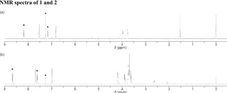
NMR spectra of 1 and 2
Fig. S1 1H NMR spectra of cyclophane 1 (a) and the linear reference compound 2 (b). Peaks corresponding to the anthracene moieties and chloroform (solvent) are marked with asterisks and a triangle, respectively.
S5
Summary of the photophysical properties
Table S1. Emission lifetimes and quantum yieldsa)
Emission decay profiles in solution
Fig. S2 Emission decay profiles of chloroform solutions of (a) the linear reference compound 2 collected at 495 nm
and (b) cyclophane 1 collected at 495 nm (orange line) and 600 nm (red line). All profiles were measured at room temperature with lex = 405 nm and concentrations of 1 ´ 10–5 M.
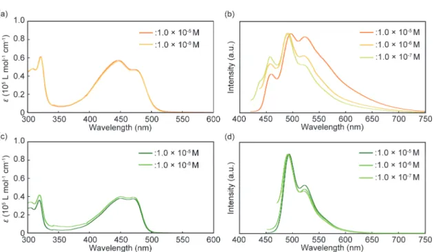
Concentration dependence of the absorption and emission spectra for 1 and 2
Fig. S3 (a,b) Absorption and emission spectra of 1 in chloroform at different concentrations. (c,d) Absorption and
emission spectra of 2 in chloroform at different concentrations. All emission spectra were measured at room temperature with λex = 400 nm.
S6
Precipitation of 1 from other solvent combinations
The YCR-form was obtained by precipitation from chloroform/hexane, whereas other solvent combinations typically afforded orange-yellow emitting powders. For instance, Fig. S3 shows the result of precipitation using chloroform/methanol.
Fig. S4 (a) An image representing the photoluminescence color of the powder obtained through precipitation from
chloroform/methanol. (b) Photoluminescence spectra of the YCR-form (yellow line), the ROCR-form (red line), and the orange-yellow emitting powder obtained by precipitation using chloroform/methanol (orange line).
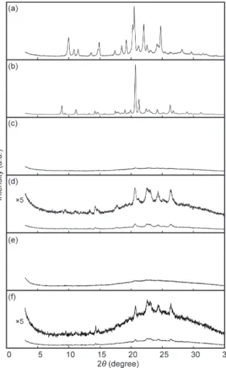
X-ray diffraction measurements
Fig. S5 Powder X-ray diffraction patterns of 1 in the YCR-form (a, precipitated into hexane), ROCR-form (b, after heating for 10 min at 250 °C), YAM1-form (c, after grinding to ROCR-form), ROLO-form (d, after subsequent heating to YAM1-form for 10 min at 250 °C), YAM2-form (e, after grinding to YCR-form), and ROLO-form (f, after subsequent heating to YAM2-form for 10 min at 250 °C). All measurements were carried out at room temperature.
S7
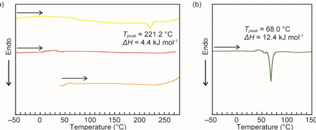
DSC measurements
Fig. S6 (a) DSC curves of the YCR-form (yellow line), ROCR-form (red line), and YAM1-form (orange line) on the first heating. (b) DSC curve of the compound 2 on the first heating.
TGA measurements
Fig. S7 Thermogravimetric analysis traces of the YCR-form (yellow line) and ROCR-form (orange line) of cyclophane 1.
Emission decay profiles in the solid state
Fig. S8 Emission decay profiles of the YCR-form (yellow line) and the ROCR-form (orange line) of cyclophane 1 collected at 600 nm. All profiles were measured at room temperature with lex = 405 nm.
S8
Emission spectral change responding to mechanical stimuli for 2
Fig. S9 Photoluminescence spectra of 2 in the solid states before grinding (green solid line) and after grinding
(green dotted line). All spectra were measured at room temperature with λex = 400 nm.
References
S1. C. V. Yelamaggad and G. Shanker, Tetrahedron, 2008, 64, 3760–3771.