European Heart Journal (1990) 11, 566-571
Is there an isolated arrhythmogenic right atrial
myocarditis?
M. FROMER, C. GENTON,* J. SCHLAEPFER, JJ. GOY, L. KAPPENBERGER
Division of Cardiology, Medical Clinic, University Hospital, Lausanne and the * Institute of Pathology, University of Lausanne, Switzerland
KEY WORDS: Atrial myocarditis, atrial tachycardia, endocardial resection.
Two cases with drug refractory ectopic atrial tachycardia are described. A map-guided partial resection of the right atrium (RA) was done after preoperative endocardial catheter mapping hadshown well-defined areas of fractionated RA potentials. Intraoperatively, there were no aneurysmal formations present as described by other authors. Histopathologic examination of the resected tissue showed atrial myocarditis in both patients. Postoperative right ventricular myocardial biopsies revealed no inflammatory tissue. A minor elevation of antibodies against echoviruses was found in one case. Postoperative electrophysiologic studies were negative.
We conclude: focal RA myocarditis without concomitant ventricular myocarditis may represent one cause of drug-resistant ectopic atrial tachycardia. Map-guided surgical intervention may cure the disease.
Introduction
The anatomic substrate of ectopic atrial tachy-cardia (AT) is rarely evaluated as the investigation of the underlying disease requires a surgical approach. Successful surgical resection was reported by a few groups'1"3'. Olsson el a/.'1' described an aneurysmal formation of the atrial tissue close to the right atrial appendix. Wyndham
et al.m reported in 1980 a case with patchy fibrosis of the right atrium close to the appendix, an increase of connective tissue and mononuclear cell infiltration. The same group reported later surgical experience with eight cases, however, the results of histologic examination were not given'31. In our report of two cases we describe the occurrence of ectopic right AT without aneurysmal formation, however with well-defined inflammation of the right atrium in the absence of concomitant myocarditis of the right ventricle. We hypothesize that myocarditis of the right atrium may be responsible for recurrent ectopic AT resistant to antiarrhythmic drug regi-mens, and map-guided resection of the abnormal area may cure the disease.
Submitted for publication on 9 August 1989, and in revised form 26 September 1989.
Martin Fromer is recipient of a SCORE grant #3.750-87 from the Swiss National Foundation of Sciences.
Address for Correspondence M. Fromer, MD. Division of
Cardiology, Centre Hospitalier Universitaire Vaudois, CHUV, 1011 Ljusanne. Switzerland.
Casel
A 20-year-old male was referred for electro-physiologic evaluation of a 1 -year history of highly symptomatic, drug-resistant supraventricular tachycardia. Clinical and echocardiographic exam-ination showed no associated cardiomyopathy. Standard techniques for electrophysiologic evalu-ation were used'4'. An atrial tachycardia with a cycle length of 300 ms and 2:1 atrioventricular conduction was present (see Fig. 1). Endocardial catheter mapping showed areas of abnormal electri-cal activity with fractionated potentials in the postero-lateral regions of the mid right atrium (Fig. 2). This area was considered to be responsible for the AT. This finding together with the patient's history motivated an attempt at surgical resection. Intraoperative epicardial mapping confirmed the presence of an ectopic right sided AT originating in the infero-posterior areas of the right atrium. The postoperative course was unremarkable and the atrial Jachycardia did not reoccur.
Case 2
A 27-year-old male has been referred for the evaluation of a 5-year history of drug-resistant supraventricular tachycardia. Drug trials with disopyramide, verapamil, beta blockers, flecainide and propafenone were not effective. Clinical and echocardiographic evaluation disclosed no cardio-vascular abnormality. The electrophysiologic study 0195-668X 90 060566 + 06 $03.00 0 © 1990 The European Society of Cardiology
Focal RA myocarditis 567
SCHC
Figure 1 12-lead standard electrocardiogram of patient 1
show-ing an atrial tachycardia with 2:1 atrioventricular conduction. Paper speed 25 mm s"'.
showed an atrial tachycardia (Fig. 3) and large zones of fractionated potentials (Fig. 4) in the mid-right atrium. The same findings were reproducible during a second electrophysiologic study done pre-operativelyand motivated the surgical intervention. Epicardial mapping confirmed the presence of right sided AT. Intra-operatively, large zones with frag-mented potentials were found around the appendix and anterolateral segments of the right atrium. A large resection including all areas of fragmented potentials was done and replaced with a pericardial patch. After this the AT was no longer inducible.
Results of histologic examination CASE 1
Six specimens were given for histologic examin-ation. The largest measuring 2 x 1 x 0 - 5 cm, the
smallest 1 -5 x 0-7 x 0-2 cm. Histologically, a moder-ate inflammatory infiltrmoder-ate was found in the myo-cardium and endomyo-cardium. This focally confluent infiltrate was essentially lymphocytic, there were no eosinophilic leucocytes nor giant cells. Obviously damaged myocytes with vacuolization were present (Fig. 5). In some areas, granulation tissue with the beginning of fibrosis was present. There were no mural thrombi. These findings are consistent with a moderate, diffuse lymphocytic myocarditis with granulation tissue and fibrosis. Eight days later, right ventricular endomyocardial biopsies were obtained from the lateral wall and septum. Examin-ation of multiple sections of the six specimens suggested an increased interstitial cellularity but immunohistochemical staining for polymorpho-nuclear leucocytes and lymphocytes were negative. The specimens were therefore considered to represent normal myocardium.
568 M. Fromer tta\.
M R A
H B E
-SCH.C. Figure 2 Representative illustration of local recordings of right atrial electrocardiograms in
patient 1 dunng atrial tachycardia with 2:1 AV-conduction. Arrows indicate nght atrial potentials with abnormal low amplitude. Note for comparison the normal right atrial potentials (HRA) with rapid rise of the intnnsic deflection and high amplitude of the local electrocardiogram and shorter duration of the signal. I, aVF, V1 and V6 represent standard surface electrocardiographic leads. Other abbreviations: HRA = high right atrium, MRA = mid right atnum, HBE = His bundle electrocardiogram, including low right atrial depolarization. Paper speed 100 mm s~'.
V * » •
Figure 3 12-lead electrocardiagrams of patient 2 showing ectopic
right atnal tachycardia with 1:1 conduction. Paper speed at left 2 5 m m s ', at right 50mm s ' .
Focal RA myocarditis 569 l t 774 aVF
—4r ~ V ^
4/
J^-MRA HRA K.P.
Figure 4 Local recordings of right atnal electrocardiograms in patient 2 during right atnal
tachycardia with 1:1 conduction. Arrows indicate abnormal fragmented nght atrial potentials as discussed in Fig. 2. For abbreviations see Fig. 2.
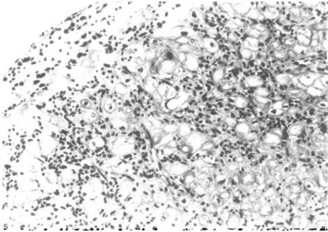
Figure 5 Confluent lymphocytic infiltrate of myocardium and endocardium accompanied by
obvious vacuolization of myocytes and beginning fibrosis. Haematoxylin and eosin x 250.
CASE 2 endocardium showed oedema and an essentially The resected part of the right atrium measured lymphocytic infiltrate which was also present in the 6 x 3-5 cm and comprised the appendix, which myocardial interstitium. There was focal vacuoliz-appeared slightly dilated. Microscopically the ation and necrosis of myocytes (Fig. 6), sometimes
570 M. Fromer eta\.
figure o hocal and connuent lymptiocytic lntiltrates in trie myocardium with vacuolization
and focal necrosis of myocytes, some of which show large hypcrchromatic nuclei. Haematoxylin and eosin x 250.
accompanied by small haemorrhages. There was also some degree of interstitial fibrosis. The micro-scopic findings are consistent with a moderate dif-fuse lymphocytic myocarditis with some interstitial fibrosis of unknown origin.
Discussion
Several reports in the literature have discussed the diagnostic, therapeutic and electrophysiologic aspects of ectopic (right) atrial tachycardia'1"3-6-71. However, the probable anatomic substrate for these tachycardias is difficult to investigate. Wyndham
et a/.'21 described the histologic findings of one patient with ectopic right AT demonstrating mono-nuclear cell infiltration and increased connective tissue. Olsson et a/.'1' described two patients with ectopic AT. In one of those patients three small aneurysms were seen located between the sinus node and right atrial appendage. After excision of this area, sinus rhythm reappeared. Results of histologic examination were not given.
In the two cases described here, no aneurysmal formations were observed. Histologic examination showed a subacute myocarditis in one case and an on-going chronic myocarditis in the other151.
Biopsies of the right ventricle showed no con-comitant ventricular myocarditis, however, proof
that the disease was only affecting the right atrium was unavailable. The etiology of the disease is not clear yet. In case 1 serologic investigations for a viral infection have not been done. In case 2 a mild elevation of antibodies to echoviruses was found. In both cases the endocardial catheter mapping showed large areas in the right atrium with abnor-mal electrical activity. After intra-operative con-firmation of these findings and resection of those abnormal zones, the ectopic tachycardia dis-appeared. We therefore conclude that in some cases with ectopic (right) AT, an isolated myocarditis of the atrium may be the anatomic substrate for the arrhythmia. Careful histologic examination of the resected areas may clarify the pathogenesis of ectopic AT in a subgroup of patients. Further investigations are necessary to clarify whether the management of these patients requires anti-inflammatory drug therapy to avoid dissemination of the disease.
References
[1] Olsson SB, Blomstroem P, Sabel K-G, William-Olsson G. Incessant ectopic atrial tachycardia: successful surgical treatment with regression of dilated cardiomyopathy pic-ture. Am J Cardiol 1984; 53: 1465-6.
[2] Wyndham CRC, Amsdorf MF, Lcvitsky S et at. Successful surgical excision of focal paroxysmal atrial tachycardia. Circulation 1980,6: 1365-72.
Focal RA myocarditis 571
[3] Seals AA, Lawrie GM, Magro S el al. Surgical treatment of right atrial focal tachycardia in adults. J Am Coll
Car-diol 1988; 11: 1111-7.
[4] Shenasa M, Fromer M, Faugere G el al. Efficacy and safety of intravenous and oral diltiazem for Wolff-Parkinson-White syndrome. Am J Cardiol 1987; 59: 301-6.
[5] Aretz HT. Myocarditis: the Dallas criteria. Hum Pathol 1987; 18:619-24.
[6] Mirowski M, Lau SH, Wit AL el al. Ectopic right atrial rhythms: experimental and clinical data. Am Heart J 1971; 81: 666-76.
[7] Scheinman MM, Basu D, Hollenberg M. Electrophysio-logic studies in patients with persistent atrial tachy-cardia. Circulation 1974; 50: 266-73.