Mechanisms of Brain Injury in Bacterial Meningitis: Workshop Summary
Hans-Walter Pfister, Adriano Fontana,
Martin G. Tauber, Alexander Tomasz,
and W. Michael ScheId
From the Department ofNeurologv, Klinikuni Gross/wdem. LlIdwig-Maxiniilians Universitv.Munich. Germanv: the Department ofClinical l mmunologv. Universitdtsspital. Zurich. Switzeriand: the Microbial
PathogenesisUnit. San Francisco General Hospital. Sail Francisco.
California. L/.\'I1: the Laboratorv oj'Microhi%gl. The Rockefdler Universit v. :Yew York. New York. USA: and the Departments of 'In tern a/ Medicine and Neurosurgerv. Universit v o] Vilxinia. ClWr/Oll£'.ITille. Virginia. USA
Morbidity and mortality associated with bacterial meningitis remain high, although antibiotic therapy has improved during recent decades. The major intracranial complications of bacterial meningitis are cerebrovascular arterial and venous involvement, brain edema, and hydrocephalus with a subsequent increase of intracranial pressure. Experiments in animal models and cell culture systems have focused on the pathogenesis and pathophysiology of bacterial meningitis in an attempt to identify the bacterial and/or host factors responsible for brain injury during the course of infection. An international workshop entitled "Bacterial Meningitis: Mechanisms of Brain Injury" was organized by the Department of Neurology at the University of Munich and was held in Eibsee, Germany, in June 1993. This conference provided a forum for the exchange of current information on bacterial meningitis, including data on the clinical spectrum of compli-cations, the associated morphological alterations, the role of soluble inflammatory mediators (in particular cytokines) and of leukocyte-endothelial cell interactions in tissue injury, and the mo-lecular mechanisms of neuronal injury, with potential mediators such as reactive oxygen species, reactive nitrogen species, and excitatory amino acids.Itis hoped that a better understanding of the pathophysiological events that take place during bacterial meningitis will lead to the develop-ment of new therapeutic regimens.
Although the recent licensure ofHaeniophilus influenzae
type b (Hib) polysaccharide conjugate protein vaccines has had a marked impact on the incidence of invasive Hib dis-ease in the United States and Western Europe. bacterial men-ingitis overall remains a significant problem worldwide. In addition, despite the introduction of new antimicrobial agents and improved diagnostic techniques, the mortality and morbidity associated with bacterial meningitis remain unacceptably high. This discrepancy between a rapid bacteri-ologic response at the site of infection (i.e .. eradication of viable bacteria from the CSF due to treatment with potent bactericidal agents) and the persistence of the associated neu-rological sequelae and mortality has prompted intense inves-tigation over the past two decades into the pathophysiology
Received 13 December 1993; revised 4 March 1994.
The workshop summarized herein was held in Eibsee, Germany. on 25 and 26 June 1993.
Reprints or correspondence: Dr. Hans-Walter Pfister. Klinikum Grossha-demo Ludwig-Maximilians University. Marchioninistrassc IS. 81336 Mun-ich. Germany.
Clinical Infectious Diseases 1994;19:463-79 © 1994 by The University of Chicago. All rights reserved. 1058-4838/94/1903-0010$02.00
of this disease. Indeed. recent clinical studies have employed adjunctive agents (e.g .. dexamethasone) in an attempt to re-duce mortality and alleviate neurological sequelae [1-4]. These studies and the resulting recommendations regarding the use of adjunctive dexamethasone have recently been re-viewed [5.6].
In line with the current emphasis on the pathogenesis and pathophysiology of bacterial meningitis. two workshops were held in California in the 1980s [7]. Recognizing that many advances have been made in this field in the several years since those meetings. the Department of Neurology at the University of Munich organized a similar workshop, which was held in Eibsce. Germanv. on 25 and 26 June 1993. AsIS evident from its title-"Bacterial Meningitis: Mechanisms of Brain Injury"-the workshopfocused on the pathophysiol-ogy of brain dysfunction during bacterial meningitis. Also considered. however, were many other areas of investigation. including pathogenesis. diagnosis. and therapy, Ihe work-shop brought together approximately 50 scientists working in diverse fields (including neurology. infectious diseases. pe-diatrics, neuropathology. microbiology. immunology. molec-ular biology. and free-radical chemistry) in a setting favor-able to the exchange of ideas. This article summarizes the presentations delivered at the workshop and.111light of these
464 Pfister etal. CIO 1994; 19(September)
presentations, suggests some fruitful areas for future investi-gation.
Clinical Aspects of Brain Injury in Bacterial Meningitis
Karen Roos (Indianapolis, Indiana, USA) presented a broad overview of the clinical aspects of bacterial meningitis, especially its complications. Roos reviewed a retrospective study documenting various complications of community-ac-quired meningitis in adults [8]. On admission or within the first24hours thereafter,29%of patients had focal seizures or focal neurological findings. Previous studies had shown that seizures occurring during acute meningitis correlate with subsequent (late) seizures and with neurological sequelae. Sixty percent of the seizures associated with the acute disease occur in the first 2days of hospitalization, and 20%-40%of all patients with bacterial meningitis develop seizures at some point. Focal and generalized seizures as well as unpro-voked seizures are documented in -- 13%of survivors within 5 years after the acute illness. The pathophysiological picture of seizures in meningitis is complex and includes fever, hy-ponatremia (due to the inappropriate secretion of antidi-uretic hormone), cerebrovascular disease (focal ischemia and/or infarction), intracranial mass lesions (brain abscess, subdural effusion, or subdural empyema), and antibiotic tox-icity [9].Roos's presentation and the subsequent discussion under-scored some practical issues in the management of seizures. One recommendation is to treat all patients who have pneu-mococcal meningitis with phenytoin at the time of presenta-tion (loading dose, 20mg/kg iv) in light of the high incidence of seizure activity in this group. Such treatment may prevent the development of status epilepticus but apparently does not reduce the incidence of focal seizures. The impact of this practice on ultimate mortality is unknown. Roos also dis-cussed the pathophysiology and management of increased intracranial pressure (ICP). The development of increasing ICP is suggested by findings on clinical examination. A pa-tient who is awake and alert does not need an ICP-monitor-ing device; however, as the level of consciousness deterio-rates from confusion to stupor and then to coma, such a device should be used. Components in the management of increased ICP include (1) hyperventilation (to lower arterial carbon dioxide pressure[Paco-]to 25 mm Hg), (2) elevation of the head of the bed to 30 degrees, and (3) induction of coma with use of pentobarbital (loading dose, 5-10 mg/kg,
with the serum level titrated to achieve a burst-suppression pattern on electroencephalography). No consensus was reached by the participants on the use of mannitol in the routine management of bacterial meningitis. Roos recom-mended the use of dexamethasone(0.15 mg/kg iv) every 6 hours for the first 4 days of antimicrobial therapy, with the first dose of dexamethasone administered20minutes before the first dose of antibiotic.
Figure 1. Right-carotid angiography (lateral view from a com-mon internal carotid artery injection) in a 66-year-old patient with pneumococcal meningitis. Angiography disclosed marked narrow-ing in the supraclinoid portion of the right internal carotid artery
(arrow);this change resembledvasospasmassociatedwith subarach-noid hemorrhage secondary to aneurysm rupture.
The results ofa prospective study of86 adult patients with bacterial meningitis were reviewed by Hans-Walter Pfister (Munich, Germany) [10].This was the first published pro-spective study of neurological complications in such pa-tients. All patients with a Glasgow coma scale score of <8, with focal deficits, or with no improvement after 3 days of antibiotic therapy underwent computed tomography (CT).If
the coma score was still <8 after 3 more days of therapy, patients underwent angiography as well. Of the 86 patients, 35 suffered intracranial complications, including angiograph-ically documented cerebrovascular involvement in 13 cases, cerebral edema in 12, hydrocephalus in 10, and cerebral her-niation in 7. Some patients had obvious narrowing of the major arteries at the base of the brain-a finding that is sug-gestive of vasospasm (figure 1) and is analogous to the situa-tion observed in some cases of subarachnoid hemorrhage. Cerebrovascular complications were associated with a very poor prognosis: 6 patients died, 5 developed neurological sequelae, and only 2 recovered completely. Hydrocephalus was documented as early as the first day after admission and up to several weeks after the initiation of therapy. The sys-temic complications during the acute illness consisted mainly of septic shock, disseminated intravascular coagula-tion, and the adult respiratory distress syndrome.
Frank Erbguth (Erlangen, Germany) stressed that patients with bacterial meningitis may have focal signs and symptoms or seizures that mask the underlying diffuse process. The differential diagnosis includes arterial lesions (involving small, medium, and/or major arterial branches), venous oc-clusion, and cerebritis. Hypodense lesions evident on CT may not appear for several days after admission. Erbguth reminded participants that patients may develop a stroke-like syndrome in the absence of fever.
Volker Schuchardt (Heidelberg, Germany) presented the results of a long-term follow-up study of 73 patients with bacterial meningitis, including 10 patients with tuberculous meningitis. A poor prognosis was associated with the ex-tremes of age, severe underlying diseases, a pneumococcal or tuberculous etiology, the presence of cerebritis or abscess (as detected byCT), and coma. This series from Heidelberg was consistent with other large series of adult patients with bacte-rial meningitis in terms of the incidence of sequelae and/or complications [1
L
12].Benjamin C.P. Lee (S1. Louis, Missouri, USA) discussed the intriguing use of magnetic resonance imaging (MRI) and magnetic resonance (MR) angiography in the diagnosis of complications of bacterial meningitis. MRI and MR angiog-raphy are very sensitive for the detection of venous and arte-rial changes, including infarction, venous sinus thrombosis, and arterial stenosis, occlusion, and spasm. MR angiogra-phy, which does not require contrast enhancement, is clearly superior to CT for the diagnosis of thrombosis of cavernous and other major dural sinuses and may be a suitable alterna-tive to conventional angiography. However, MR angiogra-phy is dependent on the rate of blood flow and cannot differ-entiate slow flow from total occlusion. Because it yields images of the internal auditory canal and the cochlear aque-duct, MRI may prove useful for differentiating the routes of infection leading to hearing impairment during bacterial meningitis.
Finally, Hilmar Prange (Gottingcn, Germany) discussed the CSF concentration of the elastase-o-J proteinase inhibi-tor as a marker for discriminating bacterial from viral menin-gitis. He described studies in which the concentration of this inhibitor was elevated in CSF from 20 patients with bacterial meningitis and from a few patients with tuberculous meningi-tis but not in CSF from patients with viral meningimeningi-tis or other neurological diseases, including Lyme neuroborreliosis. Concentrations in patients with bacterial meningitis and those in patients with viral meningitis did not overlap. Thus, measurement of this inhibitor may be useful in differentiat-ing these diseases. Follow-up evaluation of the patients sug-gested that the CSF concentration of this inhibitor correlates with CSF neutrophil concentration.
Morphological Alterations During Bacterial Meningitis Philip R. Dodge (S1. Louis) began the session with a com-plete review of the route of infection, pathogenesis, pathol-ogy, and complications of bacterial meningitis. The focus of the presentation was that-as cited in several reviews and in an editorial published several years ago by Morton N. Swartz in the New England Journal of Medicine-"meningitis in-volves more than just the meninges" [13, 14).
Cerebrovascular involvement is a frequent complication of bacterial meningitis [15]. In addition to infiltration of the vessel wall by inflammatory cells, an active vascular
re-sponse to the infection-e-i.e .. vdsospasm-may occur. As a consequence of vascular involvement. brain infarction may take place [16]: in rare cases. spinal cord infarction has been reported. Subdural effusion is not a common problem in the adult population hut in some studies has occurred in
3mr-50%of children with bacterial meningitis. Onlv > -Ic
r
ofchil-dren with subdural fluid collections develop subdural em-pyema. which usually requires surgical intervention. The cerebral ventricles frequently become enlarged. However. in only I%-2% of patients is hydrocephalus severe enough to require surgical treatment. The most common neurological complication in children is some degree of sensorineural hearing impairment. A prospective study of acute bacterial meningitis in children showed that "'-' I0% of patients had persistent bilateral or unilateral sensorineural hearing loss [17]. This impairment is most likely secondary to cochlear dysfunction due to the presence of viable bacteria in the co-chlea and coco-chlear aqueduct. Infarction due to local blood-vessel invasion may also be an important factor.
The neuropathology of experimental pneumococcal men-ingitis in an adult rat model was discussed by Martin G. Tauber (San Francisco). With his colleagues. Tauber exam-ined neuropathological features between 24 hours and ) days after infection. Early changes appeared to be confined to the subarachnoid space (SAS); later in the infection. granu-locytic infiltrates were found along cortical vessels in the Virchow-Robin spaces. Some of these infiltrates appeared to be evolving into frank abscesses. Although studies of the rat model documented extensive leukocytic infiltration along blood vessels into the cortical parenchyma, they revealed little neuronal damage adjacent to the infiltrates. The extent of SAS inflammation and vascular involvement appeared to increase with the size of the bacterial inoculum in this model. The background neuropil was altered focally. and few pyk-notic neurons were seen. Neuronal changes were detected in only a minority of animals. Some changes were found in the hippocampus. The paucity of findings indicative of neuronal injury and the focal nature of the changes that were detected were prominent points in Tauber's presentation. During the discussion, the lack of induction of heat-shock protein 72 was mentioned [18]: in light of this observation. it is doubtful that all of the changes documented were secondary to hyp-oxia alone. Furthermore. since there was virtually no change in the CSF glucose concentration, the changes were not due solely to hypoglycemia and/or hypoglycorrhachia. The correlation of neuronal changes with the bacterial concentra-tion in CSF and with the CSF leukocyte response. the im-proved resolution with electron microscopy. and the use of other bacterial pathogens (e.g., gram-negative organisms) for challenge were cited as promising areas for future investiga-tion.
The toxic effects of pneumococci on cultured rat astro-cytes and cerebral endothelial cells were reviewed by Andrea Bernatowicz (Munich). Pneumococcal cytotoxicity was
as-466 Pfister et al. CID 1994;19 (September)
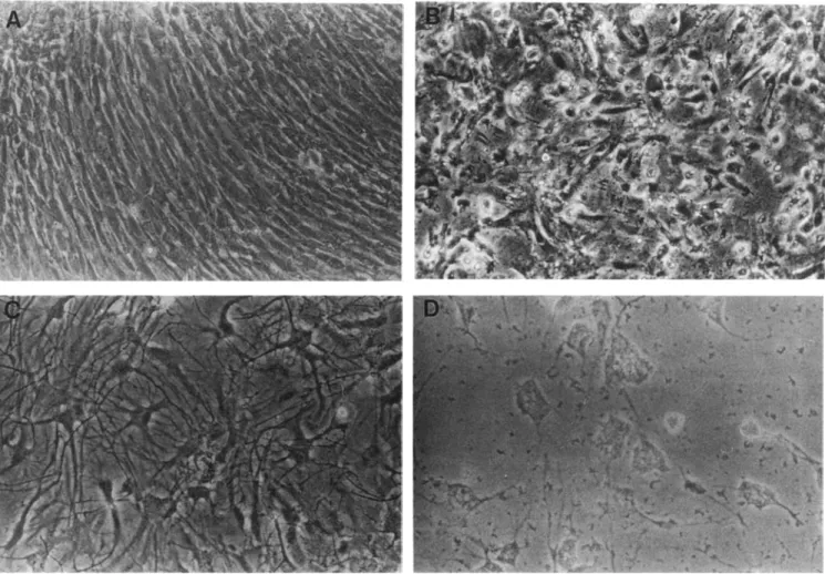
Figure 2. Cytotoxicity of live pneumococci for cultured rat cerebral endothelial cells and astrocytes. A: Confluent monolayer of primary rat endothelial cells cultured for 5 days. B:Endothelial cells 6 hours after exposure to live pneumococci ( 107
cfu/ml.),The monolayer is destroyed. Lysed cells can be identified. Some islands of endothelial cells are still recognizable. C: Primary rat astrocytes cultured for 14 days.D:Astrocytes 48 hours after exposure to live pneumococci (107cfu/rnl.). The polynuclear symplasms are disconnected, and the cell contours can no longer be recognized.
sessed by morphological studies and measurements oflactate dehydrogenase (LDH) activity. Primary cultures of unpas-saged astrocytes and of endothelial cells up to the first passage were used. Damage to astrocytes was documented morphologically after exposure to heat-inactivated pneumo-cocci. LDH activity was apparent as early as 0.5 hour after exposure of astrocytes to pneumococci. In contrast, neither morphological alterations nor LDH activity was apparent in endothelial cells after exposure to heat-inactivated pneumo-cocci for 0.5 hour to 192 hours. However, morphological changes in endothelial cells were seen after exposure to live pneumococci (figure 2). The pneumococcal strain used in such studies appears to be critical. The investigators used the S3 strain, which is noninflammatory in a rabbit model when heat inactivated, presumably because the capsule prevents recognition of the proinflammatory cell wall. It was sug-gested that the heat-inactivated, unencapsulated R6 strain be used in this assay.
Finally, Pedro M. Faustmann (Essen, Germany) pre-sented some preliminary data documenting the early appear-ance of mononuclear phagocytes in the SAS during acute meningitis. Specifically, these cells appeared in the SAS of both rabbits and rats within I hour of the intracerebral injec-tion of complement component C5a (100J-Lgin rabbits and 10J-Lg in rats). Electron microscopy revealed interaction of
the macrophages with polymorphonuclear leukocytes
(PMN s) via pseudopodia in the cisterns of the SAS. In addi-tion, an interaction of macrophages and astroglia was de-tected. Studies with monoclonal antibodies specific for mononuclear cells and/or for cells of macrophage lineage showed an increased number of ED2-positive cells within the SAS in the first hour after stimulation in rats. This very early phase of experimental meningitis was the focus of some discussion, which centered in particular on the specificity of the antibody (ED2) and the source of the mononuclear cells. Mononuclear cells or macrophages may be mobilized from
sites within the CNS or may enter the SAS from an intravas-cular source. These possibilities require further experimental assessment.
How Microorganisms Challenge the Host in Bacterial Meningitis
Alexander Tomasz (New York, New York, USA) began his review by reminding workshop participants of the rapid spread and increasing incidence of pneumococci resistant to multiple antibiotics. He cited a case in the United States in which ceftriaxone treatment of pneumococcal meningitis failed. The isolate was resistant to penicillin, third-genera-tion cephalosporins, irnipenern, erythromycin, chloram-phenicol, tetracycline, and trimethoprim-sulfamethoxazole. Ultimately, vancomycin had to be used in this instance.
There are ,....,500,000 cases of pneumococcal pneumonia and5,000 cases of meningitis annually in the United States, and the fatality rate for meningitis is still around25%-30%. A recent report from South Africa described pneumococcal meningitis in children; mortality due to penicillin-resistant pneumococci was nearly50% higher than that due to penicil-lin-susceptible strains
[19].
The high incidence of resistant pneumococcal strains in several countries and the need for bactericidal activity to control pneumococcal meningitis constitute a major challenge for the designers of new antimi-crobial agen ts.Experiments in a rabbit model indicate that CSF offers a poor environment for the multiplication of at least some pneumococcal strains. In some instances a net increase in bacterial titer was preceded by lag times as long as 20-30 hours after the intracerebral inoculation of an inoculum of
103-104
cfu. Once bacterial growth commences, the time of onset ofCSF leukocytosis is a remarkably sensitive indicator of the bacterial concentration. In studies with several capsu-lar type 2, type 3, and type
19
strains of pneumococci, the number of PMNs in the CSF began to rise sharply as the bacterial concentration reached a threshold range (a few times 106cfu).
Whether or not iv injection ofa bolus of ampicillin causes an inflammatory overload (known as the antibiotic-induced inflammatory burst) also depends on and is very sensitive to the concentration of bacteria in the CSF at the time of the injection. Ampicillin had no impact on the course of rapid pleocytosis when the bacterial concentration was already above the threshold range. The antibiotic's injection shortly before this threshold concentration was reached resulted in the inflammatory burst, while its injection several hours ear-lier actually postponed the onset of inflammation.
The nature of the inflammatory agent( s) released spon ta-neously at the inflammatory threshold bacterial concentra-tion or during treatment with ampicillin is not clear. The ultimate wall-degradation products of pneumococcal
autoly-tic amidase (the enzyme responsible for the disintegration of pneumococci during ampicillin-induced lysis) have only poor or moderate inflammatory activity. On the other hand. the in vivo triggering of this enzyme shortly after treatmen t of the bacteria with ampicillin causes a release into the me-dium of highly polymerized cell-wall structures containing an intact network of peptidoglycan and teichoic acid in a soluble form [20]. The specific inflammatory activity of this macromolecule remains to be tested. An additional intracel-lular inflammatory agent that does not behave like cell walls has been identified.
Biochemical studies on the specific inflammatory activity of well-defined cell-wall fractions from pneumococci and staphylococci have continued. Structural variants of staphy-lococcal and pneumococcal muropeptides [21] differ greatly in inflammatory potential. For instance, oligomeric rnuro-peptides from the staphylococcal peptidoglycan exhibit much less inflammatory activity than monomeric subcom-ponents. Whole cell walls from a penicillin-susceptible pneumococcal strain and its penicillin-resistant transfer-mants show different degrees of inflammatory activity, with the resistant organism's wall causing more rapid inflamma-tion. This difference is reproduced when muropeptides of
<
I,000 daltons from the two bacteria are compared. These observations suggest the existence of one or more muropep-tide receptors on a host cell in the CSF spaceIntroduction of cell-wall components or live pneumococci into the cisterna magna triggers an enormous number ofmo-lecular and pathophysiological events. Collaborative experi-ments involving Tornaszs and Pfister's groups have identi-fied one of the early events as an increase in regional cerebral blood flow, which is followed by an increase in ICP and pleocytosis. Studies with combinations of well-defined bacte-rial inflammatory components and a variety of anti-inflam-matory agents (e.g., dexamethasone, superoxide dismutase. transforming growth factor (3. antibody to ('[) I g. and cy-clooxygcnase inhibitors) may enable us to organize these events into a definitive inflammatory pathway.
Arnold L. Smith (Seattle. Washington. LfSA) next dis-cussed the mechanisms of nasopharyngeal colonization by Hib. Smith reviewed the results obtained in the infant maca-que model of Hib meningitis [22]. The first histopathologic lesion in the eNS in this model is choroid plexitis. This find-ing suggests that Hib enters the eNS through the choroid plexus. As in the rabbit model of pneumococcal meningitis. no SAS inflammation develops until bacterial concentra-tions in CSF reach ,....,10° cfu/rnl.. The use of the primate model is appropriate since Hib is specific for adhesion to primate epithelium. Smith reviewed the role of fimbriae in this adhesion. The expression of fimbriae may be switched on or offaccording to the conditions in which Hib is growing (for example, on Hel.a cells or in broth). In cell lines that mimic the nasopharyngeal epithelium, the bacteria can
468 Pfister et at. CID 1994; 19 (September)
"sense where it is," and fimbrial expression may be switched on. Furthermore, Hib is capable of accurately regulating fim-brial phase variation in vivo. For example, a-fimbriae are expressed to enhance binding to the anterior nasopharynx and IJ-fimbriae to facilitate binding to posterior ciliated naso-pharyngeal cells. Smith's presentation and the discussion that followed underscored the need for appropriate in vitro and in vivo systems in which to evaluate the pathogenesis and pathophysiology of meningeal pathogens, many of which are adapted only to primates. In vivo models are criti-cal for the delineation of complex pathophysiologicriti-cal and microbiological events. The differences between modelsmay
be subtle, and the interpretation of results obtained in rodent models may be problematic in some respects.
In the next presentation, Barbara Spellerberg (New York) discussed preliminary results of studies of the isolation of a receptor for pneumococci on human brain endothelial cells. Previous experiments had suggested that the pneumococcal cell wall plays a role in the attachment of Streptococcus
pneu-nioniae to human umbilical-vein endothelial cells [23]. In
the studies described by Spellerberg, brain endothelial-cell lysates were probed by western blots with antibodies specific for the pneumococcal cell wall, and one band was detected. Brain endothelial cells were then used to develop a cDNA library with X-GT II phage and expression in Escherichia
coli.A plaque assay with 3sS-labeled pneumococci was used to identify putative receptors for the pneumococcal cell wall on brain endothelial cells. A I 16-kD fusion protein of two clones was identified as a potential receptor. The discussion focused on the specificity of those findings. The specificity of the receptor for brain endothelial cells and for organisms other than pneumococci is unknown. No direct evidence that the receptor is surface-expressed on endothe-lium was presented. These exciting preliminary data sug-gested a variety of areas for productive investigation in the future.
A second presentation by Martin G. Tauber focused on the question: Are bacterial products neurotoxic? Primary neuronal cultures from embryonic rat brain consist of 90%-95% pure neurons, with some contamination by astrocytes and glial cells. For standard assays of neurotoxicity, primary neurons are grown in culture for ,..., 12 days and are then exposed to bacteria andjor bacterial products for 2 days. Tauber presented evidence that primary neurons are less sensitive to heat-killed pneumococci (R6 strain) than are U 373 astrocytes in tissue culture. Similarly, primary neurons are less sensitive to pneumococcal cell-wall components than are U373 cells, which are destroyed in a dose-depen-dent manner. No change in toxicity in neurons was noted after exposure to lipoteichoic acid or lipopolysaccharide (LPS) from Neisseria nieningitidis at concentrations up to 25 JLgjmL. U373 cells also exhibited minimal evidence of toxic-ity in the MTT assay after exposure to LPS. The striking
resistance of these neuronal cells to various bacterial prod-ucts was the subject of further discussion.
Finally, Hans Lassmann (Vienna, Austria) described ex-periments in a rat model of experimental autoimmune en-cephalomyelitis (EAE) [24, 25] combining immunocyto-chemical techniques, ultrastructural studies, and in situ nick translation. Lassmann and co-workers found that up to 49% of T lymphocytes in EAE lesions showed signs of apoptosis (programmed cell death) when recovered during disease. Thus apoptosis ofT lymphocytes is one possible mechanism for the elimination of these cells from inflammatory brain lesions. These intriguing results mayor may not be relevant to bacterial meningitis.
Soluble Inflammatory Mediators
Various aspects of the role ofcytokines and soluble media-tors in the development of brain injury during bacterial men-ingitis were considered. Bernhard Moser (Berne, Switzer-land) reviewed the functional and molecular characteristics of interleukin 8 (IL-8), relatedcyrokines,and their receptors. IL-8 was discovered about 6 years ago to be a chemotactic cytokine in vitro. Injection of IL-8 into the skin of rats in-duced a rather pronounced local inflammatory response-consisting mainly of PMNs-within 4 hours after challenge. The inflammatory potency of IL-8 after intradermal injec-tion was confirmed by studies of rabbits and humans. IL-8, formerly referred to as neutrophil-activating peptide I (NAP-1), belongs to the family of chemokines (chemotactic cyto-kines: for a review, see [26]). On the basis of the arrangement of the first two cysteines, the chemokines are divided into two subfamilies. In the CXC chernokines, the first two cys-teines (Cs) are separated by one amino acid (X); this group includes IL-8, the GRO (growth-related gene) peptides, and NAP-2. In the CC chernokines, the two first cysteines are adjacent. The most prominent members of this group are monocyte-chemotactic proteins I, 2, and 3 and macrophage-inflammatory proteins I(Y and I,B. TheCXCchemokines
ap-pear to be highly selective for polymorphonuclear phago-cytes, whereas the CC chemokines specifically activate mononuclear cells, including monocytes, eosinophils, baso-phils, and lymphocytes.
IL-8 is produced by various leukocytes and by a large num-ber of tissue cells upon stimulation with LPS or the proin-flammatory cytokines IL-l and tumor necrosis factor (TNF). IL-8, which is strongly chemotactic for PMNs, causes modifi-cations in the shape of'neutrophils, release ofenzymes, stimu-lation of the respiratory burst, and upregustimu-lation of IJ-inte-grins CD-IIJCD-18. IL-8 binds to at least two different PMN receptors, p44 and p70. The first receptor is more spe-cific for IL-8 than for related cytokines, while the latter has a similar affinity for IL-8, GRO-a, and NAP-2. The two IL-8 receptors have a seven-transmembrane domain architecture
typical of G protein-coupled receptors. The major form of IL-8 isolated from the medium of cultures of activated blood monocytes consists of 72 amino acids and has been identi-fied as a dimer by nuclear MR studies and X-ray diffraction of IL-8 crystals. The C terminus of the molecule is not in-volved directly in receptor binding, but deletions at the N terminus reduce the activity of IL-8. The integrity of each of the three residues that precede the first cysteine(Glut-Leu> Arg6;ELR) is critical for receptor binding and
neutrophil-sti-mulating activity. Recently, a series of analogues ofIL-8 (4-72)-the truncated form of IL-8 with the N-terminal sequence ELRC-were synthesized as potential IL-8 antago-nists [27]. The discussion of this elegant work focused on the potential role of IL-8 in CNS disease. LPS induces the ex-pression of IL-8, but the role of this cytokine in CNS infec-tion is unknown. The time course of IL-8 expression after LPS stimulation requires further study. Whether or not IL-8 receptors exist on endothelial cells also needs to be deter-mined.
The potential role of IL-8 in experimental meningitis was investigated by Rachel Dumont and Terry O'Reilly (Basel, Switzerland). The authors noted that IL-8 was present in the CSF of patients with bacterial meningitis at concentrations ranging from 0.3 to 24 ng/mL. IL-8 is capable of enhancing PMN migration across an endothelial-cell monolayer and therefore may playa role in SAS inflammation. These searchers injected IL-8 and related substances (human re-combinant IL-8, rabbit IL-8, human rere-combinant GRO, and human recombinant NAP-2) intracisternally into rabbits. The results, somewhat surprisingly, documented a lack of CSF pleocytosis during the 7 hours after challenge with all four molecules. However, the rabbits did respond to intracis-ternal challenge with LPS injected 7 hours after IL-8. The discussion of this presentation emphasized future experi-ments on topics such as the injection oflL-8 along with other cytokines, the injection of monoclonal antibodies to IL-8 along with an LPS challenge, and the potential influence of IL-8 on blood-brain barrier disruption.
The role of soluble inflammatory mediators in the patho-physiology of bacterial meningitis was reviewed by Karl Frei (Zurich, Switzerland). In collaboration with Adriano Fon-tana, Frei asked whether cytokines produced intrathecally in the CNS during experimental infection were involved in the elimination of microbes from the CNS. In a mouse model of
Listeria monocytogenesmeningitis, these investigators docu-mented that TNF and IL-6 were present in the CSF at 4 hours and that concentrations peaked at '""-'24 hours after intracerebral inoculation ofL. monocytogenes. In contrast, interferon')' (IFN-')') was not detectable until 24 hours after challenge. Perhaps the most significant finding was that IL-10 was also produced late (up to 72 hours) in the course of listeriosis in mice [28]. The authors used an in vitro system of murine macrophages (J77A-I cells) on coverslips to
investi-gate the effect of pretreatment with various cytokines on Iis-terial infection in vitro. No increase in listericidal activity was apparent after pretreatment with TNF or IL-6. However, a marked increase in the listericidal activity of J 774A-1 cells was evident after preexposure to IFN-')' (i.e., activated macro-phages) and this increase was dependent on the production of nitric oxide. An interesting finding was that IL-IO im-paired the listericidal activity of macrophages-an effect du-plicated when macrophages were exposed to CSF drawn from mice 48 hours after infection. This effect was reversed by preexposure to a monoclonal antibody to IL-I 0, which inhibits nitric oxide synthase. Although IFN-')' and IL-IO may not playa similar role in meningitis because of the pres-ence of extracellular bacteria, the potential role of cytokine modulation of the late events in infection and the complex interplay of the various cytokines are topics of interest. IL-I 0 is produced mainly by TH2 cells, including monocytes and macrophages. During the discussion period, Frei presented data documenting elevated concentrations of IL-I 0 in 959c of CSF samples from children with bacterial meningitis but in only 3% of samples from children with viral meningitis. IL-IO concentrations ranged from '""-' 100 to well over 20,000
pg/ml..The potential role oflL-1 0 in the diagnosis ofmenin-gitis and its potential anticytokine activity late in the disease clearly require further investigation.
Jay H. Tureen (San Francisco) discussed the effects of TNF on cerebral blood flow and metabolism. With his asso-ciates, Tureen challenged rabbits intracisternally with type III pneumococci at two concentrations (5X 10⁶cfu and5x
104cfu) and measured cerebral blood flow (radiolabeled mi-crosphere technique), rate of cerebral oxygen metabolism. CSF lactate level, CSF TNF level (L929 cytotoxicity assay), and CSF bacterial titers 16 hours and 20 hours later. Cef-triaxone therapy was initiated 16 hours after challenge. In-creased bacterial concentrations in CSF correlated with in-creased concentrations of lactate and TNF. CSF lactate concentrations were further increased after treatment with ceftriaxone, which also elicited a burst ofTNF activity. Per-haps most important. TNF concer.trations in ('SF correlated inversely with cerebral blood flow and cerebral oxygen up-take. These findings may reflect the increased incidence of neurological sequelae among patients with high ('SF bacte-rial densities associated with a shift to anaerobic metabolism. In discussing this presentation, participants attempted to de-fine the primary event in these pathophysiologic alterations.
It was suggested that the primary event was a decrease in brain oxygen demand coupled with a reduction in cerebral blood flow. The driving force is clearly a critical variable. Studies to measure critical variables after challenge with TNF and/or monoclonal antibody to TNF are planned.
The effect of inflammatory products on primary neurons in tissue culture was discussed by Martin G. Tauber (San Francisco). Previous experiments had suggested that the
mu-470 Pfister et al. CID 1994; 19 (September)
rine neuronal cell line HN 33.1 was sensitive to various cyto-kines, in particular TNF [29]. These studies indicated that the cytotoxicity in CSF from animals with bacterial meningi-tis was attributable to TNF since soluble TNF receptors blocked virtually all of this activity except that observed late after challenge with either live pneumococci or pneumococ-cal cell wall. Twelve-day-old primary neuronal cultures were exposed over 2 days to various cytokines, and cytotoxicity was measured with the MTT (3-[ 4,5-dimethylthiazol-2-yl]-2,5-diphenyltetrazolium bromide) assay. Although cytotoxic-ity was observed after exposure to CSF samples from animals with pneumococcal meningitis diluted in culture medium to 20%-30% (vol/vol), the cytokines examined (IL-l and TNF) produced cytotoxicity in primary neurons only at concentra-tions in the micromolar range. Furthermore, synergistic cyto-toxicity was not evident when submaximal concentrations of IL-l and TNF were used together. IL-l was marginally more active than TNF in the induction of cytotoxicity in this assay. The discussion of this presentation focused on questions re-garding the relative susceptibilities of embryonic neurons and neurons of older animals, the change of embryonic neu-rons with age in culture, the response of embryonic neuneu-rons to N-methyl-o-aspartate antagonists, and other issues. Pri-mary neurons are remarkably resistant to cytokines in this system.
Leukocyte-Endothelial Cell Interactions
The critical role ofleukocyte-endothelial cell interactions in tissue injury-and potentially in bacterial meningitis-was addressed in several presentations. The broad subject of these interactions in tissue injury was reviewed by Hans-An-ton Lehr (Munich). The crucial role of leukocyte-endothe-lial cell interactions in host defense is underscored by the development of recurrent bacterial infections in patients with leukocyte adhesion deficiency. Without the adhesion of leukocytes, there is no inflammatory response to facilitate the elimination of an invading microorganism. However, there is potentially harmful leukocyte activity. Leukocyte-endothelial cell interactions may cause a local Leukocyte-endothelial lesion, and chemotactic mediators, including leukotrienes, complement, reactive oxygen species, and platelet-activating factor, may subsequently be released from the endothelium. The liberation of these mediators leads to further recruit-ment of leukocytes at the site of endothelial-cell damage. Attracted activated leukocytes may release cytotoxic prod-ucts, such as reactive oxygen species, reactive nitrogen inter-mediates, and proteolytic enzymes, that may augment tissue damage, with subsequent edema formation. This leukocyte-induced tissue damage plays a role in a variety of disease processes, such as ischemia-reperfusion [30], atherogenesis [31], and bacterial meningitis [32, 33].
Ruggero Pardi (Milan, Italy) discussed the molecular mechanisms underlying leukocyte-endothelial cell
interac-tions. Aspects ofadhesion, including the bidirectional mecha-nism of receptor density, the ability to diffuse within mem-branes, and ligand affinity, are the focus of Pardi's experiments. Leukocyte-endothelial cell interactions are mediated by cell-surface adhesion receptors and their counter-receptors on leukocytes and endothelial cells, which include members of the integrin and selectin families and the immunoglobulin superfamily. Pardi reviewed the interac-tions of leukocytic integrins (integral membrane proteins) with intercellular adhesion molecules (ICAM) and with en-dothelial integrins; these interactions are of low affinity in the millimolar range. Conversely, selectin-carbohydrate pro-tein interactions are of high affinity in the nanomolar range, but the association-dissociation of these events is very rapid. L-selectin interacts with a vascular addressin rich in carbohy-drates and important in the homing of lymphocytes. Pardi suggested that leukocytes are never adhesive under resting conditions; thus leukocyte adhesiveness requires triggering by external stimuli. Increasing evidence supports the involve-ment of members of the selectin family (namely, L-selectin expressed by leukocytes and P-selectin on endothelial cells) in the initial rolling of leukocytes along the endothelium. This process precedes firm adhesion and transmigration [34]. The earliest detectable intracellular event associated with ad-hesion-membrane phosphoinositide breakdown followed by the production of inositol-l ,4,5-triphosphate and diacyl-glycerol-results in calcium-dependent protein kinase C ac-tivation and a transient Ca2+ increase [34]. In Pardi's pro-posed model, leukocyte adhesion is dependent on activation and mediated by lymphocyte function-associated antigen I. The discussion of this presentation centered on potential sec-ond messengers relevant to leukocyte-endothelial cell inter-actions. Pardi reported that many leukocyte adhesion mole-cules are signal transducers. For example, after the adhesion of neutrophils, the dominant neutrophil integrin (CD II b/ CD 18) triggers the generation of oscillating intracellular cal-cium transients, which are associated with exocytosis and phagosome-lysosome activity.
The relevance of these complex interactions ofleukocytes and endothelial cells for bacterial meningitis was the subject of the next presentation, which was delivered by Eva Rozd-zinski (N ew York). She described a hypothetical scheme in which the entry of bacteria into the CSF is followed by the release of cell-wall or other inflammatory stimulants whose enhancement of leukocyte adhesion to endothelial cells (with modulation by cytokines) leads to alterations in blood-brain barrier permeability. Rozdzinski also reviewed selec-tin-mediated leukocyte-endothelial cell rolling mechanisms and subsequent integrin-mediated adhesion events. The influence of these two sequential mechanisms on the micro-circulation of the CNS is poorly understood. Rozdzinski re-viewed experiments with a monoclonal antibody to adhe-sion-promoting receptors on leukocytes (IB4) in a rabbit
mg/kg) reduced CSF pleocytosis after challenge with live bac-teria (meningococci, pneumococci, or Hib), pneumococcal cell wall, or Hib LPS. IB4 did not influence the incidence of bacteremia in this model but did improve rates of survival. Evidence suggests that IB4 reduced leukocyte migration across an endothelial-cell monolayer in vitro in a dose-de-pendent manner. In studies of rabbits with pneumococcal meningitis, Rozdzinski and colleagues administered ampicil-lin 15 hours after infection and then administered IB4and
dexamethasone by the iv route. IB4 appeared to be more
effective than dexamethasone in the reduction of CSF pleo-cytosis. Furthermore, and perhaps somewhat surprisingly, IB4appeared to be more effective than the combination of IB4and dexamethasone in this model. These results conflict with data from previous studies of experimental Hib meningi-tis [33]. Inhibition of leukocyte-endothelial cell interac-tions, with the reduction of CSF pleocytosis, probably has both negative and positive consequences in the human host. This possibility must be studied carefully before relevant clinical trials are contemplated.
The IB4study demonstrated that members of the integrin
family are involved in leukocyte-endothelial cell interaction and in subsequent injury during pneumococcal meningitis. In a further study, the role of members of the selectin family in the pathophysiology ofpneumococcal meningitis was stud-ied. Selectins are C-type lectins that recognize carbohydrates on leukocytes. These proteins are expressed on injured endo-thelial cells and promote leukocyte margination. Rozdzinski presented evidence suggesting that subunits S2 and S3 of pertussis toxin contain carbohydrate recognition domains and are thus competitive inhibitors of selectin-mediated leu-kocyte-endothelial cell interactions [35]. S2 and S3 peptides inhibited the adherence of human neutrophils to human um-bilical-vein endothelial cells and induced upregulation of the integrin CR3 on neutrophils in vitro. Rozdzinski and her colleagues are searching for peptides that block endothelial-cell adhesion without upregulation of integrins. In the ani-mal model, the systemic administration of these peptides I hour after the injection of heat-killed pneumococci of strain R6 into the cisterna magna reduced CSF inflammation. The potential for the use of these agents in the systemic treatment of meningitis in patients was discussed extensively after this presentation.
Confocal laser scanning microscopy, a sophisticated new technique for the imaging of leukocyte-endothelial cell in-teractions in vivo, was described by Stefan Lorenzi (Mun-ich). Lorenzi and associates investigated dynamic character-istics of rhodamine 6G-Iabeled leukocytes in the pial microcirculation during the first 6 hours after the induction of pneumococcal meningitis in the rat [36]. Closed cranial windows were implanted for the examination of the pial ves-sels in vivo. Through optical sectioning, confocal laser scan-ning microscopy allows the study of pial vessels through an intact dura mater. A video depicting leukocyte-endothelial
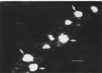
Figure 3. Leukocyte adherenceto microvascular endotheliumin a pial venule of an infected rat. A closed cranial window was im-planted in an anesthetized rat without removal of the dura mater. and confocal laserscanning microscopy wasusedforin vivo exami-nation of the cerebral microcirculation. Intravenous rhodamine 6G selectively stained white blood cellsand platelets but not endothe-lial cells. The number of rhodamine 6G-Iabeled leukocytes adher-ing to venular endothelium was increased 3 hours after intracister-nal injection of livepneumococci(arrows). Bar= 10tim.
cell interactions in real time was shown: this video docu-mented the augdocu-mented adhesion of leukocytes to the cere-bral microvasculature as early as I hour after pneumococcal infection in the rat model. Leukocyte adherence to microvas-cular endothelium occurred in pial venules but not in arteri-oles (figure 3). Pretreatment of the rats with dexamethasone decreased leukocyte adhesion and extravasation into the ex-tracellular space. Administration of the novel 21-aminoster-oid agent U74389F, a potent inhibitor of lipid peroxidation [37], also appeared to decrease leukocyte extravasation. The mechanism of the effect ofU74389F on leukocyte-endothe-lial cell interactions is unknown.
Molecular Mechanisms of Neuronal Injury
A potpourri of eight presentations examined various puta-tive mediators of neuronal injury during meningitis-currently a primary topic of investigation. The production of nitric oxide. superoxide, and peroxynitrite by activated in-flammatory cells was reviewed by Joseph Beckman (Bir-mingham, Alabama. USA). Reactive oxygen species have been implicated as mediators of brain injury in a variety of conditions, including cerebral ischernia/reperfusion injury. experimental fluid/percussion brain injury. cold-induced brain edema. cerebral arteriolar abnormalities after acute ar-terial hypertension. and bacar-terial meningitis 138--40]. Super-oxide radical and hydrogen perSuper-oxide may interact via the iron-catalyzed Habet-Weiss reaction to produce the highly active hydroxyl radical, which. in turn, may initiate lipid
per-472 Pfister et al. GO 1994; 19 (September)
oxidation and cellular injury. Beckman presented evidence suggesting that the Haber-Weiss reaction is too slow to dam-age tissue in vivo. In contrast, the interaction of superoxide with nitric oxide to form the powerful oxidant peroxynitrite anion occurs at the very high rate of 6.7 X 10⁹M-1s- 1 and therefore is ,..., I million times faster than the Haber-Weiss reaction. Pathological conditions such as inflammation, isch-emia followed by reperfusion. and sepsis can substantially upregulate the production of superoxide and nitric oxide in stimulated macrophages and neutrophils, thereby increasing peroxynitrite formation. When protonated. peroxynitrite de-composes to generate highly reactive agents-apparently hy-droxyl radical and nitrogen dioxide(N02)[41]. At
physiolog-ical
pll.
peroxynitrite has a half-life ofr -I second that allowsit to diffuse to critical cellular targets before decomposing. Thus, peroxynitrite can diffuse ,..., 10,000 times longer than the hydroxyl radical. Peroxynitrite interacts with various metals to form nitro groups on proteins as a marker of its presence. Superoxide dismutase (SOD), which inactivates superoxide anion radical. may prevent the formation of toxic peroxynitrite from superoxide and nitric oxide [41]. How-ever, it has been shown that peroxynitrite reacts with SOD to produce a species with the reactivity ofa toxic nitronium ion (NOH); as a result, tissue injury is perpetuated [42]. Since catalase scavenges nitric oxide in addition to hydrogen perox-ide, in vivo experiments with catalase must be interpreted with caution.
Beckman presented satisfactory evidence suggesting that peroxynitrite (as opposed to nitric oxide) is a neuronal toxin in vivo. The cytotoxic peroxynitrite has been implicated as a mediator of cerebral ischemic injury [41]. The concentration of nitric oxide synthase is highest in the brain-i.e., at least 20-fold higher than levels in endothelial cells. Nitric oxide concentrations of2-4J.LMin ischemic brain [43] account for ,..., 10% of oxygen consumption by the CNS during ischemia. These concentrations should be contrasted with nitric oxide concentrations within macrophages, as opposed to 0.4J.LMin endothelial cells and much less than 0.1 J.LMin most other cell types. Nitric oxide diffuses ,...,45 J.Lm during its in situ half-life ofr -1 second. In contrast, hydroxyl radicals perfuse only about a tenth of the diameter ofa protein in situ and are therefore of doubtful significance during ischemia. Beckman presented evidence suggesting that nitric oxide itself does not kill bacteria in vitro, even at I mM. In contrast, all E. coli cells were killed within 10 seconds by exposure to peroxyni-trite [44].
Nitric oxide is much more than a vasodilator. It plays im-portant roles in the control of systemic blood pressure, respi-ration, digestion, penile erection, platelet aggregation, cere-bral blood flow, and neuronal synaptic plasticity [42, 45]. There is increasing evidence that nitric oxide or a secondary oxidant derived from it is also involved in the pathophysiol-ogy of bacterial meningitis (see Kadel and Berkowitz below). Clearly, the production of nitric oxide in situ and its
appear-ance in CSF require additional study. Furthermore, potential interactions of nitric oxide and various cytokines may be promising areas of investigation. Nitric oxide was the "mole-cule of the year" in 1992 [46]; those in attendance at this presentation were left with the impression that the molecule of the year in 1994 may be peroxynitrite.
The role of reactive oxygen species, a group of potential mediators of neuronal and endothelial injury during bacte-rial meningitis, was reviewed by Hans-Walter Pfister (Mun-ich), who summarized experiments in a rat model of early pneumococcal meningitis. Continuous infusion of SOD (22,000 U[kg· h] iv) prevented the early increase in regional cerebral blood flow as well as the increase in ICP and brain water content typical of this early (6-hour) model [47]. Like-wise, polyethylene glycol-conjugated SOD (PEG-SOD; 10,000 U/kg iv) was effective in preventing increases in ICP and brain water content in meningitis induced by intracister-nal injection of pneumococcal cell-wall components [40]. The effect of SOD infusion was more pronounced than that of catalase infusion (25,000 U/[kg· h] iv) in the same model [48]. Perhaps more important, SOD reduced ICP and brain water content when given as late as 3 hours after intracere-bral inoculation of pneumococci. A role for oxygen radicals in the pathophysiological alterations that take place during bacterial meningitis was supported by recent studies by Ber-kowitz and Traystman [49]. Intravenous PEG-SOD and PEG-catalase prevented impairment of microvascular auto-regulation in Hib meningitis in rats. SOD may act by pre-venting the formation of toxic peroxynitrite from superoxide anion radical and nitric oxide [41]. Pfister cited results from studies by McKnight et al. [50] documenting an increase in pial arteriolar diameter following the topical application of group B streptococci to the brain surface in rats. Treatment with iv PEG-SOD (10,000 U/kg) and PEG-catalase (20,000 U /kg) before exposure to group B streptococci prevented va-sodilation; this result indirectly supports a role for reactive oxygen species. Nevertheless, as was evident from the discus-sion of this presentation, the exact site of oxygen radical gen-eration during bacterial meningitis is still unknown. Further-more, in all experiments to date, the Munich group has employed an early model of bacterial meningitis (within 6 hours of challenge), and the role of reactive oxygen species in the later stages of the disease remains poorly defined.
Some preliminary results with a new optical method for the detection of free radicals in vivo were presented by Ulrich Dirnagl (Munich). Dirnagl and colleagues used a cra-nial window technique in the rat with lucigenine-enhanced chemiluminescence to monitor the generation of free radi-cals in situ [51]. The photon count rate increased after intra-cisternal inoculation of live pneumococci; this effect was at-tenuated in the presence of SOD. The strength of the method is that it allows for continuous measurement of free-radical production in situ. Although methodological concerns must still be addressed, this method is promising and may provide
further information on the role of reactive oxygen species in experimental infection.
Indirect evidence for a pathophysiological effect of nitric oxide during the early phase of pneumococcal meningitis was presented by Uwe Kadel (Munich). Kodel utilized the early model of experimental pneumococcal meningitis in the rat [47]. Three hours after intracisternal injection of live pneumococci. NG-nitro-L-arginine (l-NA), a competitive in-hibitor of L-arginine, was given by bolus injection at a dose of 5 or 10mg/kg,with a subsequent continuous iv infusion of 5 or 10 mg[kg· h). Acting in a dose-dependent manner, l-NA reversed the increase in regional cerebral blood flow typical of this model. In addition, this inhibitor prevented the devel-opment of brain edema and an increase in ICP and reduced the degree of CSF pleocytosis. However, the higher dose of l-NA resulted in the death of four of five rats between the second and third hour of treatment, whereas none of the rats that were infected and then either left untreated or treated with the lower dose died. Similar increased mortality rates were reported by Cobb et al. [52], who administered NG amino-t-arginine to awake dogs after challenge with endo-toxin, and by Haberl et al. [53], who administered l-NA methyl ester to rats with experimental pneumococcal menin-gitis. Studies investigating the effect of l-NA in uninfected rats are warranted.
Ivor D. Berkowitz (Baltimore, Maryland, USA) described experiments suggesting that nitric oxide contributes to pial arteriolar dilation and impaired autoregulation of cerebral blood flow during experimental Hib meningitis in rats. Pial vessels were directly visualized through a closed cranial win-dow, and Hib was topically applied to the brain surface (at a concentration of 105cfu/rnl. of artificial CSF).
Autoregula-tion was assessed as the ability of pial arterioles to dilate in response to hemorraghic hypotension [54). In infected un-treated rats, the degree of pial arteriolar dilation documented was ,..., 150% (with baseline dilation assigned a value of
100%). Pretreatment of rats with 20 mg of l-NA methyl es-ter/kg prevented pial arteriolar dilation 4 hours later. Auto-regulation of cerebral blood flow was preserved in this model after the administration of l-NA methyl ester. The results reported are in accordance with previous studies by the Mun-ich group [53], who reported that the administration of
l-NA methyl ester attenuated the pathophysiological
alterations typical of the early phase of experimental pneu-mococcal meningitis in the rat. However, these experiments must be interpreted cautiously since l-NA methyl ester not only is thought to be a competitive antagonist of nitric oxide synthase but also has been shown to be a muscarinic receptor antagonist [55].
H. Niels Diemer (Copenhagen, Denmark) next reviewed the role of excitatory amino acids in CNS pathology. The excitotoxic hypothesis of neuronal injury proposes that glu-tamate or related endogenous excitatory neurotransmitters become toxic through their interaction with glutamate
recep-tors and that subsequent intracellular events result in neuro-nal death [56, 57). The glutamate receptors are divided into groups: the ionotropic N-methyl-D-aspartate (NM DA) re-ceptors: the non-NMDA.kainate/quisqualate receptors: and the metabotropic glutamate receptors, which instead of pos-sessing an intrinsic ion channel exert their effects through second-messenger systems hy activation of a G protein. The protective effects of glutamate receptor antagonists suggest that excitatory amino acids are involved in the pathogenesis of cerehral ischemia, profound hypoglycemia, and status cpi-lepticus. Elevated extracellular concentrations of glutamate and aspartate in rat hippocampus during transient ischemia have been documented by microdialysis techniques [58]. Re-uptake of amino acids is energy dependent at the synapse via transporters on a sodium-potassium gradient: the loss of this gradient during ischemia leads to the accumulation of these toxic amino acids. The use ofNMDA antagonists, including MK-80 I and a-amino-3-hydroxy-5-methyl-4-isoxazolepro-pionic acid (AMPA) antagonists such as 2,3-dihydroxy-6-ni-tro-7-sulfamoyl-henzo (f) quinoxaline. to protect ischemic tissues is a focus of intense investigation. The AMPA recep-tor is pentameric. Diemer presented evidence showing that the components of this receptor are encoded on different chromosomes: these components are designated gluR I through gluR4. A change in a single amino acid in gluR2 determines whether or not the channel is permeable to cal-cium. Various mechanisms that may attenuate the NMDA receptor-mediated current were discussed. For example. the accumulation of lactate in the ('SF during bacterial meningi-tis renders the extracellular milieu acidic. and this aciditv may reduce the potential for damage after meningitis and/or hypoglycemia-hypoxia. The current status of MK80 I was raised during the discussion: this compound has not been introduced into clinical practice because of its psvchomime-tic properties.
The potential role of excitatory amino acids in the patho-physiology of bacterial meningitis was reviewed by Martin G. Tauber (San Francisco). Five papers. puhlished from 1975 to 1982. documen ted elevated concentrations of amino acids-including 'Y-aminobutyric acid. glutamate. glycine, and aspartate-in CSF from patients with bacterial meningitis (cited in [59]). Concentrations of amino acids in CSF appear to peak I or 2 days after initiation of therapy. No data are available on amino-acid accumulation in the brain of patients with bacterial meningitis. Tauber reviewed the results of measurements of amino acid concentrations in the CSF and brain ofrahhits with pneumococcal meningitis [59]. Standard CSF analyses were conducted. and microdialysis techniques were used to document the accumulation ofhoth lactate and amino acids in brain interstitial fluid. Concentra-tions of glutamate, aspartate. glycine. taurine. and alanine increased after 22 hours in ('SF from infected animals: there was no concomitant increase in the level of asparagine, ser-ine, or threonine. Glutamate was the only excitatory amino
474 Pfister et al. CID 1994; 19 (September)
acid to appear in increased concentrations in brain intersti-tial fluid after 22 hours. Levels of alanine also increased sig-nificantly at this site after 22 hours. As alanine is considered a marker for anaerobic glycolysis, perhaps it was not surpris-ing that its concentrations in brain interstitial fluid were correlated with those of lactate. Tauber also noted an in-crease in levels of quinolinic acid in CSF and brain during experimental bacterial meningitis. This substance plays a po-tential role in the development of encephalopathy in pa-tients with AIDS, but its role in meningitis is unknown. Much of the discussion of this presentation was concerned with the mechanism( s) of excitatory amino-acid accumula-tion in the brain during meningitis. Alteraaccumula-tions in blood-brain barrier permeability to amino acids, the release of amino acids from cells within the CNS, a decline in mecha-nisms of amino-acid clearance from the SAS, and accen-tuated binding of amino acids to brain tissue are all possibili-ties. These issues are difficult to study in vivo with current techniques.
Adriano Fontana (Zurich) presented intriguing informa-tion on the potential neurotoxicity of macro phages. Neurons were isolated from the cerebellum of 7-day-old mice and ex-posed to macrophages at a ratio of I macrophage to 10 neu-rons [60, 61]. The neuneu-rons were destroyed within 24 hours in this in vitro system. IL-
L
TNF, IL-6, and IFN-)'-both alone and in combination-were not cytotoxic. In cocul-tures of macrophages and neurons, induced neurotoxicity was blocked by an NMDA receptor blocker but not by cata-lase or SOD. This result raised the interesting possibility that macrophages produce excitatory amino acids that contribute to neuronal toxicity. Direct evidence was provided that mac-rophages release glutamate, which can be detoxified by astro-cytes. Thus, a balance between the amount ofglutamate pro-duced and the capacity for its detoxification by astrocytes may be crucial to the development of macrophage-mediated neurotoxicity. The contribution of excitatory amino acids to this form of toxicity requires further study.Cerebral Blood Flow and Metabolism During Bacterial Meningitis
Jay H. Tureen (San Francisco) reviewed the changes in cerebral blood flow and metabolism in bacterial meningitis. A decrease in cerebral blood flow has been observed in pa-tients with this infection [62-65]. The data on cerebral blood flow in animals with experimental meningitis are extensive and sometimes conflicting. As assessed by the radiolabeled microsphere technique, cerebral blood flow varies somewhat with time after experimental infection. There is an early hy-peremic phase during experimental pneumococcal meningi-tis in the rabbit (a finding similar to those obtained by the Munich group using Doppler flow techniques in a rat model), with a decline in blood flow later in infection. Twenty hours after pneumococcal challenge, total cerebral
blood flow is reduced by --25%. Hippocampal blood flow in the same animal is reduced to -- 50% of control values. Whereas autoregulation of cerebral blood flow is maintained in normal animals, this ability is generally lost during experi-mental pneumococcal meningitis [66]. ICP remains stable despite marked alterations in cerebral blood flow in normal animals but rises progressively with changes in cerebral blood flow in animals infected with pneumococci. There-fore, artificial increases in blood pressure lead to increases in cerebral blood flow, which in turn lead to increases in ICP in animals with meningitis. Fluid-restricted rabbits have further decreases in mean arterial pressures and cerebral blood flow, with the accumulation of CSF lactate after treatment with ceftriaxone, whereas these changes do not occur in euvole-mic rabbits [67]. The implications for the policy of fluid re-striction, which may be adhered to rigidly during the early phase of meningitis because of concerns about the syndrome of inappropriate secretion of an tidiuretic hormone, were not lost on the participants during the discussion period. Further-more, cerebral blood flow declines more rapidly after antimi-crobial therapy. A more rapid reduction in the bacterial titer in CSF causes a more rapid reduction in cerebral blood flow, with obvious potential implications for therapy. Tureen also reviewed microdialysis experiments whose results supported the concept that at least some CSF lactate is produced locally in the brain parenchyma [68]. Other possible sources ofCSF lactate are bacteria and leukocytes. The rate of glucose ex-traction across the brain does not change over time, whereas the rate of cerebral oxygen metabolism falls after challenge with bacteria at high concentrations. Tureen's review raised much concern over the delicate balance among cerebral per-fusion pressure, fluid restriction, ICP, and cerebral blood flow in patients with meningitis. An additional source of concern is the possibility that a rapid decrease in bacterial titer as a consequence of rapidly bactericidal therapy may temporarily decrease cerebral blood flow.
Anthony Slater (London, United Kingdom) discussed ce-rebrovascular autoregulation during experimental Hib men-ingitis in rabbits and the role ofleukocytes in this interaction [69]. In contrast to results obtained with previous models [66], the rate of cerebral blood flow measured in these rab-bits with the microsphere technique was increased 18 hours after intracerebral Hib challenge. Pretreatment of the ani-mals with monoclonal antibody (IB4 , 1.5 mg/kg) decreased
CSF pleocytosis (consistent with previous observations [32]) but was associated with an increase in bacterial concentra-tions in the CSF. Moreover, such pretreatment augmented cerebral hyperemia in this model, and the increase in cere-bral blood flow was correlated with increased bacterial titers in CSF. Cerebrovascular autoregulation, as assessed by blood-flow measurements at graded levels of hemorrhagic hypotension, was preserved in Hib meningitis, but the capac-ity for autoregulation was lost in infected IB4-treated
prob-lems with Hib as a pathogen in the rabbit model: this organ-ism does not induce a universally lethal infection in this model.
The use of transcranial Doppler (TCD) sonography in pa-tients with bacterial meningitis was described by Hans-Peter Haring (Innsbruck, Austria). Haring and his colleagues stud-ied 110 patients with various infections of the CNS; 37% of patients had bacterial infection, 24% had viral processes, and 39% had unclassified disease [70]. TCD sonography docu-mented an increase of blood-flow velocity in the middle cere-bral artery in 77% of patients infected with pneumococci and in 65% of those with meningococcal meningitis. Flow veloci-ties were maximal 3-5 days into the illness and took 10-14 days to return to normal. Various causes of the observed increase in flow velocity through the M I segment of the mid-dle cerebral artery, including increased cerebral blood flow, decreased vessel diameter, and decreased cerebrovascular pe-ripheral resistance, were considered during the discussion of this presentation. The contribution of these processes to the documented result requires further study. The specificity of the observation is also unknown. The impact of treatment with dexamethasone or other adjunctive agents on the re-sults of TCD sonography has not been studied.
Approaches to the Treatment of Bacterial Meningitis
Erich Schmutzhard (Innsbruck) reviewed the treatment of bacterial meningitis with antimicrobial agents. Schmutzhard questioned the value of heparin and raised the possibility of administering cytokine antagonists. He discussed in some depth the potential role of dexamethasone as an adjunct to antimicrobial therapy for meningitis. A meta-analysis cover-ing data from five studies on the subject has recently been published [71]. All of the data were for cases in children and supported the contention that rates of early and late neuro-logical sequelae-particularly severe bilateral sensorineural hearing loss-are reduced by concomitant therapy with dexa-methasone. Nevertheless, only one clinical trial showed a marked effect on both parameters [4], and the incidence of neurological sequelae tended to be unusually high in the control groups in these trials. Schmutzhard suggested that further study of this issue was necessary. He also discussed the possibility that third-generation cephalosporins are not as efficacious as ampicillin and chloramphenicol in therapy for bacterial meningitis because they exacerbate the inflam-matory burst. The limitations of the data on dexamethasone were considered during the discussion. The abandonment of third-generation cephalosporins as empirical therapy for meningitis in adults was not endorsed. Moreover, the recom-mendation for a stepwise increase in the antibiotic dose used in the treatment of pneumococcal disease did not meet with favor among the clinicians in attendance.
Terry O'Reilly (Basel) presented the results of two studies. The first described a rabbit model ofLPS-induced meningitis
that mimicked magnification of the inflammatory burst f(JI-lowing antibiotic therapy. Meningitis was induced in rabbits by intracisternal inoculation of 2.5 ng of LPS. The resulting primary inflammation was followed 6 hours later by an injec-tion of25 ng ofLPS. which produced a secondary inflamma-tion simulating the accentuainflamma-tion of SAS inflammainflamma-tion after antibiotic therapy. The administration of dexamethasone (1.5 mg/kg iv) 15 minutes before the second injection ofLPS inhibited the second peak of LPS-induced CSF inflamma-tion. In addiinflamma-tion. dexamethasone tended to reduce the sec-ond peak in the TNF concentration in CSFfollowing chal-lenge with LPS. Somewhat surprisingly, brain levels ofTNF mRNA after 7 hours were similar in dexamethasone-treated rabbits and controls.
In another presentation. O'Reilly discussed adjunctive treatment in an experimental animal model of meningitis and reviewed the results of experiments conducted by Otto Zak and colleagues [72. 73]. This series of studies charted mortality and neurological sequelae in a large number of rabbits in which pneumococcal meningitis was treated with ampicillin and various anti-inflammatory agents. Treatment was started 18 hours after infection. with all agents given twice daily for 3 days. The mortality in the first 30 days was clearly lowest among animals that received ampicillin (25 mg/kg) plus oxindanac (5 mg/kg). an experimen tal nonste-roidal anti-inflammatory drug. rather than ampicillin alone. ampicillin plus dexamethasone (I mg/kg). ampicillin plus indomethacin (5rug/kg). or no treatment. Although all an ti-inflammatory drugs tended to reduce rates of neurological sequelae, the results were best for ampicillin plus oxindanac. The duration of neurological sequelae was also reduced bv this combination. The discussion of this presentation fo-cused on the mechanism( s} responsible for the observed re-sults. A "weaker" nonsteroidal anti-inflammatory agent may be more effective in this model. since indomethacin reduces concentrations of prostaglandin E2(PGE2) in ('SF whereas oxindanac does not. A reduction in PGE2levels may actually increase inflammation and therefore may be counterproduc-tive.
The role of antibiotics other than iJ-Iactam agents in ther-apy for pneumococcal meningitis was discussed by Martin G. Tauber (San Francisco). When a series of quinolones were evaluated in the rabbit model of experimental pneumo-coccal meningitis. a good correlation was found between the ratio of the peak CSF concentration to the MBC of the in-fecting strain and the ultimate outcome of treatment. CP 116,5 17 and temafloxacin were more effective in the erad-ication of pneumococci from the CSF than was either ofloxa-cin or ciprofloxaofloxa-cin. Somewhat surprisingly. the rate of kill-ing in the CSF after the infusion ofrifampin was only ""-'0.25 logs per hour-much lower than that observed with maxi-mally active iJ-lactam antibiotics-despite very favorable MBC values for rifampin. Clarithromycin was generally inet-fective despite peak CSF concentration-to-M BC' ratios of